Archives of Cardiovascular Disease (2009) 102, 593—594
IMAGE
Afterload mismatch revealed by an exercise biphasic
response in aortic stenosis
Sténose aortique hémodynamique sévère révélée par
une réponse biphasique à l’effort
Patrizio Lancellotti
∗, Marie Moonen , Christophe
Garweg , Luc A. Piérard
Department of Cardiology, University Hospital, CHU Sart Tilman, Liège, Belgium Received 12 March 2009; received in revised form 16 April 2009; accepted 16 April 2009 Available online 3 July 2009
KEYWORDS Asymptomatic severe aortic stenosis; Echocardiography; Mismatch; Exercise MOTS CLÉS Rétrécissement valvulaire aortique ; Échocardiographie ; Postcharge ; Effort
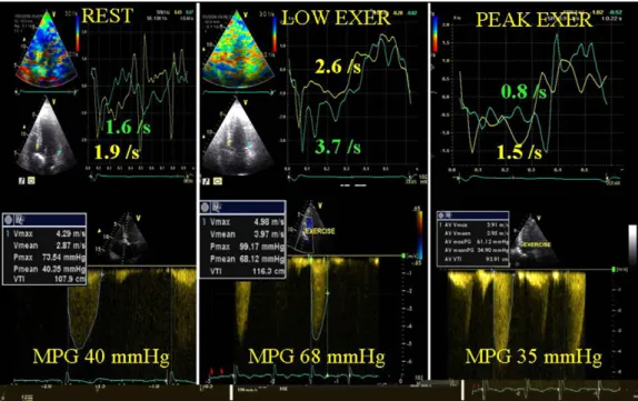
A 71-year-old man with asymptomatic severe aortic stenosis was referred to our stress echocardiography laboratory for risk stratification. He had no coronary risk factors but led a sedentary life. A symptom-limited bicycle exercise was performed in the supine posi-tion. The patient reached the target heart rate (128 beats/min) and developed moderate dyspnoea at a workload of 100 W. The systolic blood pressure initially increased from 130 to 150 mmHg whereas no further changes occurred at a higher exercise level. Baseline transthoracic echocardiography (rest) confirmed the presence of severe valvular aortic stenosis, with an aortic valve area of 0.78 cm2(outflow tract time velocity integral 20 cm)
and a mean transaortic pressure gradient of 40 mmHg. At low-level exercise, the increase in longitudinal long-axis function (peak negative strain rate obtained off-line by tissue Doppler reconstruction at the level of the septal and lateral walls) (Fig. Top panel) was accompanied by a significant rise in transaortic pressure gradients (Fig. Bottom panel). The calculated aortic valve area was 0.71 cm2(outflow tract time velocity integral 19 cm)
indicating absence of valve compliance. At peak exercise, the transaortic pressure gradi-ent decreased as a result of a significant impairmgradi-ent of left vgradi-entricular function (decrease in peak negative strain rate). The aortic valve area remained fairly unchanged (0.69 cm2)
whereas the left ventricular outflow tract time velocity integral decreased (15 cm), indi-cating reduced flow across the aortic valve. No inducible ischaemia was observed during test and no concomitant coronary artery stenosis was found at angiography.
∗Corresponding author.
E-mail address:[email protected](P. Lancellotti).
1875-2136/$ — see front matter © 2009 Elsevier Masson SAS. All rights reserved. doi:10.1016/j.acvd.2009.04.009
594 P. Lancellotti et al.
Figure 1. Quantitative exercise Doppler echocardiographic evaluation of a patient with severe asymptomatic aortic valve stenosis.
Évaluation par échocardiographie Doppler d’effort d’un patient asymptomatique porteur d’une sténose aortique sévère.
Comment
In this patient, the inadequate left ventricular adaptation to exercise is due to exercise-induced afterload mismatch. Initially, the heart adapted appropriately to the increased afterload. The recruited inotropic reserve — increase in lon-gitudinal myocardial deformation — was accompanied by a significant rise in transaortic pressure gradient. At high-level
exercise, the aortic valve compliance recruitment was fully exhausted and a mismatch between afterload and contrac-tility occurred as expressed by reduced long-axis function and aortic flow.