i
Université du Québec
Institut National de la Recherche Scientifique-
Institut Armand-Frappier
Development of Cellulose Nanocrystal (CNC)
Reinforced Bio polymeric Matrix for
Microencapsulation of Bioactive Compounds
Par
Tanzina Huq
Thèse présentée pour l’obtention du grade de Philosophiae Doctor (Ph.D.) en Biologie
Jury d’evaluation
Président du jury et examinateur interne Prof. Charles Ramassamy INRS-Institut Armand Frappier Examinateur externe Prof. George Szatmari
Université de Montreal Examinateur externe Prof. Philippe Bébin
Centre de Technologie Minerale et de Plasturgie Directrice de recherche Prof. Monique Lacroix
INRS-Institut Armand Frappier Co- directeur de recherche Prof. Bernard Riedl
Université Laval
ii
Acknowledgment
I was so fortunate to have the support of a number of people throughout my whole PhD programme without whom this thesis would have been impossible. I am grateful to all of them who have contributed towards this thesis directly or indirectly.
First and foremost, I would like to express my sincere gratitude to my PhD director Prof. Monique Lacroix for giving me an invaluable opportunity to gain knowledge and great experience in her laboratory. Her constant support, guidance, motivation and encouragement has driven me to aim higher and achieve my goals. I would like to give my sincere thanks to my PhD co-director Prof. Bernard Riedl for his valuable suggestions, comments on my thesis and also special thanks for French corrections. I am highly thankful to Dr. Jean Bouchardand Dr. Carole Fraschinifor their valuable comments and corrections of my thesis.
My sincere thanks to Dr. Ruhul A. Khan, Dr. Khanh Dang Vu, Dr. Dominic Dussault, Stephane Salmieri and Dr. Canh Le Tien for their great advice during my PhD research. I am thankful to all my fellow graduate, colleagues and trainees in the Labo-Resala Rose Roseline, Amira Ben Mabrouk and Farah Hossain for their assistance and friendship. I would like to express my deepest sense of gratitude and affection to my beloved parents and in-laws and all my family members back in Bangladesh for their blessings, inspiration and constant help. My loving and caring husband, Avik has always inspired and encouraged me to overcome all the difficulties during these times. Additionally my cute cat Tuki has made me happy, relax and feel me with joys by her innocent loving affection. Finally, I would like express my sincere thanks to Fondation Universitaire Armand-Frappier for their scholarship.
iii
Table of Contents
Acknowledgment ... ii
Table of Contents ... iii
Résumé ... x
Abstract ... xiii
List of Figures ... xv
List of Tables ... xix
List of Abbreviations ... xx Introduction ... 1 Chapter -1 ... 3 Literature Review ... 3 1.1 Introduction ... 4 1.2 Bioactive compounds ... 5
1.2.1 Essential Oils (EOs) ... 5
1.2.2 Bacteriocin: Nisin Antimicrobial Polypeptide ... 8
1.2.3 Probiotics ... 11
1.3 Microencapsulation of Bioactive Compounds ... 14
1.3.1 Extrusion ... 15
1.3.2 Emulsification ... 16
1.3.3 Drying Methods ... 17
1.3.4 Compression Method ... 21
1.4 Biopolymers Used for the Microencapsulation of Bioactive Compounds ... 22
1.4.1 Microencapsulation in Alginate System ... 22
1.4.2 Microencapsulation in Gelatin and Polysaccharide System ... 23
1.4.3 Chitosan-coated Alginate Encapsulate System ... 24
1.4.4 Encapsulation in Cellulose Derivatives ... 25
iv
1.4.6 Encapsulation of Probiotics in Whey Protein Isolate (WPI) Gel Particles ... 30
1.4.7 Encapsulation of Live Probiotics in a Modified Alginate System ... 30
1.4.8 Effect of Prebiotics for Probiotic Encapsulate System ... 31
1.5 Conclusion and Future Challenges ... 32
1.6 References ... 33
1.7 Problematic, Hypothesis and Objectives ... 44
1.7.1 Problematic ... 44
1.7.2 Hypothesis ... 44
1.7.3 Objectives ... 45
1.7.4 Methodology ... 45
1.7.4 Simplified Flowchart for the Ph.D. Thesis... 47
Chapter-2 ... 48
Publication-1 ... 48
Cellulose Nanocrystals (CNC) Reinforced Alginate Based Biodegradable Nanocomposite Film ... 49
2.1 Contribution of the Authors ... 50
2.2 Specific Objectives of Pubicaltion-1 ... 51
2.3 Résumé ... 52
2.4 Abstract ... 53
2.5 Introduction ... 54
2.6 Materials and Methods ... 55
2.6.1 Materials ... 55
2.6.2 Preparation of Alginate Based Films ... 56
2.6.3 Mechanical Properties Measurement ... 56
2.6.4 Water Vapor Permeability ... 57
2.6.5 Gel Swelling Property ... 58
2.6.6 Fourier Transform Infrared (FTIR) Spectroscopy ... 58
2.6.7 X-ray Diffraction ... 59
2.6.8 Thermo Gravimetric Analysis (TGA) ... 59
2.6.9 Differential Scanning Calorimetric (DSC) Analysis ... 59
2.6.10 Scanning Electron Microscopy (SEM) Analysis ... 59
2.6.11 Statistical analysis ... 60
v
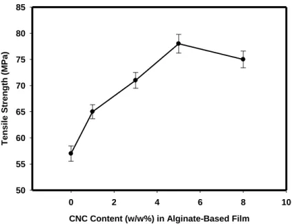
2.7.1 Effect of CNC Loading on the Mechanical Properties of Alginate-Based Films ... 60
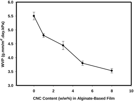
2.7.2 Water Vapor Permeability ... 61
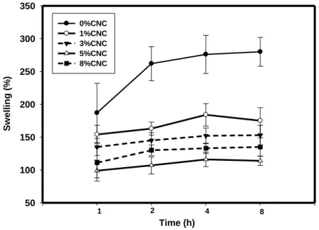
2.7.3 Gel Swelling Property ... 62
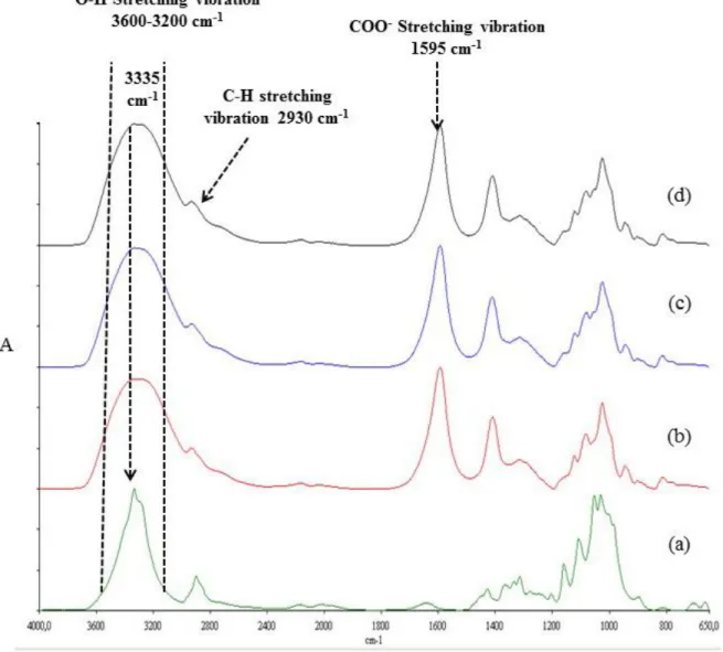
2.7.4 Fourier Transform Infrared Spectroscopy... 63
2.7.5 X-Ray Diffraction Analysis of the Films ... 64
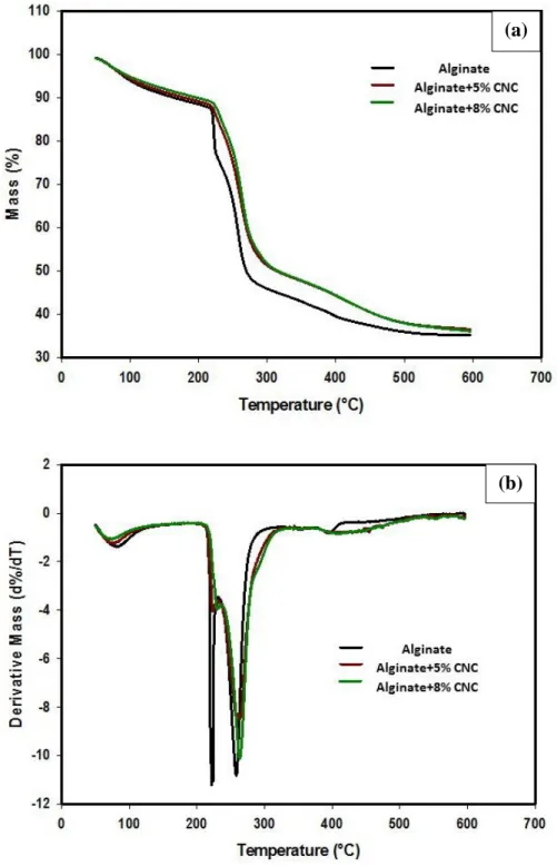
2.7.6 Thermal Properties of the Films ... 65
2.7.7 Scanning Electron Microscopy (SEM)... 66
2.8 Conclusion ... 67
2.9 Acknowledgement ... 67
2.10 References ... 68
2.11 General Discussions of Publication-1 ... 80
Chapter-3 ... 81
Publication 2 ... 81
Microencapsulation of Nisin in Alginate-Cellulose Nanocrystal (CNC) Microbeads against Listeria monocytogenes ... 82
3.1 Contributions of the Authors ... 83
3.2 Specific Objectives of the Pubicaltion-2 ... 84
3.3 Résumé ... 85
3.4 Abstract ... 86
3.5 Introduction ... 87
3.6 Materials and Methods ... 90
3.6.1 Materials ... 90
3.6.2 Preparation of Nisin Solution ... 90
3.6.3 Microencapsulation of Nisin ... 91
3.6.4 Fourier Transform Infrared (FTIR) Spectroscopy Analysis for Alginate-CaCl2-Nisin Complex ... 91
3.6.5 Bacterial Culture ... 92
3.6.6 BHI-Agar Deep-Well Model to Evaluate Depletion Activity of Nisin (in vitro Study) ... 92
3.6.7 Nisin Bioassay against L. monocytogenes ... 93
3.6.8 Preparation of Ham Samples (in situ Study) ... 93
3.6.9 Microbiological Analysis ... 94
3.6.10 pH Measurement of Ham Samples ... 94
vi
3.6.12 Statistical Analysis ... 95
3.7 Results and Discussions ... 95
3.7.1 ATR-FTIR Analysis of Alginate-CNC Microbead Containing Nisin ... 95
3.7.2 Microencapsulated and Free Nisin Availability during Storage: in vitro Study ... 96
3.7.3 Antimicrobial Activity of Microencapsulated Nisin against L. monocytogenes: in situ Test . 98 3.7.4 Effect of Microencapsulated Nisin on Ham pH During Storage ... 100
3.7.5 Colour of RTE Ham during Storage ... 101
3.8 Conclusion ... 103
3.9 Acknowledgements ... 103
3.10 References ... 103
3.11 General Discussions of the Publication-2 ... 120
Chapter 4 ... 121
Publication 3 ... 121
Synergistic Effect of Gamma (γ)-Irradiation and Microencapsulated Antimicrobials against Listeria monocytogenes on Ready-to-Eat (RTE) Meat ... 122
4.1 Contributions of the Authors ... 123
4.2 Specific Objectives of the Pubicaltion-3 ... 124
4.3 Résumé ... 125
4.4 Abstract ... 126
4.5 Introduction ... 127
4.6 Materials and Methods ... 130
4.6.1 Materials ... 130
4.6.2 Preparation of Nisin Solution ... 130
4.6.3 Microencapsulation of Nisin and EOs ... 131
4.6.4 Bacterial Culture ... 131
4.6.5 BHI-Agar Deep-Well Model to Evaluate Depletion Activity of EOs and Nisin ... 132
4.6.6 EOs and Nisin Bioassay against L. monocytogenes ... 132
4.6.7 Preparation of Ham Samples ... 133
4.6.8 Irradiation Treatment ... 133
4.6.9 Microbiological Analysis ... 134
4.6.10 Bacterial growth rate... 134
vii
4.6.12 Statistical Analysis ... 135
4.7 Results and Discussions ... 136
4.7.1 Effect of γ-irradiation on Available Antimicrobial Content (Free or Microencapsulated) during Storage: in vitro ... 136
4.7.2 Synergistic Effect of Microencapsulated Combined Antimicrobials and γ-irradiation (at 1.5 kGy) during Storage: in situ ... 137
4.7.3 Effect of Microencapsulated Antimicrobials on Radiosensitization of L. monocytogenes .. 141
4.8 Conclusion ... 143
4.9 Acknowledgements ... 143
4.10 References ... 143
4.11 General Discussions of the Publication-3 ... 159
Chapter-5 ... 160
Patent Application ... 160
Development of Cellulose Nanocrystal (CNC) Reinforced Alginate Microcapsule to Protect the Viability of Lactobacillus rhamnosus ATCC 9595 During Storage and Gastric Passage ... 161
5.1 Contributions of the Authors ... 162
5.2 Specific Objectives of the Patent Application ... 163
5.3 Résumé ... 164
5.4 Abstract ... 165
5.5 Introduction ... 166
5.6 Materials and Methods ... 169
5.6.1 Materials ... 169
5.6.2 Strain and Culture Preparation ... 169
5.6.3 Microencapsulation Procedure ... 169
5.6.4 Freeze-drying ... 170
5.6.5 Determination of Mechanical Strength ... 170
5.6.6 Swelling and Dissolution under Simulated Gastrointestinal Conditions ... 171
5.6.7 Water Activity (aw) and Water Content Measurement ... 171
5.6.8 Storage of Dried Microencapsulated Probiotic Beads ... 171
5.6.9 Determination of Survival and Inactivation Rate Constant ... 172
viii
5.6.11 Survival of Free and Microencapsulated Probiotic after Sequential Incubation in SGF and
SIF ... 173
5.6.12 Microbiological Analysis ... 173
5.6.13 Scanning Electron Microscopy (SEM) Analysis ... 174
5.6.14 Statistical Analysis ... 174
5.7 Results and Discussion ... 175
5.7.1 Optimization of CNC, Lecithin and Starch Concentration by Checking the Viability of the Probiotic during FreezeDrying ... 175
5.7.2 Mechanical Property of the Freeze Dried Beads ... 178
5.7.3 Swelling and Dissolution under Simulated Gastrointestinal Conditions ... 179
5.7.4 Storage of Dried Free and Microencapsulated L. rhamnosus ... 181
5.7.5 Survival of Free and Microencapsulated L. rhamnosus in SGF ... 184
5.7.6 Survival of Free and Microencapsulated Probiotics after Sequential Incubation from SGF to SIF ... 186
5.7.7 Scanning Electron Microscopy (SEM) Analysis ... 188
5.8 Conclusions ... 189
5.9 Acknowledgements ... 190
5.10 References ... 190
5.11 General Discussions of the Pathent Application ... 217
Chapter-6 ... 218
Patent Application ... 218
Development of Probiotic Tablet Formulation with Cellulose NanoCrystals (CNC) by using Central Composite Design (CCD)... 219
6.1 Contribution to the Authors ... 220
6.2 Specific Objectives of the Patent Application ... 221
6.3 Résumé ... 222
6.4 Abstract ... 223
6.5 Introduction ... 224
6.6 Materials and Methods ... 227
6.6.1 Materials ... 227
6.6.2 Freeze Dried Probiotic Culture Preparation (Biomass) ... 227
6.6.3 Probiotic Tablet Preparation Method ... 228
ix
6.6.5 Experimental Design for Probiotic Tablet Using CCD ... 228
6.6.6 Viability of L. rhamnosus in Probiotic Tablet after Incubation in Simulated Gastric Fluid (SGF) ... 229
6.6.7 Survival of CCD optimized Probiotic Tablet after Sequential Incubation in SGF and Simulated Intestinal Fluid (SIF) ... 230
6.6.8 Swelling Study ... 231
6.6.9 Storage of CCD Optimized Probiotic Tablets ... 231
6.6.10 Determination of Viable Cell Counts ... 231
6.6.11 Statistical Analysis ... 232
6.7 Results and Discussion ... 232
6.7.1 Data Analysis from CCD ... 232
6.7.2 Survival of CCD optimized Probiotic Tablets after Sequential Incubation in SGF and SIF ... 235
6.7.3 Swelling Behaviour of CCD optimized (alginate-pectin-CNC) Tablet ... 237
6.7.4 Storage Stability of L. rhamnosus ... 239
6.8 Conclusion ... 241
6.9 Acknowledgement ... 241
6.10 References ... 242
6.11 General Discussions of the Patent Application ... 257
Chapter 7 ... 258
General Discussions/Conclusions and Future Perspectives... 258
Annexe-I (Supplementary Data) ... 266
Annexe-II (Supplementary Data) ... 279
Annexe-III (Biography) ... 284
x
Résumé
La nanotechnologie a connu une croissance rapide en recherche, avec des applications en sciences alimentaires, comme une technologie émergente. Les potentiels de la nanotechnologie sont explorés dans divers projets de recherche pour la sécurité alimentaire et le développement d'aliments fonctionnels. La cellulose nanocristalline (CNC) est un nanomatériau cellulosique. En application dans les aliments, ce nanomatériau a été appliqué comme biopolymère alimentaire. Une matrice alginate de base renforcée de CNC a été développée pour la microencapsulation de composés bioactifs (probiotiques et les composés antimicrobiens). A un niveau de 5% en poids, la concentration en CNC a été optimisée dans une matrice d'alginate suite à l'amélioration de la résistance à la traction de 37% de films et la réduction de perméabilité à la vapeur d'eau de 31% par rapport à la matrice d'alginate. On a constaté qu’une concentration de 5% en poids dans une matrice d'alginate CNC a également amélioré les propriétés thermiques et morphologiques. Ce niveau optimisé de 5% en poids de CNC dans la matrice d'alginate a été utilisé pour la microencapsulation de composés antimicrobiens tels que la nisine et d'huiles essentielles afin d'améliorer la sécurité alimentaire des produits carnés prêts -à-manger (RTE) pendant le stockage, suite à une activité contre la bactérie Listeria monocytogenes. Il a été constaté que la nisine microencapsulée (63μg/ml) dans de l'alginate-CNC permet d’améliorer son activité et disponibilité 20 fois et a réduit la présence de L. monocytogenes par 3.04 log UFC / g par rapport à celle de la nisine libre (63μg/ml) pendant 28 jours de stockage à 4° C. Cette étude a révélé une nouveauté, soit par le développement de films d'alginate-CNC pour la microencapsulation de la nisine qui présentait un effet de protection des produits carnés prêts à consommer. Après avoir constaté l'effet antimicrobien de concentrations différentes de nisine micro-encapsulée et libre sur le système carné RTE, la plus faible concentration de nisine (16μg/ml) active a été évaluée en combinaison
xi
avec l’origan ‘compactum’ ou la cinnamome ‘cassia’ (250 μg / ml) pour vérifier l'effet synergique de l’irradiation γ ou la pasteurisation à froid.
La popularité dans le monde des produits contenant des probiotiques a augmenté énormément dont certains qui sont très reconnus comme nutraceutiques. Mais ces produits montrent une viabilité très inférieure du probiotique pendant le stockage et en état gastrique. Notre matrice d'alginate-CNC développée a été utilisée pour la microencapsulation de probiotiques (Lactobacillus rhamnosus ATCC 9595) pour améliorer sa stabilité au cours du stockage. La concentration de CNC a été une fois de plus optimisée avec une matrice d'alginate en vérifiant la viabilité pendant la lyophilisation. Les résultats ont montré que 13 % en poids de CNC dans des microbilles d'alginate a seulement réduit la viabilité de L. rhamnosus par 0.01 log alors que le log de la viabilité des microbilles d’alginate a été réduit de 0.95 log après le séchage libre. On a également constaté que 13 % en poids de CNC en combinaison avec des microbilles d'alginate -lécithine a montré une plus grande amélioration de la viabilité au cours du stockage. Les résultats ont démontré que cette formulation a augmenté de 61 % la viabilité des probiotiques après 42 jours de stockage à 25˚ C par rapport à des contrôles de microbilles d'alginate. Cette formulation de microbilles d'alginate-CNC-lécithine a amélioré la viabilité de L. rhamnosus de 52 % au cours de la transition complète à travers le passage gastrique par rapport à microbilles d'alginate. Il y a eu aussi une nouvelle formulation de revêtements développée à l'aide d'alginate, de pectine et de CNC, afin d'améliorer la stabilité de L. rhamnosus à 25˚ C en utilisant un modèle de type ‘composite central’. Cette formulation de revêtement a montré une bonne stabilité de L. rhamnosus à la température ambiante jusqu'à six mois de stockage. L'interaction observée entre les formulations de revêtement d'alginate pectine-CNC a également présenté une viabilité de 84% après le passage gastrique.
xii
En résumé, cette thèse a été entreprise pour développer des systèmes de microbilles biopolymères comestibles pour l'application dans le domaine de l’alimentation et à des fins nutraceutiques. L'utilisation de CNC a créé une voie innovante pour l'industrie alimentaire et nutraceutique.
Tanzina Huq Prof. Monique Lacroix
xiii
Abstract
Nanotechnology has been rapidly growing research in food application as an emerging technology. The potentials of nanotechnology have been explored in various food researches for food safety and development of functional foods. Cellulose Nanocrystals (CNC) are a cellulosic nanomaterial. In food applications, this nanomaterial has started its association with food biopolymers. A CNC reinforced alginate based matrix was developed for microencapsulation of bioactive compounds (probiotics and antimicrobials compounds). A 5 % wt CNC content was optimized in alginate based matrix with an improvement of tensile strength of 37% and a reduction of water vapour permeability of 31% compared to those of alginate matrix. It was found that 5 % wt CNC in alginate matrix also improved the thermal and morphological properties. This optimized 5 % wt CNC content in alginate matrix was applied for microencapsulation of antimicrobial compounds such as nisin and essential oils in order to improve the food safety of ready-to-eat (RTE) meat products during storage against Listeria monocytogenes. It was found that microencapsulated nisin (63μg/ml) in alginate-CNC beads improved the activity of nisin 20 times and reduced the counts of L. monocytogenes by 3.04 log CFU/g compared to those of free nisin (63μg/ml) during 28 days of storage at 4° C. This study revealed a novelty by the development of alginate-CNC beads for microencapsulation of nisin that exhibited protection effect of RTE meat products. After obtention of a antimicrobial effect of different concentrations of microencapsulated and free nisin on RTE meat system, the lowest concentration of nisin (16μg/ml) was evaluated in combination with Origanum Compactum or Cinnamomum Cassia (250 μg/ml) to check the synergistic effect of γ-irradiation or cold pasteurization.
The worldwide popularity of probiotic containing products have been increasing tremendously and these products are known as nutraceuticals. But the viability of probiotics in these products is much
xiv
lower during storage and gastric conditions. Our alginate-CNC matrix was used for microencapsulation of probiotics (Lactobacillus rhamnosus ATCC 9595) to improve its stability during storage. The CNC concentration was once more optimized with an alginate matrix by checking the viability during freeze drying. Results demonstrated that 13 wt% of CNC in alginate microbeads only reduced the viability of L. rhamnosus by 0.01 log whereas alginate microbead reduced the viability by 0.95 log after freeze drying. It was also found that 13 wt% of CNC in combination with alginate-lecithin microbead showed more improvement of viability during storage. Results showed this formulation increased the probiotic viability by 61% after 42 days of storage at 25˚C compared to control alginate microbead. This alginate-CNC-lecithin microbeads formulation improved the viability of L. rhamnosus by 52% during complete transition through the gastric passage as compared to alginate microbead. There was also a novel coating formulation developed by using alginate, pectin and CNC in order to improve the stability of L. rhamnosus at 25˚C by using central composite experimental design. This coating formulation showed a good stability of L. rhamnosus at room temperature up to six months of storage. The interaction found between alginate-pectin-CNC coating formulations exhibited a viability of 84% after complete transition through the gastric passage.
In summary, this thesis was undertaken to develop biopolymer edible microbeads for food applications and nutraceutical purposes. The utilization of CNC established an innovative route for the food and nutraceutical industry.
xv
List of Figures
Figure 1.1: Schematic Diagram of Spray Drying Method ... 19
Figure 1.2: Freeze Drying Method... 21
Figure 1.3: Microencapsulated bioactives a) before freeze drying and b) after freeze drying
... 21
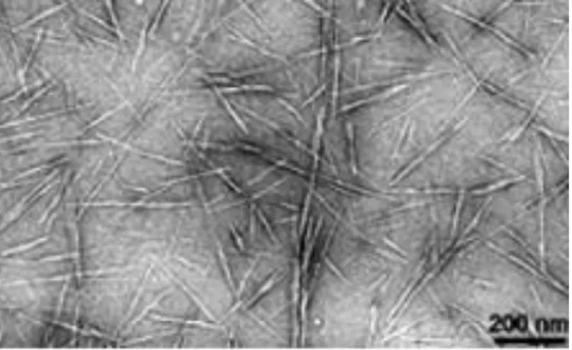
Figure -1.4: TEM image of highly anionic cellulose nanocrystals adapted from Habibi et al.
(2010). ... 29
Figure 2.1(a): Effect of CNC Content (w/w %) on Tensile Strength (MPa) of Alginate-based
Film, as a function of CNC content in dry matrix. ... 72
Figure 2.1 (b): Effect of CNC Content (w/w %) on Tensile Modulus (GPa) of Alginate-based
Film, as a function of CNC content in dry matrix. ... 72
Figure 2.1(c): Effect of CNC Content (w/w %) on Elongation at Break (%) of Alginate-based
Film, as a function of CNC content in dry matrix. ... 73
Figure 2.2: Effect of CNC Content (w/w %) on WVP of Alginate-based Film, as a function
of CNC content in dry matrix. ... 73
Figure 2.3: Effect of CNC Content (w/w %) on Swelling of Alginate-based Film, as a function
of CNC content in dry matrix. ... 74
Figure 2.4: FTIR spectra of (a) Pure CNC film, (b) Native alginate, (c) Alg+5% (w/w) CNC
and (d) Alg+8% (w/w) CNC. ... 75
Figure 2.5: X-ray Diffractograms for CNC, Alginate, Alg+5% (w/w) CNC and Alg+8%
(w/w) CNC. ... 76
Figure 2.6: (a) TGA and (b) derivative TGA curve for Alginate, Alginate +5% (w/w) CNC
and Alginate +8% (w/w) CNC Film. ... 77
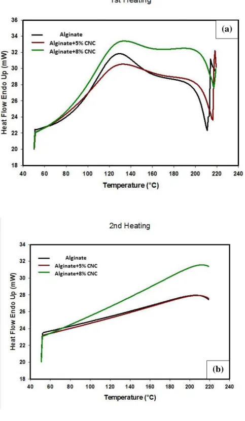
Figure 2.7: DSC curves for alginate, alginate with 5 and 8% (w/w) CNC films (a) first heating
and (b) second heating. ... 78
Figure 2.8: SEM images of the fracture surface of alginate film (a), alginate film with 5 %
xvi
Figure 3.1: BHI-agar deep-well model of peptide depletion during storage (left) and activity
bioassay against a pathogen (right) which is adapted from Bi et al. (2011a). ... 110
Figure 3.2 A: ATR-FTIR spectra of i) control polymer (alginate-CNC microbead); ii) N1-E
16 μg/ml (alginate-CNC microbead with 16 μg/ml of nisin); iii) N2-E 31μg/ml (alginate-CNC microbead with 31μg/ml of nisin); iv) N3-E 63 μg/ml (alginate-CNC microbead with 63 μg/ml of nisin). ... 111
Figure 3.2 B: ATR-FTIR spectra in the wavenumber region between 1801-1192 cm-1. .... 112 Figure 3.3: Standard curve for A) Free nisin and B) Microencapsulated nisin against L.
monocytogenes in in vitro bioassay. ... 113
Figure 3.4: Available Nisin Concentration from A) Free nisin and B) Microencapsulated nisin
against L. monocytogenes during storage at 4° C in in vitro BHI-agar deep well model. .... 114
Figure 3.5: The digital photograph of agar diffusion assay against L. monocytogenes for free
and microencapsulated nisin (N3-63 μg/ml) during storage. ... 115
Figure 3.6: Growth of L. monocytogenes on vacuum packaged cooked ham slices coated with
A) Free nisin and B) microencapsulated nisin during storage at 4° C. ... 116
Figure 3.7: pH value of A) free nisin and B) microencapsulated nisin coated RTE cooked
ham during storage at 4°C. ... 117
Figure 4.1: Standard curve of Chloramphenicol against L. monocytogenes in in vitro bioassay
... 152
Figure 4.2: Effect of γ-irradiation on available free or microencapsulated antimicrobials,
mg/ml CAM (Origanum compactum) against L. monocytogenes during storage at 4° C in in vitro BHI agar deep well model. A) without irradiation and B) with irradiation. ... 153
Figure 4.3: Effect of γ-irradiation on available free or microencapsulated antimicrobials,
mg/ml CAM (Cinnamomum cassia and nisin) against L. monocytogenes during storage at 4° C in in vitro BHI-agar deep well model. A) without irradiation and B) with irradiation. .... 154
Figure 4.4: Effect of microencapsulated Origanum compactum + nisin on RTE cooked ham
xvii
Figure 4.5: Synergistic effect of microencapsulated Origanum compactum + nisin and
γ-irradiation on RTE cooked ham during storage at 4°C ... 155
Figure 4.6: Effect of microencapsulated Cinnamomum cassia + nisin on RTE cooked ham
during storage at 4°C without irradiation ... 156
Figure 4.7: Synergistic effect of microencapsulated Cinnamomum cassia + nisin and
γ-irradiation on RTE cooked ham during storage at 4°C ... 156
Figure 5.1: A) D-mannuronic acid (M), B) L-guluronic acid (G) (George and Abraham, 2006) and C) egg-box structure of alginate as proposed by Li et al., 2007. ... 200
Figure 5.2: General microencapsulation flow diagram for L.rhamosus ATCC 9595. ... 202
Figure 5.3A: Effect of CNC (wt %) with alginate beads on the viability of L. rhamnosus
ATCC 9595 during freeze drying. ... 203
Figure 5.3B: Effect of lecithin (wt%) on the viability of L.rhamnosus ATCC 9595 in
optimized alginate + CNC beads during freeze drying. ... 204
Figure 5.3C: Effect of Starch (wt %) on the viability of L.rhamnosus ATCC 9595 in
optimized alginate + CNC+ lecithin beads during freeze drying. ... 205
Figure 5.4A: Effect of CNC content (wt%) on compression strength (MPa) of 100 wt % (3%
w/v) alginate beads, as a function of CNC content in dry matrix.. ... 206
Figure 5.4B: Compression strength (MPa) of the optimized formulations.. ... 207
Figure 5.5: Swelling behavior of freeze-dried optimized beads at two different simulated
gastric fluid pH (1.5 and 5) values and in simulated intestinal fluid (pH 7). ... 209
Figure 5.6: The storage stability of freeze-dried free and microencapsulated L. rhamnosus A)
at 25° C and B) 4° C. ... 210
Figure 5.7: Survival of L.rhamnosus ATCC 9595 in optimized microbead formulations after
exposure in SGF (2 h) + SIF (3 h) A) before freeze-drying and B) after freeze-drying. ... 214
Figure 5.8: Surface SEM micrographs of freeze-dried beads of (A) and (B) A-0: 100 % w/w
alginate; (C) and (D) AC-1: 95 % w/w alginate + 5 % w/w CNC; (E) and (F) AC-3: 87 % w/w alginate + 13 % w/w CNC; (G) and (H) ACL-1: 85 % w/w alginate +13 % w/w CNC +
xviii
2 % w/w lecithin; (I) and (J) ACLS-2: 54 % w/w alginate + 8 % w/w CNC + 2 % w/w lecithin + 36 % w/w starch, respectively. ... 216
Figure 6.1 (A): Response surface viability (%) of probiotic during SGF treatment obtained
by varying alginate (X1) and pectin (X2) concentration. ... 251 Figure 6.1(B): Response surface viability (%) of probiotic during SGF treatment obtained by varying alginate (X1) and CNC (X3) concentration. ... 251 Figure 6.1(C): Response surface viability (%) of probiotic during SGF treatment obtained by
varying pectin (X2) and CNC (X3) concentration. ... 252 Figure 6.2: Viability (%) of CCD optimized probiotic tablets during sequential transition
through SGF (2hr) to SIF (3hr). ... 253
Figure 6.3: Swelling (%) of CCD optimized formulation comparing individual biopolymers
(alginate, pectin and CNC) of probiotic tablets during complete transition from SGF to SIF. ... 255
Figure 6.4: Storage stability of L. rhamnosus ATCC 9595 containing probiotic tablet A) 25°
xix
List of Tables
Table 1.1: The major components of some EOs with antimicrobial properties are presented:
... 7
Table-1.2: Application of Nisin in whole world* ... 10 Table 3.1 A: Effect of free nisin coating on the colour coordinates L*, a* and b* in RTE cooked ham during storage at 4°C**. ... 118
Table 3.1 B: Effect of microencapsulated nisin coating on the colour coordinates L*, a* and b* in RTE cooked ham during storage at 4°C**. ... 119
Table 4.1: Growth rate (ln CFU/g/day) of L. monocytogenes of free and microencapsulated
antimicrobial compounds on RTE cooked ham. ... 157
Table 4.2: D10 and radiosensitivity (RS) of L. monocytogenes of free and microencapsulated
antimicrobial compounds on RTE cooked ham. ... 158
Table-5.1: Optimized formulations are presented according to wt%. ... 201
Table 5.2: Inactivation rate constants k (d-1) for L. rhamnosus at 25 and 4°C storage conditions. The determination coefficients (R2) of each individual k value are given in brackets next to the respective k-value. ... 211
Table 5.3 A: Water activity of freeze-dried L.rhamnosus free cell and microencapsulated L.
rhamnosus samples during storage at 25˚C. ... 212
Table 5.3B: Water activity of freeze-dried L.rhamnosus free cell and microencapsulated L. rhamnosus samples during storage at 4˚C... 212
Table 5.4: Survival of L. rhamnosus ATCC 9595 (Log CFU/g) after 2 h SGF treatment.* & **
... 213
Table 6.1: Levels of the factor tested in the CCD ... 249
Table 6.2: Results of L. rhamnosus viability (%) after 2h SGF treatment by CCD design 250
Table 6.3: Viability of L.rhamnosus in CCD optimized Tablet (Log CFU/Tablet) after
xx
List of Abbreviations
Analysis of variance (ANOVA) Brain Heart Infusion (BHI) Chloramphenicol (CAM)
Carboxymethyl cellulose (CMC) Cellulose acetate phthalate (CAP) Cellulose nanocrystals (CNC) Central composite design (CCD)
Cetyl trimethyl ammonium bromide (CTAB) Code of Federal Regulations (CFR)
Colorectal cancer (CRC)
Differential Scanning Calorimetric (DSC) Elongation at break (Eb%)
Essential Oils (EOs)
Facility Electron Microscopy Research (FEMR)
Food and Agriculture Organization (FAO) Food and Drug Administration (FDA) Fourier Transform Infrared (FTIR) Genarally recognised as safe (GRAS) Health Products and Food Branch (HPB) Hydroxyl propyl methylcellulose acetate
succinate (HPMCAS)
Hydroxypropyl methyl cellulose (HPMC) International Atomic Energy Agency (IAEA) Lactic acid bacteria (LAB)
Modified atmospheric packaging (MAP) Natural Sciences and Engineering Research
Council of Canada (NSERC) Puncture deformation (PD) Puncture strength (PS) Ready-to-eat (RTE)
Response surface methodology (RSM) Scanning Electron Microscopy (SEM) Simulated gastric fluid (SGF)
simulated intestinal fluid (SIF) Tensile modulus (TM)
Tensile strength (TS)
Thermo Gravimetric Analysis (TGA) Viscoelasticity (Y)
Water vapor permeability (WVP) Water-in-oil (W/O)
Whey Protein Isolate (WPI)
World Health Organization (WHO) X-ray Diffraction (XRD)
1
Introduction
Current interest in nanotechnologies has led to the application of Cellulose Nanocrystals (CNC) in food application. In today’s world, Canada is the world’s leading producer of CNC and ranks first in research in this area. The North American market for CNC may reach $250 million according to Canada’s forest industry news. The overall range of material applications for CNC is virtually limitless; in addition, the possibility of controlling their biosynthetic pathways opens up new windows to encouraging their use as a facile energy resource for food application. CNC has been found to be an innovative nanomaterial due to its good reinforcing properties with biopolymers over the past 20 years (Samir et al., 2005). Among the biopolymers, alginate is one of the most widely used biopolymers as a bioactive coating in food application. It has a very good applicability to prepare microcapsules owing to its good biocompatibility, biodegradability, non-toxicity, gelation and mucoadhesion properties (Lertsutthiwong et al., 2008), with the addition of some other additives.
Bioactive compounds (antimicrobial and probiotic) are of growing world wide interest due to the consumer demand on food safety and nutritional aspects for human health. Antimicrobials are used to reduce pathogenic bacteria such as Listeria monoctytogens in the food supply. Bacteria such as L. monocytogenes causes 94% hospitalization which results in a 15.6% death rate in the US each year (Scallan et al., 2011). In 2008, there was a listeriosis outbreak in Canada and 22 people died due to the contamination of Maple Leaf Food products (meat products) by L. monocytogenes. Thus it is important to add antimicrobials in food for health safety. The direct addition of antimicrobial compounds in food system showed degradation by interacting with individual food components. On the other hand, probiotics are living microorganism food ingredients that have a beneficial effect on human health. Lactic acid bacteria used as probiotics are commonly incorporated into
2
foods to provide a wide variety of health benefits but stabilization of probiotics at room temperature and under gastric environment is a challenging endeavour. However, microencapsulation has been found to be a powerful technology to protect the antimicrobials and probiotics from these adverse conditions. This technology is based on the proper utilization of biopolymers which are normally referred to as bioactive coatings. The aim of this research was to develop CNC reinforced alginate matrix for microencapsulation of antimicrobials and probtiotics.
3
Chapter -1
Literature Review
4
1.1 Introduction
This literature review provides the information about microencapsulation of bioactive compounds in biopolymeric matrix. The importance of different biopolymer matrixes or matrices described along with different microencapsulation processes. CNC is a new bio-nanomaterial which delivers interesting properties with biopolymer matrix. The part of this literature review has been published in Critical Reviews of Food Science and Nutrition. The text is reproduced from the following literature review with the addition of antimicrobial compounds, some more information about biopolymers and microencapsulation:
Huq, T., Khan, A., Khan, R.A., Riedl, B. & Lacroix, M., (2013). Encapsulation of Probiotic Bacteria in Biopolymeric System. Critical Reviews in Food Science and Nutrition, 53:909–916. The pulished literature review was prepared by Tanzina Huq with continuous direction of my director Prof. Monique Lacroix. Prof. Bernard Riedl, my co-director, helped me to correct and improve the draft. Avik Khan has helped me to write the microencapsulation technology. Dr. Ruhul A. Khan has corrected the draft.
5
1.2 Bioactive compounds
The range of food components that could be considered as bioactive compounds include vitamins, minerals, functional lipids, probiotics, amino acids, peptides and proteins, fatty acids and antioxidants/ antimicrobials. Among the bioactive compounds, antimicrobials and probiotics have been chosen due to their potential activity for the food industry. Antimicrobial compounds are used for food safety, probiotics and for nutraceutical applications. Both antimicrobials and probiotics are two important classes of bioactive compounds that need to be protected from adverse condition as antimicrobials can readily react with food components and probiotics are very sensitive to oxygen. Our present review involved essential oils and bacteriocins as antimicrobial compounds.
1.2.1 Essential Oils (EOs)
EOs are volatile, natural, complex compounds characterized by a strong odour and are formed by aromatic plants as secondary metabolites. They are usually obtained by steam or hydro-distillation. It has long been recognized that EOs have antimicrobial properties for food preservation. Besides antimicrobial properties, EOs or their components have been shown to exhibit antiviral, antimycotic, antitoxigenic, antiparasitic, and insecticidal properties. They are gaining interest for their potential as preservative ingredients or decontaminating treatments, as they have GRAS status and a wide acceptance from consumers (Burt and Reinders, 2003; Burt, 2004). The antimicrobial components that are commonly found in the essential oil fractions have a wide spectrum of antimicrobial activity, with potential for control of L. monocytogenes and spoilage bacteria within food system (Oussalah et al., 2007). Table-1 represents the major components of some EOs with antimicrobial properties.
6
1.2.1.1 Uses of EOs in Food Preservation
The potential application of EOs as natural antimicrobials and antioxidants has been found in meat, fish, fruit, vegetables and dairy products. Eugenol and coriander, clove, oregano and thyme oils were found to be effective at levels of 5-20 μl/g by inhibiting the growth of L. monocytogenes in meat products (Burt, 2004). Some packaging technologies such as vacuum, modified atmospheric packaging (MAP) have been used to enhance the antimicrobial activity of EOs on food systems. In general, MAP is a food packaging technology in which the proportion of oxygen, carbon dioxide and nitrogen are maintained in a sealed food product. Chouliara et al. (2007) investigated the combined effect of oregano oil and MAP on the shelf-life extension of fresh chicken meat stored at 4˚C. Valero et al. (2006) declared that the combination of MAP and eugenol or thymol was an interesting tool to preserve the quality, safety and functional properties of grapes. Dussault et al. (2014) showed that oregano (500 ppm) reduced around 2 log CFU/g growth of L. monocytogenes in vacuum packed RTE cooked ham compared to control ham.
7
1.2.1.2 Major Components of EOs
Table 1.1: The major components of some EOs with antimicrobial properties are presented:
Common name of EO
Latin name of plant sourcs
Major components Approximate concentration of major components of EOs (%) Antimicrobial against the bacteria References Oregano Origanum Compactum Carvacrol Thymol Trace-80% Trace-64% Listeria monocytogenes Escherichia coli Daferera et al. (2000); Turgis et al. (2012); Daferera et al. (2003). Cinnamon Cinnamomum Cassia Trans-cinnamaldehyde 65% L. monocytogenes E.coli Salmonella typhimurium Caillet et al. (2006); Turgis et al. (2012).
Thyme Thymus vulgaris Thymol
Carvacrol γ-Terpinene p- Cymene 10-64% 2-11% 2-31% 10-56% L.monocytogenes E. coli Turgis et al. (2012); Daferera et al. (2000). Rosemary Rosmarinus officinalis Camphor 1,8-cineol 2-14% 3-89% L. monocytogenes E.coli Staphylococcus aureus Daferera et al. (2003); Dussault et al. (2014).
8
1.2.2 Bacteriocin: Nisin Antimicrobial Polypeptide
Bacteriocins are defined as a protein-based substances possessing antimicrobial activities produced during the growth of Gram (+) and Gram negative (-) bacteria. The first known bacteriocin was identified in Escherichia coli as an antimicrobial protein and named colicin. Bacteriocins are often confused with antibiotics, however the main differences is that antibiotics are not ribosomally synthesized, while they also differentiate from antibiotics on the basic mode of action, antimicrobial spectrum, toxicity and resistance mechanisms (Cleveland et al., 2001). The interest in bacteriocins produced by GRAS (generally recognized as safe) microorganisms has been leading to considerable interest for nisin, being the first bacteriocin to gain widespread commercial application since 1969 after the approval from FAO/WHO (Food and Agriculture Organization/World Health Organization) (De Arauz et al., 2009). Nisin has been approved by the Food and Drug Administration (FDA) since 1988, satisfying the demands for natural foods with less chemical additives (Penna et al., 2005). It is a natural antimicrobial peptide produced by strains of Lactococcus lactis subsp. lactis that effectively inhibits Gram (+) bacteria and the outgrowth of spores of Bacilli and Clostridia (Balciunas et al., 2013). Nisin could also be effective against Gram (-) bacteria after having gone through some modification (Stevens et al., 1991). Structurally, it is a 34 amino acid polypeptide, presenting cationic and hydrophobic characteristics, with a molar mass of 3500 Da, belongs to the lantibiotic family as it contains lanthionine and methyl lanthionine groups. In addition, the unusual amino acids composition might be responsible for the important functional properties of nisin, i.e. acid tolerance, thermo stability at low pH and a specific bactericidal mode of action (De Vuyst and Vandamme, 1992). The solubility, stability and biological activity of nisin are dependent on the pH of the solution. In fermentation process, at pH < 6.0, more than 80% of nisin produced is released into the medium whereas at pH > 6.0, most of
9
the nisin is associated with the cellular membrane, but not the cytoplasm. Solubility and stability increase drastically with the lowering of pH and it was found that nisin is stable at pH 2.0. At this pH, nisin can be autoclaved at 121˚ C for 15 min without inactivation, while, on the other hand, in neutral and alkalineconditions nisin is almost insoluble (Penna et al., 2005).
1.2.2.1 Application of Nisin in Food as a Natural Antimicrobial
Nowadays, consumers prefer minimally processed foods of high quality, prepared without artificial preservatives, safe and with long shelf-life. Furthermore, nisin is the only bacteriocin that has been officially employed in the food industry and its use has been approved world-wide (Cleveland et al., 2001). There are numerous applications of nisin as a natural food preservative, including dairy products, canned foods, processed cheese and meat products. The FAO/WHO Codex Committee on milk and milk products accepted nisin as a food additive for processed cheese at a concentration of 12.5 mg pure nisin per kilogram product (De Arauz et al., 2009). Table-2 shows the world wide food application of nisin. The meat system is also one of the best examples for the use of nisin. Nitrates are commonly used to prevent clostridial growth in meat; however, safety concerns regarding the presence of nitrites have prompted the food industry to look for alternative methods of preservation. Nisin or its combination with lower levels of nitrate can prevent the growth of Clostridium (Rayman et al., 1983; Cleveland et al., 2001). During processing, this ready-to-eat (RTE) food products can readily be contaminated by pathogenic bacteria (L. monocytogenes). Nisin showed very strong antimicrobial effect in cooked ham against L. monocytogenes (Marcos et al., 2007).
10
Table-1.2: Application of nisin in world*
Country Food in which nisin is
permitted
Maximum Level (IU/g)
Argentina Processed Cheese 500
Australia Cheese, processed cheese, canned tomatoes
No limit
Belgium Cheese 100
Brazil Cheese, canned vegetables, sausages, meat products
500
France Processed Cheese No limit
Italy Cheese 500
Mexico Permitted additive 500
Netherlands Factory cheese, processed cheese, cheese powder
800
Peru Permitted additive No limit
Russia Dietetic processed cheese, canned vegetables
8000
USA Pasteurized processed cheese spread, meat products
10,000
UK Cheese, canned foods, clotted cream
No limit
11
1.2.3 Probiotics
People have been usingLAB for more than 4000 years for foods’ fermentation. Today, probiotics are also used in a variety of fermented dairy products and their manufacture involves fermentation: microbial process by which lactose is converted into lactic acid. Food and Agriculture Organization (FAO) of the United Nations and the World Health Organization (WHO) define probiotics as ‘‘Live microorganisms (bacteria or yeasts), which when ingested or locally applied in sufficient numbers confer one or more specified demonstrated health benefits for the host’’(FAO/WHO, 2001). LABs are the most important probiotic microorganisms typically associated with the human gastrointestinal tract. These bacteria are gram-positive, rod-shaped, spore-forming, catalase-negative organisms that are devoid of cytochromes and are of non-aerobic habit but are aero-tolerant, acid-tolerant and strictly fermentative; lactic acid is the major end-product of sugar fermentation. A few of the known LABs that are used as probiotics are Lactobacillus acidophilus, Lactobacillus amylovorous, Lactobacillus casei, Lactobacillus crispatus, Lactobacillus delbrueckii, Lactobacillus gasseri, Lactobacillus johnsonii, Lactobacillus paracasei, Lactobacillus plantarum, Lactobacillus reuteri, Lactobacillus rhamnosus etc. (Anal and Singh, 2007). Lactobacilli, as a part of the commensal microbial flora of humans and mammals and main representatives of the probiotic bacteria, might be useful candidates in prevention and treatment of infections caused by multi-resistant bacteria due to their ability to modulate the immune responses of the host and to protect the host from pathogens by competitive exclusion (Brachkova et al., 2010). Other common probiotic microorganisms are the bifidobacteria. Bifidobacteria are also gram-positive and rod-shaped but are strictly anaerobic. These bacteria can grow at pH in the range 4.5-8.5. Bifidobacteria actively ferment carbohydrates, producing mainly acetic acid and lactic acid in a molar ratio of 3:2 (v/v), but not carbon dioxide, butyric acid or
12
propionic acid. The most recognized species of bifidobacteria that are used as probiotic organisms are Bifidobacterium adolescentis, Bifidobacterium animalis, Bifidobacterium bifidum, Bifidobacterium breve, Bifidobacterium infantis, Bifidobacterium lactis and Bifidobacterium longum. Other than these bacteria, Bacillus cereus var. toyoi, Escherichia coli strain nissle, Propioniobacterium freudenreichii, and some types of yeasts, e.g. Saccharomyces cerevisiae and Saccharomyces boulardii have also been identified as having probiotic effects (Holzapfel et al., 2001). Some importance effect of probiotic are dicussed below.
1.2.3.1 Intestinal Tract Health
A number of studies have found probiotic consumption to be useful in the treatment of many types of diarrhoea, including antibiotic-associated diarrhoea in adults, travellers’ diarrhoea, and diarrhoeal diseases in young children caused by rotaviruses. The most commonly studied probiotic species in these studies have been Lactobacillus GG, L. casei, B. bifidum and S. thermophilus. Because diarrhoea is a major cause of infant death worldwide and can be incapacitating in adults, the widespread use of probiotics could be an important, non-invasive means to prevent and treat these diseases, particularly in developing countries. Probiotic bacteria have also been shown to preserve intestinal integrity and mediate the effects of inflammatory bowel diseases, irritable bowel syndrome, colitis, and alcoholic liver disease. In addition, LAB may improve intestinal mobility and relieve constipation (Pitino et al., 2010; Isolauri et al., 1991; Nanji et al., 1994).
1.2.3.2 Nutrient Synthesis and Bioavailability
Fermentation of food with LAB has been shown to increase the folic acid content of yogurt, bifidus milk and kefir and to increase niacin and riboflavin levels in yogurt, vitamin B12 in cottage cheese
and vitamin B6 in cheddar cheese. In addition to nutrient synthesis, probiotics may improve the
13
acid, propionic acid and butyric acid produced by lactic acid bacteria may help maintain an appropriate pH and protect against pathological changes in the colonic mucosa (Chen and Subirade, 2009; Kruis et al., 1997).
1.2.3.3 Probiotic Antimicrobial Activity
The importance of probiotics in human nutrition has been gaining recognition in recent years. Marianelli et al. (2010) proposed an improved in vitro model for the study of probiotic antimicrobial activity against enteropathogens, by attempting to re-create, in a common culture medium, environmental growth conditions comparable to those present in the small intestine. In that study, a preliminary experiment was carried out in order to find out a culture medium able to support both probiotics and pathogens. This experiment was done with the aim of obtaining correct assessment of the interaction under shared growth conditions. Brain Heart Infusion (BHI) medium was selected as the common culture medium and was therefore used in antimicrobial activity assays. The interactions between Salmonella 1344 and Lactobacillus rhamnosus-Lactobacillus reuteri were then assessed at different pH and oxygen availability conditions mimicking the small intestinal environment. L. rhamnosus GG ATCC 53103 had the strongest antimicrobial effect, in particular under anaerobic conditions and at lower pH levels. Its antagonistic activity involved both lactic acid and secreted non lactic acid molecules.
1.2.3.4 Probiotics for Cancer Prevention
Studies on the effect of probiotic consumption on cancer appear promising. Colorectal cancer (CRC) is the biggest cause of death from cancer in the Western world. Approximately 70% of CRC is associated with environmental factors most likely due to the diet. The fermented milk containing probiotic cultures can play a protective role against CRC. Interventional studies have shown a shift of intermediate markers of CRC risk in human subjects from a high to low risk
14
pattern after ingestion of fermented milks or probiotics. Animal studies consistently show a reduction in chemically induced colorectal tumor incidence and aberrant crypt formation accompanying probiotic administration. In vitro studies also provide evidence of protection, and permit a better understanding of active compounds involved, and of the mechanisms underlying their anti-carcinogenic effects. Probiotics may beneficially modulate several major intestinal functions: detoxification, colonic fermentation, transit, and immune status, which may accompany the development of colon cancer (Saikali et al., 2004; Hirayama and Rafter, 2000). LAB or a soluble compound produced by the bacteria may interact directly with tumour cells in culture and inhibit their growth. LAB significantly reduced the growth and viability of the human colon cancer cell line HT-29 in culture, and di-peptidyl peptidase IV and brush border enzymes were significantly increased, suggesting that these cells may have entered a differentiation process (Baricault et al., 1995).
1.3 Microencapsulation of Bioactive Compounds
Microencapsulation has been defined as the technology of packaging solid, liquid and gaseous materials in small capsules that release their contents at controlled rates over prolonged periods of time. A microcapsule consists of a semi permeable, spherical, thin and strong membrane surrounding a solid or liquid core, with a diameter varying from a few microns to 1 mm. It can be used for many applications in the food industry, including stabilizing the core material, controlling the oxidative reaction, providing sustained or controlled release (both temporal and time-controlled release), masking flavours, colours or odours, extending the shelf life and protecting components against nutritional loss (Champagne and Fustier, 2007). Microencapsulation can reduce the volatility of EOs and also protect the EOs from in contact with the fat components of food (Burt, 2004). The inactivation of nisin has been enhanced by the interaction with proteolytic
15
enzymes of food (meat products). Only microencapsulation can protect nisin from this inactivation during storage (Hosseini et al., 2014). Similarly, for enhancing the viability of probiotic, microencapsulation facilitates handling of cells and allows a controlled dosage. Food-grade polymers such as alginate, chitosan, carboxymethyl cellulose (CMC), hydroxypropyl methyl cellulose (HPMC), carrageenan, gelatin and pectin are mainly applied, using various encapsulation technologies (Anal and Singh, 2007).
Some of the most common microencapsulation technologies are described below.
1.3.1 Extrusion
Extrusion is a widely used technology that produces a small droplet of an encapsulation material by forcing the solution through nozzles or small openings in droplet-generating devices. The smaller the inner diameter of the nozzle or openings, the smaller will be the capsules. Industry-oriented groups quite often have the incorrect assumption that this procedure does not allow for large scale production and is only suitable for laboratory scale processes. However, enormous advances have been made in the up-scaling of encapsulation processes using extrusion technology. Large scale droplet production can be achieved by multiple-nozzle systems, spinning disc atomizer, or by jet-cutter techniques (Kailasapathy, 2002). Extrusion is mainly utilized for probiotic encapsulation. This method is a simple and cheap method with gentle operations which makes cell injuries minimal and causes relatively high viability of probiotic cells. Biocompatibility and flexibility are some of the other specifications of this method. A hydrocolloid solution is first prepared, probiotics are added and the solution is dripped through a syringe needle or nozzle. The droplets are allowed to fall into a hardening solution. In this technique, alginate, k-carrageenan, k- carrageenan plus locust bean gum, xanthan gum plus gellan, alginate plus corn starch and whey proteins have been used as wall materials for encapsulation of lactobacilli and bifidobacteria. The
16
size of the microcapsules is affected by the nozzle size. The diameter of the obtained alginate beads is also increased as the concentration of sodium alginate increases, but the alginate concentration does not significantly influence the numbers of free cells. A mixture of gellan and xanthan gum has better technological properties than k-carrageenan, or locust bean gums, but the shape and size of the gellan and xanthan gum capsules has been found to vary (Rokka and Rantamaki, 2010).
1.3.2 Emulsification
Emulsification is a very successful technique for both antimicrobial and probiotic microencapsulation. Contrary to the extrusion technique, it can be easily scaled up and the diameter of produced beads is considerably smaller (25 μm-2 mm). However, this method requires more cost for performance compared with the extrusion method due to need of using vegetable oil for emulsion formation. McClements, et al. (2009) recently reviewed and discussed all the interactions that have to be taken into account when designing an encapsulation system by emulsification for bioactive food molecules. Emulsification is defined as a process of dispersing one liquid in a second immiscible liquid. By including the core material in the first liquid we can encapsulate the bioactive component. In most cases researchers and companies choose to encapsulate the bioactive molecules in food grade (GRAS) derived molecules by applying electrostatic interactions, hydrophobic interactions, or hydrogen bonding between the bioactive molecule and an encapsulating molecule. In this technique, a small volume of bioactive/polymer slurry (as a dispersed phase) is added to the large volume of vegetable oil (as a continuous phase) such as soy-, sun flower-, corn-, milletor light paraffin oil. Resulting solution becomes quite homogeneous by proper stirring/agitating, in water-in-oil (W/O) emulsion forms. Once W/O emulsion forms, the water soluble polymer such as alginate, pectin becomes insoluble by means of cross linking agent (calcium chloride) and thus makes gel particles in the oil phase. Using
17
emulsifiers causes formation of beads with smaller diameters, because these components decrease interfacial tension of the water and oil phases. It has been claimed that by applying emulsifiers like Tween 80 and lauryl sulphate together, beads with a range of 25-35μm in diameter can be produced. In the emulsion technique relevant to alginate, a fat soluble acid such as acetic acid is usually added to the encapsulation mixture. Thereby, pH of alginate solution is reduced to approximately 6.5, at which the gelation process of alginate with calcium ions starts. After gel formation, the encapsulated mixture is poured into water to separate the oil phase by decantation. It has been reported that concentration and viscosity of the encapsulation mix before gelation and its agitation rate are the main parameters that control the diameter of the final formed microbeads. It should be noted that the bead diameter, apart from having a crucial effect on the viability of probiotic cells, affects the metabolic rate and sensory properties of the final product, and also affects the distribution and dispersion quality of the microbeads within the product (Krasaekoopt et al., 2003; Picot and Lacroix, 2004).
1.3.3 Drying Methods
Drying of the encapsulated mixture in order to produce cell powders/granules can be achieved by different methods. The most important of these methods are freeze drying, spray drying and fluidized bed drying. Typical survival rates in the spray-drying and freeze drying processes are in the range of 70–85%. Although such a survival rate may be acceptable, the prolonged storage stability of the product is often low. The presence of deoxidants and desiccants has been found to improve the cell survival. In general, the drying process causes some injuries to the microbeads, release of some cells and reduces viability of the cells.
18
1.3.3.1 Spray Drying
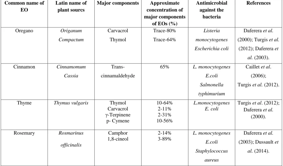
Spray drying is a one of the most commonly used microencapsulation technologies. It is being applied for both bioactive molecules and living probiotics. It is a fast and relatively cheap procedure that, when adequately performed, is highly reproducible. The principle of spray drying is dissolving the core in a dispersion of a chosen matrix or shell material. Figure 1.1 represents the spray drying method. The dispersion is subsequently atomized in heated air. This promotes fast removal of the solvent (water). The powdered particles are then separated from the drying air at the cyclone outlet at a lower temperature. The relative ease and also the low cost are the main reasons for the broad application of spray drying in industrial settings. This technique represents problems when utilized for probiotic encapsulation where the bacteria may leak with the product during hydration. It has been reported that bifidobacteria are very sensitive to high inlet temperatures (O'Riordan et al., 2001). Temperatures above 60˚ C interfered with survival of the bacteria and also influenced the spray drying process as sticky products were reported with this type of bacteria in the cyclone (Kailasapathy, 2002). Thus it would be advisable to investigate the sensitivity of a probiotic for increased temperatures before proposing spray drying as the technology of preference for a specific bacterium. But spray drying technology has many advantages over the other technologies for other bioactive food components such as EOs, bacteriocins, vitamins, minerals, flavors, unsaturated oils, and enzymes. Only water-based dispersions are applied in spray drying. Therefore the matrix should have a high solubility in water. In most instances hydrophilic carbohydrate molecules are applied. These carbohydrates undergo a transition to a so-called glass (i.e., an amorphous solid) when the dispersion is rapidly evaporated. Usually the product is very stable and allows for a significant increase in shelf life (Augustin and Hemar, 2009).
19
Figure 1.1: Schematic diagram of spray drying method.
1.3.3.2 Fluid Bed Drying
An improved spray dry technology that expands the field of application is the fluid bed coating methodology. In this technology, the bioactive food components are suspended in air and the matrix molecules are sprayed onto the bioactive components. This forms a capsule (Champagne and Fustier, 2007). The choice for matrix molecules is broader than for traditional spray drying. It may be fats, proteins, carbohydrates but also emulsifiers. Also it is useful for applying an additional layer of molecules for controlled release of bioactives during storage. In principle of fluid bed drying, the core materials should be always solid compounds. For example, this technology would be efficient for bacteriocin but not for volatile compounds like essential oils (De Vos et al., 2010).
20
1.3.3.3 Freeze Drying
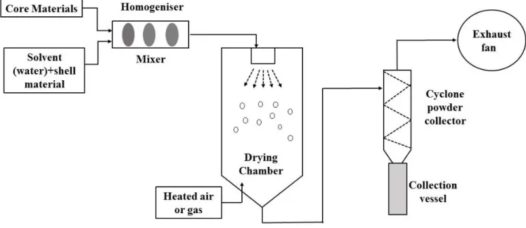
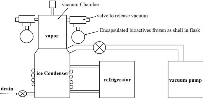
Freeze drying in combination with matrix molecules has been proposed as an alternative for spray drying of heat sensitive bacteria. A schematic representation of freeze drying method is presented in Figure 1.2. However, drying in general should not be considered to be a very efficacious methodology for preservation of bioactivity of living cells. Probiotics are exposed to damage from the process conditions such as very low freezing temperatures and dehydration. Cells are first frozen to below the critical temperature of the formulation, and then dried by sublimation under high vacuum in two phases: primary drying, during which unbound water is removed and secondary drying, during which the bound water is removed. These stages can damage the constituents of the cell wall and lead to cell death (Jalali et al., 2012). In the freeze drying technique, heat injuries to the cells are minimal compared with other techniques. Also, cryo protectants must be used to inhibit cold injuries to the cells (Augustin and Hemar, 2009) and microencapsulation using biopolymers can also protect the cells during drying (Chana et al., 2011). Figure 1.3 a) and b) represent the images of microencapsulated bioactives in alginate-CNC microbeads before and after freeze drying, respectively.
21
Figure 1.2: Freeze drying method
Figure 1.3: Microencapsulated bioactives a) before freeze drying and b) after freeze drying.
1.3.4 Compression Method
The compression coating method has been found as a novel encapsulation technique for improving the viability of living cells during storage and gastric treatment. Biopolymers (sodium alginate, pectin, gellan gum) which can form gels after being hydrated, have been exploited as the prime coating material. Probiotic containing powders are first compressed into a pellet, which is then encapsulated with the coating material by further compression. Chan and Zhang, (2002)
22
demonstrated this technology for probiotic encapsulation to keep its viability stable during gastric transition.
1.4 Biopolymers Used for the Microencapsulation of Bioactive Compounds
1.4.1 Microencapsulation in Alginate System
One of the most commonly applied polysaccharides is alginate. Alginates are linear polymers with 1-4 linked-β-D mannuronic acid (M) and ɑ-L-guluronic acid (G) residues arranged as blocks of
either type of unit or as a random distribution of each type. They can be obtained in different G/M ratios which provides different degrees of mechanical stability. Alginate is very compatible with most of the encapsulation technologies. Conventional extrusion method has been used to microencapsulate both antimicrobial and probiotic where sodium alginate is extruded in calcium chloride (CaCl2) solution. Anal and Singh, (2007) reported that probiotics encapsulated in
calcium-alginate beads are better protected, as shown by an increase in the survival of bacteria under gastric conditions, compared to the non-encapsulated state. The authors indicated that the viability of encapsulated bacteria in gastric fluid increased with an increase in capsule size. However, it was reported that very large calcium alginate beads (>1 mm) caused a coarseness of texture in live microbial feed supplements and that small beads of size less than 100mm in diameter do not significantly protect the bacteria in SGF, compared with free cells. These studies indicate that these bacteria should be encapsulated within a particular size range (Anal et al., 2003). In this wrok, alginate microbeads (20-70 µm) were prepared by the mixture of probiotic cells and sodium alginate in vegetable oil and subsequently cross-linked with CaCl2. It was found that the probiotic
loaded alginate microparticles remained stable during storage at 4ºC in 0.05 M CaCl2 and in milk
(2% fat), sour cream and yogurt for up to 16 days and in SGF (pH 2.0) for 1 h at 37ºC. Theyalso showed that B. bifidum survived in higher numbers in frozen milk in beads made from alginate