Regulation of kinetochore localization of the
Spindle checkpoint kinase Bub1
Thèse
Adeel Asghar
Doctorat en biologie cellulaire et moléculaire
Philosophiae doctor (Ph.D.)
Québec, Canada
Regulation of kinetochore localization of the
Spindle checkpoint kinase Bub1
Thèse
Adeel Asghar
Sous la direction de:
iii
Résumé
Le point de contrôle d’assemblage du fuseau mitotique (SAC) est un système de surveillance conservé chez les eucaryotes permettant un attachement précis entre les kinétochores et les microtubules. Le SAC empêche la progression mitotique jusqu'à ce que soit généré un attachement et une tension correcte entre les kinétochores et les microtubules. La dérégulation du SAC a des conséquences graves avec de l'aneuploïdie retrouvé dans la plupart des tumeurs solides. BUB1 est une kinase sérine/thréonine requise pour le fonctionnement du SAC. Elle possède à la fois des rôles dépendants et indépendants de sa fonction kinase. Ce projet définit plusieurs fonctions associées à BUB1 lors de la mitose. L'utilisation d’outils in vivo et in vitro ont permis d’identifier plusieurs sites d'autophosphorylation sur Bub1. Nous avons testé et confirmé le site T589 de BUB1 comme un site d'autophosphorylation. Un mutant de ce site (BUB1-T589A) a été exprimé de manière stable et un anticorps phosphospécifique a été généré pour étudier ce site.
Le rôle structural des domaines de BUB1 a été rapporté précédemment. Nous montrons que quand le domaine d'extension du domaine kinase (aa 724-780) située en N-terminal du domaine kinase est nécessaire pour l’autophosphorylation de BUB1-T589 et l'activité de la kinase BUB1, le TPR à l’extrémité N-terminale est localisée normalement kinétochores et n’est pas requis pour l'activité kinase. BUB1-T589A a modifié le taux de renouvellement au kinétochores. Cela conduit à la propagation des signaux de SGO1 et de H2ApT120 au niveau des bras des chromosomes. Enfin, l’autophosphorylation en T589 régule le congression des chromosomes mais pas la fonction de BUB1 pour le SAC.
De plus nous montrons que l'inhibition de PLK1, une autre kinase sérine/thréonine, augmente la localisation de BUB1 aux kinétochores après la suppression BUB3 dans les cellules humaines. Ainsi, PLK1 peut réguler la localisation de BUB1 aux kinétochores. Nous montrons également que cette régulation se produit à travers KNL1, une protéine d'échafaudage du SAC. PLK1 pourrait réglementer BUB1 kinétochore localisation pour influencer la
iv
progression mitotique. Des futures études se concentreront sur l’élucidation de mécanismes derrière ces interactions.
v
Abstract
The Spindle assembly checkpoint (SAC) is a monitoring system conserved in eukaryotes for accurate attachments between kinetochores and microtubules. The SAC precludes mitotic progression until correct attachments and tension between kinetochores and microtubules is generated. Deregulation of the SAC has the severe consequence of aneuploidy found in most solid tumors. BUB1 is a serine/threonine kinase required for the SAC function. It has both kinase-dependent and kinase-independent roles. This project defines several BUB1 associated functions during mitosis. Using in vivo and in vitro tools several autophosphorylation sites on BUB1 were identified. We tested and confirmed BUB1 T589 as an autophosphorylation site. A mutant of this site (BUB1-T589A) was stably expressed in cells and a phosphospecific antibody was generated to study this site.
The role of structural domains of BUB1 has been studied earlier. We show that while the kinase extension domain (aa 724-780) located N-terminal to the kinase domain is required for BUB1-T589 autophosphorylation and BUB1 kinase activity, the TPR at the N-terminus localizes normally to kinetochores and is not required for kinase activity. BUB1-T589A has altered turnover at kinetochores. This leads to the spread of SGO1 and H2ApT120 signal to chromosome arms. Finally, autophosphorylation at T589 regulates chromosome congression but not the SAC function of BUB1.
We further show that inhibition of PLK1, another serine/threonine kinase, increases BUB1 kinetochore localization after BUB3 depletion in human cells. Thus, PLK1 can regulate BUB1 kinetochore localization. We also show that this regulation occurs through KNL1, a scaffold protein of the SAC. It is possible that PLK1 could regulate BUB1 kinetochore localization to influence mitotic progression. Future studies will focus on elucidation of mechanism behind these interactions.
vi
Table of Contents
Résumé ... iii Abstract ... v Table of Contents ... vi List of Tables ... ix List of Figure ... x List of Abbreviations ... xi Foreword ... xiii 1. INTRODUCTION ... 11.1. Cell cycle and Checkpoints ... 1
1.1.1. G1 or gap1 phase ... 1
1.1.2. G1/S checkpoint ... 2
1.1.3. S phase (Synthesis phase) ... 3
1.1.4. G2 phase (Gap2 phase) ... 3
1.1.5. G2/M checkpoint ... 4
1.1.6. M phase (Mitosis) ... 4
1.2. Kinetochore- proteinaceous structures on chromosomes during mitosis ... 7
1.3. Spindle assembly checkpoint (SAC) ... 11
1.3.1. SAC monitors specific attachments between kinetochores and microtubules ... 11
1.3.2. SAC senses tension and kinetochore attachments ... 11
1.3.3. The strength of the SAC varies during mitosis ... 13
1.3.4. SAC Components ... 13
1.3.5. Mitotic checkpoint complex (MCC) ... 14
1.3.6. Molecular basis of APC/C inhibition and anaphase delay ... 16
1.3.7. APC/C activation and mitotic progression ... 19
1.3.8. SAC Silencing ... 19
1.3.8.1. SAC silencing through Phosphoregulation ... 19
1.3.8.2. Stripping of SAC components ... 20
1.3.8.3. MCC disassembly ... 22
1.3.9. Importance of SAC ... 23
1.3.10. Shugoshin-1 (SGO1) ... 26
1.3.10.1. Structure of SGO1 ... 26
1.3.10.2. Functions of SGO1 during mitosis ... 27
1.3.10.2.1. SGO1 acts as modulator of cohesin removal ... 27
1.3.10.2.2. SGO1 in chromosome biorientation ... 29
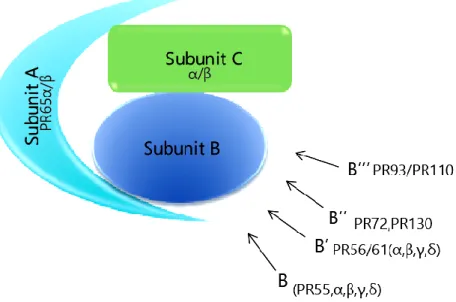
1.3.11. Protein phosphatase 2A (PP2A) ... 31
1.3.11.1. Structure of PP2A ... 32
1.3.11.2. Role of PP2A during mitosis ... 33
1.3.12. PLK1 (Polo-like kinase 1) ... 34
1.3.12.1. Functions of PLK1 in cell cycle ... 35
1.3.12.2. Function of PLK1 during mitosis ... 35
1.3.13. MPS1 (Monopolar spindle 1) ... 36
1.3.13.1. MPS1 functions ... 37
1.3.13.1.1. SAC function ... 37
1.3.13.1.2. Function at Centrosome ... 37
vii
1.4.1. Structure of BUB1 ... 39
1.4.2. BUB1 kinetochore recruitment ... 39
1.4.3. Regulation of BUB1 activation... 40
1.4.4. BUB1 Functions ... 42
1.4.4.1. BUB1 role in mitosis ... 42
1.4.4.2. BUB1 requirement for SAC ... 42
1.4.4.3. BUB1 kinase activity and SAC ... 42
1.4.4.4. BUB1 and recruitment of kinetochore components ... 44
1.4.4.5. BUB1 in chromosome congression, biorientation and segregation .... 47
1.4.4.6. BUB1 in Aneuploidy and Cancer development ... 48
1.5. Hypothesis and Objectives ... 50
2. CHAPTER2 ... 53
2.1. ABSTRACT ... 54
2.2. RÉSUMÉ ... 55
2.3. INTRODUCTION ... 56
2.4. RESULTS ... 57
2.4.1. Identification of Bub1 autophosphorylation sites ... 57
2.4.2. Regulation of Bub1 activation and autophosphorylation ... 61
2.4.3. Bub1 T589 autophosphorylation regulates mitotic progression ... 64
2.4.4. Bub1 autophosphorylation restricts H2A-pT120 to centromeres ... 67
2.4.5. Bub1-KD and -T589A display increased cytoplasmic residency ... 70
2.4.6. Kinetochore-tethered Bub1-T589A refocuses H2A-pT120 and Sgo ... 73
2.5. DISCUSSION ... 76
2.6. Materials and Methods ... 80
2.6.1. Cell culture and transfection ... 80
2.6.2. Chromosome Spreads ... 80
2.6.3. Cloning and mutagenesis ... 81
2.6.4. Immunofluorescence and antibodies ... 81
2.6.5. Fractionation, immunoprecipitation and Western Blotting ... 82
2.6.6. Microscopy, Live cell imaging, and FRAP ... 82
2.6.7. SILAC labelling and Mass spectrometry ... 83
2.6.8. Nano-LC-MS/MS Analysis ... 84
2.6.9. Data processing and analyses ... 85
2.6.10. Phosphopeptide analysis ... 86
2.6.11. Quantification and statistical analysis ... 86
2.7. ACKNOWLEDGEMENTS: ... 87 2.8. Author Contributions: ... 88 2.9. REFERENCES: ... 89 2.10. Supplementary Information ... 96 3. CHAPTER 3 ... 101 3.1. Introduction ... 102
3.2. Hypothesis and objectives ... 103
3.3. Results ... 103
3.3.1. PLK1 inhibition causes rescue of SGO1 localization in BUB1 T589A ... 103
3.3.2. PLK1 inhibition enhances BUB1 localization in Bub3 depleted HeLa cells104 3.3.3. PLK1 regulates BUB1 localization through KNL1 ... 107
3.4. Discussion and Perspectives ... 110
viii
3.5.1. Cell culture, transfection and drug treatment ... 113
3.5.2. Immunofluorescence and Microscopy ... 113
3.6. Refernces ... 115
4. CHAPTER 4 ... 118
4.1. Discussion and Perspectives ... 118
4.2. Current understanding of Bub1 activity in SAC ... 119
4.3. References ... 122
ix
List of Tables
Table1 ...47 Table S1...100
x
List of Figure
Figure1.1 Four phases of cell cycle ...2
Figure 1.2 Stage of mitosis ...5
Figure 1.3 kinetochore is a trilaminar multiprotein complex ...8
Figure 1.4 Assembly of Kinetochore proteins ...9
Figure 1.5 Attachments monitored by SAC ...12
Figure 1.6 SAC signal prevents mitotic progression ...14
Figure 1.7 MCC formation ...16
Figure 1.8 Inhibition of APC/C ...18
Figure 1.9 SAC silencing by Phosphoregulation ...20
Figure 1.10 SAC silencing by stripping ...21
Figure 1.11 Disassembly of MCC...23
Figure 1.12 Structure of human Sgo1 ...27
Figure 1.13 SGO1 and cohesin removal during mitosis ...29
Figure 1.14 SGO1 role in biorientation ...30
Figure 1.15 PP2A hologenzyme ...33
Figrue 1.16 PLK1 structure and activation ...34
Figure 1.17 MPS1 recruits SAC proteins at KNL1...38
Figure 1.18 Structural domains of human BUB1 ...40
Figure 2.1 Identification of Bub1 autophosphorylation sites ...59
Figure 2.2 Full Bub1 activation is mitotic specific and requires the kinase extension domain ...62
Figure 2.3 Loss of Bub1 phosphorylation at T589 causes chromosome congression defects...66
Figure 2.4 Uniform H2A-T120 phosphorylation, ectopic Sgo recruitment and impaired sister chromatid resolution in cells expressing Bub1-T589A ...68
Figure 2.5 Bub1-KD and Bub1-T589A display increased residency in the cytosol .71 Figure 2.6 Bub1-KD and Bub1-T589A display aberrant kinetochore shuttling dynamics ...74
Figure 2.7 Model of Bub1 activation and autoregulation ...75
Supplementary Figure 1. Characterization of the pT589 and pS679 Bub1 antibodies ...96
Supplementary Figure 2. Characterization of the isogenic MYC-GFP Bub1 WT, KD and T589A HeLa cell lines ...97
Supplementary Figure 3 Aurora recruitment and activation are normal in Bub1-T589Aexpressing cells ...98
Supplementary Figure 4. Original non-cropped Western Blots presented in this manuscript ...99
Figure 3.1. PLK1 and AURORA B inhibition recovers SGO1 loss from centromeres while H2ApT120 remains unchanged in BUB1 T589A cells ...105
Figure 3.2. PLK1 inhibition recruits cytosolic BUB1, SGO1 and H2ApT120 in BUB3 depleted cells ...106
Figure 3.3. PLK1 inhibition impairs KNL1 binding and MELTs phosphorylation at kinetochores ...108
Figure 3.4. BUB1 localization is regulated by PLK1 at KNL1 ...109
xi
List of Abbreviations
APC/C Anaphase promoting complex/cyclosomeaa Amino acid
ATM Ataxia telangiectasia mutated
ATR Ataxia telangiectasia and Rad3-related protein Bub1 Budding uninhibited by benzimidazole 1
BuBR1 Bub1-related protein 1
CCAN Constitutive Centromere-Associated Network Cdc Cell division cycle
Cdk Cyclin-dependent kinase CENP Centromere protein
CPC Chromosomal passenger complex D-Box Destruction Box
FRAP Fluorescence recovery after photo-bleaching GLEBS Gle2-binding sequence
Hec1 Highly enhanced in cancer
KD Kinase Dead
KEN-box Lys-Glu-Asn-box
KNL1 kinetochore null protein 1 Mad Mitotic arrest deficient MELT Met-Glu-Leu-Thr
MCAK Mitotic associated Kinesin MCC Mitotic checkpoint complex Mis12 Minichromosome instability 12 Mps1 Monopolar spindle 1
Ndc80 nuclear division cycle protein 80 PBD Polo-box domain
Plk1 Polo-like kinase 1 PP1 Protein phosphatase 1 PP2A Protein Phosphatase 2A SAC Spindle assembly checkpoint
SILAC Stable isotope labeling in cell culture siRNA Small interfering RNA
TPR TetratricoPeptide Repeat
xii
“Take a moment to think about the context in which your next decision will occur: You did not pick your parents or the time and place of your birth. You didn't choose your gender or most of your life experiences. You had no control whatsoever over your genome or the development of your brain. And now your brain is making choices on the basis of preferences and beliefs that have been hammered into it over a lifetime - by your genes, your physical development since the moment you were conceived, and the interactions you have had with other people, events, and ideas. Where is the freedom in this? Yes, you are free to do what you want even now. But where did your desires come from?” -Sam Harris, Free Will 2012
xiii
Foreword
I would like to express my sincere gratitude to my supervisor Dr. Sabine Elowe who welcomed me in her lab and guided me throughout my PhD research work at CHUL. I am grateful for the time and knowledge she shared with me during my research. I am also deeply indebted to Audrey Lajeunesse, a former master’s student, for her support and guidance in the lab when I started this program. Also, her initial observations on Bub1 shaped my research project. I want to say many thanks to Philippe Thebault for this help in the lab. Dr. Danielle Caron provided her support and help on numerous occasions for which I am very thankful. I especially thank Guillaume Combes for his friendship, support and ideas during these four years. I will not forget others who have helped me in one way or another. I want to say my sincere thanks to Luciano Braga Gama, Michelle Mathieu and Abrahim Alharbi for their support and friendship. Finally, I want to thank to those who participated in my research project in some way. I may not know them however, without their support this work could not have been accomplished.
The thesis presents analyses and results obtained during my Ph.D on the Bub1 kinase. The results describe the identification of autophosphorylation sites on Bub1 which was done prior to start of my Ph.D. The characterization and observation of Bub1 T589 site was done with the help of my colleagues, especially Audrey Lajeunesse. For better reading, I have divided this thesis into 4 chapters. The first chapter, the introduction, provides general discussion on past and recent literature. This chapter describes Cell cycle and its checkpoints. Then, I have given a brief summary of kinetochores since SAC activity takes place on them. Next, I have explained SAC and its importane which is followed by relevant proteins for this project: Sgo1, PP2A, Plk1 and Mps1. I have discussed past and recent literature on Bub1 in detail. Finally, I have discussed the Hypotheses and aims of this doctoral thesis.
xiv
Chapter 2 presents already published work on the Bub1 autophosphorylation site T589. The article titled “Bub1 autophosphorylation feeds back to regulate kinetochore docking and promote localized substrate phosphorylation” (Nature Communications 6, Article number: 8364 doi: 10.1038/ncomms9364) was published on 24 September 2015. I have contributed for this research article in Figure 2.1 (b,c,d), Figure 2.4 (a-d), Figure 2.5 (c-f), Figure 2.6 (b-e), Supplementary Figure 1 d, Supplementary Figure 2, Supplementary Figure 4 (except 2e). Chapter 3 provides unpublished initial results on regulation of Bub1 localization by Plk1. I have provided results in the form of a mini article. Fourth and final chapter presents discussion and future directions for research work Bub1.
1
1. Introduction
1.1. Cell cycle and Checkpoints
Most among billions of cells in human body remain in a reversible quiescent state (1, 2). The rest, cycle to duplicate their genomic content and divide into two daughter cells (3, 4). The cell cycle can be broadly divided into two phases: the actively dividing mitosis or M phase and non-dividing interphase (Figure 1.1). The cell cycle can also be divided into four distinct phases of G1, S, G2 and M (mitosis) phase (3, 5). The traverse through each phase is governed by regulatory protein complexes of cyclin dependent kinases (CDKs) and cyclins (6). A number of CDKs have been discovered e.g. CDK1, CDK2, CDK4 and CDK6 which are activated by specific cyclins e.g. cyclin B, cycline D, and Ccclin A during each phase of cell cycle. Cyclin expression is regulated throughout cell cycle to allow for controlled activation of CDKs (7-9). To protect cells from external influence that may induce errors, cells have developed “checkpoints” to regulate cell cycle timing for correction of errors (10, 11). Checkpoints are defined as “mechanisms by which the cell actively halts progression through the cell cycle until it can ensure that an earlier process, such as DNA replication or mitosis, is complete” (12).
There are three major checkpoints in eukaryotes: the G1/S checkpoint, the G2/M checkpoint and the spindle assembly checkpoint (SAC). The transition from G1 to S is monitored by G1/S checkpoint while the passage from G2 to M is controlled by G2/M checkpoint. The final major checkpoint, the SAC, lies within M phase.
1.1.1. G1 or gap1 phase
Cells enter G1 after cell division (3). During G1 phase cells synthesize proteins necessary for the next cell cycle phase called the S phase. The cells may also enter a reversible quiescent state called G0 or resting phase (5). G1 progression is regulated by CDK4/6-cyclin D and CDK2/cyclin E (9). The progression through early G1 is controlled by the regulation of retinoblastoma (Rb), a tumor suppressor protein involved in the repression of transcription factors e.g. E2F (13). Kinases CDK4 or 6, after activation by cyclin D, phosphorylate retinoblastoma (Rb) to dissociate it from histone deacetylase
2
(HDAC) proteins causing the repression of retinoblastoma and activation of E2F transcription factor to allow for gene transcription required for S Phase initiation (14).
Figure1.1 Four phases of cell cycle: G1, S, G2 and M shown with their respective activities and checkpoints. During G1 cells start to mature and grow in size. It is during G1 that cells decide whether to continue cell cycle or go into a quiescent state called G0. G0 is reversible and cells can enter cell cycle upon return of external growth factors. After G1, cells enter S phase during which DNA replication occurs. After S phase cells have made a copy of their DNA which will allow equal distribution of genomic content into daughter cells during cell division. Next, the cells enter G2 phase where they grow in size and prepare for cell division. Before entry into M phase, G2/M checkpoint checks for DNA damage. The cells enter M phase during which their genomic content is equally divided into two daughter cells, thus completing cell cycle. SAC, the third checkpoint, delays mitotic progression until chromosomes are correctly captured by microtubules during mitosis. Modified from (15)
1.1.2. G1/S checkpoint
The transition from G1 to S is monitored by G1/S checkpoint also known as Restriction -(R) point in mammalian cells and START in yeast (16). Removal of growth factors before this point brings cells into G0 or quiescent state but
3
beyond that point withdrawal of growth factors does not affect the cell cycle as the cell becomes committed to cell cycle completion (17, 18). Rb proteins play a crucial role and prevent entry into S phase by transcription factor inhibition, however, after the decision to enter S phase, CDK4 or 6/Cyclin D and CDK2/cyclin E complex phosphorylate Rb to inactivate it and promote activation transcription factor E2F for S phase entry (19-21). During G1, DNA damage resulting in double stranded breaks activates the ATM (Ataxia telangiectasia mutated) kinase that phosphorylates CHK2 and p53 and represses the activation of CDK4/6-cyclin D and CDK2/cyclin E. In case of UV exposure DNA damage, the ATR (Ataxia telangiectasia and Rad3 related) complex phosphorylates its substrates that include CHK1 which in turns phosphorylates CDC25a phosphatase and causes its degradation by ubiquitination, hence CDC25a cannot activate phosphorylated CDK2 required for cell cycle progression (12, 22).
1.1.3. S phase (Synthesis phase)
During S phase, DNA replication synthesizes another copy of its DNA that will be received by one of the two daughter cells after mitosis. E2F transcription factor activity is required for synthesis of products to enter S phase (23), while activation of CDK2 by cyclin A or E is required for the initiation of DNA synthesis and progression from late G1 to S phase (24). The termination of DNA replication is mediated by a negative feedback loop in which E2F transcription factor helps synthesize cyclin A that activates CDK2 (6, 25, 26). Near the end of S phase, CDK2/cyclin A phosphorylates E2F1 factor for its dissociation from DNA and termination of DNA replication (6, 27).
1.1.4. G2 phase (Gap2 phase)
After the successful DNA replication in S phase, cells enter G2 phase to prepare for cell division (19). CDK1/cyclin A is required for cell cycle progression during G2 and is implicated in the activation of CDK1/cyclin B required for mitotic progression (28, 29). During G2, CDK2/cyclin A and CDK1/cyclin B phosphorylate a transcription factor FoxM1 to relieve its inhibition for gene expression required during mitosis (9).
4
1.1.5. G2/M checkpoint
The G2/M checkpoint or DNA damage checkpoint monitors DNA damage at the end of S phase before cells can divide their genetic material in M phase, thus allowing time for DNA repair (30). The target of G2/M checkpoint is CDK1 whose activation is required for mitotic entry (30, 31). Kinases WEE1 and MYT phosphorylate CDK1 leading to its inactivation, while the phosphatase CDC25 reverses this phosphorylation for activation by cyclin B (32). Upon DNA damage, both ATM and ATR pathways are activated depending on the nature of DNA damage (33). Phosphorylation of CDC25 phosphatase by ATM and ATR kinases binds it to 14-3-3 for its sequestration in cytoplasm and degradation. Meanwhile, WEE1 kinase phosphorylates CDK1 to keep it inactivated and cell cycle arrest is achieved (34, 35).
1.1.6. M phase (Mitosis)
M phase or mitosis is an important step for equal distribution of genetic material i.e. chromosomes, into two daughter cells (Figure1.2) (36). The progression of M phase is monitored by CDK1/cyclin B also known as the mitotic promotion factor/maturation promoting factor (MPF) (8). CDK1/cyclin B kinase function promotes chromosome condensation, spindle generation and nuclear envelope breakdown (37).
There are five distinct steps of mitosis: prophase, prometaphase, metaphase, anaphase and telophase followed by cytokinesis for cytoplasmic content division into two daughter cells (Figure 1.2)(4). Prophase is the first distinct phase of mitosis identified by condensation of chromatin, a complex of DNA and its proteins (38). Further compaction of chromatin results in the formation of chromosomes which are held together by a protein complex known as cohesin (39). Cohesin is wrapped around chromatids during S phase and acts as a “glue” to keep sister chromatids together (Figure 1.2). It is removed from chromosome arms during prophase and later from centromeres during anaphase (40-42). At later stages of mitosis, chromosomes are captured by microtubules, the hollow cylindrical structures, arising from two centrosomes which are the microtubule organizing centers consisting of a pair of centrioles (Figure 1.2a) (43, 44). During prophase,
5
centrosomes start moving to the opposite poles and microtubules start to radiate from them.
The nuclear envelope is broken down during prometaphase to allow microtubules to access chromosomes to align them in the center by search and capture, a process during which microtubule growth and shrinkage allows them to search and bind special proteinaceous structures on centromeres called kinetochores (Figure 1.2b) (45, 46). Search and capture is a complex process that also requires motor proteins kinesin activity for kinetochore-microtubule interactions (45, 47, 48). The next stage, metaphase follows (Figure 1.2c) during which microtubules bind kinetochores from opposite poles in a bi-oriented manner and chromosomes align in the center of the cell (49, 50). Tension is generated due to pulling force of microtubules from opposite ends and resistance of cohesion between sister chromatids, thus stabilizing chromosomes in the middle.
Figure 1.2 Stage of mitosis: a) Prophase is the first stage of mitosis. During this phase the chromatin becomes compact and takes the shape of a visible chromosome. The chromosomes appear to have x-shape connected by centromeres consisting of sister chromatids, the two identical copies forming a chromosome. The two centrosomes from opposite pole have their microtubule nucleation increased forming a dynamic spindle. b) Prometaphase: The nuclear envelop breaks down and microtubule can access chromosomes. Microtubules start to attach chromosomes at kinetochores. c) During Metaphase chromosomes after attaching with microtubules form metaphase plate, a plane perpendicular to the spindle, in the middle. d) Once
6
correct kinetochore-microtubule attachments are established the sister chromatids are separated during anaphase. e) The separated sister chromatids reach opposite poles and nuclear membrane starts to form around them during telophase. f) The cytoplasmic content of the cell are divided into two daughter cells during cytokinesis, thus completes division of
genetic and cellular contents. Modified from
http://www.nature.com/scitable/topicpage/mitosis-and-cell-division-205#
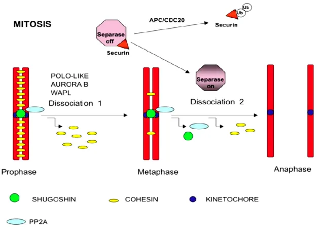
Anaphase ensues after correct chromosome alignment in the center. The sister chromatids are separated by cleavage of cohesin around them (Figure 1.2d) (51). There are two stages of anaphase: A and B (52). During Anaphase A, kinetochore microtubules (kMT) are shortened due to depolymerisation resulting in a force that drives poleward movement of separated sister chromatids. During anaphase B, centrosomes move further toward the periphery which pulls separated chromatids apart even more (53, 54). The separated chromatids reach opposite poles which marks the last step, the telophase (Figure 1.2e). The nuclear envelope starts to form around them and the newly separated chromosomes start to decondense (55). The final step, cytokinesis separate and equally divide cytoplasmic content into two nascent daughter cells thus, completing the cell division (Figure 1.2f).
7
1.2. Kinetochore- proteinaceous structures on chromosomes
during mitosis
Kinetochores are proteinaceous structures formed on chromosomes during mitosis to mediate binding between chromosomes and microtubules, thus are crucial for proper chromosome segregation (55, 56). They are assembled on special regions on chromosomes called centromeres (57, 58). Specifically, kinetochores assemble on inner centromeres composed of DNA and proteins that are collectively called constitutive centromere-associated network (CCAN), and are required for kinetochore assembly (59, 60). Microtubule bind kinetochores during mitosis and the number of microtubules binding to each kinetochore vary among organisms. While in budding yeast (Saccharomyces cerevisiae) only one microtubule binds a kinetochore (61, 62), it is estimated that approximately 15-30 microtubules attach each kinetochore in humans (63). The number of microtubules also determines the strength of SAC on each kinetochore in organisms where more than one microtubule binds to a kinetochore (64).
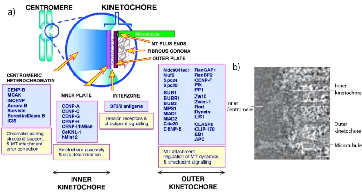
Vertebrate kinetochores appear to have a “trilaminar” structure (Figure 1.3a, b) i.e. having three distinct layers: the inner, the middle and the outer layer (65, 66). The inner kinetochore (inner plate) is located next to the inner centromere. The middle layer follows to the exterior and outer kinetochore (outer plate) is first to encounter kinetochore microtubules (kMT). Fibrous structures emerging away from outer kinetochores are called the fibrous corona (67).
There are about 100 kinetochore proteins identified in humans (55, 68). The outer kinetochore layer (Figure 1.3) contains SAC proteins that include MAD1 (mitotic arrest deficient 1), MAD2 (mitotic arrest deficient 2), RZZ ( Rod-Zwilch-ZW10) complex, CENP-E (Centromere Protein-E) MPS1(monopolar spindle 1), BUB1(budding uninhibited by benzimidazoles 1), BUBR1(BUB1-related 1) and BUB3 (budding uninhibited by benzimidazoles 3) as well as KMN network proteins: KNL1 (kinetochore null protein 1) complex, MIS12 (mis-segregation 12) complex or MIS12C and NDC80 (nuclear division cycle protein 80) complex or NDC80C (Figure 1.3) important for microtubule binding and spindle assembly checkpoint (69).
8
Figure 1.3 Kinetochore is a trilaminar multiprotein complex: a) Kinetochores are trimlaminar structures i.e. having three layers, an inner, middle and outer region. They host a variety of proteins required for its assembly and microtubule binding. The CENP proteins at the inner kinetochores are required for kinetochore structure and assembly while the outer kinetochore contains SAC proteins for correct attachment kinetochore-microrubule attachments. Image from (69). b) Electron micrograph showing trilaminar human kinetochore bound to microtubules. Scale bar, 100 nm. Modified from (55).
KMN network proteins are composed of several subunits. KNL1 forms a complex with ZWINT (Zeste white 10 interactor). The MIS12 complex is composed of 4 subunits: MIS12, NSL1, DSN1 and NNF1 while the NDC80C also contains 4 subunits: SPC24, SPC25, NUF2 and NDC80 (70). The inner plate hosts various CENP proteins e.g. CENP-A, CENP-C, CENP-H/-I, required for kinetochore structural integrity (69, 71, 72).
Once at kinetochores, KMN proteins bind stably at kinetochores which are important for various functions during mitosis (72). The Spc24 and Spc25 heterodimer is required for end-on kinetochore attachment whereas NDC80
9
and NUF2 contact microtubules (Figure 1.4) (73, 74).The NDC80C interacts with MAD1 to promote its recruitment and also supports recruitment of MAD2, MPS1 and RZZ, and therefore, acts as a scaffold for checkpoint protein recruitment (75-78).
Figure 1.4 Assembly of kinetochore proteins: Kinetochores assembled on the inner centromere contain CCAN proteins. In vitro analyses have shown that CENP-T forms a complex with CENP-W,-S, and –X required for DNA supercoiling. NDC80C is recruited through interaction with CENP-T and also binds microtubules. NDC80C also binds DAM1 complex (SKA in humans) through its loop region. Together with NDC80C, SKA complex is required for stable kinetochore-microtubule binding. CENP-A connects inner kinetochore components and CCCENP-AN network via CENP-C. CENP-C recruits MIS12C at kinetochores. Once at kinetochores, MIS12C acts a scaffold for KNL1 and NDC80C. KNL1 also acts as scaffold for core SAC proteins and forms interactions with microtubule via its N-terminus. Image from (58).
MIS12C binds directly with centromeric DNA and CCAN to promote its recruitment and acts as a scaffold for KNL1C and NDC80C to mediate kinetochore assembly (Figure 1.4) (68, 74). KNL1 is recruited to kinetochores by MIS12C where it heterodimerizes with ZWINT and is required for
10
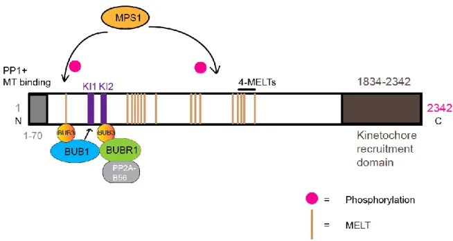
recruitment of CENP-F and protein phosphatase PP1 (68, 79-81). In vitro analyses have shown that KNL1 can bind microtubules that could lead to SAC silencing (74, 82). In addition to microtubule binding, KNL1 is a well-known anchor for components of SAC that include BUB3, BUB1 and BUBR1, thus promote SAC activation (79, 83). More specifically, KNL1 is a target of MPS1 kinase whose phosphorylation is required to recruit these proteins which is explained in more detail in MPS1 section (84-87).
11
1.3. Spindle assembly checkpoint (SAC)
The SAC also known as mitotic or spindle checkpoint is a surveillance mechanism that delays mitotic progression until correct attachments between kinetochores and microtubules are established (88-90). For this reason, the SAC signal is emitted from unattached kinetochores to ensure accurate attachments and equal distribution of chromosomes into daughter cells and thus it preserves genomic integrity (88-91).
1.3.1. SAC monitors specific attachments between kinetochores
and microtubules
The attachments between microtubules and kinetochores are very specific to allow for equal division of chromosomes i.e. each kinetochore must bind microtubules only from one pole (amphitelic attachment) to achieve bi-orientation (92-94) (Figure 1.5). However, incorrect attachments may occur which include syntelic attachments, when both kinetochores of a chromosome are attached to microtubules from the same pole; merotelic attachments occur when kinetochores attach microtubules from both poles. Merotelic attachments eventually segregate chromosome normally but when they persist till anaphase they can cause aneuploidy (95-97). Monotelic attachments predominantly prevail during prometaphase before bi-orientation (92, 93). Hence, the goal of SAC is to achieve amphitelic attachments or bi-orientation between chromosomes and microtubules.
1.3.2. SAC senses tension and kinetochore attachments
The establishment of bi-orientation causes tension across kinetochores due to microtubule driven pulling forces from opposite poles which stabilizes these attachments (98). Therefore, nature of SAC regarding tension and attachment has been a matter of debate (99). Pioneering work on kinetochore-microtubule attachments discovered that kinetochores emitted a “wait anaphase signal” in the presence of mono-oriented chromosomes that can be relieved by laser induced destruction of unattached kinetochore suggesting that SAC senses attachment at kinetochores (100). Meanwhile, studies in preying mantid spermatocytes revealed that tension exerted by micromanipulator needle on misoriented chromosome reduced mitotic delay (101). Later, micro-needle manipulation studies demonstrated that a
mono-12
oriented chromosome created from bi-oriented chromosome lost tension however, the number of microtubules on the sister kinetochores were also reduced (102). A recent study in budding yeast using isolated kinetochores and their interaction with microtubules concluded that tension increases the stability of kinetochore-microtubule attachments (98). Hence, tension encourages stable kinetochore-microtubule attachments and the SAC responds to both tension and kinetochore-microtubule attachments (99).
Figure 1.5 Attachments monitored by SAC: a) Monotelic attachments occur in prometaphase before amphitelic attachment is achieved. b) Syntelic attachments are attachments when both kinetochores bind microtubules from the same pole. c) The amphitelic attachments are correct attachments between chromosomes and microtubules needed for genomic integrity during which two sister kinetochore on a chromosomes are captured by microtubule from opposite poles, thus each kinetochore experiences tension from one centrosome. d) Merotelic attachments occur when one kinetochore is bound by microtubules from both centrosomes. Image from (97) .
13
1.3.3. The strength of the SAC varies during mitosis
Recent studies have shown that SAC is more dynamic than it was believed to be and functions more like a rheostat than a toggle switch i.e. having variation in its strength rather than working in an on/off fashion (103, 104). Previously, it was demonstrated that a single kinetochore was enough to arrest cells in mitosis for “wait anaphase” signal (100). However, in agreement with the rheostat model recent mouse studies have shown that after depletion of CENP-E, a protein required for stable kinetochore-microtubule binding, SAC strength diminishes and cells exit mitosis in the presence of one or few unattached kinetochores suggesting that the strength of SAC is graded (103). Studies by laser microsurgery for chromosome detachment in human cells revealed that individual chromosomes did not impose SAC efficiently compared to controls in which complete spindle disruption occurred (105). Furthermore, a recent study concluded that APC/C (anaphase promoting complex/cyclosome) strength can modulate SAC in humans i.e. the strength of APC/C can dictate the SAC strength (106). APC/C is activated by CDC20 (cell division cycle 20) to promote mitotic exit, whereas MCC (mitotic checkpoint complex) inhibits APC/C and mitotic exit. CDH1(CDC20 homolog 1) is another APC/C adaptor protein that is required for the activation of APC/C; however it is not needed for mitotic exit for it is required in G1 phase (107) and thus will not be discussed in this manuscript. Analyses of APC/C and MCC revealed that MCC can bind two CDC20 molecules to inhibit APC/C activation pointing to a rapid response upon reactivation of spindle assembly checkpoint (108). Data in C.elegans embryogenesis have demonstrated that SAC strength depends on cell size rather than development stage (109). In summary, above studies clearly show that SAC does not work in a switch like manner on the contrary SAC strength is graded.
1.3.4. SAC Components
SAC components were discovered in the 1990s in budding yeast genetic screens in which cells could not arrest in mitosis when challenged with spindle poisons to induce spindle damage (110-112) . It was suggested that failure to arrest in these conditions i.e. spindle damage, which normally
14
induce mitotic arrest was a result of a dysfunctional checkpoint and the mutant genes isolated were components of that checkpoint (110). The core mitotic checkpoint proteins include BUB1, BUBR1 and BUB3 (110, 111, 113); MAD1 and MAD2 (111, 114, 115), MPS1 (112), PLK1 (polo like kinase 1) (116, 117), AURORA B, RZZ complex (118, 119) and CENP-E (120). BUB1 and MAD1 are stably bound kinetochore proteins while others e.g. BUBR1, MPS1 are exchanged frequently (89, 121). The main target of SAC is APC/C, a multiprotein E3 ubiquitin ligase (89, 108, 122, 123). SAC members converge on unattached kinetochore to orchestrate MCC formation required for inhibition of APC/C activation (122, 124) (Figure 1.6).
Figure 1.6 SAC signal prevents mitotic progression: SAC proteins accumulate at unattached kinetochores. Assembly of core SAC proteins MPS1, MAD1, MAD2, BUB1, BUB3, and BUBR1 promotes the formation of an inhibitory complex, MCC (Mitotic checkpoint complex) to prevent activation of APC required for mitotic progression. APC/C requires CDC20 cofactor for its activation. As a result mitosis is halted by SAC proteins until unattached kinetochores achieve correct attachments. Modified from (57)
1.3.5. Mitotic checkpoint complex (MCC)
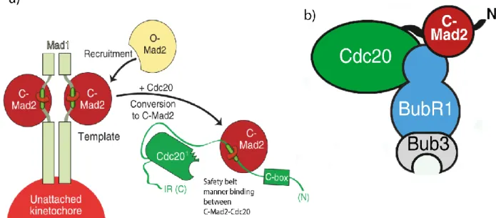
MCC is composed of checkpoint proteins MAD2, BUBR1, BUB3 and CDC20 (122, 125, 126). Both MAD2 and BUBR1 can directly bind CDC20 (122, 124). The “template model” of MAD2 activation (Figure 1.7a) is the contemporary model that explains MCC formation and inhibition of APC/C activation (127).
15
MAD2 has the ability to adopt two distinct topological conformations: Open MAD2 (O-MAD2) and Closed MAD2 (C-MAD2) (128, 129). O-MAD2 is cytosolic, inactive and cannot bind to MAD1 or CDC20, thus cannot inhibit APC/C activation (130, 131). The other conformation, C-MAD2, is active and bound MAD2 conformation that makes adjustments in its carboxy-terminal β-sheet to wrap around and lock MAD1 or CDC20 in a “safety belt” manner. Therefore, this region of MAD2 is termed as safety belt (Figure 1.7a) (132-134). A kinetochore bound MAD1-C-MAD2 complex catalyzes the conformational change of inactive O-MAD2 to an intermediate MAD2 (I-MAD2), a transition state, before it can become active C-MAD2, capable of binding CDC20 (129-131, 135). In vitro studies showed that CDC20-C-MAD2 complex is identical to MAD1-C-MAD2 complex and can promote conversion of O-MAD2 to C-MAD2 in the cytosol like MAD1-C-MAD2 does on unattached kinetochores (127, 136). However, C-MAD2 surface in the cytosol is involved in interactions with BUBR1 or p31comet, a SAC silencing protein (137, 138) and may not provide for conformational conversion of MAD2 (129, 139). Finally, BUBR1-BUB3 also joins CDC20 and C-MAD2 (Figure 1.7b) and completes the formation of MCC (124, 129, 138).
The SAC kinases, MPS1 and BUB1 also contribute to the formation of MCC. It has been shown that MPS1 inhibition abrogates MCC formation and BUB1, MAD1, MAD2 and BUBR1 localization is reduced (140). Furthermore, MPS1 kinase function recruits O-MAD2 to kinetochore bound MAD1-C-MAD2 and inhibition of MPS1 leads to the eviction of MAD2 from partially intact MCC (CDC20-BUBR1) causing SAC defects (140-143). In budding yeast, MPS1 phosphorylates BUB1 to promote BUB1-MAD1 complex at kinetochores (144). BUB1 kinase is also implicated in the recruitment of MAD1 and MAD2 at kinetochores to facilitate MCC formation (145-148). BUB1 phosphorylates MAD1 in vitro (149), however, a number of studies have reported that while BUB1 is required for MAD1 recruitment its kinase activity is redundant for this function (145, 148, 150, 151). Both MPS1 and BUB1 are required for the recruitment of BUBR1 at unattached kinetochores and thus contribute in MCC formation (140, 146).
16
Figure 1.7 MCC formation: a) MAD2 exists in two conformations: O-MAD2 (Open MAD2) and C-MAD2 (Closed MAD2). MAD2 in C-MAD2 conformation can bind MAD1 and CDC20. MAD1-C-MAD2 complex at unattached kinetochores recruits O-MAD1 and converts into C-MAD2 that can bind CDC20. The interaction of O-MAD2 with CDC20 is facilitated by MAD1-MAD2 complex. b) CDC20-C-MAD2 becomes part of a functional MCC by incorporation of BUBR1/BUB3 complex that binds between CDC20 and C-MAD2 and forms simultaneously interactions with CDC20 and C-C-MAD2. Modified from (88).
1.3.6. Molecular basis of APC/C inhibition and anaphase delay
Rapidly exchanging CDC20 at kinetochore has major structural domains that include seven WD40 domains arranged into a β-propeller structure for protein-protein interactions; a C-Box; a KEN (lysine-glutamate-asparagine) box; a CRY box (Cysteine, Argnine, Tyrosine); a MAD2 interacting motif (MIM); and a C terminal IR (isoleucine-arginine) tail (152, 153). The N-terminus, C-Box and C-terminus IR-tail are required to bind CDC20 with APC/C (134, 154). KEN and CRY boxes are involved in regulation of CDC20 stability (155, 156).
17
Protein degradation is regulated through recognition of degradation signal or degron defined “as a minimal element within a protein that is sufficient for recognition and degradation by a proteolytic apparatus” (157). CDC20 has dual activity towards APC/C i.e. it acts as a suppressor when bound to MCC and as an activator of APC/C near the start of anaphase (122, 152, 154, 158). When acting as an activator of APC/C, CDC20 recruits substrates and activates APC/C through recognition of two degrons called destruction (D-box) box and KEN box of substrates which allows for their destruction by ubiquitylation (126, 152, 156, 159, 160).
CDC20 acts an inhibitor of APC/C when present in complex with C-MAD2 and BUBR1. It blocks substrate recognition sites thus preventing APC/C activation (124, 161). The C-terminal “safety belt” of C-MAD2 wraps around CDC20 MIM, a region that overlaps with APC/C binding region (124, 162-164). A recent study showed that MAD2 can bind to a short conserved motif known as KILR (Lysine-Isoleusine-leucine-argnine) present in MIM required for APC/C activation and thus competes with APC/C for CDC20 binding (134). Mutagenesis studies have shown that MIM can separate activator and repressor function of CDC20 (88). MIM mutant unable to bind MAD2 overrides the SAC and has efficient APC/C activity (162, 165). C-MAD2 also promotes BUBR1-CDC20 binding and acts synergistically with BUBR1 for APC/C inhibition (125, 166). However, BUBR1:CDC20 complex can be formed without MAD2 suggesting MAD2-CDC20 acts as diffusible amplifier of BUBR1-CDC20 to sustain MCC activity (167-170).
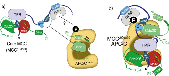
BUBR1 interacts with both CDC20 and C-MAD2 (Figure 1.8)(88). Through its N-terminus region, it binds two conserved residues (Arg133 and Gln134) of C-MAD2 for inhibition of APC/C (138), while its two KEN boxes, KEN1 and KEN2 have been studied for interaction with CDC20 (160, 171-174). BUBR1 KEN1 box is essential for MCC formation and inhibiting APC/C activity by blocking KEN degron binding on CDC20, thus acting as a pseudosubstrate inhibitor of CDC20 (124, 160, 175). However, a recent study in human cells has shown that mutating KEN1 box does not affect CDC20 kinetochore recruitment although it severely reduces MCC formation (176). Instead
18
another region termed as internal CDC20 binding domain (IC20BD) (aa 490– 560) that encompasses a Phe box (contains two phenylalanines) and a D-box binds CDC20 and promotes SAC silencing (176). The second BUBR1 KEN box (KEN2) is required for inhibition of activated APC/C-CDC20 (124, 160). It was confirmed recently that KEN2 along with D-box is required to bind a second CDC20, thus forming MCC2Cdc20 for rapid inactivation of activated APC/C
(Figure 1.8)(108). Studies have confirmed that the BUBR1 middle region (BUBR1M) containing D-box and an ABBA (Cyclin A, BUBR1, BUB1 and Acm1) motif, also known as Phe box or A box binds CDC20 for maintaining MCC (161, 177). In summary, inactivation of APC/C requires extensive interactions of BUBR1 and C-MAD2 with CDC20.
Figure 1.8 Inhibition of APC/C: a) BUBR1 KEN box motifs occupy CDC20 KEN1 box recognition motif thus blocking its activity toward APC/C substrate. BUBR1 also binds to MAD2 via its N-terminus TPR region KEN box motif. C-MAD2 binds CDC20 through “safety belt” thus; forming a Core MCC with only 1 CDC20 that can bind to APC/C for its inhibition. b) BUBR1 has a second KEN box that binds a second CDC20 and forming an MCC with 2 CDC20 molecules (MCC2Cdc20) for effective inhibition of
APC/C. The binding of a second CDC20 may also help when rapid inactivation of APC/C is needed after its initial activation. Modified from (88).
19
1.3.7. APC/C activation and mitotic progression
Upon accurate kinetochore attachments, SAC is turn-off and APC/C is activated by CDC20 (Figure 1.8) (89, 129, 178). Activated APC/C-CDC20 promotes transition from metaphase to anaphase through two key regulatory events that involve destruction of APC/C substrates: a) APC/C polyubiquitylates cyclin B for its degradation to inactivate CDK1. b) APC/C polyubiquitylation of securin, an inhibitor of separase enzyme, for its destruction (Figure 1.8). Separase enzyme then cleaves cohesin between two sister chromatids to separate them (88, 89, 159).
1.3.8. SAC Silencing
1.3.8.1. SAC silencing through Phosphoregulation
SAC activity is immensely promoted by the activity of kinases, thus its inactivation must require phosphatase activity (Figure 1.9). For this reason, a tight balance between phosphorylation and dephosphorylation has been observed during mitosis (179). SAC proteins orchestrate their own removal from kinetochores by recruiting phosphatases through a negative feedback mechanism (180, 181). Several studies have highlighted the role of PP1 (Protein phosphatase 1) and PP2A (Protein phosphatase 2A) in the regulation of SAC (80, 180-186).
The studies in budding and fission yeast have shown that PP1 activity is required for SAC silencing by reversing the phosphorylation needed for SAC activation (182, 183). PP1 binds at KNL1 N-terminus conserved SILK (Serine, Isoleucine, Leucine and Lysine) and RVSF (Argnine, Valine, Serine, Proline) motifs to negatively regulate the recruitment of BUB1 and other downstream SAC proteins thus, promotes SAC silencing (80, 184, 185, 187).
BUBR1 binds PP2A for regulation of mitotic progression and kinetochore-microtubule attachments (188-190). Kinetochore recruitment of BUBR1-PP2A complex is promoted by MPS1 and PLK1 kinase activity at KNL1 (84, 85, 191-194). Studies in human cells have demonstrated that PP2A removes KNL1 phosphorylation needed for SAC activation, thus can efficiently counter SAC activation (181). Recent studies have proposed that the negative feedback mechanism sufficiently explains rapid SAC On/Off when an opposing
20
phosphatase is already bound to core SAC protein but it will pose a problem for an efficient SAC signaling (195). For this reason, negative feedback phosphatase PP2A does not directly antagonize SAC at KNL1, instead it opposes AURORA B substrate phosphorylation that promotes PP1 binding to KNL1 which then promotes SAC silencing that eventually removes PP2A-B56 form kinetochores (Figure 1.9) (180, 195, 196). This added layer of control may provide enough temporal separation between SAC activation and silencing (195).
Figure.1.9 SAC silencing by Phosphoregulation: Phosphorylation of KNL1 by MPS1 helps mount SAC by recruiting BUB3 that binds BUB1 and BUBR1 which later recruit downstream SAC proteins including MAD1, MAD2 and CDC20 etc. BUBR1 binds PP2A-B56 phosphatase and recruits it to kinetochores. AURORA B activity counteracts PP1 kinetochore localization at SILK and RVSF motifs. Upon correct attachments, phosphatase activity of PP2A opposes AURORA B activity leading to PP1 binding to KNL1. PP1 then antagonizes MPS1 activity thus, removing BUB3, BUB1, BUBR1 and PP2A-B56 from kinetochores. In this negative feedback mechanism PP2A promotes PP1 recruitment that effectively silences SAC. Overall, SAC proteins recruited by kinase function, bring phosphatases for their own regulation at kinetochore leading to SAC silencing. Modified from (195).
1.3.8.2. Stripping of SAC components
Localization of core SAC proteins MAD1, MAD2, BUB1, BUBR1 and BUB3 is reduced at kinetochores during anaphase providing evidence that SAC proteins are removed from kinetochores for SAC silencing (113, 115,
197-21
201). Stripping or physical removal of SAC proteins has been proposed to achieve SAC silencing via the minus-end directed microtubule motor Dynein complex (202, 203).
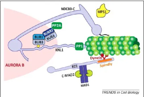
Dynein binds its cofactor Dynactin, a multisubunit activator complex, required for its motor function (204-207). In addition, Dynein-dynactin complex requires RZZ, a complex essential for functional SAC, and Spindly, a Dynein recruitment factor, for its kinetochore binding (Figure 1.10) (208-211).
Figure 1.10 SAC silencing by stripping: Dynein, a motor protein complex, is recruited to kinetochores through interactions with RZZ and Spindly and moves towards minus ends (polewards) on microtubules. Dynein complex is important for stripping of MAD1-MAD2 and therefore also for silencing the SAC. Retention of Spindly suppresses SAC silencing for this reason Dynein mediated removal of Spindly and RZZ is required for SAC silencing. Image from (212).
MAD1 and MAD2 are carried towards microtubule minus-ends by Dynein-Dynactin complex after accurate kinetochore-attachments as inhibition of Dynein complex leads to retention of residual MAD2 and persistent checkpoint activity (213-215). Although, these studies explain SAC protein stripping from kinetochores, others have suggested that stripping is more likely an auxiliary pathway for SAC silencing because the depletion of human Spindly causes Dynein recruitment defects yet MAD1, MAD2, BUBR1, Zwilch, and CENP-E are still removed and SAC is silenced suggesting a
Dynein-22
independent SAC silencing mechanism (210, 214). Interestingly, mutants of Spindly that impaired Dynein recruitment and localized normally to kinetochores still had MAD1, MAD2 and RZZ complex and defective SAC silencing (214). Therefore, SAC silencing occurs only after removal of Spindly, suggesting that Spindly may suppress the SAC protein stripping mechanism, and removal of both Spindly and RZZ by Dynein is crucial for SAC silencing (214, 216).
1.3.8.3. MCC disassembly
MCC disassembly provides another way of SAC silencing. p31comet was
discovered in yeast as a MAD2 partner whose overexpression caused premature securin destruction leading to precocious mitotic exit and its RNAi-mediated depletion promoted anaphase onset delay (217, 218). p31comet can
bind C-MAD2 on the same surface where BUBR1 or O-MAD2 binds, thus p31comet promotes MCC disassembly and SAC silencing (Figure 1.11) (137,
219). A “capping model” has been proposed in which p31comet caps C-MAD2 in
MAD1-C-MAD2 complex and interferes with the recruitment of O-MAD2 (137, 220, 221). However, later studies suggested that capping model is not efficient in explaining SAC silencing as depletion or overexpression of p31comet
does not change O-MAD2 levels at kinetochores (222, 223). p31comet also
binds C-MAD2 in complex with CDC20 to cause disruption of MCC complex and activation of APC/C (Figure 1.11) which is considered more plausible for p31comet mediated SAC silencing (222, 224). p31comet binding to MAD2 also
encourages conformational changes in CDC20 that leads to CDK phosphorylation of CDC20 and promotes its dissociation from BUBR1 (225). Recent analyses of MCC disassembly points to a two-step process: BUBR1 is released from MCC by p31comet that binds to the same surface on C-MAD2 and
requires CDK activity. In the next step CDC20-C-MAD2 subcomplex disassembly requires p31comet and ATPase Thyroid Receptor Interacting
23
Figure1.11 Disassembly of MCC: a) MAD1-C-MAD2 complex recruits O-MAD2 to convert it into C-MAD2 by binding to CDC20 and effectively promotes APC/C inhibition. b) During SAC silencing, p31comet competes with MAD2 and interferes in
the process of O-MAD2 recruitment to MAD1-C-MAD2 thus inhibits the conversion of active C-MAD2. p31comet also directly binds to CDC20 and antagonizes MAD2 binding,
thus inhibiting MCC formation (modified from (219).
1.3.9. Importance of SAC
Aneuploidy refers to “an abnormal karyotype that is not a multiple of haploid complement” (227). It is reported that aneuploidy is present in over 90% of tumors and has the potential to cause cancer (228, 229). While the SAC ensures genomic integrity due to its ability to halt the progression of mitosis in case of incorrect microtubule attachments with kinetochores, its deregulation has been implicated in aneuploidy and tumorigenesis reviewed in (230, 231). Partial loss of the SAC manifested in deregulation of SAC protein expression contributes to chromosome instability rather than complete loss of SAC (232, 233). A complete loss of the SAC has been implicated in embryonic mortality at early stages which seems to be conserved in Drosophila and mouse models. Studies involving a null MAD2 resulted in embryonic death and initial studies on Drosophila showed embryonic mortality in BUB1 null embryos (234, 235). Thus, the CIN effects are only detected in partially defective checkpoint perhaps due to requirement the SAC for cell survival during early development. One of the earliest studies on SAC reported that mutations in BUB1 contributed to chromosomal instability (CIN) (236). The mutations of BUB1 paralog BUBR1
24
have been associated with growth defects and cancers (237). Moreover, altered MAD2 expression in breast cancer has been reported (238). The SAC genes overexpression is also reported as a cause of aneuploidy. Upregulation of Bub1, Bub3/BubR1 has been reported in breast and gastric cancers respectively (239). Mad2 overexpression has also been shown to promote aneuploidy (240). Studies have reported a role of parallel signaling pathways that can contribute to altered SAC gene expression. Studies in human cells have shown that mutations in proto-oncogene p53 could alter the expression of Mad1 SAC genes (241, 242). The overexpression of SAC components induces persistent mitotic arrest, change in mitotic timing and merotelic attachments (reviewed in (232). Kinase function of bona fide mitotic kinases has also been implicated in progression of cancer. For example, MPS1 kinase is shown to be overexpressed in many human cancers and may promote proliferation and survival of tumor cells (243-245). Overexpression of AURORA kinases can promote polyploidy and chromosome instability while overexpression of PLK1 has been associated with human cancer and weak prognosis (reviewed in(246). MPS1 inhibition in combination with drugs affecting microtubules known as microtubule targeting agents (MTAs) has been suggested as anti-cancer strategy due to increased in chromosome segregation defects that cannot sustain survival of cancer cells. Indeed, MPS1 inhibition in combination with low doses of Taxol drug has been effective in tumor cell sensitization (247). BUB1 kinase function has been shown to promote cell growth through transforming growth factor-β (TGFβ) receptor and BUB1 inhibition severely affected TGFβ pathway in human cancer cells (248). Therefore, inhibition of these core SAC kinases by chemical inhibitors provides an attractive strategy against tumor proliferation and growth and is a subject of intense current research.
SAC components that include BUB1, MPS1 and MAD2 are also involved in the control of accurate division during meiosis, a process of germ cell division (249-252). Similar to mitosis, knockdown of MAD2 and BUB1 leads to precocious meiosis I, chromosome misalignment and aneuploidy in mouse (253) and BUB1 depletion causes centromeric cohesion defects in both mouse and yeast (252, 254). Also mutations in Bub1 gene cause age-related aneuploidy in mouse (255). Studies on human oocytes have shown that
25
younger females had better chromosome alignment than older females and showed reduced expression of Bub1 and Mad2 genes that could causes age-related aneuploidy leading to birth defects such as Down’s syndrome (256, 257). Despite the role SAC role in meiosis, it is suggested that the SAC of meiosis is less robust than the SAC of mitosis because the cells with a misaligned X chromosome spends the same time in meiosis I as in controls in female mice whereas a single unattached kinetochore is sufficient to halt mitotic progression (258). In summary, studies mentioned above underline the importance of SAC as a defense against cellular anomalies.
26
1.3.10. Shugoshin-1 (SGO1)
The equal distribution of chromosomes into daughter cells dictates that sister chromatids remain together till anaphase during which they are separated and move towards opposite poles (259, 260). The Mei-S332 gene in Drosophila was defined as protector of chromosome cohesion during meiosis and mutants of mei-S332 had premature loss of centromeric cohesion (261). Mei-S322 homologs were later discovered in budding and fission yeast as protectors of cohesion and named “Shugoshins” (meaning guardian spirit in Japanese) (262-264). Among eukaryotes, fission yeast and humans have two SGO proteins: SGO1 or SGOL1 (Shugoshin-Like 1) and SGO2 or SGOL2 (Shugoshin-Like 2), whereas budding yeast and Drosophila have only one SGO protein (265, 266). During fission yeast meiosis, SGO1 is only required for meiosis I whereas, SGO2 paralog is associated with centromere region in both cell divisions of meiosis and mitosis (260, 266, 267). In humans, although both SGO1 and SGO2 are present, SGO1 is primarily required for cohesion protection during mitosis as depletion of it causes chromosome missegregation defects (268-271). BUB1 kinase activity is required for proper localization of SGO1 because preventing BUB1 kinase activity mislocalizes SGO1 from centromeres. Moreover, SGO1 forms a complex with PP2A (protein phosphatase 2A) and localizes to centromeres in BUB1 kinase dependent manner for protection of cohesion (more detail in BUB1 section) (269, 270, 272-274).
1.3.10.1. Structure of SGO1
Human SGO1 is encoded by Sgo1 gene is a paralog of SGO2 protein (264). It contains an N-terminal coiled coil (CC) domain and a C-terminal SGO region (Figure 1.12) (264). CC domain and SGO are conserved throughout eukaryotes, whereas other motifs are variable (260). SGO1 CC is required for PP2A binding and depletion or mutation in this region impairs SGO1-PP2A interaction (270). K492 (Lysine 492) of SGO recognizes and binds Histone H2A phosphorylated by BUB1 at T120 (275). Thus, CC and SGO region are required for PP2A and H2A binding respectively. SGO1 also has a KEN box (aa 310-312) and three D-boxes: D-Box1 aa 192-200, D-Box-2(aa 438-446) and D-Box3 (aa 457-465) (276, 277). The D-and KEN-Boxes are implicated
27
in SGO1 degradation by APC/C during mitotic exit (276). Finally, SGO1 has a CDK1 phosphorylation site at T346 (Threonine 346) required for cohesin binding. Therefore, SGO1 T346 mutant is unable to bind cohesin and has defective chromosome cohesion protection (275).
Figure.1.12 Structure of human SGO1: N-terminus Coiled coil (CC) domain is a PP2A binding site. SGO1 is phosphorylated by CDK1 at conserved motif T346 required for SGO1 cohesin interaction. KEN box and D-box serve to regulate SGO1 expression and are likely recognition site for APC/C ubiquitylation. C-terminus Basic region “SGO motif” contains K492 residue needed for H2ApT120 interaction. SGO and CC motifs are conserved in yeast, drosophila, mice, xenopus and humans. Adapted from (267, 275, 276).
1.3.10.2. Functions of SGO1 during mitosis
1.3.10.2.1. SGO1 acts as modulator of cohesin removal
Cohesion between sister chromatids is required to achieve accurate attachments between kinetochores and microtubules. Therefore, cohesion plays a crucial role for proper distribution of chromosomes in daughter cells (278, 279). The cohesion between sister chromatids depends on cohesin proteins assembled onto chromosomes during S phase (39, 280). Cohesin comprises SMC1, SMC3 (structural maintenance of chromosome 1 and 3), a Kleisin subunit SCC1 (Sister chromatid cohesion protein 1)/RAD21 and SCC3 subunit (SA in animal cells, REC8 in meiosis) (36, 41, 281). The presence of cohesin is essential to keep a balance between tension generated due to kinetochore-microtubule attachment and cohesion on sister chromatids (282). While the cohesin complex is central to chromosome cohesion, other
28
cohesin-associated proteins are also required for cohesion including PDS5 (precocious dissociation of sisters protein 5), WAPL (wings apart-like), a protein needed for cohesin removal in prophase and Sororin, a protein required for maintaining cohesin. Vertebrate PDS5 binds to cohesin to maintain and establish sister chromatid cohesion (283, 284). Sororin forms a complex with PDS5 to stabilize cohesin on chromosomes by opposing WAPL and depletion of Sororin causes sister chromatid cohesion loss (285, 286). WAPL also interacts with PDS5 leading to assembly of a cohesin releasing complex called “releasin” for cohesin removal from chromosome arms in prophase (287-289). Recent evidence has shown that WAPL and Sororin share a conserved motif required for PDS5 binding and thus WAPL and Sororin antagonize each other through binding competition to regulate chromosome cohesion (290, 291).
In vertebrates, cohesin assembly at chromosomes is temporally regulated by a two-step sequestration of cohesin from chromosomes in mitosis to allow for preservation of chromatin integrity (Figure 1.13) (42, 292). A large portion of cohesin is removed from chromosome arms but retained at centromeres to prevent premature sister chromatids separation (293, 294). The removal of cohesin in this step is separase and cleavage independent and SCC1 remains unaffected (295). The second removal of cohesin depends upon entry into anaphase and APC/C activation and results in cleavage of SCC1 by separase which removes cohesin from centromeres (295).
Phosphorylation catalyzed by kinases PLK1, CDK1 and AURORA B, is the molecular trigger governing the first step removal of cohesin from chromosome arms (39, 296-298). During this step, SGO1-PP2A helps maintain centromere PDS5-Sororin complex by preventing Sororin phosphorylation and therefore, inhibits PDS5-WAPL formation and cohesin removal (273, 298). The SGO1-PP2A complex also counters PLK1 activity towards SA2 (Scc3 homolog 2) to prevent cohesin removal (268, 292). Finally, SGO1 competes with WAPL for SA2 binding counters WAPL binding, and thus acts as protector of cohesion during mitosis (267, 292, 299).
29
Figure 1.13 SGO1 and cohesin removal during mitosis: During prophase cohesin binds chromosome along the arms and on centromeres. Most of the cohesin is removed from the arms by the end of prophase due to phosphorylation of cohesin subunit SA2 by kinases PLK1 and AURORA B and CDK1. WAPL binds cohesin at PDS5 (not shown) and destabilizes cohesin. At centromeres, the presence of SGO1-PP2A is sufficient to counteract phosphorylation of kinases due to presence of PP2A hence; SGO1-PP2A complex protects cohesin at centromeres. By the end of metaphase, when correct attachments between kinetochores and microtubules are established, SGO1-PP2A complex is delocalized as a result of SAC silencing. APC/C is activated and destroys separase enzyme inhibitor securin by promoting its ubiquitlyation to release separase which cleaves cohesin. Image from (300).
1.3.10.2.2. SGO1 in chromosome biorientation
Tension generated between sister kinetochores due to resistance of cohesin in response to microtubule pull is an indicator of establishment of chromosome biorientation (93, 267). Lack of kinetochore-microtubule attachments due to cohesin defects promotes the activity of AURORA B kinase to remove defective kinetochore-microtubule attachments and activate SAC to stabilizes PDS1(securin), an indicator of APC/C inactivation (301). SGO proteins have been described as sensors of tension at kinetochores and