I
Université de Montréal
Impact of genetic polymorphisms determining leukocyte/neutrophil count on chemotherapy toxicity
par
Sanja Joksimovic Glisovic Département de Pharmacologie
Faculté de Médecine
Mémoire présenté à la Faculté de Médecine
En vue de l’obtention du grade de Maîtrise science (M.Sc.) en Pharmacologie
option Pharmacogénomique
Décembre 2015
II
Université de Montréal Ce mémoire intitule :
Impact of genetic polymorphisms determining leukocyte/neutrophil count on chemotherapy toxicity
présenté par : Sanja Joksimovic Glisovic
a été évalué par un jury composé des personnes suivantes : Dr. René Cardinal, président-rapporteur
Dr. Maja Krajinovic, directeur de recherche Dr. Yves Pastore, codirecteur
III
RÉSUMÉ
Nous avons investigué la relation entre les polymorphismes de nucléotides simples (SNPs) chez trois gènes/loci candidats : DARC, CXCL2 et le loci ORMDL3-GSDMA-CSF3 situés sur le chromosome 17q21 et les complications neutropéniques et infectieuses qui en résultent durant la chimiothérapie chez les patients atteints de la leucémie lymphoblastique aigue. Ces loci codent pour certaines composantes du système immunitaire altérant la concentration de chémokines et leur distribution (DARC), stimulant le relâchement et la migration des neutophiles de la moelle épinière (CXCL2) et régulant la prolifération et la survie des granulocytes (G-CSF). Il est possible que des polymorphismes dans ces loci lorsqu’associés à de la chimiothérapie puissent mettre des individus suceptibles à un risque plus élevé de complication reliées à la chimiothérapie. Une sélection des marqueurs SNPs dans ces gènes ont été génotypés chez des enfants traités au CHU Ste-Justine pour une ALL entre 1989 et 2005. Après correction pour tests multiples, un polymorphisme DARC rs3027012 situé dans le 5’UTR a été associé à un compte phagocytaire peu élevé (APC<500 et <1000 cellules/µL, p=0.001 and p=0.0005, respectivement) ainsi qu’une hospitalisation due à une neutropénie (p=0.007) ou due à une infection et/ou neutropénie (p=0.007). Un effet protecteur a été identifié pour la mutation non sense Gly42Asp variant rs12075 (p=0.006). Des polymorphismes sur le chromosome 17q2 étaient associés à une hospitalisation due à une infection (rs3859192, p= 0.004) et à une neutropénie (rs17609240, p=0.006) L’infection était aussi modulée par CXCL2 (rs16850408, p=0.008) Cette étude identifie pour la première fois que les loci modulant le décompte des leucocytes et des neutrophiles pourraient jouer un rôle dans de déclenchement de complications dues à la chimiothérapie et pourraient ainsi servir de marqueurs pour un ajustement et un suivi du traitement.
IV
ABSTRACT
We investigated the relationship between single nucleotide polymorphisms (SNPs) in 3 candidate genes/chromosomal loci: DARC, CXCL2 and ORMDL3-GSDMA-CSF3 locus on chromosome 17q21, and neutropenic and infectious complications during chemotherapy in pediatric acute lymphoblastic leukemia (ALL) patients. These loci encode the components of immune system altering chemokine concentration and distribution (DARC), stimulating neutrophil release from bone marrow and migration (CXCL2) and regulating granulocyte proliferation and survival (G-CSF). It is possible that polymorphisms of these loci when associated with chemotherapy may put susceptible individuals at higher risk of chemotherapy complications. Selected tag SNPs in these genes are genotyped in children treated at the CHU Sainte-Justine for (ALL) between 1989 and 2005. After correction for multiple testing, DARC polymorphism rs3027012 in 5’UTR was associated with low absolute phagocyte count (APC<500 and <1000 cells/µL, p=0.001 and p=0.0005, respectively) and hospitalisation due to febrile neutropenia (p=0.007) or due to infection and/or febrile neutropenia (p=0.007). A protective effect was instead noted for missense Gly42Asp variant rs12075 (p=0.006). The polymorphisms on chromosome 17q2 were associated with hospitalisation due to infection (rs3859192, p= 0.004) and neutropenia (rs17609240, p=0.006). Infection was also modulated by CXCL2 (rs16850408, p=0.008) This study identifies for the first time that the loci modulating white blood cell and neutrophil count may play a role in the onset of chemotherapy complications and may thus serve as markers for adjustment or follow-up of the treatment.
V
TABLE OF CONTENTS
RÉSUMÉ ... III ABSTRACT ...IV TABLE OF CONTENTS ... V LIST OF TABLES ... VII LIST OF FIGURES ... VIII LIST OF ABBREVIATIONS ...IX ACKNOWLEDGEMENT ...XI
CHAPTER 1: INTRODUCTION ... 1
A. Acute Lymphoblastic Leukemia (ALL) ... 1
A.1. Introduction to ALL ... 1
A.2. Etiology of ALL ... 2
A.3. Prognostic factors ... 4
A.4. Treatment of ALL ... 6
B. Pharmacogenomics ... 8
B.1. Introduction to pharmacogenomics ... 8
B.2. Role of pharmacogenomics ... 9
B.2.1. Polymorphisms that influence drug pharmacokinetics ... 10
B.2.2. Polymorphisms that influence drug pharmacodynamics ... 11
B.2.3. Combination of polymorphisms that influence pharmacokinetics and pharmacodynamics ... 12
B.2.4. Genetic Polymorphisms with Indirect Effects on Drug Response ... 13
B.3. Genetic variants ... 14
B.4. Association studies ... 14
B.4.1. The genome wide association study (GWAS) ... 15
B.4.2. Candidate gene study ... 16
B.5. Genotyping ... 16
C. Neutropenia ... 17
C.1. Introduction to neutropenia ... 17
C.2. Chemotherapy-induced neutropenia and infection ... 19
VI
D. Gene selection ... 21
D.1.1. Candidate genes and polymorphisms on chromosome 17q21 ... 22
D.1.2. G-CSF and its role ... 23
D.1.3. ORMDL3 and physiological function ... 24
D.1.4. GSDMA and its role ... 25
D.2.1. CXCL2 gene ... 25
D.2.2. CXCL2 and its role ... 26
D.3.1. DARC gene ... 27
D.3.2. DARC and its role ... 28
CHAPTER 2: HYPOTHESIS AND OBJECTIVES ... 31
CHAPTER 3: PRESENTATION BY ARTICLE ... 32
CHAPTER 4: DISCUSSION ... 53
VII
LIST OF TABLES
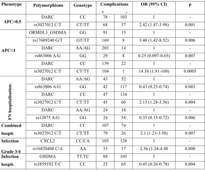
Table 1. The polymorphisms associated with neutropeniac and infectious complications ... 41 Supplementary Table 1. Identity of polymorphisms, details of PCR and ASO hybridisation..45
VIII
LIST OF FIGURES
Figure A. Estimated frequency of specific genotypes in childhood ALL ... .6 Figure B. Allele Specific Oligonucleotide Hybridization ... .17 Figure 1. The polymorphisms in DARC and GSDMA genes in relation to the frequency of neutropenia and duration of hospitalisation. ... .43 Supplementary Figure 1. Genomic structure and selected tag SNPs of neutropenia pathway genes ... ………44
IX
LIST OF ABBREVIATIONS
ALL: Acute Lymphoblastic Leukemia AML: Acute Myeloid Leukemia ANC: Absolute Neutrophil Count APC: Absolute Phagocyte Count ASO: Allele Specific Oligonucleotide CEU:Northern Europeans from Utah CI: Confidence Interval
CNS: Central Nervous System CR: Complete Remission
CSF3: Colony Stimulating Factor gene
CTCAE: Common Terminology Criteria for Adverse Events (CTCAE), DARC: Duffy antigen Receptor for Chemokines
DFCI: Dana Farber Cancer Institute EC: Endothelial Cells
EFS: Event -Free Survival
FDA: Food and Drug Administration FDR: False Discovery Rate
FN: Febrile Neutropenia
G-CSF: Granulocyte-Colony Stimulating Factor GSDMA: Gasdermin protein
GWAS: Genome Wide Association Study HR: High Risk
X LD: Linkage Disequilibrium
MAF: Minor Allele Frequency
MCP-1: Monocyte Chemoattractant Protein-1 MLL: Mixed Linkage Leukemia
MTX: Methotrexate OR: Odds Ratio
ORMDL3: Orosomucoid 1-like 3 protein OS: Overall Survival
PPBL2: Pro-Platelet Basic protein-like 2 SNP: Single Nucleotide Polymorphisms SR: Standard Risk
TGN: Thioguanine Nucleotide
TPMT: Thiopurine S-Methyl Transferase TRM: Treatment Related Mortality TS: Thymidylate Synthase
TSS: Transcription Start Site VTE: Venous Thromboembolism WBC: White Blood Cell
XI
ACKNOWLEDGEMENT
I would like to thank my research director Dr Maja Krajinovic and my co-director Dr Yves Pastore, for the patient guidance, encouragement and advice they have provided throughout my time as their student.
I would also like to thank all the members of Dr Krajinovic laboratory, especially, Aziz Mohamed Rezgui, Maria Plesa, Rasheed Obaji and Vincent Gagné for their support and friendship.
I would also like to acknowledge all members of the jury for their valuable comments on this thesis.
I must express gratitude to my son Andrej and husband Petar, for providing me with unfailing support and continuous encouragement throughout my years of study and through the process of researching and writing this thesis. This accomplishment would not have been possible without them. Thank you.
1
CHAPTER 1: INTRODUCTION
A. Acute Lymphoblastic Leukemia (ALL) A.1. Introduction to ALL
Pediatric cancers affect approximately 1 in 500 children before the age of 15 years and are the leading cause of death by disease in this population.
Acute lymphoblastic leukemia (ALL) is the most common pediatric cancer (26%) and the main type of leukemia. ALL accounts for 80% of childhood leukemia [Ward et al., 2014] Leukemia is a biologically and clinically heterogeneous disease of the blood cells with many different types and subtypes. The important characteristics of acute leukemia is the rapid growth of abnormal white blood cells that accumulate in the bone marrow and interfere with the production of normal blood cells.
The acute leukaemia is divided by morphological and cytochemical criteria into myeloid and lymphoid subtype. For acute leukemia, the flow cytometry is the method of choice for determining the blast lineage that has been affected.
Lymphoid malignancies can be classified by cell surface antigen phenotype as derived from either T or B cell lineage [Amylon et al., 1999]. Further, ALL has been classified into precursor T, precursor B, and B-cell (Burkitt) phenotypes, which are then further subdivided according to recurrent karyotypic abnormalities, including aneuploidy and translocations.
T-acute lymphoblastic leukemia (T-ALL) is a neoplasm of immature T-cell precursors or lymphoblasts and accounts for 10% to 15% of newly diagnosed cases of childhood acute lymphoblastic leukemia (ALL) [Onciu, 2009]. Historically, T-ALL patients have had a worse
2
prognosis than other ALL patients. Patients with T-ALL remain at increased risk for induction failure, early relapse, and isolated CNS relapse [You et al., 2015].
In about 80% to 85% of children with ALL, the leukemia starts in B cells. There are several subtypes, and the most common is precursor B-cell (B-lymphoblastic) leukemia. Children diagnosed with B-precursor ALL have a good prognosis [Smith et al., 1996].
Mature B-cell ALL (also called Burkitt leukemia) is rare, accounting for only about 2% to 3% of childhood ALL [Meinhardt et al., 2010].
Acute myeloid leukemia (AML) is the second-most common form of leukemia in children. AML comprises almost 20% of pediatric leukemia [Ries et al., 1999].
In the World Health Organization (WHO) classification, the term “myeloid” includes all cells belonging to the granulocytic (neutrophil, eosinophil, basophil), monocyte/macrophage, erythroid, megakaryocytic and mast cell lineages. A minority of children have ambiguous or mixed lympho-myeloid phenotypes [Vardiman et al., 2009].
ALL could affect children of all ages, however the highest incidence is seen between ages 2 and 5 years [Pui et al., 1998]. The incidence in children under the age of five years old was 5.7 per 100,000 person years, followed by the 5- to 9-years old (2.7) and the 10- to 14-years old (1.6) [Siesling et al., 2003].
A.2. Etiology of ALL
Epidemiological studies of leukemia have examined a number of possible risk factors (environmental, genetic, and infectious) in an effort to determine the etiology of the disease. Various factors have been reported to confer an increased risk for this disease, including high birth
3
weight, parental occupation, maternal reproductive history, parental tobacco or alcohol use, maternal diet, exposure to pesticides or solvents and exposure to the highest levels of residential, power-frequency magnetic field [Hjalgrim et al., 2003; Buckley et al., 1989; Infante-Rivard et al., 2005].
Recent studies have also suggested that early exposure to infection may be protective to childhood ALL [Urayama et al., 2011]. To date, however, no direct gene-environment interactions have been established convincingly.
Certain genetic conditions as Down syndrome, Bloom syndrome and ataxia-telangiectasia are associated with an increased risk for acute leukemia. [Fung et al., 1987; Taylor et al., 1996]. A higher risk exists for other less common chromosomal abnormalities including Klinefelter's syndrome, trisomy G, neurofibromatosis, and Schwachmann's syndrome [Margolin et al., 2002]. There are a number of constitutional chromosomal abnormalities associated with childhood leukemia. Chromosomal abnormalities and translocation could have an important role in the genesis of leukemia, and they also have prognostic and therapeutic implication. All patients with ALL can be classified according to specific genetic abnormalities (Fig A). [Pui et al., 2012].
Recurrent chromosomal abnormalities are a hallmark of lymphoblastic leukemia. The most common translocation found in childhood B-precursor ALL is the t(12;21)(p13;q22). The presence of this translocation has been demonstrated in approximately 25% of childhood ALL [Yeoh et al., 2002]. The TEL-AML1 fusion protein generated by the t(12;21) contains the basic helix-loop-helix domain of TEL, fused to the DNA-binding and transactivation domains of AML1. Both the TEL and AML1 genes are found in other leukemia-associated translocations. TEL-AML1 expression is associated with an excellent prognosis, with event-free survival rates approaching 90% [Rubnitz et al., 1999].
4
Another common chromosomal aberration found in B-precursor ALL is the presence of more than 46 chromosomes (hyperdiploid ALL). Patients with this aberration have a very good prognosis with event-free survival rates near 90%.
The t(1;19)(q23;p13) encoding the E2A-PBX fusion protein is present in about 6% of all B-precursor leukemia.
The most chromosomal translocations in T-ALL patients lead to inappropriate activation of structurally intact cellular proto-oncogenes such as MYC, TAL1, HOX11 or LMO2. Although some translocations can produce fusion genes. MLL-ENL fusion results from the translocation t(11;19)(q23;p13), and is associated with AML, B-cell precursor ALL, and T-ALL. The translocation encoding the BCR-ABL fusion protein is found in only 5% of childhood cases [Pui et al., 1995].
A.3. Prognostic factors
The treatment of childhood acute lymphoblastic leukemia has advanced significantly over the past 3 decades, with overall survival rates progressing from 20% to 75%. The identification of biologic and clinical prognostic factors allowed the definition of patient subgroups with distinct relapse risks and the realization of risk-adapted treatment strategies. This improvement can be attributed, in part, to “risk-adapted therapy”. Risk is defined based on clinical and biologic variables, cytogenetic and immunophenotypic characteristic of leukemic cells. Treatment intensity is then modified according to expected event- free survival (EFS) to maximize cure while minimizing toxicity [Silverman et al., 2010].
Age at diagnosis, WBC count, gender, and DNA index have strong prognostic effect. Age and WBC count are ideal parameters as these are always available. Therefore, in the classification of
5
the National Cancer Institute, standard risk (SR) is defined as age 1-9 years old and WBC <50,000/µL and high risk (HR) as WBC >50,000/µL or/and age 10 years and more.
DNA index >1.16 and hyperdiploidy (>50 chromosomes per leukemic cell) is associated with more favorable outcome. Overall survival (OS) in girls is more favorable than with boys.
African-American and Hispanic children with ALL tend to have a lower cure rate than children of other races.
Primary genetic abnormalities can be identified in 75% to 80% of childhood ALL cases with standard chromosomal and molecular genetic analyses. Molecular screening for fusion genes and cytogenetics at diagnosis are useful methods to identify some of the high risk patients. Hyperdiploidy, TEL-AML1 fusion, and trisomy 4, 10 and 17 are associated with favorable prognosis, whereas the Philadelphia chromosome, t(4;11), and hypodiploidy, MLL rearrangements and fusion genes the E2A-PBX have less favorable outcome [Pui et al., 2012]. The MLL gene (also termed ALL-1, HRX, and TRX1) located at chromosome band 11q23 is a recurrent target of chromosomal translocations in acute leukemias.
Rearrangements of the MLL gene occur in both acute lymphoblastic and acute myeloid leukemia and are associated with aggressive acute leukemias.
6
Figure A. Estimated frequency of specific genotypes in childhood ALL [Pui et al., 2012].
A.4. Treatment of ALL
Progress in the treatment of patients with ALL has led to better survival rates, but children have benefited more from improved treatment than adults. The prognosis in children with ALL is one of the most favourable of all disseminated cancers: more than 95% achieve complete remission (CR) and disease-free survival rates (5 years) are 63 to 83%.
The rate of success in the treatment of ALL has increased steadily since the 1960s. The five-year event-free survival rate is nearly 80 percent for children with ALL [Pui et al., 2004]. In the United
7
States, ALL is more common in boys than in girls and Hispanic and white than black children [Ward et al., 2014].
Progress has been incremental, from the introduction of combination chemotherapy and central nervous system treatment for pre symptomatic leukemia to newer, intensive treatment regimens for patients at high risk for relapse.
Patients enrolled in our study underwent treatment with Dana Farber Cancer Institute Consortium protocols DFCI 87-01, 91-01, 95-01 and 2000-01. At the time of diagnosis, all patients were classified as standard or high risk based upon their presenting features, including age, white blood cell (WBC) count, immunophenotype and presence or absence of CNS leukemia. In general, each protocol consisted of four phases: Induction (4 weeks), Central Nervous System therapy (3 weeks), Intensification (30 weeks), and Continuation therapy (74 weeks).
Induction Therapy: All patients received multi-agent remission induction to eradicate more than 99 percent of the initial number of leukemia cells and to restore normal hematopoiesis. This treatment phase almost always includes the administration of a glucocorticoid (prednisone, prednisolone, or dexamethasone), vincristine, and at least one other agent (usually asparaginase, an anthracycline, or both). Children with high-risk or very-high-risk ALL receive four or more drugs during remission-induction therapy.
CNS Therapy consists of intrathecal cytarabine instilled immediately after a diagnostic lumbar puncture, and triple intrathecal chemotherapy used in all subsequent treatments. Depending on the presenting patient characteristics and the CNS status, patients with low-risk disease received 13 to 18 intrathecal treatments, and patients with standard-risk disease received 16 to 25 intrathecal treatments.
8
CNS relapse remains a major obstacle to cure, accounting for 30% to 40% of initial relapses in some clinical trials [Bostrom et al., 2003].
CNS treatment prevents relapse avoiding cranial irradiation that has been associated with long term sequelae [Pui et al., 2006].
Intensification (Consolidation) Therapy: When normal hematopoiesis is restored, patients in remission become candidates for intensification therapy. Commonly used regimens for childhood ALL include high-dose methotrexate with mercaptopurine, high-dose asparaginase given for an extended period and corticosteroid.
Continuation Therapy: Continuation therapy for all patients has included every 3 week cycles of vincristine, methotrexate, 6-MP and corticosteroid. Dosing of corticosteroid during intensification and continuation therapy has been based upon risk group status, with high risk patients receiving 3 times the dose of steroids as standard risk patients.
B. Pharmacogenomics
B.1. Introduction to pharmacogenomics
Pharmacogenomics is the study of the role of genetics in drug response. The response to pharmacologic therapy is characterized by a considerable interindividual variability. Although many factors can contribute to variation in response to drug therapy as age, gender, diet, drug interactions and nature of the disease, there are now numerous examples of cases in which interindividual differences in drug response are due to the variants in genes encoding drug-metabolizing enzymes, drug transporters, or drug targets. It is estimated that genetics can account for 20% to 95 % of variability in drug disposition and effects [Kalow et al., 1998].
9
The interindividual variability of drug response is a major problem in clinical practice and drug development [Meyer, 2000]. It can lead to therapeutic failure or adverse effects of drugs in individuals or subpopulations of patients. Pharmacogenomics can help to maximize treatment efficacy by targeting the use of certain drugs only to patients who will respond appropriately. In addition, the use of pharmacogenetics can identify individuals who may be at higher risk of adverse side effects of drugs. In such individuals, it is possible to change the treatment modality using a different drug or a different drug dose [Krajinovic et al., 2002].
The words pharmacogenomics and pharmacogenetics are often used interchangeably. The distinction between the two terms has become arbitrary in the literature.
In general, pharmacogenetics usually refers to how variation in one single gene influences the response to a single drug. Pharmacogenomics is a broader term, which studies how all of the genes (the genome) can influence responses to drugs.
B.2. Role of pharmacogenomics
Interindividual differences in drug response could be due to variants in genes encoding drug-metabolizing enzymes, drug transporters, or drug targets. Subsequently, these variants affect the pharmacokinetics or pharmacodynamics of medications and interindividual variability in treatment response. Knowing that most drug effects are determined by the interplay of several gene products that influence the pharmacokinetics and pharmacodynamics of medications, pharmacogenomics research aims to elucidate these polygenic determinants of drug effects. Also, polymorphisms in genes encoding proteins that are neither direct targets of medications nor involved in their disposition have been shown to alter the response to treatment in certain situations.
10
B.2.1. Polymorphisms that influence drug pharmacokinetics
One important example is the polymorphism of the thiopurine S-methyltransferase (TPMT) gene, which encodes the key enzyme in the metabolism of thiopurine. Thiopurine is an essential component of treatment for acute lymphoblastic leukemia in children and for intestinal inflammatory diseases because of its clinically significant influence on the toxicity of the anticancer and immunosuppressive agents: mercaptopurine and azathioprine [Dulucq et al., 2014]. These medications are metabolized into active cytotoxic thioguanine nucleotides (TGNs), and cytotoxicity occurs mainly by incorporation of TGNs into DNA or RNA resulting in cell cycle arrest and apoptosis[Relling et al., 1999]. The most frequent variants are TPMT*2, TPMT*3A and TPMT*3C [Cheok et al., 2009; Gervasini et al., 2012].
Hematopoietic toxicity risk (myelosuppression) depends on the intracellular concentration of TGN, which is inversely proportional to the activity of TPMT. About 30 polymorphisms in the TPMT gene have been identified. The most frequent TPMT variants are TPMT * 2 (c.238G> C), TPMT * 3A (c.460G> A and c.719A> G), and TPMT * 3C (c.719A> G) [Krynetski et al., 1995; McLeod et al., 2000]. Since these three alleles represent over 90% of TPMT inactive alleles, genotyping tests have a high probability of being informative. Approximately 10% of the population is heterozygous for non-functional variants and accordingly TPMT intermediate enzyme activity, while one in 300 inherits two non-functional alleles whereby TPMT has minimal enzymatic activity [Cheok et al., 2009].
The use of conventional doses of thiopurine in individuals who inherit two inactive alleles of TPMT causes severe myelosuppression. A significant proportion of heterozygous individuals will show a moderate myelosuppression. Individuals homozygous for the TPMT wild type allele have lower concentrations of TGN metabolite and, therefore, present a minor risk of myelosuppression.
11
The existence of numerous studies demonstrating a correlation between TPMT genotype and intolerance to 6-MP have prompted the FDA to recommend genotyping for TPMT in patients with clinical evidence of myelosuppression. A reduction to 10% of standard dose of 6-MP is recommended for individuals who are homozygous for the TPMT variant allele [Relling et al., 2011].
Heterozygous individuals can also benefit from a reduction of the dose. The adjustment of the dose based on the genotype of TPMT has reduced side effects induced by thiopurine without compromising the anti-tumor effects and immunosuppression [Ansari and Krajinovic, 2007]. B.2.2. Polymorphisms that influence drug pharmacodynamics
During the last 10 years, a number of polymorphisms of genes that encode drug targets have also been discovered and shown to alter drug responses.
Methotrexate (MTX) is an antimetabolite and antifolate agent with antineoplastic and immunosuppressant activities. MTX is a key medication for ALL treatment. Many candidate genes in the MTX pathway have been studied and shown to impact interindividual differences observed in ALL patients. One of the most studied is the thymidylate synthase (TS) gene. TS encodes an enzyme which is a key target for the antileukemic role of MTX.
TS catalyses conversion of deoxyuridylate to deoxythymidylate. Suppression of this enzyme by glutamylated MTX results in thymidine depletion and increase in uracil incorporation into DNA, which subsequently results in chromosome breaks and cell death [Allegra et al., 1985; Krajinovic et al., 2002]. Different polymorphisms in corresponding genes have been described and are associated with different TS expression: a double (2R) or triple (3R) 28-bp repeat sequence in the enhancer element of the 5´UTR gene region, a G to C substitution of the 3R allele and a 6-bp variation in the 3´UTR [Krajinovic et al., 2002]. Children diagnosed with ALL (n = 200) treated
12
according to the DFCI treatment protocol and who are homozygous for the 3R allele, associated with higher TS expression, have been shown to have a poorer event free survival (EFS) compared to patients without this genotype [Costea et al., 2006].
B.2.3. Combination of polymorphisms that influence pharmacokinetics and pharmacodynamics
Warfarin is used for the prophylaxis and treatment of thromboembolic diseases [Hirsch et al., 1998]. The effectiveness and safety of warfarin is critically dependent on maintaining the prothrombin time, expressed as the international normalized ratio (INR), within the therapeutic range. Therapy with warfarin is complicated by the large interindividual variability in the dose required for proper anticoagulation [Cannegieter et al., 1995].
Two important candidate genes are associated with the response to warfarin, epoxy reductase complex vitamin K 1 (VKORC1) and cytochrome P4502C9 (CYP2C9). The enzyme CYP2C9 metabolizes the active S-enantiomer of warfarin. The VKORC1 is the target protein of warfarin. Specifically, warfarin inhibits VKORC1 and thus prevents the conversion of vitamin K to its reduced form, a cofactor necessary for the activation of coagulation factors. CYP2C9 alleles * 2 and * 3 are the main dysfunctional alleles among Europeans with a frequency of 13% and 6%, respectively. Compared to individuals homozygous for CYP2C9 * 1, individuals treated with warfarin having one or two copies of CYP2C9 * 2 or * 3 have a high risk of bleeding and need more time to acquire a stable INR if treated with standard doses warfarin. Holders of CYP2C9 * 2 or * 3 alleles require lower doses to achieve similar levels of anticoagulation than individuals carrying the CYP2C9 * 1 * 1 [Sconce et al., 2005]. A polymorphism in the regulatory region of VKORC1, (G-1639A), has also been identified in order to improve the prediction of the warfarin dose to be used. The minor allele A in this position is associated with decreased gene expression
13
and a significant decrease in the dose of warfarin required for anticoagulation compared to the major allele G [D’Andrea et al., 2005].
Candidate gene studies analyzing these two genes together show that CYP2C9 and VKORC1 genotypes explain up to 45% of the variability in warfarin dose [Yin and Miyata, 2007].
These associations were also confirmed by several genome-wide studies [Takeuchi et al., 2009; Perera et al., 2013; Parra et al., 2015].
Recognition of the importance of polymorphisms CYP2C9 / VKORC1 led the FDA to change the label of warfarin to include the statement that the information of the patient's genotype can be helpful for dose adjustment [Gage and Lesko, 2008].
B.2.4. Genetic Polymorphisms with Indirect Effects on Drug Response
Polymorphisms in genes encoding proteins that are neither direct targets of medications nor involved in their disposition have been shown to alter the response to treatment in certain situations. This approach aligns with the hypothesis of our study, considering that our goal was to show that polymorphisms in genes associated with neutropenia may worsen neutropenia caused by chemotherapy.
Similarly, inherited differences in coagulation factors can predispose women taking oral contraceptives to venous thromboembolism (VTE). It has been discovered that the molecular basis of resistance to activated protein C is G1691A variation in the Factor V gene. This polymorphism determines the substitution Arg506Gln of the factor V protein. The mutant protein, called factor V Leiden, is less susceptible to inactivation by activated protein C. FV Leiden is more frequent among patients with thromboembolism and confers a 5 to 7 fold increase in risk of VTE [Koster et al., 1994].
14
The risk of venous thrombosis of the lower extremities is increased by factors that cause hypercoagulability or venous stasis, such as the use of oral contraceptives. In women who use oral contraceptives and have factor V Leiden, risk of VTE is increased 20 to 30 times [Martinelli et al., 2003].
B.3. Genetic variants
Humans are 99.8% identical at the DNA level. This 0.2% variation, could explain the molecular basis of differences between individuals. Some of these variations are expressed at the phenotypic level (morphology, physiology, behavior, etc.), whereas others remain "hidden", and highlighting these variations requires the use of appropriate techniques (variability proteins or sequences DNA). Single Nucleotide Polymorphisms (SNPs) are the most studied variants in pharmacogenetics, constituting about 90% of all human genome variations and occurring every 100 to 300 bp. SNP is defined as a variant found in more than 1 % of the population. SNPs can be classified as structural RNA polymorphisms (srSNPs), regulatory polymorphisms (rSNPs), or polymorphisms in coding regions (cSNPs): srSNPs alter mRNA processing and translation, rSNPs alter transcription, and cSNPs alter protein sequence and function.
B.4. Association studies
Genetic association studies aim to detect association between one or more genetic polymorphisms and a trait. A detected polymorphism could have a causal role, or if no causal role exists the polymorphism may be associated with a nearby causal variant; or the association is due to some underlying stratification or admixture of the population [Cordell and Clayton, 2005].
Direct association studies target polymorphisms which are assumed to be causal variants. This type of study is the easiest to analyse and the most powerful, but the difficulty is the identification of candidate polymorphisms. A mutation in a codon which leads to an amino acid change is a candidate causal variant. However, it is likely that many causal variants responsible for heritability of common complex disorders will be non-coding. For example, such variants may cause variation
15
in gene regulation and expression, or differential splicing. We do not know enough to predict which variants may have such effects. Thus, direct association studies only have the potential to discover some of the genetic causes of disease and disease-related traits [Cordele and Clayton, 2005].
In indirect association studies, the polymorphism is a surrogate for the causal locus and it is selected on the basis of linkage disequilibrium (LD). Linkage disequilibrium is the non-random association of alleles at different loci i.e. the presence of statistical associations between alleles at different loci that are different from what would be expected if alleles were independently, randomly sampled based on their individual allele frequencies. These studies have shown that most of the common SNPs in the genome have groups of neighbors that are all nearly perfectly correlated with each other. The genotype of one SNP perfectly predicts those of correlated neighboring SNPs. One SNP can thereby serve as a proxy for many others in an association screen. The genes that underlie common disease and quantitative traits may be mapped by two main approaches: Genome Wide studies and Candidate Gene studies.
B.4.1. The genome wide association study (GWAS)
The genome-wide association approach is an association study that screen most of the genome for causal genetic variants. A large number of SNPs across the whole genome may be screened in search for disease susceptibility genes. This study represents an unbiased approach, because it may be performed even in the absence of convincing evidence regarding the function or location of the causal genes [Horschhorh and Daly, 2005]. Genome-wide association studies (GWASs) have identified large numbers of loci that contribute to the genetic basis of complex traits, but in almost all cases account for only a small fraction of observed heritability of the trait under study [Schork et al., 2009]. GWASs are expensive and require great effort to genotype hundreds of thousands of SNPs per individual enrolled in the study.
16 B.4.2. Candidate gene study
Association studies with candidate genes have been widely used for the study of complex diseases. The candidate-gene approach can be defined as the study of the genetic influences on a complex trait by identifying candidate genes that might have a role in the etiology of the disease; or identifying variants in or near those genes that might either cause a change in the protein or its expression, or be in linkage disequilibrium with functional changes [Tabor et al., 2002].
Furthermore, a candidate-gene study is usually conducted in a population-based sample of affected and unaffected individuals (a case–control study). A candidate-gene study therefore takes advantage of both the increased statistical efficiency of association analysis of complex diseases and the biological understanding of the phenotype, tissues, genes and proteins that are likely to be involved in the disease.
B.5. Genotyping
An allele specific oligonucleotide (ASO) hybridization was performed as a method of genotyping in our analysis. This technique is coupled with Polymerase Chain Reaction (PCR). PCR enabled us to amplify a specific target region on the DNA strand. The PCR product is then transferred on the membranes. Following cross-linking and denaturation of the amplified DNA, the hybridization steps can follow.
17
Figure B. Allele Specific Oligonucleotide Hybridization.
Oligoprobes for wild type and mutation under investigation are usually designed to be 120bp long. The ASO is labelled before hybridization using radioactive isotope of phosphorus ³²P. Membranes are placed on photographic film for a few hours, and later exposed. By comparing the presence of signal on both films, the genotype for certain loci may be determined [Labuda et al., 1999].
C. Neutropenia
C.1. Introduction to neutropenia
Neutrophils (neutrophilic granulocytes or polymorphonuclear leukocytes) are the most abundant white blood cells (>70%), and act as first defenders against infection [Von Vietinghoft and Ley, 2008]. They play a crucial role against bacterial and fungal pathogens, and participate in the development of the inflammatory reaction [Nathan, 2006].
Their main function is to phagocytize and kill pathogens, while also producing various inflammatory mediators.
Neutrophils are rapidly produced in the bone marrow but after entering the peripheral blood they have a half-time of only 6 to 8 hours, and usually die performing their antimicrobial function. Also,
18
neutrophils release cytokines and contribute to orchestrating the immune response [Bazzoni et al., 1991].
Neutrophils are the first to be recruited to a site of infection or a diseased site. Among various inflammatory mediators, CXC chemokines including IL-8 (CXCL8), MIP-2 (CXCL2), and KC (CXCL1) are the most critical for such recruitment. Neutrophils function as effector cells that kill bacteria or destroy affected tissues mainly through the production of reactive oxygen species. Recent studies, however, revealed that neutrophils are involved in the production of chemokines in response to a variety of stimulants including LPS, TNF-alpha, and IFN-gamma, thereby contributing to immunomodulation. These functions are also regulated by selectins during infiltration into various sites [Kobayashi, 2008].
Neutrophil homeostasis in the blood is achieved through a balance of neutrophil production, release from bone marrow and clearance from circulation.
Neutropenia is defined as an abnormally low number of circulating neutrophils in the peripheral blood. According to Common Terminology Criteria for Adverse Events (CTCAE), neutropenia is classified from Grade 1 through 4. The absolute neutrophil counts (ANC) below 500 cells/µL are categorised as grade 4, between 500 and 1000 as grade 3, between 1000 and 1500 as grade 2, and the least severe – between 1500 and 2000 cells//µL –as grade 1.
Febrile neutropenia is characterized by an ANC<1000/µL and fever of unknown origin without clinically or microbiologically documented infection (≥38.3°C). Fever may be the only clinical sign of bacteremia, which then may progress to septicemia with a potentially fatal outcome.
Patients with leukemia often present with neutropenia. The factors that impact the risk of developing chemotherapy- induced neutropenia include host genetic, their primary disease – leukemia and treatment related factors.
19
C.2. Chemotherapy-induced neutropenia and infection
Febrile neutropenia and infection are the common cause of hospitalisation for pediatric leukemia patients. Also, they are major dose-limiting side effect of chemotherapy in malignant disease patients [Crawford et al., 1991].
The degree and duration of neutropenia is determined by the intensity of the chemotherapy regiment Current standard therapy for patients with fever in association with neutropenia includes hospitalization and immediate use of broad-spectrum intravenous antibiotics [Nijhuis et al., 2002]. Because fever may be the first and only manifestation of infection, it has been standard practice for all patients who present with fever in the setting of neutropenia to receive broad-spectrum antibiotics. Usually this has been accompanied by hospitalization, although certain favorable subgroups of patients may possibly be treated as outpatients. Traditionally, patients have remained hospitalized, on antibiotic therapy, until fever and any sign of active infection have resolved and the ANC has recovered.
Increased susceptibility to chemotherapeutic agents through prolonged neutropenia represents a dose-limiting factor in the proper administration of the chemotherapy regimen. 24% of patients treated on the Dana-Farber Cancer Institute ALL Consortium 05-01 required reduction of 6-MP/MTX administrations during consolidation due to leukopenia and neutropenia [Merryman et al., 2012].
Furthermore, frequent reductions in chemotherapy dose intensity may compromise disease control and survival [Lyman, 2009].
20
A prolonged episode of neutropenia is also associated with increased risk of infectious complications [Palazzi, 2011]. Infections are the most common treatment related mortality (TRM) among pediatric patients with ALL [O'Connor et al., 2014].
Bacteremia occurs in approximately 10-20% of febrile neutropenic patients aggressively treated for acute leukemia [Castagnola et al., 2007].
Bacteria commonly associated with infection in these patients include Streptococcus pyogenes, S. pneumonia, Staphylococcus aureus, S epidermidis, viridians streptococci, enterococci, Enterobacteriaceae and Pseudomonas aeruginosa.
Candida species are the most common pathogens causing invasive fungal infection in febrile neutropenic children. Common viral infection in children with ALL are caused by influenza, herpes simplex and herpes zoster virus.
More than 40 years ago Bodey et al showed a direct correlation between the duration of neutropenia and the risk of infection in patients undergoing chemotherapy for acute leukemia. They reported that the most important factor for the increased risk is an absolute neutrophil count ANC <0.5x10⁹/L [Bodey et al., 1966].
The actual risk of severe infection and the likelihood of recovery depend not only on the level of the ANC, but on the duration of the neutropenia. There are many factors predisposing to infections in these patients, including malignant processes and deficiencies in host defense mechanisms secondary to cancer chemotherapy [Bodey, 1986].
Genetic differences in production of cytokines and other components of the innate immune response are considered to be important host factors in individual variability of the clinical response to infection.
21 C.3. Ethnical and racial differences
Ethnicity is an important contributor to variation in leukocyte and neutrophil count. Asymptomatic or benign reductions in neutrophils are observed in individuals of all ethnic backgrounds but may be more common in those of African descent. A number of studies have found that total WBC and neutrophil count are lower in Africans and those of African ancestry [Rippey, 1967; Haddy et al., 1999; Kourtis et al., 2005].
Also, the mean neutrophil count in Arabs is lower than in individuals of European origin [Denic et al., 2009].
Neutropenia associated with race/ethnicity may affect therapy for cancer or other illnesses. Racial and ethnic differences in survival in childhood ALL are reported in various studies, with poorer outcomes reported for black children than for white children [Rivera et al., 1993; Kadan-Lottick et al., 2003].
Outcomes for Hispanic children were intermediate between those of white and black children, whereas Asian children had outcomes better than all other racial and ethnic groups (Bhatia et al., 2002].
African American women who had received adjuvant chemotherapy for breast cancer had statistically significantly lower WBC counts and longer chemotherapy duration than white women [Hershman et al., 2003].
D. Gene selection
Neutrophil production, release from bone marrow and clearance from the circulation are tightly controlled and regulated processes. Myelopoiesis is regulated by a number of cytokines, chemokines, growth factors and their receptors. GWA studies identified SNPs, located in regions
22
encoding some of these components of the immune system, to be associated with WBC and WBC subtype counts.
D.1.1. Candidate genes and polymorphisms on chromosome 17q21
G-CSF is encoded by the CSF3 gene on chromosome 17. Several genetic polymorphisms on chromosome 17q21were recently associated with total WBC and neutrophil count in European population. [Soranzo et al., 2009; Crosslin et al., 2012; Nalls et al., 2011]. Several of the identified polymorphisms are in ORMDL3-GSDMA region, a known susceptibility locus for childhood asthma [Moffatt et al., 2010].
Regulation of the 17q21 locus and its immunologic relevance have not been well characterized.
Notably, this locus contains the CSF3 gene, which encodes granulocyte colony stimulating factor 3, a cytokine controlling the production, differentiation and function of granulocytes. CSF3 is the most likely biological candidate in this region responsible for phenotypic variation in WBC count [Reiner et al., 2011].
Soranzo et al. applied a genome–wide meta-analysis and identified 22 loci associated with eight hematological parameters in the HaemGen consortium. This consortium contains 13,943 samples from six European population-based studies. Only one common variant (rs17609240), located in the region between ORMDL3 and GSDMA genes, was associated with total white blood cell count (Tabela) [Soranzo et al., 2009].
A GWA analysis, led by Crosslin and coworkers, revealed two SNPs in the GSDMA gene (rs3894194 and rs3859192) associated with the WBC count [Crosslin et al., 2012].
23
In addition, Nalls et al. performed a meta-analysis and identified one SNP rs4794822 linked to the WBC count and rs8078723 associated with the neutrophil count.
D.1.2. G-CSF and its role
The cytokine granulocyte-colony stimulating factor (G-CSF) is critically involved in granulopoiesis by stimulating proliferation, differentiation and survival of neutrophil precursor in the bone marrow. Also, G-CSF has an important role in neutrophil release from bone marrow into the circulation.
Neutrophil release and production is regulated by G-CSF in physiological stress such as infection and in non-inflammatory conditions. It is assumed that G-GSF is made locally in the bone marrow under homeostatic conditions, however cellular sources of this cytokine and regulating factors are unknown.
G-CSF is expressed by numerous different cell types upon stimulation with appropriate immunogenic factors such as LPS, IFN-gamma, TNF-alpha.
Accumulating evidence suggests that neutrophil egress from bone marrow is antagonistically regulated by CXC chemokine receptors CXCR2 and CXCR4, which are both expressed on neutrophils [Semerad et al., 2002; Eash et al., 2010; Wengner et al., 2008].
CXCR2 ligand (CXCL2) and CXCR4 ligand (CXCL12) are both constitutively expressed by osteoblast and endothelial cells in the bone marrow. The CXCR2/CXCL2 axis promotes release, while the CXCR4/CXCL12 axis is a key retention signal for neutrophils in the bone marrow [Eash et al., 2010].
In acute inflammation, G-CSF mobilizes neutrophils into the periphery indirectly by shifting the balance between retention (CXCR4/CXCL12) and egress (CXCR2/CXCL2) signals. G-CSF reduces the number of osteoblasts which are a major source of CXCL12, while increasing
24
expression of CXCR2 ligands by endothelial cells in the bone marrow [Semerad et al., 2002; Wengner et al., 2008].
A recent study by Kohler and colleagues has shown that G-CSF induces CXCR2 ligand secretion through thrombopoietin (Tpo). These investigators determined that G-CSF stimulates Tpo expression in the bone marrow, which results in the secretion of CXCR2 ligands (CXCL1 and CXCL2) by megakaryocytes and possibly endothelial cells [Kohler et al., 2011].
G-CSF and CXCL2 produced locally in the BM and at a distance in the peripheral tissues could influence neutrophil release from bone marrow synergistically [Wengner et al., 2008].
G-CSF generated peripherally promote neutrophil release from the bone marrow, without effect on neutrophil infiltration into tissue.
D.1.3. ORMDL3 and physiological function
ORMDL3 (orosomucoid 1-like 3) gene is a member of a gene family that encodes transmembrane proteins ORMDL3 anchored in the endoplasmic reticulum (ER) [Hjelmqvist et al., 2002]. ORMDL3 is expressed in many tissues, particularly liver and peripheral blood lymphocytes and its function is generally unknown. Recent reports suggested the involvement of ORMDL3 on ER-mediated Ca2+ signaling and facilitation of ER-mediated inflammatory responses [Cantero-Recasens et al., 2010].
Additionally, Miller and colleagues demonstrated that in mice ORMDL3 is an allergen and cytokine (IL-4 or IL-13) inducible gene, expressed predominantly in airway epithelial cells. Allergens induce a 127-fold increase in ORMDL3 mRNA in bronchial epithelium in WT mice [Miller et al., 2012].
25 D.1.4. GSDMA and its role
The Gasdermin (GSDM) gene family is composed of four genes, Gasdermin A (GSDMA), Gasdermin B (GSDMB), Gasdermin C (GSDMC), and Gasdermin D (GSDMD).
GSDMA is expressed in the upper gastrointestinal tract but frequently silenced in gastric cancers (GCs), and regulates apoptosis of the gastric epithelium.
Recently it was revealed that risk alleles of SNPs within the 17q21 locus were associated with increased ORMDL3 and GSDMA gene expression after stimulation. Children homozygous for four risk alleles within the 17q21 locus, showed increased expression for ORMDL3 and GSDMA and increased IL-17 secretion [Lluis et al., 2011].
D.2.1. CXCL2 gene
The CXCL2 gene encodes CXCL2/GRO β, a macrophage–derived chemotactic cytokine for polymorphnuclear leukocytes.
Reiner et al, identified a novel SNP on chromosome 4q13 associated with WBC through a GWA study in African Americans. The lead SNP rs9131 is located in the 3’ UTR of the CXCL2 gene. The results were replicated in a European cohort confirming association with lower WBC for individuals with T allele of rs9131 (P=0.004). Additionally, another SNP rs16850408 showed stronger association with WBC signal (P=8.04 ×10⁻⁶) than rs9131. This SNP is located in an intergenic region between CXCL2 and the pro-platelet basic protein-like 2 gene (PPBL2) [Reiner et al., 2011].
26 D.2.2. CXCL2 and its role
Neutrophils are the first to be recruited to a site of infection or a diseased site. Among various inflammatory mediators, CXC chemokines including IL-8 (CXCL8), MIP-2 (CXCL2), and KC (CXCL1) are the most critical for such recruitment [Kobayashi, 2008]
Chemokines are generated locally at the site of infection and direct the recruitment of leukocytes from the bone marrow into tissues. Approaching the injured site, neutrophils leave the peripheral blood and transmigrate into peripheral tissue mainly at the post- capillary venules.
In human models, chemokines induce neutrophil migration mainly via the activation of the CXC chemokine receptor 1 (CXCR1) and CXCR2. Both are 7-transmembrane domain-type proteins functionally coupled to G proteins.
The major hallmark of chemokine mobilization is rapid onset, with response occurring in minutes and hours. CXC chemokines are generally selective for neutrophils. CXCL2 is a selective ligand for the CXCR2 receptor. This ligand is secreted by monocytes, macrophages, neutrophils, and endothelial cells.
It has been shown that a single i.v. injection of chemokine CXCL2 significantly increases the number of neutrophils in the bone marrow sinusoids after only 15 minutes [Burdon et al., 2008]. In the bone marrow, CXCL2 is produced mainly by endothelial cells, and this process is influenced by G-CSF. CXCR2/CXCL2 promote egress of neutrophils from the bone marrow into blood [Semerad et al., 2002].
CXCL2 that is secreted in infected tissue may act locally in the circulation recruiting neutrophils from blood, and at distance in the bone marrow. Neutrophils leave peripheral blood and transmigrate into peripheral tissue mainly at the postcapillary venules. CXCL2 generated in
27
peripheral tissue, has a dual role, namely to recruit neutrophils locally and to mobilize neutrophils systematically.
D.3.1. DARC gene
Duffy antigen/chemokine receptor (DARC), also known as Fy glycoprotein (FY) or CD234 (Cluster of Differentiation 234), is a protein that in humans is encoded by the DARC gene. Nalls and coworkers reported an association between SNP rs2814778 of DARC and white blood cell (WBC) count among African Americans through admixture mapping [Nalls et al., 2011]. Lower WBC in African population has been attributed to this variant.
Polymorphisms in this gene are the basis of the Duffy blood group system. Two SNPs (rs12075 and rs2812778) determine the Duffy antigen phenotype.
Fy(a ) and Fy(b) differ at a single amino acid at position 42: glycine in Fy-a and aspartic acid in Fy(b) due to the guanine to adenosine substitution at position 125 (rs12075).
The genetic basis for the Duffy “null” phenotype is a point mutation (rs2814778) in the erythroid specific promoter (a T -> C mutation at position -33 in the GATA box ).
Individuals with Fy (b) allele who are homozygous for rs2814778 do not express Duffy antigen on their red blood cells.
Further it has been shown that low neutrophil count is predominantly responsible for low WBC. Neutrophil count was more strongly associated to the Duffy variant than to ancestry, suggesting that the variant itself causes benign ethnic neutropenia. The African, or “null,” form of this variant abolishes expression of the “Duffy Antigen Receptor for Chemokines” on red blood cells, perhaps altering the concentrations and distribution of chemokines that regulate neutrophil production or migration [Reich et al., 2009].
28
Duffy null individuals, mostly African descents, have lower neutrophil count and worse clinical outcomes compared to Europeans descents [Kangelaris et al., 2012].
This variant is very rare among subjects of Europeans ancestry, however no other polymorphism in DARC gene was strongly associated with WBC or neutrophils in Europeans. Although, Schnabel et al. performed a GWAS of monocyte chemoattractant protein-1 (MCP-1) in three independent cohorts of European descendants and identified the strongest association with rs12075 variant [Schnabel et al., 2010].
D.3.2. DARC and its role
Duffy blood group system was first reported by Catbush and Mollisson, 1950. The Fy gene that encodes Duffy glycoprotein is located in the 1q22-q23 region of chromosome 1. The system consists of four alleles, five phenotypes, and five antigens. FYA, FYB, FYBES, and FYBWK are the alleles; Fy(a + b −), Fy(a − b +), Fy(a + b +), Fy(a − b + wK), and Fy(a − b −) are the phenotypes, and Fya, Fyb, Fy3, Fy5, and Fy6 are the antigens. Fy(a − b −), or Duffy-negative individuals, lack the Duffy protein on erythrocytes and are predominantly African and American blacks. They have the FYBES allele with a mutation in the promoter region, which abolishes the expression of the
protein in erythrocytes only. In the few cases of non-black Fy(a − b −) individuals, a nonsense mutation prevents the synthesis of gp-Fy. In Fy(a − b + wK) erythrocytes, the Fyb antigen is weakly expressed due to a reduced amount of the protein. The Fy5 antigen includes the Rh protein, and the Fy6 antigen is defined by a murine monoclonal antibody. Gp-Fy is produced in several cell types, including endothelial cells of capillary and postcapillary venules, epithelial cells of kidney collecting ducts, and lung alveoli, as well as Purkinje cells of the cerebellum [Pogo and Chaudhuri, 2000].
29
In 1991, Darbonne et al, discovered that Duffy antigen on red blood cells (RBC) binds members of the CXC and CC cytokine families with high affinity and it has been subsequently named Duffy antigen receptor for chemokines (DARC) [Darbonne et al., 1991].
The DARC is expressed mainly on erythrocytes and endothelial cells of the postcapillary venules, but also has been found on Purkinje cells in cerebellum, lung alveoli and epithelial cells of kidney.
DARC belongs to the family of rhodopsin-like seven-helix transmembrane proteins. Unlike all other seven-transmembrane chemokine receptors, DARC lacks the highly conserved G-protein coupling motif located in the second cytoplasmic loop, and it cannot couple with G-protein and transduce subsequent signaling cascades. Thus, it belongs to ‘silent’ receptors known as interceptors (internalizing receptors).
Pruenster and Rot suggest that DARC on erythrocytes acts as a sink and a long-term blood reservoir for chemokines. This limits their dissemination through blood into distant tissues and organs, and prevents desensitization and activation of plasma leukocytes [Colditz et al., 2007].
DARC on erythrocytes serves also as a receptor for malaria parasites. Plasmodium vivax (P. vivax) and Plasmodium falciparum (P. falciparum) are the most prevalent species of human malaria. The P. vivax is primarily dependent on Duffy antigen to invade RBC, where P. falciparum enters through multiple receptor pathways.
The FY(ES) allele with mutation (rs2814778) in the promoter region results in lack of DARC on erythrocytes and subsequent resistance against P. vivax malaria. The Duffy-negative phenotype is highly prevalent in individuals of African ancestry, and this phenomenon is explained as an evolutionary advantage against malaria for populations with a long history of the disease.
30
Nalls and coworkers reported an association between SNP rs2814778 and low WBC count among African Americans [Nalls et al., 2011]. This variant is very rare among subjects of European ancestry.
The African or “null” form of this variant abolishes expression of DARC on red blood cells, perhaps altering the concentration and distribution of chemokines that regulate neutrophil production or migration.
Duffy negative individuals lacking erythrocyte receptor still express DARC on endothelial cells (EC). DARC on EC supports promigratory function of chemokines, where it acts as an unidirectional transporter. Chemokines transported by DARC from the basal to the luminal side of EC can be presented to the adherent leukocytes. This could lead to firm adhesion of the leukocytes and their subsequent emigration [Pruenster et al., 2009].
31
CHAPTER 2: HYPOTHESIS AND OBJECTIVES
White blood cell and neutrophil counts can be influenced by individual genetic factors. Recent publications provided evidence that genetic variants in DARC gene, ORMDL3-GSDMA-CSF3 locus on chromosome 17q21 (chr17q21), and CXCL2 gene influence total white blood cell and neutrophil count. These polymorphisms could impact the susceptibility to neutropenia and its complications in children undergoing chemotherapy.
Our objectives were to determine if SNPs in candidate genes influence:
toxicity of chemotherapy, defined as the need to postpone the treatment due to severe myelosuppression;
32
CHAPTER 3: PRESENTATION BY ARTICLE
Impact of genetic polymorphisms determining leukocyte/neutrophil count
on chemotherapy toxicity
Sanja Joksimovic Glisovic 1,3, Yves D Pastore 1,3*, Gagne,V, Plesa M, Caroline Leverdière 1,3, Jean-Marie Leclerc 1,3, Daniel Sinnett 1-3, Maja Krajinovic 1-3*
¹ Charles-Bruneau Cancer Center, CHU Sainte-Justine Research Center, Montreal, QC, 2 Department of Pediatrics, Faculty of Medicine, Université de Montréal
3 Department of Pharmacology, Université de Montréal
33
Abstract
Neutropenia and infection are major dose-limiting side effects of chemotherapy. The risk of initial infection and subsequent complications are directly related to the depth and duration of neutropenia. Recent publications provided evidences that genetic variants in DARC gene, ORMDL3-GSDMA-CSF3 locus on chromosome 17q21 (chr17q21), and CXCL2 gene influence total white blood cell (WBC) and neutrophil count irrespective of the treatment. It is possible that polymorphisms of these loci when associated with chemotherapy may put susceptible individuals at higher risk of chemotherapy complications. To test this we investigated the influence of 12 tagSNPs in DARC gene, 8 SNPs in chr17q21 locus and 2 SNPs in CXCL2 gene on neutropenic and infectious complications during chemotherapy in 286 Caucasian children with acute lymphoblastic leukemia (ALL). After correction for multiple testing, DARC polymorphism rs3027012 in 5’UTR was associated with low absolute phagocyte count (APC<500 and <1000 cells/µL, p=0.001 and p=0.0005, respectively) and hospitalisation due to febrile neutropenia (p=0.004) and hospitalisation due to infection or FN, or both (p=0.007) Protective effect was instead noted for missense Gly42Asp variant rs12075 (p=0.006). The polymorphisms on chromosome 17q2 were associated with hospitalisation due to infection (rs3859192, p= 0.004) and neutropenia (rs17609240, p=0.006, Infection was also modulated by CXCL2 (rs16850408, p=0.008). This study identifies for the first time that the loci modulating WBC and neutrophil count may play a role in the onset of chemotherapy complications and may thus serve as markers for adjustment or follow-up of the treatment.
34
Introduction
Acute lymphoblastic leukemia (ALL) is the most common childhood malignancy accounting for more than 26% of paediatric cancer cases. The implementation of multi-agent treatment protocol has increased remarkably prognosis of children with ALL [Pui et al., 2012]. Despite this improvement, treatment-related toxicity remains unacceptably high. Chemotherapy-induced neutropenia is the most common hematological toxicity in children with ALL, febrile neutropenia (FN) and infection are frequent cause of hospitalisation and the infection is the major factor contributing to treatment related mortality [O’Connor et al., 2014]. Increased susceptibility to chemotherapeutic agents through prolonged neutropenia represents a dose-limiting factor in the administration of chemotherapy regimens; 24% of patients treated on the Dana-Farber Cancer Institute (DFCI) ALL Consortium 05-01 protocol required reduction of 6-MP/MTX administration during consolidation due to leukopenia and neutropenia [Merryman, 2012]. Furthermore, frequent reductions in chemotherapy dose intensity may compromise disease control and survival [Lyman, 2009], whereas longer duration of neutropenia leads to the increased risk of infection. [Palazzi, 2011]. Severity of neutropenia can be influenced by a variety of factors, including chemotherapy regimen. White blood cell (WBC) and absolute neutrophil counts (ANC) can also be influenced by individual genetic factors. Lower counts are commonly observed in certain ethnic groups, in particular African-American descents [Rippey, 1967; Haddy et al., 1999; Kourtis et al., 2005] and to a lower extent in Arabic peninsula populations [Denic et al., 2009]. Single nucleotide polymorphisms (SNP) in the Duffy antigen receptor for chemokine (DARC) were the first to be associated with lower WBC, ANC and monocyte count [Nalls et al., 2008]. DARC rs2814778 SNP, which influences the expression of the Duffy antigen, has been linked to neutropenia in African-Americans [Reiner et al., 2011; Crosslin et al., 2012]. By abolishing the Duffy expression, the concentrations and distribution of chemokines in the blood and tissues are altered, which may
35
explain the association of this variation with response to infections or to the development and severity of certain inflammatory conditions [Kangelaris et al., 2012]. While the DARC rs2814778 SNP is rare in other ethnic groups, a recent genome wide association study (GWAS) [Reiner et al., 2011] identified SNPs linked to the lower WBC and ANC across different populations in the CXCL2 gene, a chemokine implicated in neutrophil mobilisation and recruitment through granulocyte-colony stimulating factor (GCSF or CSF-3). Interestingly, SNPs in the ORMDL3-GSDMA-CSF3 locus, were linked to lower WBC in Europeans [Soranzo et al., 2009; Nalls et al., 2011; Crosslin et al., 2012].
In the present study, we sought to investigate whether the polymorphisms in ORMDL3-GSDMA-CSF3 as well as in DARC and CXCL2 genes due to their impact on WBC and ANC may predispose childhood ALL patients to higher risk of chemotherapy- related neutropenia and infection.
Methodology Patient population
The study population was same as reported previously [Tanfous et al., 2015] and consisted of 286 Caucasian children diagnosed with ALL at the Sainte-Justine University Hospital Center (UHC), Montreal between January 1989 and July 2005. The consecutively accrued patients for whom DNA and clinical data were available underwent treatment with the DFCI ALL Consortium protocols DFCI 87-01, 91-01, 95-01, or 00-01 [Silverman et al., 2010]. The study is approved by Ethics Committee of Sainte Justine UHC and all participants signed informed consent prior the enrollment to the study.
36
SNPs selection
SNPs in the DARC and CSF3 gene were selected from the 1000 Genomes database [http://1000genomes.org] 17 based on their minor allele frequency (MAF) in a Caucasians. The tagging variants (tagSNPs) were then selected by pairwise linkage disequilibrium r² statistics [Carlson et al., 2004] implemented in Haploview among SNPs with MAF ≥ 5% located in coding and regulatory gene regions spanning ~2kB upstream and downstream of the gene. Twelve tag SNPs were identified in the DARC gene and 5 such SNPs were retained in the CSF3 gene (Supplemental Figure 1a and 1b respectively). Among five polymorphisms associated previously with WBC in ORMDL3-GSDMA locus, two (rs4794822 and rs8078723) were in strong LD (r2=1) with tag SNP (rs1042658) in CSF3, and three remaining (rs17609240, rs3894194, rs3859192) were added to the analysis. Polymorphisms in CXCL2 locus (rs9131 and rs16850498,) due to their reported association with WBC, were also analyzed. 11 Genotypes were obtained using genotyping platforms at Genome Quebec and McGill Innovation Center or by allele-specific oligonucleotide hybridization as previously described [Labuda et al., 1999] (Supplemental Table 1).
Outcome measures
Clinical outcomes were defined as neutropenic or infectious complication and included episodes of febrile neutropenia, low absolute phagocyte count (APC) that delayed the treatment, high grade infections and hospitalisation due to infection and/or neutropenia. The data were collected during consolidation and maintenance treatment through medical chart reviews. The occurrence of these episodes (at least one vs. none) and their severity, assessed by the frequency in a total number of chemotherapy cycles, or by the length of hospitalisation per total length of treatment, were both used in association analysis.
The absolute phagocyte count (APC) was calculated as the sum of the absolute neutrophil count (ANC) and absolute monocyte count (AMC). APC<1000 per µL and APC<500 cells per µL
37
defining the delay in treatment were both used in the analysis. Hospitalization due to FN included children with leukemia who present with fever and neutropenia and are hospitalized for parenteral antibiotics. Severity of infection was graded using National Cancer Institute Common Terminology Criteria for Adverse Events (CTCAE), version 3.0 [http://www.ctep.cancer.gov]. Higher grades (grades 3 and 4) were considered; grade 3 assumes infection that requires intravenous administration of antibiotics, and grade 4 is infection that causes sepsis or death. Hospitalisation due to infection (viral, bacterial, and fungal) was defined based on clinical and/or microbiological evidence of infection, but without neutropenia that required hospitalization and use of oral or parenteral antibiotics. Hospitalization due to either infection or neutropenia only and all hospitalizations due to infections and/or neutropenia combined were recorded and analyzed.
Statistics
Associations of genotypes with binary clinical outcomes were evaluated using Fisher’s exact test (the two-sided). Based on frequency distribution, and for the odds ratio (OR) estimates, the genotypes were grouped according to the appropriate genetic model (dominant or recessive in relation to minor allele). Mann-Whitney U test was performed to assess an association between genotypes and the frequency of neutropenic or infectious complications or with the length of hospitalization. Benjamini-Hochberg false discovery rate (FDR) [Benjamini and Hochberg, 1995] was used to adjust for multiple testing. The cut off p value, when controlling FDR at 0.1 level, was 0.009. Multivariate analysis performed by logistic regression included beside genotypes, age, sex, risk group, treatment protocol and WBC at diagnosis.
![Figure A. Estimated frequency of specific genotypes in childhood ALL [Pui et al., 2012]](https://thumb-eu.123doks.com/thumbv2/123doknet/2049527.5282/17.914.117.756.100.648/figure-estimated-frequency-specific-genotypes-childhood-pui-et.webp)