You are only coming through in waves: wakefulness variability and
assessment in patients with impaired consciousness
Tristan Bekinschtein1,2, Victor Cologan3, Brigitte Dahmen3 and Diego Golombek4 1MRC Cognition and Brain Sciences Unit, Cambridge, UK
2Impaired Consciousness Research Group, University of Cambridge, UK
3Coma Science Group, Cyclotron Research Centre, University of Liège, Liège, Belgium 4Chronobiology Lab, University of Quilmes/CONICET, Buenos Aires, Argentina
Abstract
The vegetative state (VS) is defined as a condition of wakefulness without awareness. Being awake and being asleep are two behavioral and physiological manifestations of the daily cycles of vigilance and metabolism. International guidelines for the diagnosis of VS propose that a patient fulfills criteria for wakefulness if he/she exhibits cycles of eye closure and eye opening giving the impression of a preserved sleep-wake cycle. We argue that these criteria are insufficient and we suggest guidelines to address wakefulness in a more comprehensive manner in this complex and heterogeneous group of patients. Four factors underlying wakefulness, as well as their interactions, are considered: arousal/ responsiveness, circadian rhythms, sleep cycle, and homeostasis. The first refers to the arousability and capacity to, consciously or not, respond to external stimuli. The second deals with the circadian clock as a synchronizer of physiological functions to environmental cyclic changes. The third evaluates general sleep patterns, while homeostasis refers to the capacity of the body to regulate its internal state and maintain a stable condition. We present examples of reflex responses, activity rhythms, and
electroencephalographic (EEG) measurements from patients with disorders of consciousness (DOC) to illustrate these factors of wakefulness. If properly assessed, they would help in the evaluation of consciousness by informing when and in which context the patient is likely to exhibit maximal responsiveness. This evaluation has the potential to improve diagnosis and treatment and may also add prognostic value to the multimodal
assessment in DOC.
Keywords: disorders of consciousness; wakefulness; circadian rhythms; arousal variability; sleep patterns;
homeostasis
It could be worst/I could be alone/I could be locked in here on my own. Like a stone that certainly drops/and it never stops/I could be lost or I could be saved. Calling out from beneath the waves. (Crests of waves, Coldplay, 2002)
WAKEFULNESS
Every morning most people in this world wake up (Dylan, 1979; Jewel, 1995; Brown, 1970). Waking up is composed of several processes, of which the most obvious is that of regaining consciousness. However, the process of waking up starts well before we regain consciousness, since our internal circadian clock
unconsciously times our body rhythms to be prepared for future events. Before the actual time of waking up, both body temperature and some hormone levels (e.g., Cortisol) rise, while other nocturnal variables decrease (e.g., melatonin), thus preparing the arousal system to leave the arms of Morpheus — the god of sleep. But what happens if this highly synchronized process is disrupted by brain injury? Is it possible to "wake up" without regaining consciousness?
Wakefulness is a key feature in the diagnosis of disorders of consciousness (DOC), but it is rarely assessed in full and is commonly taken for granted (Multi-Society Task Force on PVS, 1994). The vegetative state (VS) was originally defined as "wakefulness without awareness" (Jennett and Plum, 1972) and is now widely
acknowledged by both the scientific and medical communities and even by the general public (The Sunday
Times, 2007). In disorder of consciousness patients, wakefulness refers to "preserved sleep-wake cycles in
patients without awareness." We propose that this definition is insufficient to describe wakefulness in full and does not help the clinician in determining the state of the patient in the intensive care unit (ICU) (during the acute state) nor during the postacute and chronic states. Despite the considerable experience gained in the 1980s
and 1990s, the Multi-Society Task Force on PVS (1994) did not improve or extend the definition, nor proposed any thorough approach to the assessment of wakefulness in DOC patients.
The Multi-Society Task Force on PVS (1994) created a consensus statement summarizing the current knowledge of the medical aspects of the persistent VS in adults and children. It stated that "The vegetative state can be diagnosed according to the following criteria: (1) no evidence of awareness of self or environment and an inability to interact with others; (2) no evidence of sustained, reproducible, purposeful, or voluntary behavioral responses to visual, auditory, tactile, or noxious stimuli; (3) no evidence of language comprehension or expression; (4) intermittent wakefulness manifested by the presence of sleep-wake cycles; (5) sufficiently preserved hypothalamic and brain-stem autonomic functions to permit survival with medical and nursing care; (6) bowel and bladder incontinence; and (7) variably preserved cranial-nerve reflexes (pupillary, oculocephalic, corneal, vestibulo-ocu-lar, and gag) and spinal reflexes." While the first three points deal with the awareness part of the definition, points (4)-(7) refer to the criteria in terms of wakefulness and responsiveness. In the course of this chapter we will attempt to show that point (4) is rarely addressed, point (5) is partially tested concerning brainstem, but not hypothalamic autonomic functions, point (6) is seldom or never tested, and, finally, point (7) (the only wakelfulness variable frequently tested in the neurological examination) is not enough to define wakefulness in DOC.
Since the "wakefulness" part of the definition is loosely defined and rarely discussed in the literature, we will make it the focus of this chapter. We argue that a new framework is needed to characterize wakefulness in DOC patients, and that the main concepts of wakefulness to bear in mind will be (1) arousal/responsiveness, (2) sleep patterns, (3) circadian rhythms, and (4) homeostasis. Indeed, these four concepts are well defined and widely accepted and, in addition, they provide objective criteria to define bonafide measures in clinical practice. We will also propose a few simple tests and measurements to try to characterize the different factors of wakefulness. Moreover, since these factors are heavily interconnected, when measuring one physiological parameter, usually several factors are scrutinized.
AROUSAL AND THE INTENSITY OF RESPONSES
Arousal is defined as a state of responsiveness to sensory stimulation (Mosby's Medical Dictionary, 2009) and a condition of sensory alertness. The difference between responsiveness and sensory alertness is that the former refers to the capacity of the system to respond while the latter is the threshold at which sensory stimuli can affect the system. A patient in VS may be alert from a sensory point of view but unable to control his/her
responsiveness and therefore fail to react to incoming stimuli albeit spared capacity to process them.
The arousal system can be considered at three levels: (1) an upper level encompassing cerebral cortex and white matter; (2) a middle level including thalamus and upper brainstem; and (3) a lower level encompassing lower midbrain and pons. Although the upper level does not seem to be necessary to sustain arousal and is instead linked to awareness, brain damage at any of these levels may result in coma or various arousal alterations (Brenner, 2005; Evans, 2002). Moreover, arousal has been related to performance by the Yerkes-Dodson Law, which dictates that performance increases with arousal up to a point where it starts to decay (inverted U-shaped curve) (Yerkes and Dodson, 1908). The classic approach of arousal in the transition from sleep to wake proposes that arousal will slowly increase from the stages of slow-wave sleep (SWS) to stages 2 and 1, and increases even more when fully awake. However, it takes time to reach a full arousal level since sleep inertia carries a low arousal lag into the wake state; not surprisingly, performance in different tasks increases with "more
wakefulness" (more arousal). Nevertheless, the arousal curve is not the same for all tasks, and there seems to be a distinction between cognitive tasks and automatic tasks. The former seems to require lower arousal for more difficult or intellectually challenging tasks (the subject needs to concentrate on the material), while automatic tasks require higher arousal levels for activities involving endurance and persistence (the subject needs higher and sustained motivation) (Teigen, 1994). For the purposes of defining arousal in an allegedly
unconscious or minimally conscious subject, we should keep in mind that responsiveness and sensory alertness could be severely impaired in this population. The responses to be assessed to establish arousal levels in these patients should be kept simple to avoid misinterpretations.
For the clinician the challenge is to define a few simple bedside tests that may inform about the level of arousal of the DOC patient before starting a full neurological, behavioral, cognitive, or neurophysiological assessment. One approach has been to relate pain to autonomic arousal. By taking measures of pulse rate, skin conductance, and skin temperature, it may be possible to measure the physiological arousal caused by experiencing pain (Rhudy et al., 2008). It is also possible to measure brain activity by EEG in order to determine the extent to which an individual is experiencing pain. Responses to nociceptive stimuli are frequently assessed in DOC
behavioral scales (Coma recovery scale-revised (CRS-R), GCS, SMART, WHIM) and can be easily recorded when taking measures of pulse rate, skin conductance, and skin temperature. These more sensitive methods to measure internal parameters have the potential of mapping the autonomic reactions to the stimuli but also provide the opportunity to assess arousal using objective and quantifiable methods.
Another evaluation, although rarely used in DOC, concerns arousal organization, which is linked to sleep-wake organization. A general "basic-rest activity cycle" (BRAC) has been defined and proposed to occur during both sleep and wakefulness stages (Kleitman, 1982). In other temporal scales, arousal can be defined as fast
simultaneous changes in the EEG along with autonomic and somatic activity (Halasz et al., 2004). Compared to healthy individuals, patients with DOC do not exhibit the normal arousal alternations also known as "standard cyclical alternating patterns" (Freedman et al., 2001).
In these patients, changes in brain activation may be very slow, lasting a number of seconds or even minutes, and are not always rhythmic. These arousal alternations are often more extreme than in the healthy brain and may even be life-threatening, especially those occurring in the vegetative system (e.g., involving cerebrospinal fluid pressure increases, see Evans, 2002). The contrast between the extreme changes in sleep microstructure in the damaged brain as compared to the normal brain indicates the profound impairment in arousal control
mechanisms in DOC.
A systematic approach to test arousal (responsiveness) variability in a simple manner would be to assess one particular reflex in the course of several hours. If the stimulus is frequently presented in a short period of time, it could lead to habituation effects; but if it is repeated one time every 30 s for 2h, the response could unmask waves of different arousability levels. Figure 1 depicts eyeblink reflex responses to an air puff to the cornea in three DOC patients. Although the first few trials evidenced habituation effects, after stabilization, waves of responses varying in strength and latency appeared sporadically, illustrating how variable arousal can be in different DOC patients.
SLEEP PATTERNS (OR HOW TO ASSESS BRAIN NETWORK FUNCTIONS)
It has been proposed that one of the functions of sleep is to restore general homeostasis (including, more specifically, subcortical brain structures) (Hobson, 1996), and to stabilize the synaptic weight of recent neural connections (Gilestro et al., 2009). Accordingly, the lack of sleep cycles in DOC might predict a lower probability of recovery.
Sleep is usually characterized by the adoption of a typical posture and the absence of response to external stimuli due to transient but reversible periods of unconsciousness which, in healthy individuals, are accompanied by well-defined EEG changes (Rechtschaffen & Kales, 1968). The present section focuses on the importance of neurophysiology for the evaluation of sleep in DOC, in relation with diagnostic and prognostic criteria. However, it will be stressed that in DOC the electrophysiological definition of wakefulness and sleep is problematic because oscillations recorded by EEG no longer reflect the same cellular mechanisms as in normal physiological sleep. For example, large amplitude slow waves do not necessarily indicate deep nonrapid eye movement (NREM) or 'SWS as they do in normal sleeping individuals. Indeed, a clear definition of sleep stage criteria (and therefore sleep staging) remains to be established in DOC. We will first review the available data on sleep in coma and VS in order to propose some specific recommendations. Unfortunately, there are no studies on sleep in minimaly conscious state (MCS), probably due to the recent definition of this state of consciousness (Giacino et al., 2002).
Although it is well-known that sleep abnormalities are extremely common in critically ill patients (Parthasarathy and Tobin, 2004; Cabello et al., 2007), their mechanisms and distinctive features remain poorly understood. From a behavioral point of view, normal sleep is usually preceded by the search for a safe place and a
progressive but reversible decrease in response to external stimuli, as well a decrease in motor activity. In DOC, assessing these behavioral criteria is challenging.
One way to indirectly monitor sleep-wake cycles and circadian rhythms inexpensively and over long time periods is to record motor activity with a wrist actimeter. Motor activity measured with an actimeter has shown to be correlated to sleep detection in polysomnography (DeSouza et al., 2003; Berger et al., 2008) and can give a better account of the total sleep time than sleep diaries, even though sleep periods and variables such as sleep onset latency might be overestimated (Ancoli-Israel et al., 2003). Especially for patient groups, for whom traditional sleep monitoring such as polysomnography might not be applicable and rest-activity cycles should be evaluated over longer time periods such as weeks or months, actimetry can serve as a potential alternative
(Morgenthaler et al., 2007). Actimetry has been used to investigate rest-activity cycles in different psychiatric and neurologic patient groups as for example depression (e.g., Benedetti et al., 2008) or dementia (e.g., Werth et al., 2002; Paaivilainen et al., 2005). Furthermore, it has been used to evaluate treatment effects of
nonpharmacological (e.g., Alessi et al., 2005) or pharmacological intervention (e.g., Daurat et al., 2000) to restore rest-activity rhythms. Recently, its feasibility has also been demonstrated for tetraplegic patients (Spivak et al., 2007). We recorded actimetry data in DOC patients to try to assess its feasibility to detect a near 24-h rhythm as a marker of a spared circadian clock and sleep-wake cycle across several days. Averaged movement data of a healthy control, an MCS patient, and a VS patient across 24 h are displayed in Fig. 2. What is evident from this figure is a clear sleep-wake cycle in the healthy control, as indicated by the increased movement during the day and the reduction during the hours of night. In the MCS patient this pattern is less clear but also
detectable (at least indicating a rest-wake cycle), while it is also identifiable but even more deteriorated in the VS patient.
From a physiological point of view, normal sleep is associated with well-defined cycles, stages, arousals, and microstructures (e.g., K-complexes, spindles). In DOC the existence of such poly-somnographic (PSG) elements is a matter of debate. Another characteristic aspect of sleep is its regulation by homeostatic and circadian processes. In DOC the available evidence for such regulators is scarce. As we will show in the subsequent section, circadian rhythms may be severely disrupted in DOC and, therefore, the evaluation of circadian rhythmicity should be evaluated independently from sleep since it may well be the case that a DOC patient shows sleep patterns sparsely along the day without apparent circadian control.
Early studies on coma suggested that the presence of EEG patterns resembling sleep may be reliable markers for a favorable outcome (Bergamasco et al., 1968; Chatrian et al., 1963). It was reported that sleep patterns become more complex during rehabilitation therapy, paralleling cognitive recovery (Ron et al., 1980). Some authors have used standard sleep criteria to analyze PSG data in DOC (Oksenberg et al., 2001). However, as many forms of brain damage may result in a relatively similar clinical state of unconsciousness, and cerebral activity changes in DOC may differ substantially from physiological sleep patterns, those criteria are probably not applicable for sleep staging in severely brain damaged patients. We suggest these scoring criteria need to be adapted for the study of sleep-wake patterns in DOC. To this end the visual adaptive scoring system, which describes vigilance levels with a higher resolution (Himanen and Hasan, 2000), or the analysis of microarousals (Halasz et al., 2004) may be useful alternatives.
In coma the EEG often shows a generalized slowing in the delta or theta range. Other EEG patterns that can be encountered include alpha-coma, burst-suppression, and epileptic-like activity (Brenner, 2005). No
differentiation between normal and pathological slow sleep waves have been described. However, continuous delta activity in coma should not be mistaken for normal SWS. Coma can be considered as a dysregulation of the brain's arousal system caused by diffuse brain damage or by focal brainstem lesions (Adams et al., 2000). Some earlier studies have also indicated that sleep spindles may carry prognostic information. It was subsequently shown that the presence of spindle activity after hypoxic or anoxic injury does not always indicate a good outcome. However, the absence of spindles or EEG background reactivity does predict a poor outcome (Hulihan and Syna, 1994). A more recent study supports these findings in comatose children and concludes that the reappearance of sleep patterns and sleep spindles is a sign of a good prognosis. In traumatic coma, these sleep elements are more frequently observed than in anoxic cases (Cheliout-Heraut et al., 2001).
It is assumed that spindle coma represents a combination of physiological sleep and coma, the latter accounting for the failure of arousal. In humans, the pathophysiological mechanism of spindle coma is presumed to be the preservation of pontine raphe nuclei and thalamocortical circuits subserving sleep spindle activity, together with the impairment of ascending reticular activating pathways at the midbrain level that maintain wakefulness (Britt et al., 1980; Britt, 1981; Seet et al., 2005). The predictability of different levels of sleep-wake organization in traumatic coma was compared to other indexes such as neuroradiological findings, age, or Glasgow Coma Scale (GCS) scores (Valente et al., 2002). It is interesting that PSG recordings were better predictors of outcome when compared to the GCS scores. NREM sleep elements such as K-complexes and sleep spindles as well as rapid eye movement (REM) sleep elements alternating with NREM sleep elements were also indicators of a better outcome. In contrast, a poor outcome was indicated for patients who had only mono-phasic EEG or an absence of sleep elements.
Fig. 1. Arousal changes reflected eyeblink variability to an airpuff and a startle tone. Top graph: electromyographic eyeblink responses to an airpuff to the cornea in a severely disabled (SD) patient (left), a VS patient of TBI origin (middle), and a VS patient of anoxic origin (right). Lower graph: electromyographic eyeblink responses to a loud noise burst in the same patients. Each plot is a raster of 60-100 trials occurring every 12-18 s. SD and VS-TBI patients showed habituation effects in the first 10-15 trials, while patient VS-anoxic showed no habituation effects. Nevertheless, SD patient showed nearly no changes after the 20th trial, while the two VS patients exhibited big changes in responsiveness suggesting changes in arousal. The bottom graph shows changes for SD and VS-TBI but no response for the patient from anoxic origin.
Fig. 2. Wrist activity may reveal circadian rhythmicity and sleep-wake cycling in DOC patients. Actigraphy watches measured ballistic movements in one arm in each participant for five consecutive days. While the normal volunteer (top graph) and the MCS patient (middle graph) show clear differences between day (white stripes) and night (black stripes), the VS patient shows a weaker effect (nonetheless still significant), albeit with a significantly lower effect size. The VS patient showed also more variability between days suggesting a weaker control of the circadian clock.
Compared to a healthy control group, only minor sleep alterations were found in nine traumatic patients with good outcome and no sleep patterns were found in one permanent VS patient (Giubilei et al., 1995). In another study it was reported that patients "in the last remission stages" went through all sleep stages with an increase of total sleep time in comparison to patients "in the first remission stages" of VS (D'Aleo et al., 1994b). As discussed above, spindle activity may be related to both injury severity and recovery. Evidence of spindles, although always reduced in density and duration, was found in 11 out of 20 traumatic and 3 out of 10 hypoxic VS patients (D'Aleo et al., 1994a). In addition, the authors showed an increase of spindle density from 5 to 12 per minute paralleling the clinical recovery of traumatic patients. As what has been shown for coma, these results suggest spindles as potential markers of good outcome in VS, but more studies are warranted.
Other authors focused on REM sleep in VS patients. An early study has shown the occurrence of nystagmus in wake and REM stages of six vegetative patients (Gordon and Oksenberg, 1993). The same authors also showed both a degradation of REM sleep and specific phasic events such as the number of REMs in 11 traumatic VS patients (Oksenberg et al., 2001). These findings might reflect possible damage or dysregulation in the
pedunculopontine tegmental cholinergic structures in VS. Nevertheless, other phasic events such as sleep-related erections (Oksenberg et al., 2000) seem to be preserved. At present, no correlation between REM parameters and recovery from VS has been founded. However, it is clear that the more the comatose patient's brain activity resembles normal healthy sleep, the better the prognosis. It appears that there may be indicators of good outcome such as spindles, phasic arousal activity, and conservation of sleep stages. This would justify the use of PSG in the clinical routine. Disruptions of sleep patterns and NREM phasic events (e.g., spindles) are often found in the early stage of coma. Given that human spindle generators are located in the thalamus, it is tempting to
hypothesize that the absence of spindles in coma results from the interruption of either the ascending reticular thalamocortical pathway or of thalamocortical loops. The absence of sleep-wake cycles seems associated with brainstem or hypothalamus dysfunction, and preliminary evidence suggests this may be associated with a poor outcome.
Since DOC patients do not show the normal behavioral, physiological, and regulatory signs of sleep, the characterization of their putative sleep-wake cycles is a challenging issue. Support for the presence of
homeostatic sleep regulation in DOC could be provided by sleep deprivation protocols (e.g., by maintaining their eyes open) in which EEG comparisons could be performed. Another criterion for sleep state is an increased arousal threshold. If between different EEG patterns in DOC patients, arousal threshold changes significantly, this might be signaling sleep staging and therefore some degree of sleep preservation. Arousal level can be measured with auditory stimuli and EEG or with other reflexes, as we show in Fig. 1.
Overall, in the reviewed literature, data on patients' sleep appear to be insufficient and standardized methods were rarely used to assess behaviorally the level of consciousness. In summary, large cohort studies of well-documented VS patients are needed to (1) provide a better understanding of the presence (or absence) of sleep-wake cycles in VS (and MCS), and (2) to reveal more detailed relationships between brain injury, sleep parameters, and clinical outcome.
In conclusion, the study of sleep is of particular interest in DOC with various different etiologies as it can provide relationships between neurophy-siological measures and functional neuroanatomy, whereas waking patterns in noncomatose patients only indicate the persistence of the reticular activating system. As an example, spindles may reflect the preserved functional integrity of the thalamus; SWS and REM sleep may reflect residual functioning of brainstem nuclei; and the circadian organization of sleep patterns are informative of residual hypothalamic functioning. Nevertheless, the analysis of waking EEG, filtered for muscular artefacts (which are often exacerbated in DOC) and eyeblinks may also yield useful diagnostic and prognostic information. To illustrate this point we show in Fig. 3 two patients with very different sleep structure. Patient 1 shows no EEG differences between the night hours (represented by the horizontal gray bar) and the day hours, and no eye-movement differences, despite his diagnosis of VS and subsequent evolution to MCS a few months later. On the other side, patient 2 was in MCS at the time of the assessment and showed a relatively well-structured sleep rhythm for a severe brain injury patient. This patient also showed a difference between day and night
electrooculography (EOG) (and in electrocardiographic recordings) patterns suggesting spared functioning not only in the thalamocortical loop but in brainstem structures too.
Fig. 3. 24-h EEG and EOG reveal sleep patterns preservation in DOC patients. Top graph: VS patient without
sleep patterns or differences between day and night times (horizontal bar signals lights off). Bottom graph: MCS patient exhibiting clear differences in EEG and EOG between day and night times.
CIRCADIAN RHYTHMS (OR WHEN IS IT BEST TO TEST FOR CONSCIOUSNESS)
Together with evidence of eye opening, the presence of a sleep-wake cycle defines the threshold for the progression from a comatose state (Multi-Society Task Force on PVS, 1994) to VS or MCS (Report of a
working party of the Royal College of Physicians, 2003). However, despite the importance of a sleep-wake cycle for differential diagnosis, there is very little empirical evidence that DOC patients actually exhibit sleep
phenomena or display a circadian rhythm. Sleep-wake cycles are typically inferred by behavioral observations of long periods of eye closure.
A true cycle would only be possible if the clock in the brain is functioning properly. Circadian rhythms are endogenously generated by a biological clock located in the hypothalamic suprachias-matic nuclei (SCN). SCN lesions eliminate such rhythms in physiology and behavior, while transplants of the nuclei restore rhythmicity to many variables. However, this master clock is actually the organizer of the activity of "peripheral" clocks which are present in most tissues (Reppert and Weaver, 2002). The circa-24h (circadian) activity of the SCN is coupled through neural and humoral pathways with output relay stations that control rhythmic behavior and physiology (Buijs et al., 2006). One of the key features of circadian rhythms is their plasticity in terms of entrainment to the environment by adjusting to daily cues or "zeitge-ber". The most important of these signals is the light-dark cycle, which is transduced through a retinohypothalamic tract leading to a cascade of neurochemical and genetic changes in the SCN (Golombek et al., 2003). This entrainment-SCN-output pathway regulates chronobiological parameters, including the timing of the sleep-wake cycle (Zee and Manthena, 2007). Circadian entrainment is achieved through a retinohypothalamic photoreception system led by retinal cells that give rise to an unconscious neural pathway independent from "normal" vision (Brainard and Hanifin, 2005).
Circadian and diurnal rhythms in mental performance in normal subjects have been described extensively (see Blatter and Cajochen, 2007; Carrier and Monk, 2000; Monk et al., 1997; Valdez et al., 2008; Waterhouse et al., 2001). Indeed, the diurnal variation in cognitive, mental, and physical abilities is fundamental for the
determination of risk levels in different tasks and activities (Åkerstedt, 2007; Dinges, 1995; Folkard, 1990; Waterhouse et al., 2001) as well as for educational prospects (Golombek and Cardinali, 2008; Cajochen et al., 2004). There are several variables that confer interindividual variability to circadian variation in cognitive tasks. One of them is the preferred/innate habits of temporality or chronotype, that is, the tendency toward morn-ingness or evenmorn-ingness in an individual's behavior and physiology (Home and Ostberg, 1976; Zavada et al., 2005). Extreme morning or evening chronotypes are accompanied by concomitant changes in physiological and behavioral variables (i.e., body temperature or melatonin onset, mental performance tasks), and there have been suggestions of specific polymorphisms in clock genes underlying such chronotypes (Allebrandt and Roenneberg, 2008). However, there is no profound insight into the relationship of chronotypes (even in their extreme form) and subtle changes in consciousness levels throughout the day.
On the contrary, there is strong evidence of diurnal changes in self-rated subjective feelings and mental performance tasks in normal subjects, which usually correlate with the endogenous cycle of body temperature (e.g., Blatter and Cajochen, 2007; Folkard, 1990). Indeed, mental performance is significantly worse in sleep deprivation conditions and during the night (Monk et al., 1997). It is important to state that these changes are related to the two main mechanisms responsible for the sleep-wake cycle, that is, the homeostatic (fatigue) component and the circadian (endogenous) proclivity to wakefulness or sleep (Beersma and Gordijn, 2007; Boivin et al., 1997; Borbely, 1982).
Although sleep states and, certainly, pharmacological manipulations, have traditionally been linked to different consciousness levels (Broughton, 1982; Cantero and Atienza, 2005; Tung and Mendelson, 2004), no formal approach to circadian modulation of consciousness or, on the other hand, circadian alterations in altered consciousness states has been performed, except for a few studies in patients with different degrees of brain damage. In this aspect, core body temperature rhythms are significantly affected by both impaired physical activity and brain lesions (Takekawa et al., 2002) and, more importantly, circadian rhythms have been associated with diagnosis and neurological findings in patients with altered consciousness states derived from brain damage (Dauch and Bauer, 1990). The pineal hormone melatonin, which is controlled by the circadian clock and might serve as one of its humoral outputs, has been proposed as a putative regulator of diverse plastic events in the brain, ultimately related to conscious mental processing (Bob and Fedor-Freybergh, 2008).
A handful of studies have shown 24-h variability (or lack of) in different physiological parameters in patients with impaired consciousness. Although none of them can be considered circadian studies but day-night variations (with only one day of measurements), they are still informative for our purposes. One study reported day-night brain state differences (Isono et al., 2002) in 8 out of 12 VS patients using continuous EEG; the remaining 4 patients showed the same EEG pattern during day and night. Three other studies measured blood pressure, heart rate, temperature and urinary excretion hormones, blood pressure and heart rate (Fukudome et al., 1996), and growth hormone, prolactin and Cortisol, finding significant day-night changes in body temperature and urine hormones but not in blood pressure (Pattoneri et al., 2005). DOC patients showed significantly lower day-night difference in blood pressure as compared to normal volunteers, and alterations in the rhythm of hormonal levels (Vogel et al., 1990). These results point to a large variability in DOC patients in their ability to react to external temporal variations and suggests that a subpopulation might have the circadian system disrupted.
Another condition that results in impaired consciousness due to profound organic failure is severe sepsis, one of the principal causes of mortality and morbidity in ICUs. Mortality rates are consistently estimated as between 30% and 40% (Friedman et al., 1998). The systemic response to major infections involves massive
inflammation, coagulation, and antifibrinolytic mechanisms ultimately leading to organ dysfunction. In addition, sepsis-related encephalopaties are common causes for altered consciousness states, ranging from sleep
alterations to semiconscious vegetative or comatose situations (Davies et al., 2006, Sanap and Worthley, 2002; Papadopoulos et al., 2000). Moreover, circadian rhythm disruption, often exaggerated in ICU environments, probably helps in the development of disease (Herdegen, 2002). In addition, circadian disruptions of temperature and activity rhythms are predictors of mortality rates in murine septic models (Vlach et al., 2000). A recent report suggests that an environment deprived of circadian cues (e.g., no strong light-dark cycle) — which closely resembles the situation in most ICUs during recovery from sepsis significantly impairs survival in animal models of disease (Carlson and Chiu, 2008).
Endocrine circadian rhythms are also severely disrupted in septic patients (Mundigler et al., 2002; Bornstein et al., 1988; Joosten et al., 2000; Dennhardt et al., 1989). In particular, cyclic melatonin secretion, which is usually interpreted as a close indicator of the hands of the circadian clock, is impaired in critically ill patients due to the nature of the disease, continuous drug administration, and loss of external zeitgeber cues (Mundigler et al., 2002). Chronic illness, including sepsis and most situations which require a prolonged stay in ICUs, results in sleep disruption and deprivation which in turn is a strong morbid factor decreasing quality of life and potential recovery (Friese et al., 2009; Weinhouse and Schwab, 2006). It has also been suggested that exogenous melatonin treatment might be effective for treating sleep and circadian dysfunction in chronically ill patients (Bourne and Mills, 2006).
Rhythm robustness could be used as a predictor for disease severity, including mortality rates, in septic patients (Joosten et al., 2000).
Taking these ideas into consideration we have performed studies in a murine model of sepsis (by endotoxin — LPS — administration) and found that not only LPS toxic effect is time-dependent, but also that the eventual outcome in terms of morbidity and mortality is modulated by the circadian state of the animal (Marpegan et al., 2009). In addition, when analyzing circadian rhythms in temperature, blood pressure, and heart rate in septic patients, there is a clear correlation between rhythm amplitude and robustness and the degree of sepsis severity and survival rate (Katz et al., 2002).
In short, circadian rhythms could be a good prognostic marker in the acute phase of DOC but also a good measure of the physiological state of the patient in general. The first step is to obtain a rhythm that may represent the output of the clock, and second, to measure the circadian system capacity to react to external stimuli. We have recently measured skin temperature for two weeks in a small group of patients in an attempt to characterize true rhythmicity beyond 24h (Bekinschtein et al., accepted). This study showed preserved circadian rhythmicity in two traumatic brain injury (TBI) patients, but no true rhythms in three patients of anoxic origin. Figure 2 shows patterns of activity variation (wrist actigraphy) in a normal volunteer, an MCS patient, and a VS patient. Although significantly smaller, the difference between day and night activity was significant even in the low-activity VS patient. Following these first few attempts of characterize the circadian patterns in DOC patients, we propose skin temperature measurements and wrist actigraphy as easy-to-use methods to address long-term rhythms in these patients, and melatonin or Cortisol sampling to further test other outputs of the central clock. To define whether patients do really come through in waves and how rhythmic these are, we suggest: (1) at least four days of recording of continuous temperature and motor activity, and (2) three days (and nights) blood sampling every 6h (with a higher sampling frequency at critical times) to assess Cortisol and melatonin day-night differences. These measurements, together with 24-h EEG, should give enough information on whether there are rhythms and if behavioral assessment should be planned accordingly.
HOMEOSTASIS (OR THE SEARCH FOR BALANCE IN SEVERE BRAIN INJURY)
Immediately after a brain insult, a restoration of the basic bodily functions will be intended, by mechanical and/or chemical means. Severely injured patients frequently have a general unbalance of the whole homeostatic regulation of the body (Varon and Fromm, 2001). Those patients who are stabilized, can breathe by themselves, and have a regular heart function might appear homeostatically regulated but may as well still have lesions in the hypothalamus, pituitary gland, or other organs or tissues, putting them at a higher risk of contracting disease (Katikireddy and Kuschner, 2006a). Sometimes homeostasis assessment becomes a detective-like work wherein glucose levels, hydration, sweating, temperature, and urine composition are be tested regularly.
Several common illnesses, including diabetes, dehydration, gout, and central hyper- or hypothermia, may appear when a patient is homeostatically unbalanced (Katikireddy and Kuschner, 2006a, b). In most cases the treatment is symptomatic, and it is difficult to reach a stable point since these patients tend to deteriorate with time and it becomes more difficult to restore homeostasis. In this specific patient population, homeostasis assessment determines the optimal time to measure response capacity both in terms of arousal and in terms of conscious behavior. A metabolic imbalance has a huge impact on arousal and behavior and may hinder the true cognitive capacity of the patient by decreasing responsiveness.
To illustrate this point we present the case of a VS patient with a large brainstem and hypothalamic (spreading to the left thalamus) lesion. The patient's autonomic failure makes the behavioral assessment extremely difficult. The patient does not close her eyelids anymore and suffers from extreme sweating attacks lasting 40-90min. During these attacks her right-hand withdrawal responses are higher than when she is not in
the autonomic storm. Her temperature rhythm and heartbeat proved to be very arrhythmic and she had a variation in Coma Recovery Scores between 3 and 8 in six consecutive assessments along three days (one in the morning and one in the afternoon). These results demonstrate the difficulty in determining her responsiveness and advocate for a full wakefulness assessment before a diagnostic decision regarding the level of awareness in DOC is achieved.
After brain insult some TBI patients may develop homeostatic imbalance (Sacho and Childs, 2008). It has been suggested that either excitatory center/s located in the upper brainstem and diencephalon drive paroxysms or that the causative brainstem/diencephalic centers are inhibitory in nature, with damage releasing excitatory spinal cord processes (excitatory/ inhibitory ratio model) (Baguley et al., 2008). Dysautonomic attacks seem to be more closely associated with mesencephalic rather than diencephalic damage. Many reports suggest that paroxysmal episodes can be triggered by environmental events.
The remaining question is what to test for homeostasis assessment. Unlike arousal, circadian rhythms, or sleep patterns, homeostasis is considered more frequently in DOC. Temperature, blood pressure, and glucose levels are assessed regularly. But the levels of hydration, hormones, and electrolytes (potassium, calcium, and sodium) are not tested, although they might certainly impact the patient's responsiveness. We propose that the patient should be assessed by an endocrinologist in the early postacute period to define a set of tests to be taken regularly to keep in mind homeostatic variations in the course of a day or a week. This could avoid conflicting reports arising from a homeostatic imbalance on the responsiveness levels of the patient and therefore reduce the level of misdiagnosis (Andrews et al., 1996).
ASSESSMENT OF WAKEFULNESS
The following recommendations may serve as a reference for clinicians involved in the examination and treatment of patients with DOC. They are based on our clinical experience with this patient population and the current state of knowledge about wakefulness from a clinical point of view.
Some of the recommendations for the accurate behavioral assessment of DOC patients (Majerus et al., 2005) may also apply to the evaluation of wakefulness:
• The patient should be healthy.
• The patient should be in a good nutritional state. • Sedating drugs should be withdrawn whenever possible.
• Complications and consequences of neurological imbalance should be prevented. • Controlled posture is important.
While these recommendations are certainly fundamental for the assessment of awareness, they also have a direct implication on the proposed four factors underlying wakefulness. Indeed, health is an issue at stake, since these patients are prone to infections and if this is the case the wakefulness assessment may be informative to decide whether it is worth to continue with a full behavioral and/or physiological assessment. A similar criterion applies to nutritional state: checking homeostatic responses and arousal/ responsiveness should indicate the body's metabolic state and its capacity to respond to external stimulation. In addition, control of spasticity and postures is important in the behavioral assessment, as it has been shown patients in VS and MCS score much higher when assessed at 85° at a tilt table as compared to bed position (Elliott et al., 2005). Most likely the ascending reticular activating system is stimulated by the change in position. In our opinion, any procedure that may increase arousal should be considered for DOC patients since it may unveil behaviors consistent with conscious awareness in VS patients or capacity to communicate in MCS patients.
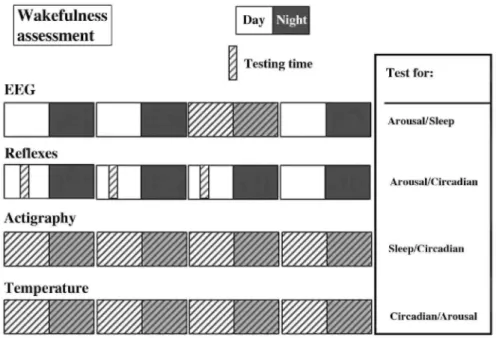
For a full understanding of the wakefulness capacity of a DOC patient, we propose a series of tests to assess arousal, circadian rhythms, and sleep patterns. We recommend starting this assessment when the patient is systemically healthy and homeostatically stable. Figure 4 shows a summary of the tests proposed and when to start each measure. Arousal is behaviorally stratified in the CRS-R scale ranging from "unarousable" to "attention"; however, this is useful for an initial test but does not inform about arousal variability in the patient. To this aim we propose to choose one reflex in the patient that can be rated (i.e., very low, low, medium, high, very high responses) and to assess it systematically (every 5min) in the course of 2-3 h for at least three days (testing at the same day and time). This approach should inform about the variability in arousal both within and between days.
To test for circadian control we propose to continuously record a simple variable for several days. To define if the central clock in the brain is functioning, the external environment should be controlled, in particular
temperature and light-dark variations. We have shown that skin temperature and motor activity may be easy to test and have the capacity to unveil the day-to-day variability. Both methods are cheap and require minimal maintenance, and if rhythms are assessed for at least four days, the clock capacity to be entrained can be determined. This test, together with the arousal assessment, will give an indication on when it is best to test for awareness in order to increase the likelihood of obtaining the maximal responses from each DOC patient. Sleep evaluation may be indicative of the degree of damage in different brain networks. A 24-h EEG assessment may relate to reticular activating function if state transitions are present; spindles may reflect the preserved functional integrity of the thalamus; and SWS and REM sleep may reflect residual functioning of brainstem nuclei.
Fig. 4. Wakefulness assessment for DOC patients. For each test the proposed testing time is shown, as well as
the factor it addresses.
CONCLUSION
In the second part of the report from the Muti-Society Task Force on PVS (1994), recovery is divided into two dimensions: recovery of consciousness and recovery of function. The first one refers to the capacity to detect awareness (emergence from VS), and the second deals with the patient's capacity to communicate, to learn, and to perform adaptative tasks. Surprisingly there is no mention of the capacity to recover from decreased
wakefulness or the problems caused by lower and/or erratic responsiveness in the assessment of recovery of consciousness or recovery of function. As we pointed out in this chapter, wakefulness may not appear as a key aspect in the evaluation of DOC patients, but the level of wakefulness may act as an enabling condition for conscious processing (Dehaene and Changeaux, 2004).
The clear relationship between circadian rhythms and sleep states (Winfree, 1982) could and should be extended to different conscious states. Although it is difficult to measure fatigue (as the homeostatic process of sleep) in DOC patients, it is possible to measure some physiological variables that are under circadian control and, in the process, responsiveness variability (changes in arousal) can be obtained. For the assessment of wakefulness and awareness, the temperature rhythm (if found) could be a good starting point to decide when, during the day, it is best to assess the cognitive processing of the patient. The understanding of the relationships between arousal, circadian rhythms, and sleep in DOCs is essential from a descriptive point of view and, most importantly, as putative diagnostic and prognostic tools that might help to aid therapeutic alternatives.
for most of the aspects of wakefulness in DOC patients. Knowing when the patient does seem to be more active, more responsive, and fully awake may decrease the misdiagnosis (false negatives) of true MCS patients
classified as VS and true severely disabled patients classified as MCS. The detailed assessment of wakefulness — whether coming through in waves, in tides, or completely absent — will help in the characterization of this neglected aspect of consciousness and may have prognostic value for DOC patients.
Acknowledgments
We thank the patients and their families for their participation. We specially thank Dr. Laureys, Dr. Delorme, Dr. Manes, and Dr. Owen for helping in our research with DOC patients. We also thank Thanh Dang-Vu for
providing the normal volunteer actigraphy data. We (the authors) declare having no conflicts of interest. TB is supported by an IIF Marie Curie Fellowship, VC by FRIA-FNRS, BD by the German National Merit
Foundation, and DG by CON-ICET, ANPCyT and Universidad de Quilmes.
References
Adams, J. H., Graham, D. I., & Jennett, B. (2000). The neuropathology of the vegetative state after an acute brain insult. Brain, 123, 1327-1338.
Åkerstedt, T. (2007). Altered sleep/wake patterns and mental performance. Physiology and Behavior, 90, 209-218.
Alessi, C., Martin, J. L., Webber, A. P., Kim, E. C., Harker, J. O., & Josephson, K. R. (2005). Randomized, controlled trial of a nonpharmacological intervention to improve abnormal sleep/wake patterns in nursing home residents. JAGS, 53, 803-810.
Allebrandt, K. V., & Roenneberg, T. (2008). The search for circadian clock components in humans: New perspectives for association studies.
Brazilian Journal of Medical and Biological Research, 41, 716—721.
Ancoli-Israel, S., Cole, R., Alessi, C., Chambers, M., Moorcroft, W., & Pollak, C. P. (2003). The role of actigraphy in the study of sleep and circadian rhythm. Sleep, 26, 342-359.
Andrews, K., Murphy, L., Munday, R., & Littlewood, C. (1996). Misdiagnosis of the vegetative state: Retrospective study in a rehabilitation unit. BMJ, 313, 13-16.
Baguley, I. J., Heriseanu, R. E., Cameron, I. D., Nott, M. T., & Slewa-Younan, S. (2008). A critical review of the pathophysiology of dysautonomia following traumatic brain injury. Neurocritical Care, 8, 293-300.
Beersma, D., & Gordijn, M. (2007). Circadian control of the sleep-wake cycle. Physiology and Behavior, 90, 190-195.
Bekinschtein, T. A., Golombek, D. A., Simonetta, S. H., Coleman, M. R., & Manes, F. F. (in press). Circadian rhythms in the vegetative state.
Benedetti, F., Radaelli, D., Bernasconi, A., Dallaspezia, S., Falini, A., Scotti, G., Lorenzi, C., Colombo, C., & Smeraldi, E. (2008). Clock genes beyond the clock: CLOCK genotype biases neural correlates of moral valence decision in depressed patients. Genes Brain Behavior, 7(1), 20-25.
Bergamasco, B., Bergamini, L., Doriguzzi, T., & Sacerdote, I. (1968). The sleep cycle in coma: Prognostic value. Electroencephalography
and Clinical Neurophysiology, 25, 87.
Berger, A. M., Wielgus, K. K., Young-McCaughan, S., Fischer, P., Farr, L., & Lee, K. A. (2008). Methodological challenges when using actigraphy in research. Journal of Pain and Symptom Management, 36, 191-199.
Blatter, K., & Cajochen, C. (2007). Circadian rhythms in cognitive performance: Methodological constraints, protocols, theoretical underpinnings. Physiology and Behavior, 90, 196-208.
Bob, P., & Fedor-Freybergh, P. (2008). Melatonin, consciousness, and traumatic stress. Journal of Pineal Research, 44, 341-347. Boivin, D., Czeisler, C., Dijk, D. J., Duffy, C., Folkard, S., Minors, D., et al. (1997). Complex interaction of the sleep-wake cycle and circadian phase modulates self-rated subjective feelings in healthy subjects. Archives of General Psychiatry, 54, 145-152.
Borbely, A. (1982). Sleep regulation: Circadian rhythm and homeostasis. In D. Ganten & D. Pfaff (Eds.), Sleep. Clinical and experimental
aspects (pp. 83-104). Berlin: Springer Verlag.
Bornstein, S. R., Licinio, J., Tauchnitz, R., Engelmann, L., Negrao, A. B., Gold, P., et al. (1988). Plasma leptin levels are increased in survivors of acute sepsis: Associated loss of diurnal rhythm, in Cortisol and leptin secretion. The Journal of Clinical Endocrinology and
Bourne, R. S., & Mills, G. H. (2006). Melatonin: Possible implications for the postoperative and critically ill patient. Intensive Care
Medicine, 32, 371-379.
Brainard, G. C., & Hanifin, J. P. (2005). Photons, clocks, and consciousness. Journal of Biological Rhythms, 20, 314-325. Brenner, R. P. (2005). The interpretation of the EEG in stupor and coma. Neurologist, 11, 271-284.
Britt, C. W., Jr. (1981). Nontraumatic "spindle coma": Clinical, EEG, and prognostic features. Neurology, 31, 393-397.
Britt, C. W., Jr., Raso, E., & Gerson, L. P. (1980). Spindle coma, secondary to primary traumatic midbrain hemorrhage. Electroencep halo
graphic Clinical Neurophysiology, 49, 406-408.
Broughton, R. (1982). Human consciousness and sleep/ waking rhythms: A review and some neuropsychological considerations. Journal of
Clinical Neuropsychology, 4, 193-218.
Brown, J. (1970). "Get up." From the single "Get Up (I Feel Like Being A) Sex Machine". Live at the Bell Auditorium (USA).
Buijs, R. M., Scheer, F. A., Kreier, F., Yi, C., Bos, N., Goncharuk, V. D., et al. (2006). Organization of circadian functions: Interaction with the body. Progress in Brain Research, 153, 341-360.
Cabello, B., Parthasarathy, S., & Mancebo, J. (2007). Mechanical ventilation: Let us minimize sleep disturbances. Current Opinion in
Critical Care, 13, 20-26.
Cajochen, C., Knoblauch, V., Wirz-Justice, A., Kräuchi, K., Graw, P., & Wallach, D. (2004). Circadian modulation of sequence learning under high and low sleep pressure conditions. Behavioural Brain Research, 151, 167-176.
Cantero, J. L., & Atienza, M. (2005). The role of neural synchronization in the emergence of cognition across the wake-sleep cycle. Review
of Neuroscience, 6, 69-83.
Carlson, D. E., & Chiu, W. C. (2008). The absence of circadian cues during recovery from sepsis modifies pituitary-adreno-cortical function and impairs survival. Shock, 29, 127-132.
Carrier, J., & Monk, T. (2000). Circadian rhythms of performance: new trends. Chronobiology International, 17, 719-732.
Chatrian, G. E., White, L. E., Jr., & Daly, D. (1963). Electroencephalographic patterns resembling those of sleep in certain comatose states after injuries to the head. Electroencephalography and Clinical Neurophysiology, 15, 272-280.
Cheliout-Heraut, F., Rubinsztajn, R., loos, C., & Estournet, B. (2001). Prognostic value of evoked potentials and sleep recordings in the prolonged comatose state of children. Preliminary data. Neurophysiologie Clinique, 31, 283-292.
D'Aleo, G., Bramanti, P., Silvestri, R., Saltuari, L., Gersten-brand, F., & Di Perri, R. (1994a). Sleep spindles in the initial stages of the vegetative state. Italian Journal of Neurological Sciences, 15, 347-351.
D'Aleo, G., Saltuari, L., Gerstenbrand, F., & Bramenti, P. (1994b). Sleep in the last remission stages of vegetative state of traumatic nature.
Functional Neurology, 9, 189-192.
Dauch, W. A., & Bauer, S. (1990). Circadian rhythms in the body temperatures of intensive care patients with brain lesions. Journal of
Neurology, Neurosurgery and Psychiatry, 53, 345-347.
Daurat, A., Benoit, O., & Buguet, A. (2000). Effects of zopiclone on the rest/activity rhythm after a westward flight across five time zones.
Psychopharmacology, 149, 241-245.
Davies, N. W., Sharief, M. K., & Howard, R. S. (2006). Infection-associated encephalopathies: Their investigation, diagnosis, and treatment.
Journal of Neurology, 253, 833-845.
Dehaene, S., & Changeux, J. P. (2004). Neural mechanisms for access to consciousness. In M. Gazzaniga (Ed.), The cognitive
neurosciences. (3rd ed., volume in press).
Dennhardt, R., Gramm, H. J., Meinhold, K., & Voigt, K. (1989). Patterns of endocrine secretion during sepsis. Progress in Clinical and
Biological Research, 308, 751-756.
DeSouza, L., Benedito-Silva, A. A., Noguiera Pires, M. L., Poyares, D., Tufik, S., & Calil, H. M. (2003). Further validation of actigraphy in sleep studies. Sleep, 26, 81-85.
Dylan, B. (1979). "When you gonna wake up?" From the Album "Slow Train Coming". CMS Digital Studios (California, USA).
Elliott, L., Coleman, M., Shiel, A., Wilson, B. A., Badwan, D., Menon, D., et al. (2005). Effect of posture on levels of arousal and awareness in vegetative and minimally conscious state patients: A preliminary investigation. Journal of Neurology, Neurosurgery & Psychiatry, 76, 298-299.
Evans, B. M. (2002). What does brain damage tell us about the mechanisms of sleep? Journal of the Royal Society of Medicine, 95, 591-597. Folkard, S. (1990). Circadian performance rhythms: Some practical and theoretical implications. Philosophical Transactions of the Royal
Society of London. Series B, Biological Sciences, 327, 543-553.
Freedman, N. S., Gazendam, J., Levan, L., Pack, A. I., & Schwab, R. J. (2001). Abnormal sleep/wake cycles and the effect of environmental noise on sleep disruption in the intensive care unit. American Journal of Respiratory and Critical Care Medicine, 163, 451-457.
Friedman, G., Eliezer, S., & Vincet, J. L. (1998). Has the mortality of septic shock changed with time? Critical Care Medicine, 26, 2078-2086.
Friese, R. S., Bruns, B., & Sinton, C. M. (2009). Sleep deprivation after septic insult increases mortality independent of age. Journal of
Trauma, 66, 50-54.
Fukudome, Y., Abe, I., Saku, Y., Matsumura, K., Sadoshima, S., Utunomiya, H., et al. (1996). Circadian blood pressure in patients in a persistent vegetative state. American Journal of Physiology, 270, 1109-1114.
Giacino, J. T., Ashwal, S., Childs, N., Cranford, R., Jennett, B., Katz, D. I., et al. (2002). The minimally conscious state: Definition and diagnostic criteria. Neurology, 58, 349-353.
Gilestro, G. F., Tononi, G., & Cirelli, C. (2009). Widespread changes in synaptic markers as a function of sleep and wakefulness in Drosophila. Science, 324, 109-112.
Giubilei, F., Formisano, R., Fiorini, M., Vitale, A., Faroni, J., Toni, D., et al. (1995). Sleep abnormalities in traumatic apallic syndrome.
Journal of Neurology, Neurosurgery and Psychiatry, 58, 484-486.
Golombek, D. A., & Cardinali, D. P. (2008). Mind, brain, education and biological timing. Mind, Brain and Education, 2, 1-6.
Golombek, D. A., Ferreyra, G. A., Agostino, P. V., Murad, A. D., Rubio, M. F., Pizzio, G. A., et al. (2003). From light to genes: Moving the hands of the circadian clock. Frontiers in Bioscience, 8, 285-293.
Gordon, C. R., & Oksenberg, A. (1993). Spontaneous nystagmus across the sleep-wake cycle in vegetative state patients. Electroencephalography and Clinical Neurophysiology, 86, 132-137.
Halasz, P., Terzano, M., Parrino, L., & Bodizs, R. (2004). The nature of arousal in sleep. Journal of Sleep Research, 13, 1-23. Herdegen, J. J. (2002). Intensive care unit sleep disruption: Can the cycle be restored? Critical Care Medicine, 30, 709-710. Himanen, S. L., & Hasan, J. (2000). Limitations of Rechtschaf-fen and Kales. Sleep Medicine Reviews, 4, 149-167. Hobson, J. A. (1996). How the brain goes out of its mind. Endeavour, 20, 86-89.
Home, J. A., & Ostberg, O. (1976). A self-assessment questionnaire to determine morningness in human circadian rhythms. International
Journal of Chronobiology, 4, 97-110.
Hulihan, J. F., Jr., & Syna, D. R. (1994). Electroencephalo-graphic sleep patterns in post-anoxic stupor and coma. Neurology, 44, 758-760. Isono, M., Wakabayashi, Y., Fujiki, M. M., Kamida, T., & Kobayashi, H. (2002). Sleep cycle in patients in a state of permanent unconsciousness. Brain Injury, 16, 705-712.
Jennett, B., & Plum, F. (1972). Persistent vegetative state after brain damage. A syndrome in search of a name. Lancet, 7753, 734-737 Jewel, A. (1995). From the Album "Pieces of You". Atlantic (New York, USA).
Joosten, K. F., de Kleijn, E. D., Westerterp, M., de Hoog, M., Eijck, F. C., Hop, W. C. J., et al. (2000). Endocrine and metabolic responses in children with meningoccocal sepsis: Striking differences between survivors and nonsurvivors. The Journal of Clinical Endocrinology and
Metabolism, 85, 3746-3753.
Katikireddy, C. K., & Kuschner, W. G. (2006a). Critical care medicine update: Essentials for the nonintensivist, part 2. Comprehensive
Katikireddy, C. K., & Kuschner, W. G. (2006b). Critical care medicine update: Essentials for the nonintensivist, part 1. Comprehensive
Therapy, 32, 74-81.
Katz, M. E., Grizzo, M. E., Salim, M., Gonzalez Ley, B., Merino, D., Golombek, D. A., et al. (2002). Sleep-wake cycle after renal transplant and cyclosporin treatment (abst). Transplantation, 74(4), 760.
Kleitman, N. (1982). Basic rest-activity cycle 22 years later. Sleep, 5, 311-317.
Majerus, S., Gill-Thwaites, H., Andrews, K., & Laureys, S. (2005). Behavioral evaluation of consciousness in severe brain damage. Progress
in Brain Research, 150, 397-413.
Marpegan, L., Leone, M. J., Katz, M. E., Sobrero, P., Bekinschtein, T. A., & Golombek, D. A. (in press). Diurnal variation in endotoxin-induced mortality in mice: Correlation with proinflammatory factors.
Monk, T., Buysse, D., Reynolds, C., Berga, S., Jarrett, D., Begley, A., et al. (1997). Circadian rhythms in human performance and self-rated subjective feelings under constant conditions. Journal of Sleep Research, 6, 9-18.
Morgenthaler, T., Alessi, C., Friedman, L., Owens, J., Kapur, V., Boehlecke, B., et al. (2007). Practice parameters for the use of actimetry in the assessment of sleep and sleep disorders: An update for 2007. Sleep, 30, 519-529.
Mosby's Medical Dictionary. (2009). 8th edn. San Diego: Elsevier Inc.
Multi-Society Task Force on PVS. (1994). Medical aspects of the persistent vegetative state (1 and 2). The New England Journal of
Medicine, 330, 1572.
Mundigler, G., Delle-Karth, G., Koreny, M., Zehetgruber, M., Steindl-Munda, P., Marktl, W., et al. (2002). Impaired circadian rhythm of melatonin secretion in sedated critically ill patients with severe sepsis. Critical Care Medicine, 30, 536-540.
Oksenberg, A., Arons, E., Sazbon, L., Mizrahi, A., & Radwan, H. (2000). Sleep-related erections in vegetative state patients. Sleep, 23, 953-957.
Oksenberg, A., Gordon, C., Arons, E., & Sazbon, L. (2001). Phasic activities of rapid eye movement sleep in vegetative state patients. Sleep,
24, 703-706.
Paaivilainen, P., Korhonen, I., Lotjonen, J., Cluitmans, L., Julha, M., Sarela, A., et al. (2005). Circadian activity rhythm in demented and non-demented nursing-home residents measured by telemetric actigraphy. Journal of Sleep Research, 14, 61-68.
Papadopoulos, M. C., Davies, D. C., Moss, R. F., Tighe, D., & Bennett, E. D. (2000). Pathophysiology of septic encephalopathy: A review.
Critical Care Medicine, 28, 3019-3024.
Parthasarathy, S., & Tobin, M. J. (2004). Sleep in the intensive care unit. Intensive Care Medicine, 30, 197-206.
Pattoneri, P., Tirabassi, G., Pela, G., Astorri, E., Mazzuchi, A., & Borghetti, A. (2005). Circadian blood pressure and heart rate changes in patients in a persistent vegetative state after traumatic brain injury. Journal of Clinical Hypertension (Greenwich), 7, 734-739.
Rechtschaffen, A., & Kales, A. (1968). A manual of standardized terminology, techniques and scoring system for sleep stages of human
subjects. Bethesda, MD: US Dept. of Health, Education, and Welfare, p. 12.
Reppert, S. M., & Weaver, D. R. (2002). Coordination of circadian timing in mammals. Nature, 418, 935-941.
Rhudy, J. L., Williams, A. E., McCabe, K. M., Russell, J. L., & Maynard, L. J. (2008). Emotional control of nociceptive reactions (ECON): Do affective valence and arousal play a role?. Pain, 136, 250-261.
Ron, S., Algom, D., Hary, D., & Cohen, M. (1980). Time-related changes in the distribution of sleep stages in brain injured patients.
Electroencephalography and Clinical Neurophysiology, 48, 432-441.
Sacho, R. H., & Childs, C. (2008). The significance of altered temperature after traumatic brain injury: An analysis of investigations in experimental and human studies: Part 2. British Journal of Neurosurgery, 22, 497-507.
Sanap, M. N., & Worthley, L. I. (2002). Neurologic complications of critical illness: Part I. Altered states of consciousness and metabolic encephalopathies. Critical Care and Resuscitation, 4, 119-132.
Seet, R. C., Lim, E. C., & Wilder-Smith, E. P. (2005). Spindle coma from acute midbrain infarction. Neurology, 64, 2159-2160.
Spivak, E., Oksenberg, A., & Catz, A. (2007). The feasibility of sleep assessment by actigraph in patients with tetraplegia. Spinal Cord, 45, 765-770.
of patients with acute stroke. Psychiatry and Clinical Neuroscience, 56, 221-222.
Teigen, K. H. (1994). Yerkes-Dodson: A law for all Seasons. Theory and Psychology, 4(4), 525-547.
The Sunday Times. (2007). 40% of coma patients in a 'vegetative state' may be misdiagnosed, says a new report. Available at: http://www.timesonline.co.uk/tol/life_and_ style/health/article3004892.ece
Tung, A., & Mendelson, W. B. (2004). Anesthesia and sleep. Sleep Medicine Reviews, 8, 213-225.
Valdez, P., Reilly, T., & Waterhouse, J. (2008). Rhythms in mental performance. Mind, Brain and Education, 2, 7-16.
Valente, M., Placidi, F., Oliveira, A. J., Bigagli, A., Morghen, I., Poietti, R., et al. (2002). Sleep organization pattern as a prognostic marker at the subacute stage of post-traumatic coma. Clinical Neurophysiology, 113, 1798-1805.
Varon, J., & Fromm, R. E. (2001). Handbook of practical critical care medicine. Berlin: Springer.
Vlach, K. D., Boles, J. W., & Stiles, B. G. (2000). Telemetric evaluation of body temperature and physical activity as predictors of mortality in a murine model of staphylococcal enterotoxic shock. Journal of Comparative Medicine, 50, 160-166.
Vogel, H. P., Kroll, M., Fritschka, E., & Quabbe, H. J. (1990). Twenty-four-hour profiles of growth hormone, prolactin and Cortisol in the chronic vegetative state. Clinical Endocrinology (Oxford), 33, 631-643.
Waterhouse, J., Minors, D., Åkerstedt, T., Reilly, T., & Atkinson, G (2001). Rhythms of human performance. In J. Takahashi, F. Turek, & R. Moore (Eds.), Handbook of behavioral neurobiology: Circadian clocks (pp. 571-601). New York: Kluwer Academic/Plenum Publishers. Weinhouse, G. L., & Schwab, R. J. (2006). Sleep in the critically ill patient. Sleep, 29, 707-716.
Werth, E., Savaskan, E., Knoblauch, V., Fontana Gasio, P., Van Someren, E. J. W., Hock, C., et al. (2002). Decline in long-term circadian rest-activity cycle organization in a patient with dementia. Journal of Geriatric Psychiatry and Neurology, 15, 55-59.
Winfree, A. T. (1982). The tides of human consciousness: Descriptions and questions. American Journal of Physiology, 242, R163-R166. Yerkes, R. M., & Dodson, J. D. (1908). The relation of strength of stimulus to rapidity of habit-formation. Journal of
Comparative Neurology and Psychology, 18, 459-482.
Zavada, A., Gordijn, M. C., Beersma, D. G., Daan, S., & Roenneberg, T. (2005). Comparison of the Munich Chronotype Questionnaire with the Horne-Ostberg's Morn-ingness-Eveningness Score. Chronobiology International, 22, 267-278.
Zee, P. C, & Manthena, P. (2007). The brain's master circadian clock: Implications and opportunities for therapy of sleep disorders. Sleep