Université du Québec
Institut National de la Recherche Scientifique Institut Armand-Frappier
Centre for Host-Parasite Interactions
INTRACELLULAR TRAFFICKING AND BIOLOGICAL
IMPACT OF LEISHMANIA VIRULENCE FACTORS ON
MACROPHAGES
By
Guillermo Arango Duque
A dissertation submitted in partial fulfillment of the requirements for the degree of
Philosophiae Doctor (PhD) in the Virology and Immunology Program
Evaluation Committee
Committee president and Maritza Jaramillo-Patiño
internal examiner INRS-Institut Armand-Frappier
External examiners Sachiko Sato
Centre Hospitalier de l'Université Laval Dave Richard
To my mother Leyda Libia, for whom I hold utmost admiration and gratitude To my family
ACKNOWLEDGEMENTS
If I were asked whether I would do it all over again, I would say yes! And I do not mean that lightly, because this great journey for the pursuit of knowledge and self-discovery has been facilitated by very remarkable people.
Let me start with my supervisor Dr. Albert Descoteaux, who has been an excellent mentor from day one. I remember very well that snowy January morning when I had the honour of setting foot in his laboratory. Since that day, Albert’s support and confidence in me have been exemplar. The knowledge and insight that I have gained from him have made me grow as a scientist and human being. I deeply admire Albert’s dedication, work ethic, leadership, confidence and ease for explaining complex science. Once again, I thank you for being such an influential person in my life. I am also very appreciative of my laboratory colleagues and summer students, both past and present, for their pedagogical support, patience and camaraderie. I thank Hamlet, Roman and Fernando for helping me to prepare for my thesis defence.
What would my life be without the love and unwavering support of my family and friends? I would not be where I am today! My parents Leyda Libia and Guillermo León have been great role models and I am forever indebted to them for inculcating in me the principles by which I live today. I thank my sweet grandmother Aura Libia for her prayers, advice and positive energy. To my family, I love you and I dedicate this thesis to you. I hold kindred the great friendships I have forged during all these years; you know who you are. To my great friends and scientific collaborators Ahmed M. Fahmy and Hamlet A. Acevedo Ospina, I thank you for always being there for me and for your
I take this opportunity to thank my scientific collaborators Drs. Simona Stäger, Mitsunori Fukuda, Armando Jardim and Étienne Gagnon. Their precious support and expertise have been essential to the successful completion of my dissertation research. I am grateful to and salute my thesis committee members for their valuable contributions towards my thesis and overall scientific education.
Finally, I am grateful to the Canadian Institutes of Health Research for having funded my laboratory and my Banting and Best Doctoral Award. I acknowledge the Centre for Host-Parasite Interactions and the Institut Pasteur International Network for having granted me the opportunity to actively participate in the numerous international congresses that have enriched my education so much.
ABSTRACT
Leishmania parasites cause a spectrum of debilitating diseases found worldwide. The Leishmania life cycle is digenetic, and starts with sandflies that inoculate metacyclic
promastigotes into a vertebrate host. Promastigotes are internalized by tissue phagocytes where they transform into amastigotes. Phagosomes mature into highly microbicidal phagolysosomes via membrane exchanges with lysosomes, and with organelles in the secretory pathway. Remarkably, Leishmania remodels phagolysosomes into parasitophorous vacuoles that promote parasite growth. To achieve this feat, the parasite employs an armament of abundant surface-bound glycoconjugates that include the GP63 metalloprotease and lipophosphoglycan. GP63 cleaves multiple host substrates, thereby enabling Leishmania to subvert phagocyte functions such as transcription, translation, lipid metabolism, cytokine secretion, LC3-associated phagocytosis and antigen cross-presentation. Lipophosphoglycan, a complex glycophospholipid, promotes parasite survival by inhibiting phagolysosomal maturation, hence quenching the microbicidal power of the phagosome.
The effector functions of a macrophage rely on a very active endomembrane trafficking system that regulates how the cell responds to environmental stimuli. Vesicle fusion is regulated by members of the soluble N-ethylmaleimide-sensitive factor activating protein receptor, and Synaptotagmin families. In this work, we characterized Syt XI, an inhibitory member of the Synaptotagmin family whose function had not been reported. We discovered that Syt XI is a recycling endosome and lysosome-associated protein that controls phagocytosis, as well as the phagosome’s killing capacity [primary article
also leads to increased TNF and IL-6 secretion and to an augmented influx of neutrophils and inflammatory monocytes to the infection site [primary article no. 2]. Unlike bacteria, Leishmania is not known to inject its virulence factors across the phagosome membrane. This raises the question of how the parasite’s virulence effectors reach their targets. Due to the reported importance of the host’s secretory pathway on parasite survival, we hypothesized that Leishmania’s virulence factors co-opt this pathway in order to egress from the phagosome into the host cell cytoplasm. Using biochemistry and microscopy-based assays, we demonstrated that GP63 and the phosphoglycans are rapidly redistributed throughout the cytoplasm in vesicles containing markers of the endoplasmic reticulum, and of the endoplasmic reticulum-Golgi intermediate compartment. Importantly, chemical inhibition of the secretory pathway hinders the redistribution of GP63 and the phosphoglycans, thereby impeding the cleavage of GP63 targets. This prompted us to study the role of Sec22b, which regulates endoplasmic reticulum-Golgi trafficking, on the intracellular trafficking of GP63 and phosphoglycans. We found that Sec22b promotes the redistribution of these virulence factors and enables GP63 to reach host proteins [primary article no. 3].
In sum, the work presented in this dissertation sheds light into how Leishmania‘s virulence molecules enter the host cell in order to exert their functions. We also revealed that GP63 induces TNF and IL-6 release in vitro and in vivo, thereby contributing to the accrual of inflammatory phagocytes to the infection site. Remarkably, GP63 and the phosphoglycans access their targets by hijacking cellular organelles and their resident vesicle fusion molecules. These findings provide important insight into how Leishmania sabotages macrophage biology.
12/13/2018
X
Guillermo ARANGO DUQUE, HBSc Senior PhD candidate
Signed by: Guillermo Arango Duque
X
Albert DESCOTEAUX, PhD Thesis advisor
TABLE OF CONTENTS
ACKNOWLEDGEMENTS ... III ABSTRACT ... V TABLE OF CONTENTS ... VII LIST OF FIGURES AND TABLES... XV LIST OF ABBREVIATIONS ... XXI
CHAPTER 1: LITERATURE REVIEW ... 25
1 THE ROLE OF PHAGOCYTES IN IMMUNITY ... 26
1.1 INTRODUCTION AND DISCOVERY ... 26
1.2 PROFESSIONAL PHAGOCYTES ... 27 1.2.1 Neutrophils ... 29 1.2.2 Mast cells ... 29 1.2.3 Monocytes ... 30 1.2.4 Dendritic cells ... 31 1.2.5 Macrophages ... 32 1.3 NON-PROFESSIONAL PHAGOCYTES ... 34
2 THE MOLECULAR MACHINERY THAT REGULATES VESICULAR TRAFFIC IN EUKARYOTIC CELLS ... 35
2.1 INTRODUCTION AND HISTORICAL CONTEXT ... 35
2.2 THE IMPORT ROUTE: ENDOCYTOSIS ... 37
2.3 THE SECRETORY PATHWAY: EXOCYTOSIS ... 39
2.3.1 The machinery involved in anterograde and retrograde transport ... 40
2.4 THE ROLE OF SNARES IN VESICLE TARGETING AND DOCKING ... 42
2.4.1 SNARE complex assembly and dissociation ... 43
3.1.3 Apoptotic cell receptors ... 59
3.2 PHAGOSOME MATURATION ... 59
3.2.1 The early phagosome ... 59
3.2.2 The late phagosome and phagolysosome ... 61
3.2.3 The killing machinery of the phagolysosome ... 62
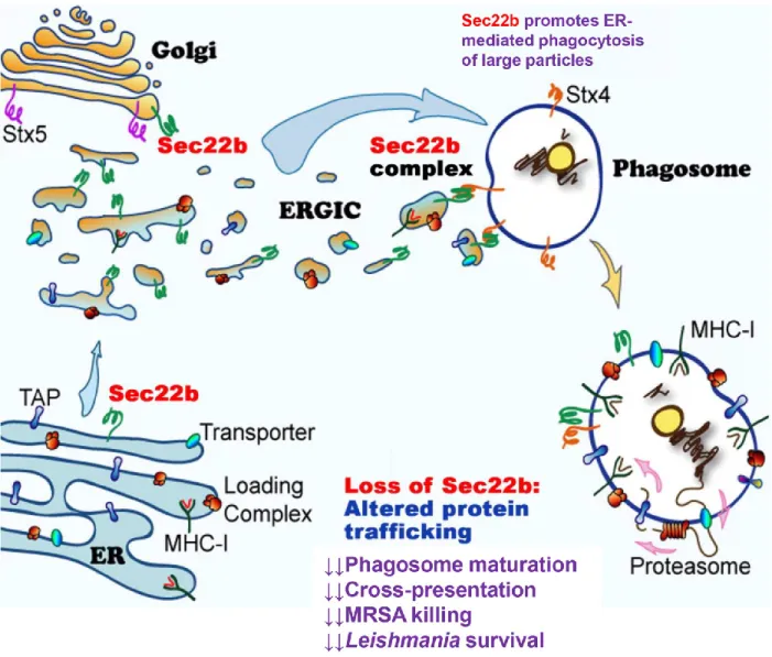
3.3 ROLES OF THE ER/ERGIC AND THE SNARESEC22B ON PHAGOSOME BIOLOGY ... 64
3.4 PHAGOCYTOSIS BY NEUTROPHILS ... 68
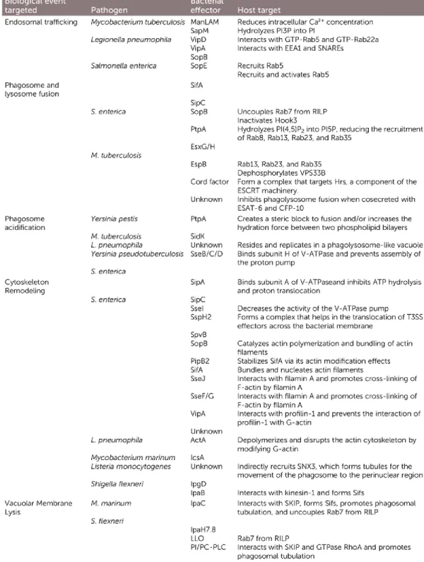
3.5 HOW PATHOGENS EVADE KILLING BY THE PHAGOLYSOSOME ... 68
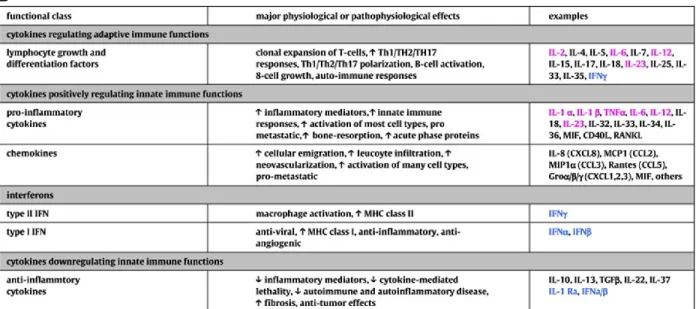
4 CYTOKINE SECRETION BY MACROPHAGES ... 72
4.1 INTRODUCTION TO CYTOKINE BIOLOGY ... 72
4.1.1 Cytokines involved in innate and adaptive immunity ... 73
4.1.2 Chemokines: cytokines that induce cell migration ... 75
4.1.3 Hematopoietins: cytokines that modulate immune cell development ... 76
4.2 MACROPHAGE CYTOKINES ... 76
4.3 VESICLE FUSION PROTEINS THAT REGULATE CYTOKINE SECRETION IN MACROPHAGES ... 79
4.3.1 The trafficking and release of model cytokines TNF, IL-6 and IL-10 ... 79
5 THE LEISHMANIA PARASITE AND ITS INTERACTION WITH THE HOST MACROPHAGE ... 83
5.1 INTRODUCTION AND EPIDEMIOLOGY ... 83
5.2 THE LEISHMANIA LIFE CYCLE ... 86
5.3 HOW LEISHMANIA SETTLES IN THE HOST MACROPHAGE ... 87
5.3.1 Life in the vertebrate host after inoculation ... 87
5.3.2 Leishmania entry into host macrophages ... 88
5.3.3 Contribution of the ER/ERGIC to the biogenesis of Leishmania PVs ... 90
5.4 LPG AND ITS INHIBITORY ROLES ON PHAGOSOME MATURATION ... 91
5.4.1 The structure of LPG ... 91
5.4.2 The importance of LPG in intracellular survival ... 92
5.4.3 LPG inhibits intraphagosomal oxidation and acidification ... 94
5.5 THE GP63 METALLOPROTEASE ... 97
5.5.1 Discovery and biochemical properties ... 97
5.5.2 The impact of GP63 on host cell biology ... 99
CHAPTER 2: HYPOTHESES AND OBJECTIVES ... 103
ARTICLE NO. 1: SYNAPTOTAGMIN XI REGULATES PHAGOCYTOSIS AND
CYTOKINE SECRETION IN MACROPHAGES ... 108
1 ABSTRACT ... 110
2 INTRODUCTION ... 111
3 MATERIALS AND METHODS ... 114
3.1 ETHICS STATEMENT ... 114
3.2 ANTIBODIES AND PLASMIDS ... 114
3.3 CELL CULTURE ... 114
3.4 TRANSFECTIONS ... 115
3.5 RT-PCR ... 116
3.6 CYTOKINE SECRETION MEASUREMENTS ... 116
3.7 PHAGOSOME ISOLATION AND PHAGOCYTOSIS ASSAYS ... 116
3.8 BACTERIA KILLING ASSAYS ... 117
3.9 CONFOCAL IMMUNOFLUORESCENCE MICROSCOPY ... 118
3.10 DATA ANALYSIS ... 119
4 RESULTS ... 120
4.1 MACROPHAGES EXPRESS SYT XI ... 120
4.2 SYT XI LOCALIZES TO RECYCLING ENDOSOMES AND LYSOSOMES ... 122
4.3 PHAGOSOMES RECRUIT SYT XI ... 124
4.4 KNOCKDOWN OF SYT XI LEADS TO AN INCREASE IN CYTOKINE SECRETION AND PHAGOCYTOSIS ... 126
4.5 OVEREXPRESSION OF SYT XI LEADS TO A DECREASE IN CYTOKINE SECRETION AND PHAGOCYTOSIS ... 128
4.6 SYT XI REGULATES THE RECRUITMENT OF GP91PHOX AND LAMP-1 TO THE PHAGOSOME ... 130
4.7 KNOCKDOWN OF SYT XI LEADS TO INCREASED INTRACELLULAR SURVIVAL OF E. COLI ... 132
5 DISCUSSION ... 134
6 ACKNOWLEDGMENTS ... 138 ARTICLE NO. 2: LEISHMANIA PROMASTIGOTES INDUCE CYTOKINE SECRETION
3.1 ETHICS STATEMENT ... 145
3.2 ANTIBODIES AND PLASMIDS ... 145
3.3 CELL CULTURE ... 146
3.4 TRANSFECTIONS, INFECTIONS AND CYTOKINE QUANTIFICATION ... 147
3.5 SYNCHRONIZED PHAGOCYTOSIS ASSAYS ... 147
3.6 CONFOCAL IMMUNOFLUORESCENCE MICROSCOPY ... 148
3.7 LYSES, SDS-PAGE AND WESTERN BLOTTING ... 148
3.8 PROTEIN PURIFICATION AND IN VITRO DEGRADATION ASSAYS ... 149
3.9 INTRAPERITONEAL INFECTIONS AND FACS ANALYSIS... 150
3.10 DATA ANALYSIS ... 150
4 RESULTS ... 151
4.1 LEISHMANIA DEGRADES SYT XI IN A GP63-DEPENDENT MANNER ... 151
4.2 LPG MEDIATES EXCLUSION OF SYT XI FROM PVS ... 156
4.3 LEISHMANIA INDUCES TNF AND IL-6 SECRETION THROUGH DEGRADATION OF SYT XI ... 159
4.4 GP63-EXPRESSING PROMASTIGOTES ELICIT INCREASED TNF AND IL-6 RELEASE IN VIVO, AS WELL AS INCREASED NEUTROPHIL AND INFLAMMATORY MONOCYTE RECRUITMENT ... 162
5 DISCUSSION ... 164
6 ACKNOWLEDGEMENTS ... 168
ARTICLE NO. 3: THE HOST CELL SECRETORY PATHWAY MEDIATES THE EXPORT OF LEISHMANIA VIRULENCE FACTORS OUT OF THE PARASITOPHOROUS VACUOLE ... 169
1 ABSTRACT ... 171
2 RESULTS AND DISCUSSION ... 172
3 MATERIALS AND METHODS ... 183
3.1 ETHICS STATEMENT ... 183
3.2 ANTIBODIES, PLASMIDS AND INHIBITORS ... 183
3.3 CELL CULTURE ... 184
3.4 INFECTIONS... 185
3.5 ELECTROPHORESIS, WESTERN BLOTTING AND ZYMOGRAPHY ... 186
3.6 SUCROSE GRADIENT FLOTATION ASSAYS ... 187
4 SUPPLEMENTARY FIGURES... 190
5 ACKNOWLEDGEMENTS ... 200
6 COMPETING INTERESTS ... 200
CHAPTER 4: DISCUSSION AND CONCLUSIONS ... 201
1 THE INVOLVEMENT OF SYT XI IN MACROPHAGE BIOLOGY AND BEYOND ... 202
1.1 SYTS REGULATE THE EFFECTOR FUNCTIONS OF THE IMMUNE SYSTEM ... 202
1.2 SYT XI CONTROLS THE EXOCYTOSIS OF PROINFLAMMATORY CYTOKINES ... 203
1.3 HOW DOES SYT XI REGULATE VESICULAR TRAFFIC IN MACROPHAGES? ... 205
2 THE IMPACT OF GP63 ON CYTOKINE RELEASE ... 208
2.1 THE EVOLUTION OF PARASITIC PROTEASES ... 208
2.2 THE PRESSURE TO INACTIVATE IS CIRCUMVENTED BY THE LEISHMANIA GP63 METALLOPROTEASE ... 209
3 INVOLVEMENT OF THE HOST CELL SECRETORY PATHWAY ON THE TRAFFICKING OF LEISHMANIA VIRULENCE FACTORS ... 212
3.1 HOW DO LEISHMANIA VIRULENCE FACTORS EXIT THE PV? ... 212
3.2 LEISHMANIA CO-OPTS THE HOST CELL SNARE SEC22B ... 214
3.3 HOW DO LEISHMANIA VIRULENCE FACTORS REACH HOST CELL ORGANELLES?... 217
4 SEC22B AND THE CONTRADICTORY ROLES OF THE ER/ERGIC DURING INFECTION ... 218
5 IS IT FEASIBLE TO TARGET VESICLE TRAFFICKING PATHWAYS DURING LEISHMANIA INFECTION? ... 220
CHAPITRE 5: RÉSUMÉ EN FRANÇAIS ... 223
2.3 TROISIÈME ARTICLE : LA VOIE SÉCRÉTOIRE PERMET L'EXPORTATION DES FACTEURS DE VIRULENCE
DE LEISHMANIA HORS DE LA VACUOLE PARASITOPHORE ... 228
APPENDIX 1: OTHER PRIMARY ARTICLES ... 229
1 ARTICLE NO. 4: LEISHMANIA INFANTUM LIPOPHOSPHOGLYCAN-DEFICIENT MUTANTS: A TOOL TO STUDY HOST CELL-PARASITE INTERPLAY 230 1.1 ABSTRACT ... 232 1.2 INTRODUCTION ... 233 1.3 METHODS ... 235 1.4 RESULTS ... 242 1.5 DISCUSSION ... 249
1.6 CONFLICT OF INTEREST STATEMENT ... 252
1.7 FUNDING INFORMATION... 252
1.8 ACKNOWLEDGEMENTS ... 252
APPENDIX 2: REVIEW ARTICLES ... 253
1 REVIEW ARTICLE NO. 1: LEISHMANIA SURVIVAL IN THE MACROPHAGE: WHERE THE ENDS JUSTIFY THE MEANS ... 254
1.1 ABSTRACT ... 255
1.2 HIGHLIGHTS ... 255
1.3 INTRODUCTION ... 256
1.3.1 Macrophages: sentinels of the immune system ... 256
Leishmania parasites have evolved to conquer macrophages ... 256
1.4 THE ONSLAUGHT OF THE GP63 PROTEASE: FROM MEMBRANE TRAFFICKING TO NUCLEAR PORE DYNAMICS ... 258
1.4.1 Manipulation of membrane trafficking to subvert antigen presentation and cytokine secretion ... 258
1.4.2 Impact of GP63 on nuclear integrity and physiology ... 263
1.5 LEISHMANIA HIJACKS METABOLIC PATHWAYS IN THE MACROPHAGE TO PROMOTE SURVIVAL ... 264
1.5.1 Retention of intracellular iron fuels amastigote survival... 264
1.5.2 Disruption of cholesterol dynamics favours Leishmania growth ... 266
1.5.3 Manipulation of the host’s energy resources promotes parasite survival ... 267
1.6 DNA METHYLATION IS A STRATEGY TO SHUT DOWN GENES INVOLVED IN HOST DEFENCE ... 268
2 REVIEW ARTICLE NO. 2: MACROCHAGE CYTOKINES: INVOLVEMENT IN
IMMUNITY AND INFECTIOUS DISEASES ... 271
2.1 ABSTRACT ... 272
2.2 INTRODUCTION: CYTOKINES AND MACROPHAGES ... 273
2.3 THE MACROPHAGE CYTOKINE PORTFOLIO ... 276
2.3.1 Proinflammatory cytokines ... 276
2.3.2 Anti-inflammatory cytokines ... 284
2.3.3 Chemokines ... 285
2.4 ALTERNATIVELY ACTIVATED MACROPHAGES AND THEIR CYTOKINES ... 287
2.5 HOW PATHOGENS DISRUPT CYTOKINE SECRETION FROM MACROPHAGES ... 290
2.5.1 Mycobacterium ulcerans uses mycolactone to inhibit cytokine production ... 291
2.5.2 Leishmania promastigotes employ GP63 to augment TNF and IL-6 release ... 292
2.6 ACKNOWLEDGEMENTS ... 295
3 REVIEW ARTICLE NO. 3: UNDERSTANDING TGEV-ETEC COINFECTION THROUGH THE LENS OF PROTEOMICS: A TALE OF PORCINE DIARRHEA ... 296
3.1 ABSTRACT ... 297
3.2 MAIN TEXT ... 298
3.3 ACKNOWLEDGEMENTS ... 302
3.4 CONFLICT OF INTEREST STATEMENT ... 302
4 REVIEW ARTICLE NO. 4: MACROPHAGES TELL THE NON-PROFESSIONALS WHAT TO DO... 303
4.1 ABSTRACT ... 304
4.2 MAIN TEXT ... 305
5 REVIEW ARTICLE NO. 5: LEISHMANIA DICES AWAY CHOLESTEROL FOR SURVIVAL ... 310
LIST OF FIGURES AND TABLES
Chapter 1: Literature review
Copyrighted material was used with permission and licence agreements can be obtained upon request. Figure 1. The ontogeny of professional phagocytes
Figure 2. Diversity of professional and non-professional at selected anatomical sites Figure 3. Vesicle trafficking in the secretory pathway
Figure 4. SNARE proteins promote vesicle fusion through quaternary structure formation
Figure 5. The role of SNAREs in the vesicle fusion cycle
Figure 6. SNARE-mediated regulation of ER-Golgi membrane trafficking
Figure 7. Syts are conserved type I membrane proteins that drive membrane fusion Figure 8. Syt XI does not bind Ca2+ and is conserved across species
Figure 9. Phagocytosis is a multifunctional and highly dynamic process
Figure 10. Receptor-mediated recognition of phagocytic particles induces actin polymerization and phagosome formation
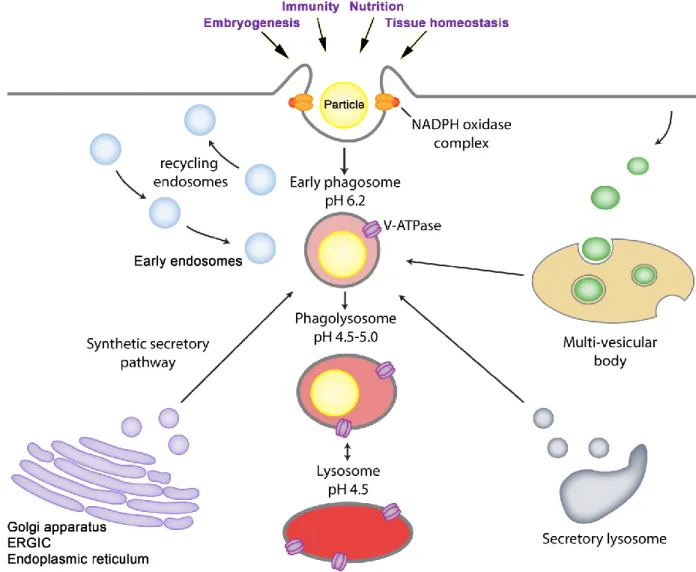
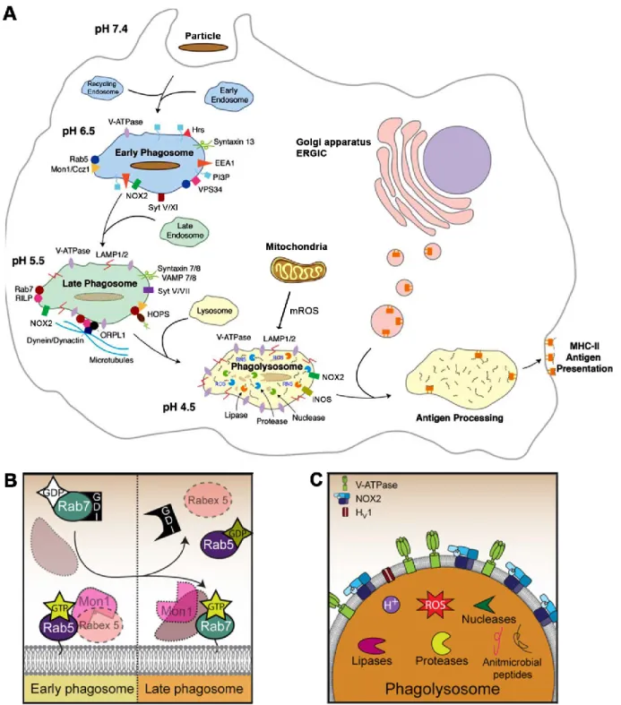
Figure 11. The molecular machinery regulating the transition from early phagosome to phagolysosome in macrophages
Figure 12. The ER/ERGIC-resident SNARE Sec22b modulates phagosomal biogenesis and function
Figure 16. Membrane trafficking pathways and molecules involved in the release of TNF, IL-6 and IL-10 in macrophages
Figure 17. Overview of the Leishmania parasite and the leishmaniases Figure 18. The structure and function of LPG and related glycoconjugates Figure 19. The structure and function of the GP63 metalloprotease
Chapter 3: Primary articles Article no. 1
Figure 1. Syt XI is expressed in macrophages
Figure 2. Syt XI associates with recycling endosomes and lysosomes Figure 3. Syt XI is recruited to early phagosomes
Figure 4. Knockdown of Syt XI leads to increased cytokine secretion and phagocytosis Figure 5. Overexpression of Syt XI leads to decreased cytokine secretion and phagocytosis
Figure 6. Syt XI regulates the recruitment of gp91phox and LAMP-1
Figure 7. Knockdown of Syt XI lowers the microbicidal activity of macrophages
Article no. 2
Figure 1. GP63 lowers Syt XI levels in infected macrophages
Supplementary Figure 1. Syt XI levels in Leishmania-infected cells are modulated by GP63
Figure 2. Leishmania promastigotes use GP63 to cleave Syt XI
Figure 3. Syt XI degradation is direct and does not occur during lysate processing Figure 4. LPG mediates exclusion of Syt XI from the phagosome
Figure 6. L. major induces TNF and IL-6 secretion via degradation of Syt XI
Figure 7. In vivo modulation of cytokine secretion and phagocyte infiltration by GP63
Article no. 3
Figure 1. GP63 and PGs are redistributed within infected cells
Figure 2. Redistribution of PGs and GP63 requires parasite internalization
Figure 3. GP63 and PGs are present in vesicles that co-occur with ER and ERGIC markers
Figure 4. Perturbation of ER-Golgi trafficking hampers the redistribution of GP63 and PGs and the cleavage of Syt XI
Supplementary Figure 1. Redistribution of GP63 and PGs in macrophages infected with
L. major and L. donovani
Supplementary Figure 2. The redistribution of PGs is similar to that of LPG
Supplementary Figure 3. GP63 activity has no impact on the redistribution of GP63 and PGs
Supplementary Figure 4. GP63 and PGs cofractionate with vesicles and ER/ERGIC markers
Supplementary Figure 5. GP63 and PGs colocalize with ER markers
Supplementary Figure 6. Sec22b promotes the redistribution of GP63 and PGs
Supplementary Figure 7. Flotation of GP63 and PGs in lysates of infected JAWS-II cells Supplementary Figure 8. Host cell organelles and proteins mediate the trafficking and
Chapter 4: Discussion and conclusions
Table I. Syts control membrane trafficking in myeloid and lymphoid cells
Figure 1. Syt XI regulates processes of great importance in immunity and neurotransmission
Figure 2. The interacting partners of Syt XI will provide insight into its mechanism of action
Figure 3. Macrophage cytokines are targeted by metalloproteases of pathogen origin Figure 4. CRT coimmunoprecipitates GP63
Figure 5. The host cell provides the molecular machinery that promotes the intracellular spread of Leishmania virulence molecules
Figure 6. Retro-2 is an inhibitor of the retrograde pathway that hinders the redistribution of GP63 and PGs
Appendix 1: Other primary articles Article no. 4
Figure 1. Constructs for the targeted deletion and complementation of the LPG1 gene in
Leishmania infantum
Figure 2. Growth curve and morphology of the ∆lpg1 mutant
Figure 3. Deletion of LPG1 does not alter LD formation in Leishmania infantum Figure 4. LPG1 promotes intraphagosomal survival in infected macrophages
Figure 5. L. infantum promastigotes evade NF-κB-dependent iNOS in an LPG-dependent manner in RAW 264.7 cells
Appendix 2: Review articles Review no. 1
Figure 2. Leishmania prevents iron efflux via hepcidin upregulation
Figure 3. Leishmania induces changes in the host macrophage methylome for survival
Review no. 2
Figure 1. Monocytes can become phenotypically distinct macrophages
Figure 2. Modulation of macrophage cytokine secretion by Mycobacterium ulcerans bacteria and Leishmania promastigotes
Review no. 3
Figure 1. TGEV and ETEC elicit context-dependent host cell responses
Review no. 4
Figure 1. IGF-1 modulates inflammation and phagocytosis by non-professional phagocytes
Review no. 5
LIST OF ABBREVIATIONS
ADRP: Adipose differentiation related protein AP-1: Activator protein-1
Arf6: ADP-ribosylation factor 6 BFA: Brefeldin A
BMM: Bone marrow-derived macrophages CNS: Central nervous system
CNX: Calnexin CRT: Calreticulin
CTD: C-terminal domain
DAPI: 4',6-diamidino-2-phenylindole DC: Dendritic cells
DIC: Differential intensity contrast DMSO: Dimethyl sulfoxide
DNA: Deoxyribonucleic acid Dvl: Dishevelled
EEA1: Early endosome antigen 1 EM: Electron microscopy
EMT: Epithelial-mesenchymal transition EPO: Erythropoietin
ER: Endoplasmic reticulum
ERGIC: Endoplasmic reticulum-Golgi intermediate compartment ETEC: Enterotoxigenic Escherichia coli K88
FAM129B: Family with sequence similarity 129 member B FBS: Fetal bovine serum
GTPase: Guanosine triphosphatase HCS: High-content screening
HDM: House dust mite
HMGCoA: Hydroxymethylglutaryl-coenzyme A
HOPS: Homotypic fusion and protein sorting complex HSPG2: Heparan sulfate proteoglycan 2
IFN: Interferon
IGF: Insulin-like growth factor IL: Interleukin
IP: Immunoprecipitation
IPEC-J2: Intestinal columnar epithelial cells IRE1α: Inositol requiring enzyme 1α
iTRAQ: Isobaric tags for relative and absolute quantification LAMC2: Laminin gamma 2
LAMP: Lysosomal-associated membrane protein LC3B: Microtubule-associated protein1 light chain 3
LC-MS/MS: Liquid chromatography coupled to tandem mass spectrometry
LD: Lipid droplet
LDL: Low-density lipoprotein LPG: Lipophosphoglycan LPS: Lipopolysaccharide MCS: Membrane contact site
M-CSF: Macrophage colony-stimulating factor MHC: Major histocompatibility complex
MIG: Monokine induced by gamma interferon, or CXCL9 MIP-2α: Macrophage inflammatory protein 2-α, or CXCL1/2 miRNA: Micro RNA
mROS: Mitochondrial reactive oxygen species MV: Microvesicle
NF-κB: Nuclear factor kappa-light-chain-enhancer of activated B cells
NO: Nitric oxide
NOX2: NADH oxidase 2 NTD: N-terminal domain
PAMP: Pathogen-associated molecular pattern PDI: Protein disulfide isomerase
PG: Phosphoglycan(s)
PGF2α: Prostaglandin F2α
Phox: Phagocytic oxidase
PI(3,4,5)P3: Phosphatidylinositol-3,4,5-trisphosphate
PI(4,5)P2: Phosphatidylinositol-4,5-bisphosphate
PI3K: Phosphoinositide 3-kinase(s) PKC: Protein kinase C
PPG: Proteophosphoglycan
PRR: Pathogen recognition receptor PV: Parasitophorous vacuole
RANTES: Regulated upon activation normal T cell expressed and secreted, or CCL5 RE: recycling endosome
RNS: Reactive nitrogen species ROS: Reactive oxygen species SEM: Scanning electron microscopy shRNA: Small hairpin RNA
siRNA: Small interfering RNA
SNARE: Soluble N-ethylmaleimide-sensitive factor attachment protein receptors Stx: Syntaxin
Tfr1: Transferrin receptor 1
TGEV: Transmissible gastroenteritis virus TGF: Transforming growth factor
TGN: Trans-Golgi network TNF: Tumour necrosis factor
VAMP: Vesicle-associated membrane protein VL: Visceral leishmaniasis
1 THE ROLE OF PHAGOCYTES IN IMMUNITY
1.1 Introduction and discovery
Phagocytes are cells of myeloid origin that circulate in the blood and reside in all animal tissues. They are endowed with the capacity to perform phagocytosis, the process by which phagocytes engulf and destroy microorganisms, cellular debris and particulate foreign bodies (Desjardins et al., 2005, Gordon, 2016). The term phagocyte comes from the term phagein (to eat) and –cito (cell). Many cell types are capable of performing phagocytosis, with immune system phagocytes being crucial in organismal development and antimicrobial defense (Banoub et al., 2017). In mammals, phagocytes have evolved specialized functions that depend on the microenvironment in which they are found. Indeed, one litre of human blood is estimated to contain 6x10⁹ of these cells, and many more reside in all organs (Banoub et al., 2017). Phagocytes are classified into professional (Figure 1) and non-professional (Figure 2) depending on the specificity and rapidity with which they engulf particles (Banoub et al., 2017, Rabinovitch, 1995, Tauber, 2003).
Phagocytes were originally a theory of Russian zoologist Ilya Ilich Metchnikoff (Илья
Ильич Мечников, 1845-1916) who suspected the existence of cells that defend against
microbial infections (Silverstein, 2011, Tauber, 2003). In 1882, Metchnikoff observed that when he pricked starfish larvae, a series of amoebal cells were recruited to and attached to the pricking agent at the wounding site. Upon presentation of his observations in Vienna, his colleague Carl Friedrich Claus suggested the term ‘phagocyte’ to denote those cells (Chernyak et al., 1988, Silverstein, 2011, Tauber, 2003). Metchnikoff then extended his studies onto other systems. He observed that fungal spores were attacked and destroyed by specialized cells that are present in the model organism Daphnia, and that Bacillus anthracis spores were engulfed by phagocytic cells in mammals. These data confirmed Metchnikoff’s initial observation
At the time, Metchnikoff’s work was met with skepticism and strife, which was in part due to the work of Paul Ehrlich in the 1890s (Kaufmann, 2008). The latter postulated that immunity was dependent on the binding of specialized host cell-surface molecules to specific chemical groups on toxins. Ehrlich’s theory was that if host cells survived an initial encounter with a toxin, the cells would start liberating some the molecule that initially bound and neutralized the toxin, thence allowing the organism to survive future encounters with the same toxin. Such molecules are nowadays known as antibodies (Kaufmann, 2008). In 1904, Almroth Wright helped to bridge Metchnikoff’s and Ehrlich’s work by suggesting that the phagocytosis of bacteria was increased in the presence of opsonins and antibodies coating the microorganisms (Gordon, 2016, Kaufmann, 2008, Silverstein, 2011). Although Metchnikoff’s work was accepted at the beginning of the 20th century, the roles of phagocytes within the immune system started – and still continue – to be understood in the 1980s. Along with Paul Ehrlich, Metchnikoff’s seminal discoveries were celebrated with the 1908 Nobel Prize in Physiology or Medicine (Kaufmann, 2008).
1.2 Professional phagocytes
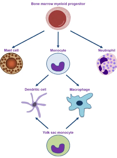
Professional phagocytes are characterized for their enhanced phagocytic efficiency and for their central role in antimicrobial defence and organismal development. This family is constituted by monocytes, macrophages, dendritic cells, neutrophils and mast cells (Gordon, 2016, Rabinovitch, 1995). Most of these phagocytes descend from a myeloid precursor in the bone marrow (Figure 1). Additionally, embryonic stem cells from the
Figure 1 of literature review. The ontogeny of professional phagocytes. Professional phagocytes descend from a myeloid precursor in the bone marrow or from stem cells in the developing embryo’s yolk sac. Monocytes are usually found in the blood stream. Upon encounter with antigens or inflammatory stimuli, they can develop into macrophages or dendritic cells, depending on the context. The phagocytic efficiency of professional phagocytes is enhanced by the presence of surface receptors that recognize a wide variety of endogenous and exogenous particles.
This section introduces professional phagocytes in the context of their particular functions in the host. Although these cells differ widely in the context-specific functions that they perform, they are attracted to and migrate to tissues when they come in contact with endogenous chemoattractants such as chemokines and pathogen
inflammatory insult and the resolution of the response (Banoub et al., 2017, Cybulsky et
al., 2016, Faurschou et al., 2003, Hellebrekers et al., 2018).
1.2.1 Neutrophils
Neutrophils (9-12 µm) are highly abundant (60-70% of circulating phagocytes) and short-lived granulocytic cells whose primary function is the active ingestion of invading bacteria and fungi (Hellebrekers et al., 2018). They are very motile and possess a characteristic multi-lobed nucleus of highly compacted chromatin (Figure 1). The abundant granules in their cytoplasm contain lytic enzymes and antimicrobial molecules that help degrade phagocytosed microbes (Faurschou et al., 2003, Hellebrekers et al., 2018). Their abundance and small size allow them to rapidly infiltrate – when needed – tissues via diapedesis. Indeed, neutrophils are the first responders during an infection, where they are attracted by the cytokines, chemokines and histamine liberated by tissue macrophages and mast cells (Hellebrekers et al., 2018). Those molecules include TNF, IL-6 and IL-8, which in turn trigger chemokine secretion and the expression of selectins on endothelial cells (Starckx et al., 2002). Neutrophils bind to selectins, which permits the extravasation of neutrophils from the blood to the site of infection. Once there, neutrophils recognize their target via toll-like receptors (TLRs), which bind to microbial molecules such as lipopolysaccharide (LPS) and peptidoglycan (Gouwy et al., 2004). Neutrophils can also externalize their chromatin in order to form neutrophil extracellular traps (NETs) that tangle and kill microbes, or hold them in place so that macrophages can come and ingest them (Sollberger et al., 2018).
tissue remodelling. This allows for cells such as neutrophils to emigrate into inflammatory sites (Sollberger et al., 2018). In allergy and anaphylaxis, excess histamine causes persistent inflammation and tissue damage. Mast cells display TLRs and other recognition receptors at their plasmalemma, which in turn facilitate the clearance of damaged erythrocytes (Sharma et al., 2018) and the killing of pathogens (Lin et al., 1999). They can also present antigens to lymphocytes, and improve the antigen-presenting capacity of dendritic cells via small extracellular vesicles known as exosomes (Skokos et al., 2003).
1.2.3 Monocytes
Monocytes are mononuclear phagocytes characterized by a kidney-shaped nucleus (Figure 1) and measuring up to 18 µm (Ginhoux et al., 2014). They originate in the bone marrow and the yolk sac, and enter the circulation to infiltrate organs such as the liver, spleen and lungs (Ginhoux et al., 2014, van de Laar et al., 2016, Yang et al., 2014). There, they differentiate into tissue macrophages or dendritic cells. Monocytes can be inflammatory (Ly6Chi) and anti-inflammatory (Ly6Clo), a dichotomy that allows them to
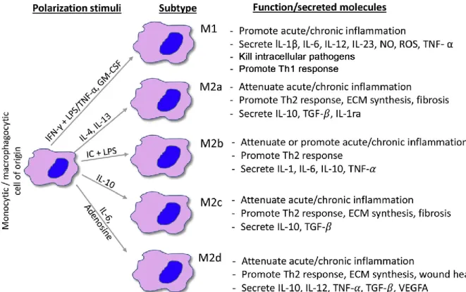
originate very distinct types of macrophages. Under physiological conditions, inflammatory monocytes monitor extravascular tissues and present antigens to lymphocytes. Stress by sterile or non-sterile inflammatory stimuli triggers the release of the CCR2 chemokine, which triggers migration of inflammatory monocytes to the injury site (Ginhoux et al., 2014). There, these cells develop into highly inflammatory and microbicidal ‘M1’ macrophages (Yang et al., 2014). On the other hand, CCR7/8-dependent migration to the lymph nodes can trigger differentiation into dendritic cells (Qu et al., 2004). Non-inflammatory monocytes patrol the luminal site of the endothelium and eliminate dying cells and debris (Thomas et al., 2015). Although they express low levels of CCR2, they can travel to inflamed tissues to become anti-inflammatory and reparative ‘M2’ macrophages. Inflammatory monocytes can also become non-inflammatory macrophages, illustrating the fact that monocyte
1.2.4 Dendritic cells
Dendritic cells are characterized by their neuronal-like protrusions that can measure up to 10 µm (Figure 1) (Banoub et al., 2017, Collin et al., 2018, Cybulsky et al., 2016, Steinman et al., 1973). They are found in great quantities in the skin, intestines, and other mucosal sites (Figure 2). Although dendritic cells are specialized in phagocytosis, their main function is to use phagocytosis as a way of processing antigens for subsequent presentation at the cell surface. Antigens are loaded onto class II major histocompatibility complexes (MHC) and presented to specialized lymphocytes, B and T cells, in order to mount an adaptive immune response to the antigen (Mellman, 2013). In this manner, dendritic cells link innate and adaptive immunity in eukaryotes. They mediate strong adaptive responses to foreign agents while maintaining a tolerogenic response to self (Mellman, 2013). These cells exist in various states of maturity, with immature dendritic cells patrolling peripheral sites for antigen. Once an antigen is phagocytosed, dendritic cells are activated and migrate towards lymph nodes. There, they mature and augment their expression of MHC molecules and other costimulatory ligands that are required for antigen presentation. They can also secrete various cytokines such as TNF, IL-1 and IL-12 (Collin et al., 2018, Mellman, 2013, Steinman et
al., 1973).
Dendritic cells were originally observed by Paul Langerhans in 1868 when he was studying human cutaneous epithelium (Steinman et al., 1994). Langerhans mistook these cells for neurons, and it was not until the 1970s that Ralph M. Steinman and Zanvil A. Cohn correctly characterized them via phase-contrast microscopy and
1.2.5 Macrophages
Macrophages, a term derived from the Greek ‘large eaters’, are monocyte-derived cells that exist in most tissues (Figures 1 and 2) (Cybulsky et al., 2016, Sieweke et al., 2013). They are the primary destroyers of particulate matter, as well as bacteria, fungi, protozoans, senescent cells and apoptotic bodies (Arandjelovic et al., 2015, Gordon, 2016). As mentioned, when monocytes infiltrate tissues, they can transform into macrophages via the action of the granulocyte macrophage colony-stimulating factor (GM-CSF) in conjunction with other cytokines such as IL-10, IL-12 and other factors such as apoptotic cells (Ginhoux et al., 2014, Sieweke et al., 2013). When monocytes become macrophages, cell size, phagocytic capacity, and intraphagosomal antimicrobial properties increase dramatically (Ginhoux et al., 2014). Although tissue macrophages are usually found in a quiescent state, they can be activated by a variety of stimuli during the immune response. Contact with and phagocytosis of antigens serves as the initial stimulus. This can include microbial molecules such as LPS, which binds to macrophages via the LPS-binding protein to TLR4 on the macrophage surface (Cybulsky et al., 2016). Activation and microbicidal capacity are further amplified by cytokines such as interferon gamma (IFN-γ), which are produced by T cells (Biswas et
al., 2010, Nathan et al., 1983, Sieweke et al., 2013, Tam et al., 2014).
Macrophages were originally observed by Metchnikoff during his experiments with starfish. However, the term ‘macrophage’ was coined by Aschoff in 1924 when he was studying these cells as part of the reticulo-endothelial system (Silverstein, 2011, Tauber, 2003). In the 1960s, the role of macrophages was defined more precisely by the work of Zanvil A. Cohn and colleagues (Steinman et al., 1994). They characterized the macrophage as a secretory cell able to kill inside (through phagocytosis) and outside of the cell, and capable of releasing over 50 products. The latter include numerous molecules that participate in inflammation, such as cytokines, prostaglandins and leukotrienes (Cybulsky et al., 2016, Steinman et al., 1994).
1.3 Non-professional phagocytes
Non-professional phagocytes are non-immune cells whose main function is not phagocytosis. They have evolved very specific functions that are crucial in homeostatic regulation and tissue remodelling at sites such as the eye and the testis, where it is detrimental to have inflammatory professional phagocytes (Figure 2) (Penberthy et al., Rabinovitch, 1995). Non-professional phagocytes are comprised of endothelial, epithelial cells and fibroblasts, which clear dead cells and debris in the tissues where they reside (Figure 2). For instance, testicular sertoli cells phagocytose senescent germ cells; in the eye, retina pigment epithelial cells clear used photoreceptor outer segments (Penberthy et al.). Non-professional phagocytes cells do not possess the recognition receptors or the intracellular machinery necessary for efficient phagocytosis. They produce low levels of reactive oxygen species (ROS) or antimicrobial peptides compared to professional phagocytes (Penberthy et al., 2018, Rabinovitch, 1995).
Recently, it was discovered that professional and non-professional phagocytes communicate via the insulin growth factor 1 (IGF-1), a pleiotropic hormone involved in muscle building (Han et al., 2016). Lung epithelial cells phagocytose apoptotic bodies and are in constant contact with allergens such as house dust mite (HDM). HDM is internalized and induces inflammation in lung epithelial cells, which then triggers the release of IL-4 and IL-13 by immune cells. Those cytokines then elicit the release of anti-inflammatory extracellular vesicles and IGF-1 by lung macrophages (Han et al., 2016). IGF-1 then signals lung epithelial cells to stop ingesting apoptotic cells and to start the uptake of anti-inflammatory macrophage-derived extracellular vesicles (Bourdonnay et al., 2015, Han et al., 2016), thus ensuing in the control of airway inflammation. Much remains to be discovered about how professional and non-professional phagocytes collaborate to promote organismal homeostasis (Penberthy et
2 THE MOLECULAR MACHINERY THAT REGULATES VESICULAR
TRAFFIC IN EUKARYOTIC CELLS
2.1 Introduction and historical context
Eukaryotic cells are factories that produce and secrete a multitude of molecules of essential function, from hormones and neurotransmitters to cytokines (Blank et al., 2014, Südhof, 2012). In the year 2013, Drs. James E. Rothman, Randy W. Schekman and Thomas C. Südhof were awarded the Nobel Prize in Physiology or Medicine in commemoration for their discoveries on the molecular machinery that regulates vesicular traffic, an intracellular transport system of pivotal importance in health and disease. Starting in the 1970s and 1980s, the three laureates worked on separate aspects of the mechanisms by which molecules such as enzymes and neurotransmitters are transported within, and out of cells. In the 1970s, Dr. Schekman worked on budding yeast and identified the genes that produce the proteins necessary for the transport of these vesicles. Using classical genetic analysis, he characterized 23 genes involved in the vesicular transport that takes place from the perinuclear region of the endoplasmic reticulum (ER) to the Golgi apparatus and the cell membrane (Novick
et al., 1980, Novick et al., 1979). On the other hand, Dr. Rothman worked with
mammalian cells. His group characterized the N-ethylmaleimide-sensitive fusion (NSF) protein, which allows the transport of proteins to the Golgi apparatus, the synaptosomal-associated protein (SNAP), and the NSF attachment protein receptor (SNARE) (Wilson
et al., 1989). He discovered how proteins make it into vesicles that reach and attach
onto a target membrane, ensuring that they are delivered to the right compartment (Rothman, 1994). The last mechanism of this cellular transport pathway was to identify
importance is their discovery of Synaptotagmin (Syt), which catalyzes this fusion step (Perin et al., 1990, Südhof, 2012).
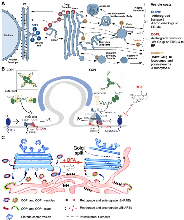
Cellular products must be transported, with great precision, from their site of synthesis to the extracellular milieu. For these reasons, the organization of cellular trafficking is fundamental, and is a process that is carried out by vesicles and their molecular adaptors. Indeed, eukaryotic cells are characterized by the orderly and precise distribution of molecules in different cellular compartments (Pang et al., 2010, Südhof, 2012). If the vesicular transport system essential for its functioning and survival does not work, the cell ceases to be a complex and precise biological machine and collapses into chaos. Vesicles act as vehicles that transport molecules among cellular organelles, and from organelles to the plasma membrane. Vesicles execute inter-organelle communication by carrying molecules as part of their lipid bilayer or lumen. Vesicular transport is spatiotemporally regulated by a wide array of membrane proteins that reside in the various cellular organelles. In turn, such molecules define the identity and content of those cellular organelles (Jahn et al., 2006, Malsam et al., 2011). For example, acidic pH and hydrolytic enzymes should be present only in lysosomes. However, when a cell ingests a particle via phagocytosis, the vacuole is acidified via a vesicle fusion process that leads to the accrual and fusion of lysosomes with phagosomes (Levin et al., 2016). Vesicles are formed in a source compartment and loaded with cargo molecules that must be transported. Once released into the cytosol, the vesicles are specifically directed towards a target compartment to which they eventually finally fuse with. Intracellular vesicles can originate at virtually all cellular organelles (Jahn et al., 2006, Malsam et al., 2011). Oftentimes, these vesicles originate at the Golgi and the ER or at the plasma membrane (Figure 3) (Bonifacino et al., 2004, Jahn et al., 2006). Indeed, crossing the plasma membrane is one of the main ways in which compounds enter or leave the cell.
2.2 The import route: endocytosis
Endocytosis is the process by which large macromolecules and small particles enter the cell in an ATP-dependent manner (Figure 3) (Bonifacino et al., 2004). In this process, material that enters the cell induces an invagination in the plasma membrane that leads to the formation of a vesicle encasing the material. Endocytosis is subdivided according to the nature of the internalized material. In receptor-mediated endocytosis, the internalization process is initiated by recognition of extracellular ligands by receptors on the cell surface. Recognition of the ligand starts a signalling program that induces the internalization of the ligand-receptor complex in a vesicle. This signalling leads to the active recruitment of vesicle coat proteins known as clathrin and caveolae, which facilitate membrane curvature and budding (Nichols et al., 2001, Traub, 2011). Caveolae are lipid microdomains that consist of small cholesterol-rich invaginations at the plasma membrane. These structures are delineated by caveolin-1, possess the ganglioside GM-1, and contain receptors such as CR3 (Harris et al., 2002, Kenworthy, 2002). Endocytosis contributes to the recycling of those cell membrane receptors, which can be triggered by the attachment of a single ubiquitin molecule to the receptor (Bonifacino et al., 2004, Ghaddar et al., 2014). On the other hand, pinocytosis refers to the endocytosis of liquid and any solutes found therein. It is a dominant feature of cells in the intestinal mucosa, which specialize in nutrient acquisition (Doherty et al., 2009). Pinocytosis can occur independently of clathrin coats through the constitutive uptake of micropinosomes (Nichols et al., 2001). Internalization of large particles is termed phagocytosis (Desjardins et al., 2005, Gordon, 2016) and will be discussed in section ‘3’ of this Chapter. Once endocytosed vesicles are formed in the cytoplasm, they may fuse with lysosomes, which induce digestion of intravesicular contents. The products of digestion are then recycled in the cell (Bonifacino et al., 2004, Levin et al., 2016).
Figure 3 of literature review. Vesicle trafficking in the secretory pathway. (A) The secretory pathway is constituted by a group of organelles through which synthesized proteins and lipids are transported in or on vesicles. This transport process is spatiotemporally regulated by coat proteins (COPI, COPII and clathrin) and vesicle fusion proteins that regulate cargo destination. (B) In retrograde and anterograde transport, vesicle buddying from the ER is
activity of Gea1/Gea2, and may also block anterograde transport. (C) This blockage halts ER-Golgi traffic, eventually causing the Golgi to collapse onto the ER. Adapted with permission from (Barlowe et al., 2013, Bonifacino et al., 2004, Nebenführ et al., 2002).
2.3 The secretory pathway: exocytosis
Exocytosis is the process by which cytoplasmic molecules are secreted outside the cell (Figure 3), and is a key feature of secretory cells such as neurons, pancreatic islets and cells of the immune system (Bonifacino et al., 2004, Huynh et al., 2007b, Stow et al., 2013). In this highly regulated process, vesicles encasing cellular products usually originate from the Golgi network, travel to the plasma membrane, and fuse to unload their contents. After proteins are processed in the ER, they are transported to the Golgi network via COPII-coated vesicles in a process known as anterograde transport (Bonifacino et al., 2004, Stow et al., 2013). There, proteins mature as they become post-translationally modified in the various cisternae of this organelle. Eventually, proteins reach the trans-Golgi network (TGN), where they are packaged and sorted into clathrin-coated vesicles (Traub, 2011), and sent to different cellular compartments or the outside of the cell (Bonifacino et al., 2004, Huynh et al., 2007b, Stow et al., 2013). There are two types of exocytosis – constitutive and regulated. Constitutive exocytosis is feature of all eukaryotic cells. In this process, vesicles are secreted in a continuous fashion, making it essential for the renewal of the plasmalemmal membrane and its proteins (Bonifacino et al., 2004, Huynh et al., 2007b). In macrophages, the secretion of cytokines such as TNF is a constitutive process that is triggered by cellular activation (Beutler, 1999, Stow et al., 2013). In regulated exocytosis, proteins are synthesized and stored in a vesicle until the cell receives a regulatory signal to initiate secretion; this is a feature of cells that secrete hormones such insulin (Bonifacino et al., 2004, Huynh et al.,
2.3.1 The machinery involved in anterograde and retrograde transport
When vesicles bud from a donor compartment, they are coated with proteins that are necessary for inducing the membrane deformity required for vesicle formation, for selecting the cargo of the vesicle, and for determining the delivery site. Assembly of these proteins induces membrane buckling and budding. Eventually, the protein coat comes off when the vesicle fuses with its target compartment (Bonifacino et al., 2004, Nichols et al., 2001, Traub, 2011). Many of these proteins were initially found in the yeast-based genetic screens performed in Dr. Schekman’s laboratory. Mutations in the components of this machinery gave rise to swollen intracellular compartments and hindered secretion (Novick et al., 1980, Novick et al., 1979). The blockages occurred in cellular locations that were later traced to where these proteins exert their functions (Bonifacino et al., 2004).
Anterograde transport, or forward pathway, describes the passage that takes place from the ER to the ER-intermediate compartment (ERGIC) and onto the cis-Golgi (Barlowe et
al., 2013, Bonifacino et al., 2004). Vesicle formation at the ER starts with recruitment of
the Sar1/2 GTPase, whose function is to recruit adaptors that are involved in cargo selection and coat formation (Figure 3). Sar1/2 is initially recruited in its inactive GDP-bound form, and through interaction with the guanine exchange factor (GEF) Sec12, Sar is activated into its GTP-bound form. This activation triggers a conformational change in Sar1/2 that unmasks a myristylated anchor, which permits Sar1/2-GTP to anchor itself into the vesicle’s membrane. Sar1/2 initiates membrane deformation and mediates the recruitment of the adaptor complex Sec23-Sec24. Those proteins possess binding sites for vesicular cargo, which can be membrane-bound or luminal. The assembling Sec23-Sec24 complexes recruit Sec13-Sec31 dimers, which form the outer part of the vesicle’s coat and induce further membrane curvature. Eventually, loaded vesicles bud off. Coat proteins are shed when GTD is hydrolyzed into Sar1/2-GDP by GTPase-activating proteins (GAPs) (Barlowe et al., 2013, Bonifacino et al., 2004).
In contrast, retrograde transport describes the traffic that originates in the Golgi or ERGIC and moves to the ER (Figure 3). It is essential for recycling vesicle trafficking-associated proteins, such as SNAREs and coat proteins, back to the ER. It also returns misfolded proteins for chaperone-assisted quality control in the ER lumen. Similar to retrograde transport, cargo selection and coat assembly is initiated by the GEF-mediated switch of Arf1-GDP into its GTP-bound form. This mediates the recruitment of inner coat complex Sec21-Sec26-Ret1-Ret3, which leads to accrual of outer coat proteins Ret1 and Sec27. This assembly forms a triskelion-like complex that mediates vesicle pinching (Barlowe et al., 2013, Bonifacino et al., 2004). The importance of vesicle trafficking in the secretory pathway is exemplified by the fact that it can be targeted by several toxins of fungal and synthetic origin. Brefeldin A (BFA) is a toxin from the Eupenicillium brefeldianum fungus that blocks the secretory pathway and is used to study endomembrane trafficking and protein transport (Nebenführ et al., 2002, Sciaky et al., 1997) (Figure 3). BFA is a reversible and non-competitive inhibitor of the GEF activity of GBF1 (Gea1/Gea2), thereby blocking the activation of Arf1 into its GTP-bound form. The ensuing blockage at the ERGIC/cis-Golgi leads to an accumulation of SNAREs and other vesicle fusion proteins. This leads to abnormal fusion of the ERGIC and Golgi with the ER, thereby triggering the unfolded protein response (Nebenführ et
al., 2002). In immune cells, BFA effectively inhibits cytokine secretion and
autophagosome biogenesis, which are actively dependent on the secretory pathway (Ge et al., 2013, Stow et al., 2013). It can also inhibit the transit of several viruses whose proteins undergo maturation in the host cell’s ER and Golgi (Tamura et al., 1968).
2014). Indeed, it has been found that vesicular transport by motor proteins requires Rab activity. The next step in vesicle trafficking is the tethering and fusion that must occur when vesicles arrive at their destination (Bonifacino et al., 2004, Murray et al., 2014).
2.4 The role of SNAREs in vesicle targeting and docking
The destination to which a vesicle travels is determined by components of the vesicle such as coat proteins, Rab effectors and importantly, by proteins that catalyze membrane fusion (Bonifacino et al., 2004, Stow et al., 2006). Indeed, vesicle docking and fusion rely on members of the SNARE family. In mammalian cells, over 60 SNAREs make up this group of conserved membrane proteins that mediate membrane traffic in most cellular organelles (Figure 4) (Bonifacino et al., 2004, Hong et al., 2014, Rothman, 1994, Stow et al., 2006). The SNARE family can be subdivided in two subfamilies, vesicular (v-) SNAREs and target (t-) SNAREs, the latter being present in acceptor or target membranes (Rothman, 1994, Wilson et al., 1989). Data suggest that t-SNAREs form stable subcomplexes that pair with incoming v-SNAREs in order to form a SNARE complex prior to fusion. SNAREs possess a characteristic SNARE motif that consists of at least 72 amino acids arranged in repetitive heptads, giving rise to a helical domain. T- and v-SNAREs interact through their SNARE domains to form a right-handed four-helix bundle known as a trans-SNARE complex. The quaternary structure of the bundle is highly conserved among species (Hong et al., 2014). Since multiple SNAREs are found on both vesicles and target membranes, SNAREs were classified according to their structural characteristics. In that nomenclature, R-SNAREs contribute an arginine (R) to the core of an assembled trans-SNARE helix bundle; R-SNAREs are often v-SNAREs such as the vesicle-associated membrane protein 8 (VAMP8) and Sec22b (Fasshauer
et al., 1998, Hong et al., 2014). Q-SNAREs are often t-SNAREs and contribute a
glutamine (Q) to the core of a complex. Q-SNAREs can be further classified into Qa, Qb and Qc depending on the location of the Q in a SNARE complex. Examples include the syntaxins (Stx) and SNAP-25 (Fasshauer et al., 1998, Hong et al., 2014). Interacting
layer, which is the site where the Q and R residues interact (Golebiewska et al., 2014, Hong et al., 2014). The function of many SNAREs was initially characterized in neuronal cells where their absence translated into crippling neurotransmission defects. Indeed, the importance of SNAREs in vesicle fusion is exemplified by the disease phenotypes observed when the expression or integrity of these proteins is compromised (Fasshauer
et al., 1998). For example, synaptic vesicle transmission, which is mediated by
SNAP-25 and VAMP1/2, can be inhibited by the botulinum toxin-mediated cleavage of those SNAREs (Blasi et al., 1993). Moreover, the tetanus toxin is a zinc metalloprotease that cleaves VAMP2 and blocks exocytosis of vesicles that cluster at the synapse (Verderio
et al., 1999). These examples also allude to the fact that several microbes have evolved
the capacity to inhibit SNARE function to promote their survival. In the case of dendritic cells, cleavage of the SNARE VAMP8 by the Leishmania GP63 metalloprotease inhibits antigen cross-presentation (Matheoud et al., 2013).
2.4.1 SNARE complex assembly and dissociation
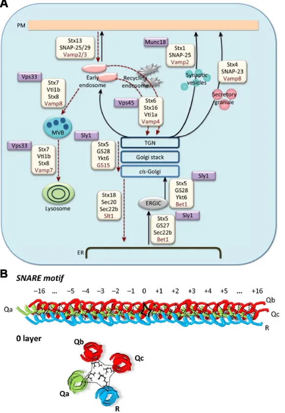
Vesicle fusion is a process that requires energy in order to overcome the electrostatic forces of repulsion between fusing membranes, and to form a fusion pore adjoining the two membranes (Hong et al., 2014, Südhof, 2012). SNARE proteins generate energy through their protein-protein and protein-lipid actions that synergize to promote membrane fusion. In the assembly stage of a SNARE complex, the adoption of a trans complex among Q- and R-SNAREs is necessary in order to bring two fusing membranes in close physical proximity to one another. As previously mentioned, SNARE complex assembly requires the intertwining of SNARE domains into a helical bundle (Golebiewska et al., 2014, Hong et al., 2014, Südhof, 2012) (Figure 4). The
Figure 4 of literature review. SNARE proteins promote vesicle fusion through quaternary structure formation. (A) SNARE proteins are diverse and reside in cellular organelles to mediate specific vesicle fusion events. V-SNAREs are in red and t-V-SNAREs in black; Munc18 and proteins stabilizing SNARE complex formation are noted in purple boxes. (B) Vesicle docking is mediated by the interaction of multiple SNAREs on donor and receptor
In order to generate the force necessary for fusion, Q- and R-SNAREs must form a
trans complex (Golebiewska et al., 2014). Indeed, part of the energy liberated during the
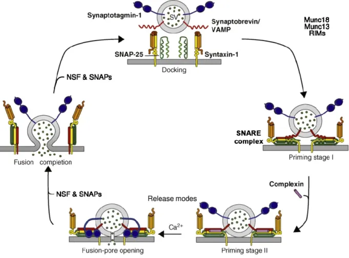
formation of a trans-SNARE complex is stored as mechanical energy in the semi-rigid zones that connect the SNAREs’ transmembrane domains with the helical bundle in a SNARE complex (Golebiewska et al., 2014, Hong et al., 2014, Südhof, 2012). That energetically unfavourable conformation is relieved when the vesicle and target membranes align with the SNARE complex (Figure 5). In this manner, this decrease in tension helps overcome the energy barrier for fusion, and the two membranes come together. At this point, trans-SNARE complexes are further stabilized in order for fusion to occur. This is mediated by complexin, which binds to the groove between the helices formed by Q- and R-SNAREs and stabilize the C-terminal region of the complex (Südhof, 2012, Südhof et al., 2011). In turn, this allows Syts (Syt) to trigger membrane fusion, an event that depends on Ca2+ levels. Syts bind Ca2+, which induces a conformational change that displaces complexin from the SNARE complex (Figure 5). Calcium binding also induces Syt to interact with the target membrane in order to form a fusion pore. After fusion occurs, SNARE complexes must be dissociated. This event is mediated by NSF, a member of the AAA+ family of ATPases. In conjunction with α-SNAP, NSF mediates the disassembly of trans-SNARE complexes in an ATP-dependent manner, a process that allows R-SNAREs to be recycled for future fusion events (Südhof, 2012, Südhof et al., 2011).
Figure 5 of literature review. The role of SNAREs in the vesicle fusion cycle. The natural repulsion that occurs when two membranes are in close proximity is overcome by SNARE interactions. Munc18 and complexin proteins mediate and stabilize trans-SNARE complex formation. This interaction brings the apposed membranes into very close proximity. Subsequently, a calcium influx activates Syt, which mediates fusion pore formation and content mixing. Following fusion, SNARE complexes are untangled by NSF and SNAP proteins, thereby permitting those SNAREs to participate in future fusion cycles. Adapted with permission from (Rodrigues et al., 2016, Südhof, 2012).
2.4.2 SNARE complexes that regulate ER ↔ Golgi transport
Trafficking among the ER, ERGIC and the Golgi apparatus is finely attuned by protein complexes that dictate the specificity and directionality of vesicle fusion events (Figures 4 and 6) (Appenzeller-Herzog et al., 2006, Malsam et al., 2011). Vesicle reconstitution assays have revealed the possibility of at least 147 Q/R-SNARE complexes that could regulate fusion among these organelles, of which five have been experimentally validated (Malsam et al., 2011, Parlati et al., 2002). In anterograde transport from the ER, COPII vesicle components have been found to interact with and select the SNAREs
components have been found to bind to peptide signals in the v-SNARE GOS28. When GOS28 is complexed with Stx5, the peptide signal is no longer accessible and cannot be recruited by COPII (Mossessova et al., 2003). Passage from the ER to the ERGIC and Golgi is governed by two complexes (Malsam et al., 2011, Volchuk et al., 2004). The first one is the Stx5-Membrin-Sec22b t-SNARE subcomplex, which pairs with v-SNARE rBet1. The second one regulates retrograde transport within the Golgi cisternae, and is constituted by Stx5-GOS28-Ykt6 (t-SNARE subcomplex) and Gs15 (v-SNARE).
In anterograde transport, COPI vesicle components have also been found to mediate the accrual of SNAREs. In particular, Rein and colleagues showed that the Arf1 GTPase recruits GOS28, and Sec22b; the latter acts as a v-SNARE in anterograde transport (Rein et al., 2002, Spang et al., 1998). In this regard, Sec22b pairs with Stx5/Stx18/Sec20/USE1 as possible cognate t-SNAREs (Dilcher et al., 2003, Spang et
al., 1998). Finally, arrival of material from early and late endosomes is packaged in
VAMP3 and VAMP4-positive vesicles, respectively. Those v-SNAREs pair with t-SNARE complex at the TGN and consist of Vti1a-Stx10 (VAMP3) and Stx16-Vti1a-Stx6 (VAMP4) (Figure 6) (Malsam et al., 2011).
2.1 The role of Syts in vesicle fusion
As mentioned previously, the SNARE-mediated targeting and docking of two membranes is not enough for vesicle fusion to occur (Fukuda, 2007, Pang et al., 2010, Südhof, 2012). Data from studies on neurotransmission has revealed that vesicles fuse with the synaptic cleft when an action potential occurs. This voltage stimulus triggers the opening of Ca2+ channels, which in turn increases intracellular [Ca2+] up to 100 nM, or ~15000X the concentration of extracellular calcium. This [Ca2+] is sensed by the Syt
protein, which in turn drives vesicle fusion at the synaptic cleft (Perin et al., 1990, Südhof, 2012). Syts regulate vesicle fusion in processes ranging from the exocytosis of synaptic vesicles and cytokines to phagocytosis and autophagy (Bento et al., 2016, Pang et al., 2010, Vinet et al., 2008). In neurons, Syt I and II are the most abundant. The importance of Syt is highlighted by knockout experiments where absence of Syt I/II severely decreased neurotransmission in fruit flies, worms and mice (Fukuda, 2007, Südhof, 2012). Indeed, Syts compose a family of more than 20 proteins that exist in eukaryotic organisms including plants and animals (Figure 7).
Figure 7 of literature review. Syts are conserved type I membrane proteins that drive membrane fusion. (A) Evolutionary tree of Syts and Syt-like proteins (Slp) showing the presence of calcium-binding C2A and C2B domains. Exceptionally, the C2 domains of Syt IV and XI do not bind calcium. (B) Structure of the C2A and C2B domains showing the calcium-binding regions, and the lipid-binding polybasic region. (C) Simplified mechanism of action by which Syt mediates calcium-mediated membrane fusion. Adapted with permission from (Fukuda, 2007, Lai et al., 2015).
All Syts are type I membrane proteins with their N-terminal domain (NTD) in the vesicle’s lumen (Fukuda, 2007, Pang et al., 2010, Perin et al., 1990, Südhof, 2012). The NTD is followed by a membrane-spanning α-helix that connects to its C-terminal domains (CTD) in the cytoplasmic side. The CTD in Syts is composed of two β-sandwich C2 domains, C2A and C2B (Figure 7) (Lai et al., 2015). These domains were originally identified in protein kinase C (PKC) and are connected by a nine amino acid linker (Perin et al., 1990). C2A binds to three Ca2+ cooperatively, whereas C2B domains bind to two Ca2+ ions (Lai et al., 2015, Südhof, 2012). Calcium binding to Syts induces an increase in the electrical potential of the C2B domain, thereby allowing it to bind to SNAREs and to the negatively-charged lipid phosphatidylinositol-4,5-bisphosphate (PI(4,5)P2) at the target membrane (Bai et al., 2003). Lipid interactions are mediated by
a polybasic region on the C2B domain (Lai et al., 2015, Perin et al., 1990). Moreover, calcium binding also allows the C2A domain to bind to phosphatidylserine (PS) on the target membrane. These CTD-lipid interactions allow Syt to bind to the target membrane, which causes curvature stress and membrane buckling. This membrane deformation is thought to induce contact and fusion of the two interacting membranes (Figure 7) (Perin et al., 1990, Südhof, 2012, Südhof et al., 2011).
2.1.1 Syt XI: an inhibitory Syt that does not bind calcium
Most members of the Syt family function as Ca2+ sensors of vesicle fusion by virtue of the C2 domains present in these Syts. However, Syts IV and XI possess a conserved serine in their C2A domain that precludes it from binding to Ca2+ and phospholipids (Pang et al., 2010, von Poser et al., 1997, Wang et al., 2010) (Figures 7 and 8). A Kyte-Doolittle plot revealed that murine SYTXI had a hydrophobicity profile similar to that of
other previously characterized Syts; of note is the highly hydrophobic N-terminal TM domain (Figure 8).