Université de Montréal
Characterization of the Mammalian homologs of the
Drosophila Melanogaster Endocytic Protein Lethal (2)
Giant Discs 1
by
Andréa Hébert-Losier
Molecular Biology Medecine
Masters’ thesis presented to the faculty of Medecine in partial fulfilment of the requirements for the degree of Master of Medecine
in Molecular Biology
April, 2010
Université de Montréal Faculté de médecine
This Master’s Thesis is named :
Characterization of the Mammalian homologs of the Drosophila Melanogaster Endocytic Protein Lethal (2) Giant Discs 1
Presented by: Andréa Hébert-Losier
has been evaluated by a jury composed of the following personel:
Sébastien Carréno, président-rapporteur Gregory Emery, Research director Michel Cayouette, member of the jury
Résumé
Endocytose joue un rôle dans l'activation du récepteur Notch. Des mutations dans le gène drosophilien lethal giant discs (lgd), provoque une prolifération cellulaire en perturbant l’endocytose de Notch. Les orthologues murins mlgd1 et 2 peuvent sauver ce phénotype, démontrant une fonction conservée. Cependant, des publications récentes suggèrent que les orthologs humains de lgd (hgd1/2) sont nucléaires. Dans cette étude, il est démontré que chez la Drosophile, le mutant dlgd08 provoque l'accumulation de Notch dans des vésicules et une surprolifération de neuroblastes . Ceci suggère que Notch est activé a l'intérieur des endosomes dans les neuroblastes. L’immunohistochimie de cellules Hela indique que hlgd1 et 2 ne sont pas nucléaires, mais associés à des strctures endosomales. Enfin, la baisse d’expression par shRNA des gènes murins mlgd1 et mlgd2 provoque une différenciation accélérée des cellules souches hématopoïétiques dans la lignée lymphopoïèse T et bloque la transition DN3 / CD4+CD8+, suggérant une suractivation de Notch.
Mots-clés : Endocytose, Notch, lethal giant discs (lgd), neuroblats, hématopoïèses, lymphopoïèse T
Abstract
Endocytosis plays a role in the activation of the Notch receptor. Mutations in the
Drosophila gene lethal giant discs (lgd), causes cellular overgrowth by perturbing Notch
endocytosis. This Drosophila phenotype is rescued by the murine orthologs mlgd1 and 2, indicating conserved function. However, recent publications suggest that the human orthologs (hlgd1/2) are nuclear. This study demonstrates that the dlgd08mutant in
Drosophila causes accumulation of Notch in vesicles and the overproliferation of
neuroblasts. This suggests Notch is activated from within endosomes in neuroblasts. Immunohistochemistry of Hela cells indicates that hlgd1 is associated with early endosome while, hlgd2 with later endosome and lysosome, and not with the nucleus. Finally, down regulation of murine mlgd1 and mlgd2 by shRNA caused an accelerated differentiation of hematopoietic stem cell into the T lymphopoiesis lineage and blocked the DN3 to CD4+CD8+ transition, suggesting that Notch is overactivated in these cells.
Keywords : Endocytosis, Notch, lethal giant discs (lgd), neuroblasts, haematopoiesis, T lymphopoiesis
Table of content
Page
Résumé ………... iii
Abstract ……….. iv
List of abbreviations ……….. vii
List of figures……….. xiii
Acknowledgments……….. xiv
Chapter 1: INTRODUCTION ………..……….. 1
1.1. Background and rationale for the current study ………... 2
1.2. Endocytosis……….... 4
1.2.1. Clathrin-Mediated Endocytosis……… 4
1.2.2. Ubiquitin-Regulated endocytosis………. 9
1.2.3. Caveolae-dependent endocytosis………. 10
1.2.4. The Early endosome……….… 11
1.2.5. The recycling endosome……….. 12
1.2.6. The ESCRT complex and sorting for the lysosomal degradation……... 13
1.3. Notch signalling………. 15
1.3.1. The role of endocytosis in Notch signalling……… 17
1.3.2. Notch and the hematopoietic system……….. 22
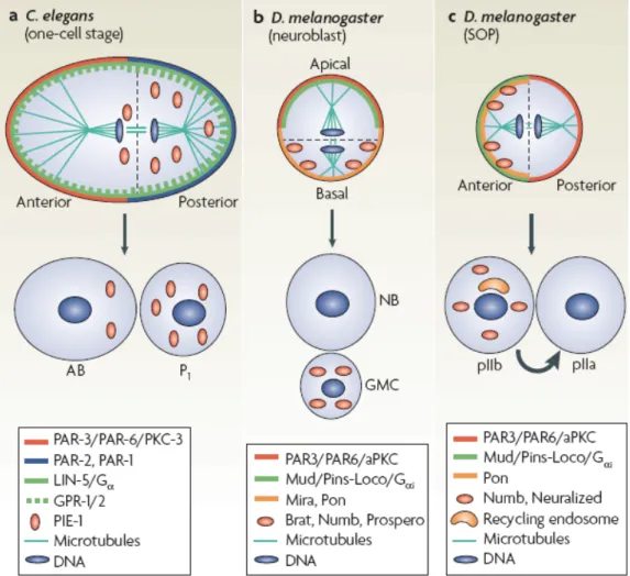
1.3.3. Asymmetric cell division and Notch Signalling………. 28
1.3.3.1. Asymmetric cell division of sensory organs ……… 29
precursor cells (SOP) 1.3.3.2. Asymmetric cell division of neuroblast……… 30
1.4. Notch and mammalian brain development……… 33
1.5. The drosophila l(2)giant discs (lgd) protein……….. 36
1.6. The mammalian lgd orthologs, coiled-coil and C2 domain……….. 38 containing 1 (CC2D1) protein A and B
Chapter 2: MATERIALS AND METHODS………. 41
2.1. Fly strains………..…. 42
2.2. Tissue staining and antibodies…………...……….. 42
2.3. Cell culture……… 42
2.4. Endocytosis assay……….. 43
2.5. Image acquisition………... 43
2.6. Lentiviral production and infection………... 43
2.7. RNA extraction, reverse transcription and RT-qPCR………... 44
2.8. Western blot……….. 45
3.9. T-cell differentiation assay……… 45
3.10. Flow-cytometry analysis and cell sorting……….. 46
Chapter 3: RESULTS………... 47
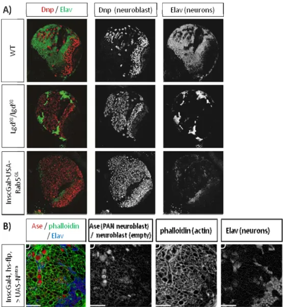
3.1. Perturbations of dlgd induce stem-cell tumours in the larval brain……… 48
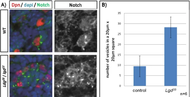
which resembles Notch overexpression phenotype 3.2. Expression of dlgd08 causes vesicular accumulation of Notch in neuroblasts…... 48
3.3. Proximal co-localization of hLgd1 with the early endosome and hLgd2 ………. 51
With the late endsome 3.4. Lentivirus production and knockdown of the murine mlgd1 and mlgd2……….. 54
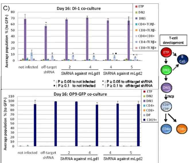
mRNA and protein levels 3.5. mlgd1 and mlgd2 modulates T-cell lineage development, most-like due………. 58
To Notch activation Chapter 4: DISCUSSION ……… 62
Bibliography……… 71 Appendices……….. I
List of abbreviations
µg Micro gram
5-HT1A Serotonin receptor ACD Asymmetric cell division
ADAM A disintegrin and metalloprotease AGL Apoptosis-linked gene
AGM region Aorta, gonads, and mesonephros region Akt serine/threonine protein kinase
AP-2 Adaptor-protein-2
aPKC Atypical protein kinase C
ARH Autosomal recessive hypercholesterolemia protein Arp Actin-Related Proteins
Ase Asense
ATP Adenosine triphosphate BAR Bin–Amphiphysin–Rvs domain bHLH Basic helix-loop-helix
C2 domain Calcium-binding motif
Ca2+ Calcium
CADASIL Cerebral autosomal dominant arteriopathy with subcortical infarcts and leukoencephalopathy
Cav Caveolin
CBP cAMP-response element-binding (CREB) binding protein CC2D1 Coiled-coil and C2 domain containing 1
CC2D1A Coil-coil containing domain
cDNA complementary deoxyribonucleic acid CLP Common lymphoid progenitors
CME Clathrin-mediated endocytosis Cre Cyclization recombination
CSL CBF1/Drosophila Su(H)/C. elegans LAG-1 C-terminus Carboxyl-terminus
DC Dendritic cell
Dl Delta-like
Dlg Discs large
dlgd Drosophila lethal giant discs DN Double-negative
DP Double positive
Dpn Deadpan
DRE Dual repressor element DSL Delta, Serrate and Lag 2
dx Deltex
E embryonic day
E’(Spl) Enhancer of Split
E. Coli Escherichia coli
E1 Ubiquitin-activating enzyme E2 Ubiquitin-conjugating enzyme E3 Ubiquitin-protein ligase
EE Early endosome
EEA1 Early Endosome Antigen 1 protein
EFC/F-BAR Extended FCH Homology / FCH- Bin–Amphiphysin–Rvs domain, EGF Epidermal growth factor
EHD Eps15-homology domain-containing protein ENTH Epsin N-terminal homology domain
ept Erupted
ESCRT Endosomal sorting complex for transport ETP Early T-cell precursor
FACS Fluorescence-activated cell sorting
Fbw F-box and tryptophan-aspartic acid (WD) repeat domain-containing Freud Five repressor element under dual repression-binding
FYVE domain Fab 1, YOTB, Vac 1 (vesicle transport protein), and EEA1 (Early Endosome Antigen 1 protein)
GLUE domain GRAM like ubiquitin binding in EAP45 domain
GMC Ganglion mother cell
GSL Glycosphingolipid
GST glutathione S-transferase
GTP Guanosine triphosphate
Gαi G protein subunit
HECT Homologous to E6-associated protein carboxyl-terminus domain Hes5 Hairy and enhancer of split 5
HIP1/R Huntingtin-interacting protein 1-related protein hlgd Human lethal giant discs
HOPS Homotypic fusion and vacuole protein sorting Hprt Hypoxanthine guanine phosphoribosyl transferase
Hrs Hepatocyte growth factor regulated tyrosine kinase substrate HSC Hematopoietic stem cell
Hsc70 Heat shock protein cognate of 70kDa
ILV Intralumenal vesicles
Insc Inscuteable
JAK Janus kinase
Kda kilodaltons
KO Knockout
LAMP Lysosomal-associated membrane protein
LBPA Lysobisphosphatidic acid LDL Low-density lipoprotein lgd Lethal giant discs
Lgl Lethal (2) giant larvae
LMPP Lymphoid primed multipotent progenitors LRF Leukemia/lymphoma-Related Factor
LTR-HSC Long-term repopulating hematopoietic stem cell
M/G myeloid
Mam Mastermind
Mib Mind bomb
Mira Miranda
ml Milli litter
mlgd Murine lgd lethal giant discs MOI Multiplicity of infection MPP Multipotent progenitors
Mvb Multi-vesicular body sorting factor MVB Multivesicular bodies
N1 Notch1 NB Neuroblast
Neur Neuralized
NEXT Notch extracellular truncation NICD Notch intracellular domain NK cell Natural-killer cell
NSCL Neuronal stem cell leukemia NSMR Non-syndromic mental retardation N-terminus Amino-terminus
Nuf Nuclear fallout
nuroD neurogenic differentiation
N-WASP Neural Wiskott-Aldrich syndrome protein PCAF P300/CBP-associated factor
PDK 3-phosphoinositide-dependent protein kinase
PEST domain Proline (P), glutamic acid (E), serine (S), and threonine (T) PH domain Pleckstrin homology domain
PI3K Phosphoinositide 3-kinase Pins Partner of inscuteable
Pon Partner of Numb
Pros Prospero
PSAP proline, serine, alanine, and proline amino acid motif PtdIns-3-P Phosphotidylinositol-3-phosphate
PX domain Phosphoinositide-binding structural domain Rab Ras-associated binding
Rab11FIP Rab11 family-interacting protein
RE Recycling endosomes
RING Really interesting new gene domain RNAi Ribonucleic acid interference SCD Spondylocostal dysostosis SEL-10 Suppressor/Enhancer of Lin-12 Ser Serine SH3 Src homology 3 domain
shRNA Small/short hairpin ribonucleic acid SIN-LTR Self inactivating long terminal repeats siRNA Small interfering ribonucleic acid
SJ1 Synaptojanin-1
SNARE proteins Soluble NSF attachment protein receptors
SNX Sorting nexin
SOP Organs precursor cells
SP Signal positive
STAM Signal transducing adaptor molecule
STAT Signal transducers and activators of transcription Su(H) Suppressor of Hairless
SV40 Simian virus 40 SVZ Subventricular zone
T-ALL Cell Acute Lymphoblastic Leukemia Tbp TATA box binding protein
TCR Pre-T-cell receptor
Thr Tyrosine Tsg101 Tumour susceptibility gene 101
TSP Thymus-seeding progenitor
TTK69 Tramtrack p69
UEV domains Ubiquitin-conjugating E2 variant domain UIM Ubiquitin interacting motif
Vps Vacuolar protein sorting factor
VZ Ventricular zone
WT Wild-type
WW domain Proline-rich peptide motifs WWC WW domain-containing protein
List of figures
Introduction:
Figure 1: Different types of endocytosis.
Figure 2: Clathrin (A) and Caveolin (B) mediated vesicles.
Figure 3: Models of Notch trafficking and activation in the endocytic pathway. Figure 4: Notch signalling in T- and B-cell development.
Figure 5: Asymmetric cell division in worms and flies.
Figure 6: Notch signalling in the developing and adult central nervous system.
Table:
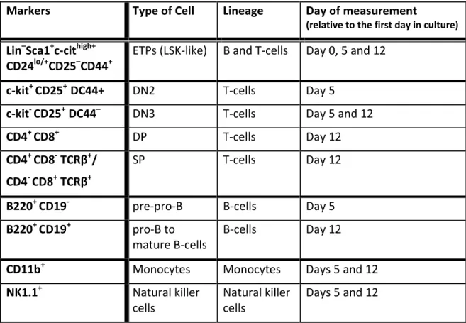
Table 1: In vitro lymphopoiesis of HMCs by OP-9 co-culture and flow cytometry markers used to identify the different lineages
Results:
Figure 7: Perturbations of the endocytic pathway induce stem-cell tumours in the larval brain which resembles Notch over expression phenotype.
Figure 8: Vesicular accumulation of Notch.
Figure 9: Proximal co-localization of hLgd1 with the early endosome and hLgd2 with the late endsome.
Figure 10: Down regulation of the mLgd1 protein and the mLgd2 mRNA after stable expression of ShRNA against mLdg1 and mLgd2 in NIH-3T3 cells.
Figure 11: shRNA knockdown of mLgd1 and mLgd2 in foetal liver cells speeds up development of progenitors into the T-cell lineage, but causes a block at DN3 stage.
Acknowledgments
I would first like to thank my supervisor Dr. Gregory Emery for the opportunity he provided me. Under his guidance and encouragement I was able to explore my scientific curiosities and learn new laboratory research techniques.
There are many people I would like to thank for their knowledge, guidance and regents they have provided to help me establishment of the lentivirus protocol. This includes Audette Karine from the Genomics Facility, Martin Blain from Dr. Sylvain Meloche’s lab and Jana Krosl from Dr. Guy Sauvageau’s. Also thanks to Marie-Elyse Lafaille-Magnan, a summer student in the lab, which work on this aspect of my project during the summer of 2009.
I would like to thank Dr. Claude Perreault and his post doctorate student Heinonen Krista for their help in establishing the T cell differentiation assay. Dr. Claude Perreault’s lab provided the cells and reagents needed to test out our hypothesis. Many thanks to Krista for showing me the experimental technique, how to analyse the collected data and the numerous hours problem solving the kinks of this experiment. Also thank-you to Danièle Gagné, from the Flow Cymometry Facility, for teaching me how to operate the FASC machine.
Thank-you to the members of my own lab, in particular the lab manager, Kris Ogoudikpe, for her assistance and feedback. I would also like to acknowledge my other colleagues, Gloria Assaker and Ramraj Velmurugan, who are no longer part of the lab, Carl Laflamme and Carole Iampietro of the Emery lab for the many scientific discussions, encouragements and shared laughs on late nights and weekends at the lab. Being able to share this experience with all of you made my time very enjoyable. Also thanks to the members of the IRIC Fly meeting, were the discussions issuing after my presentation have been most helpful insightful.
Finally, I would like to express a heartfelt thanks to my friends and family. First I would like to acknowledge my roommate and friend Sabrina Di Fulvio, who had supper ready when I got home late from the lab and whom I could went when my experiments were not working. Last but not least, thanks to my parents, Lynn and Ron, and to my
sisters, Lisa and Kim and my brother, Francis, for their encouragement. Though you may not quite understand what I do, it is thanks to your guidance and love throughout the years that has brought me to where I am today. Thank you.
1.1. Rationale of the current study
Endocytosis is the process where cell surface receptors and soluble molecules present at the plasma membrane or in the extracellular medium are internalized into intracellular compartments. It has been demonstrated that endocytosis can regulate numerous processes, including: nutrient uptake, receptor signalling[1], cell adhesion and migration[2], cell polarity[3], pathogen entry[4], antigen presentation[5], neurotransmission[6], mitosis[7], growth and differentiation[8], and drug delivery. The Notch signalling cascade has been shown to be regulated by endocytosis. The transmembrane Notch receptor has been demonstrated involved in cell fate specification in every animal species studied so far[9]. Notch signalling is essential to many developmental processes; this includes haematopoiesis[10], neurogenesis[11], and vasculogenesis[12] in mammals. It also regulates stem cell self-renewal, cell proliferation, cell differentiation and apoptosis. Loss of function of various components of the Notch signalling pathway causes inherited genetic diseases such as Alagille syndrome, spondylocostal dysostosis (SCD), and cerebral autosomal dominant arteriopathy with subcortical infarcts and leukoencephalopathy (CADASIL)[13]. Notch is also a known oncogene and tumour suppressor in mammals[14, 15]. In human, mutations in one of the Notch homolog (NOTCH1) are involved in 50% of T-Cell Acute Lymphoblastic Leukemia (T-ALL)[16]. Levels of the different Notch isoforms have been shown to be increased in almost all T-ALL. Growth arrest occurs in several T-ALL derived cell lines when Notch signalling is blocked, suggesting that modulating Notch activity may be a potent treatment strategy for some cancers[17].
Notch signalling is regulated at numerous levels. The Notch receptor itself undergoes a series of modification to insure proper response to the different ligands (Delta and Serrate in Drosophila and Delta-like and Jagged in mammals) and activation[18]. The expression and activities of the ligands are also tightly regulated. It has been demonstrated that endocytosis is necessary both in the signal-receiving cell and in the signal sending cell. Endocytosis of the ligands seems to produce a force on Notch that allows its photolytic cleavage by an ADAM-like proteases at the plasma membrane[19, 20]. This leads to another cleavage by presenilin which occurs most-likely in endosomes[21, 22].
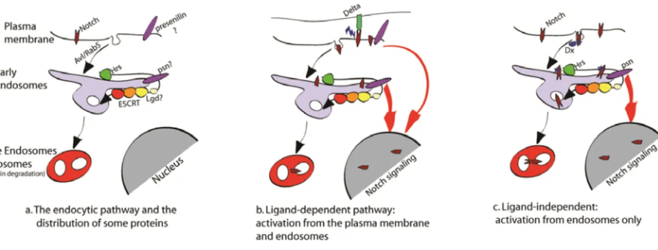
Furthermore, Notch has been shown to be downregulated in some cells by Numb, a protein that recruits the AP-2 adaptor complex, which in turn recruits clathrin and causes endocytosis of Notch[23, 24]. In some Notch expressing tissues, the absence of Numb can induce an accumulation of the receptor at the cell surface and an overactivation of Notch. This can lead to an overproliferation of cells and a tumour-like growth of tissues[25, 26]. Surprisingly, accumulation of Notch in endocytic compartments can also cause a Notch overactivation and overgrowth of proliferating tissues. For example, this is the case in
Drosophila when the endocytic proteins vps25 or erupted (ept) are missing[27, 28].
Interestingly, ept is the homolog of the tumour susceptibility gene 101 (tsg101) that has been shown to be implicated in numerous types of human cancer[29-33]. Finally, in some circumstances, Notch can also be activated through a ligand-independent mechanism. Accumulation of Notch in endosomes could favour this unusual mechanism[34-36].
The Drosophila gene lethal giant discs (lgd), named after its loss of function phenotype has been shown to play a role in the regulation of the Notch signalling. Mutations in this gene cause overgrowth of the imaginal discs through a perturbation in Notch endocytosis[37-40]. The mammalian homologs of lgd are named CC2D1A and B or Freud-1 and -2 or mLgd1 and 2. For homogeneity, the Drosophila Lgd will be named dLgd, the mouse homologs respectively mLgd1 and 2 and the human homologs hLgd1 and 2.
Though dLgd has been characterized to function in the endocytic pathway, its precise function is unknown. Even less is known of the mammalian lgd1 and 2. In a Drosophila loss-of-function experiment, both mlgd1 and 2 were able to rescue the dlgd loss-of-function phenotype, thus demonstrating a conservation of function between homologs[39]. However, recent publications suggest that they may act differently in mammals than in Drosophila[41-44]. In mammals, published data shows or strongly suggest that the Lgd homologs are not endocytic proteins but nuclear[41, 43, 44].
The aim of this study was to determine the role of the Drosophila and mammalian Lgd in the endocytic pathway and to verify the conservation of its function in mammals.
The literature review below provides a brief overview on the various forms of endocytosis and the role of endocytosis on Notch signalling. The role of Notch signalling in the hematopoietic system, brain and cancer development will also be looked at. A synopsis on what is known on Drosophila and mammalian Lgd will conclude this section.
1.2. Endocytosis
The plasma membrane is a dynamic structure that segregates the intracellular milieu (the cytoplasm) from the extracellular environment. It regulates and coordinates the entry and exit of small and large molecules from the cell. Small molecules, such as amino acids, sugars and ions, can pass through the plasma membrane via passive diffusion or through the action of integral membrane protein pumps or channels. The process of invagination and pinching-off of pieces of the plasma membrane, known as endocytosis, is needed to internalize macromolecules in membrane bound vesicles into the cell. There are multiple forms of endocytosis which can be place into two broad categories, ‘phagocytosis’ or cell eating (the uptake of large particles) and ‘pinocytosis’ or cell drinking (the uptake of fluid and solutes)[45]. Phagocytosis usually occurs in specialized mammalian cells such as macrophages, monocytes and neutrophils that function to clear large pathogens like bacteria, yeast, or large debris such as the remnants of dead cells and arterial deposits of fat[46, 47]. Pinocytosis occurs in all cells by at least one of four basic mechanisms: macropinocytosis, clathrin-mediated endocytosis (CME), caveolae-mediated endocytosis, and clathrin- and caveolae-independent endocytosis[45]. These mechanistically diverse and highly regulated endocytic pathways function to control such complex physiological processes as nutrient uptake, receptor signalling[1], cell adhesion and migration[2], cell polarity[3], pathogen entry[4], antigen presentation[5], neurotransmission[6], mitosis[7], growth and differentiation[8], and drug delivery.
1.2.1. Clathrin-Mediated Endocytosis
Clathrin-dependent endocytosis is the major pathway for the uptake of nutrients and signalling molecules in higher eukaryotic cells. The formation of endocytic-coated vesicles start by the recruitment of clathrin, adaptors and endocytic accessory proteins to the plasma membrane where they form lipid-rafts ranging from 10 to 500 nm in diameter[48]. Proteins to be endocytosed are linked to the plasma membrane by coat proteins that bind both phosphatidylinositol(4,5)bisphosphate (PtdIns-4,5-P2), which is the prevalent
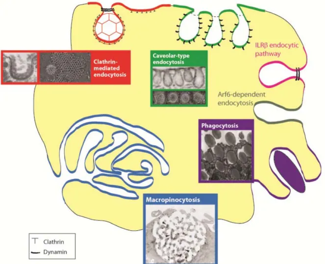
Figure 1: Different types of endocytosis. Transmission and scanning electron micrographs, and fluorescence micrographs, of structures known or thought to be involved in endocytic events. This figure was modified from Doherty and McMahon (2009)[50]. Note that there is controversy surrounding the morphologies of the IL2Rβ pathway and Arf6-dependent pathway. The arrowheads in the macropinocytic picture indicate cytoskeletal elements.
are four types of internalization signals identified located in the cytoplasmic tails of transmembrane proteins that are recognized by the endocytic adaptor proteins: the YXXΦ-, the [DE]XXXL[LI]-, the FXNPXY-type internalization signals and polyubiquitination. These signals are recognized by two types of endocytic adaptor proteins: the tetrameric adaptor-protein-2 (AP-2) and the monomeric adaptors ARH (autosomal recessive hypercholesterolemia protein), Dab2, β-arrestin, numb and epsin[51, 52]. Most adaptors are able to bind clathrin and the monomeric adaptors can also associate with AP-2. These adaptors are targeted to the plasma membrane through a phosphoinositide binding modules that associate preferentially with PtdIns-4,5-P2[49]. The AP-2 adaptor plays a central role
since it can associate with most endocytic proteins[53]. The loss-of-function of AP-2 subunits was proven to be lethal in Drosophila, C. Elegans and mice[54-56]. Also siRNA or RNAi mediated knockdown of AP-2 expression in HeLa cells eliminates about 90% of the endocytic clathrin-coated structures and blocks transferring uptake[57, 58]. However, even in the absence of AP-2 the Dab2 and epsin adaptors continue to mediate the uptake of LDL[58] and ubiquitinated cargo[59], respectively. These observations suggest that clathrin-mediated endocytosis can occur without AP-2.
There are several ways of bending a membrane which includes the enrichment of cone-shaped lipids in the cytoplasmic leaflet of the membrane, insertion of protein into the cytoplasmic leaflet, binding of coat proteins with intrinsic curvature, and force exerted by the cytoskeleton[60]. The presence of clathrin and AP-2 was demonstrated to be essential for the invagination of coated structures, however it is not sufficient[61]. Members of the ENTH, BAR and EFC/F-BAR protein families have been shown to induce, sense and stabilize membrane curvature. For example, upon binding to PtdIns-4,5-P2 Epsin inserts a
short amphipathic helix into the cytoplasmic leaflet of the plasma membrane thus bending the bilayer. Transient over-expression of the epsin ENTH domains cause tubular membrane structures in cultured cells[62]. However, when clathrin is depleted in HeLa cells, the presence of epsin and AP-2 is not sufficient to induce curvature[61]. This may be due to a requirement of clathrin for epsin to be able to curve the membrane or epsin may not be present in sufficient concentrations to bend the plasma membrane in the clathrin-deficient Hela cells. Depletion by RNAi of epsin in HeLa cells does not impede clathrin-dependent endocytosis of transferring but interfere with the uptake of ubiquitinated cargo[59].
It is thought that the clathrin lattice induces, directly or indirectly, changes in the curvature of coated membrane patches by its inherent property to form a curved polyhedral network. It is theorized that synergistic contributions obtained from interactions between adaptors and clathrin, and from proteins within the cytosolic membrane leaflet are required to effective bend the membrane. The elastic spring-like linkage between the clathrin lattice and the membrane might allow for local changes in the curvature of either the membrane or the clathrin lattice without too much resistance by the respective other. In order to convert the hexagonal facets of clathrin into pentagonal ones clathrin needs to associate with local promoter to increase the lattice curvature. There is evidence that the uncoating ATPase Hsc70 and its co-chaperone GAK/auxilin7 may be involved in this process. It has been demonstrated that low levels of GAK/auxilin [63] and of the 170 Kda isoform of synaptojanin-1 (SJ1), a phosphatidylinositol phosphatise, are recruited together with clathrin in growing coated patches at the plasma membrane[64].
The first event leading to scission and the release of a coated vesicle into the cytoplasm is the recruitment of dynamin GTPase. This most-likely occurs through curvature-sensing proteins[63, 65, 66]. Then cortactin, N-WASP, Arp2/3, actin, endophilin and the 145 kD isoform of Sj1 are recruited[64, 67]. GAK/auxilin is recruited in a short burst afterward[63]. The accessory protein HIP1/R, which is recruited together with clathrin into growing coated structures, connects the clathrin coat to actin filaments[68].
There are two theories on how these actin filaments help in scission of the coated vesicle into the cytoplasm. 1) The formation of actin filaments at the constricted neck of the budding vesicle may push the bud deeper into the cytoplasm and the increasing strain on the stalk may cause it to sever[49]. 2) Recent findings implicate the involvement of actin motor proteins in endocytosis. Myosin VI was shown to attach to coated vesicles through to the accessory protein Dab2 and PtdIns-4,5-P2 [69] and myosin 1E to dynamin and SJ1
through its SH3 domain[70]. So it is thought that the plus-end motor myosin 1E may pull the dynamin ring toward the plasma membrane while the minus-end motor myosin VI pulls the vesicle into the cytoplasm. The strain produced from these opposing forces would sever the constricted stalk beneath the dynamin ring[49].
Figure 2: Clathrin (A) and Caveolin (B) mediated vesicles. A) Diagram of the assembly and abscission of clathrin coated pits taken from Ungewickell & Hinrichsen, 2007[49]. The adaptor proteins, in this case AP-2, interact with both cargo protein and the cathrin lattice. Both the insertion of certain adaptor proteins into the membrane and the clathrin lattice help bends the membrane into the cell. Constriction of the vesicle neck by dynamin and additional pulling force provided by myosin motor proteins allow for vesicle fusion. B) Diagram of a caveola taken from Parton et al., 2007[71]. It demonstrates how caveolin is inserted into the caveolar membrane, with the N and C termini facing the cytoplasm and a putative ‘hairpin’ intramembrane domain embedded within the membrane bilayer. The scaffolding domain, a highly conserved region of caveolin, might have a role in cholesterol interactions through conserved basic (+) and bulky hydrophobic residues (red circles). The C-terminal domain, which is close to the intramembrane domain, is modified by palmitoyl groups that insert into the lipid bilayer.
1.2.2. Ubiquitin-Regulated endocytosis
Ubiquitination is a reversible, post-translational modification that is the result of the conjugation of small 76 amino acids ubiquitin (Ub) protein to a lysine residue on a target protein.[72, 73] Ubiquitin itself can be ubiquitinated on one of its 7 lysines and polymerize into either short chains (oligoubiquitination) or longer chains (polyubiquitination). The length and the topology of these chains determine the fate of the ubiquitylated protein. Modification of a protein by ubiquitin (Ub) can cause a remodelling of the targeted protein by affecting their stability, interaction with other proteins, enzymatic activity, and their subcellular localization[72, 73].
Successive action of the ubiquitin-activating enzyme (E1), ubiquitin-conjugating enzyme (E2) and ubiquitin-protein ligase (E3) is required for ubiquitylation of proteins. Based on structural features, E3s can be classified into two categories: 1) the E3s with a RING domain (Really Interesting New Gene), which act as molecular scaffolds to bridge the E2 and substrates together to allow the transfer of ubiquitin, and 2) the E3s with a HECT domain (Homologous to E6-AP C-Terminus), where the ubiquitin moiety is first covalently attaches to the E3 before it is transferred onto the substrate[72, 73]. Both E3s dictating the specificity of the ubiquitylation reaction since they usually interact directly with the substrate.
The Nedd4/Rsp5p family of ubiquitin-protein ligases regulate the stability of several yeast and mammalian transmembrane proteins by ubiquitination, and targets them for subsequent endocytosis. The Nedd4/Rsp5p family belongs to the Hect-domain superfamily of E3 enzymes. They are all composed of a variable N terminus, a C2 domain, 2 to 4 WW domains and a C-terminal Hect domain[74]. The C2 domain binds phospholipids and membranes in a Ca2+-dependent manner and plays a role in membrane targeting, intracellular localization and trafficking of proteins[75]. The WW (or WWP) domain is a small (~40 aa) protein-protein interaction module that allows Nedd4/Rsp5 to form multiple interactions and complexes with various proteins simultaneously. It usually recognizes and ubiquinates proteins that contains a conserved PPxY motif.
Once targeted plasma membrane proteins are ubiquitinated, they are recognized by Ub binding domains (UBDs) containing proteins such as the endocytic adaptors eps15 or epsin[76]. The ubiquitinated proteins are then endocytosed in a clathrin-dependent manner.
1.2.3
Caveolae-dependent endocytosis
Caveolae, which are flask-shaped invaginations of around 60–80 nm in diameter, are found in many mammalian cells including in smooth muscle, type I pneumocytes, fibroblasts, adipocytes, and endothelial cells[71]. Caveolin-1 (Cav1) and Cav2 are abundant in non-muscle cells, while Cav3 is found in skeletal muscle and in some smooth-muscle cells[77]. Transient expression of Cav1 is sufficient for de novo formation of caveolae in cells that do not express caveolin[78]. Ablation of CAV1 or CAV3 causes loss of caveolae formation in their respective cell types[79, 80]. Loss of CAV2 has no effect on caveola formation in vivo however there is some evidence that it may contribute to caveola formation in certain cell types[81]. All three caveolins have an unusual topology. They have a 33-amino acid intramembrane that forms a hairpin loop that inserts into the plasma membrane which leaves the N and C terminals in the cytoplasm[82]. Caveola formation by Cav1 and Cav3 involves oligomerization and association with cholesterol-rich lipid-raft domains. Cav1 binds to 1–2 cholesterol molecules[83] and is also palmitoylated in the C-terminal region[84]. The depletion of cholesterol has been demonstrated to disrupt caveolae structure[85]. Cav1 has been shown to bind the fatty acid tails of the glycosphingolipid (GSL) GM1 and can colocalize with GM1 and another “raft-associated” GSL, Gb3, in cellular membranes[86]. Caveolae endocytosis is dependent on dynamin. In endothelial cells dynamin is constitutively localized to the neck of caveolae[87], or is recruited in response to specific signals, such as stimulation by the SV40 virus[88]. The SV40 virus enters the cell partially through caveolae. It binds to GM1 and causes a transient recruitment of dynamin, which is followed by a burst of actin polymerization. The actin forms a tail that pushes the caveolae into the cell[88]. This sequence of events is similar to that observed in clathrin endocytosis.
Phosphorylation appears to play an important part in caveolae budding. It has been shown that the use of the general phosphatase inhibitor okadaic acid and the tyrosine
phosphatase inhibitor vanadate can stimulate caveolae internalization[88, 89]. However in contrast, the Src family of kinases has been demonstrated to be able to phosphorylate both Cav1 and dynamin and that phosporylation of dynamin is directly involved in caveolae internalization[88, 90]. Thus, the precise role of these phosphorylations is presently unclear.
1.2.4. The Early endosome
The early endosome (EE) is the first endocytic compartment to receive incoming internalized cargo from the plasma membrane. It is a highly dynamic structure that can undergo homotypic fusion[91]. The EE is composed of two distinct regions: 1) thin tubular extensions and 2) large vesicles that have membrane invaginations. These EE sub-domains appear to have different function. It is thought that proteins targeted for recycling may cluster within the tubular membranes, while proteins destined for degradation concentrates on intralumenal vesicles that accumulate within the vacuolar domains, giving rise to multivesicular bodies (MVBs)[92]. Vesicles generated from these two regions have different acidification properties. In the lumen of MVB, the pH decreases from 6.2 to ~5.5, while in the tubular recycling endosomes it increases to ~6.5[92].
The Ras-associated binding (Rab) proteins are GTP-binding proteins that are important endocytic regulators. In their active GTP-bound state, they can recruit and interact with Rab effectors. The Rab proteins have multiple roles in endocytic trafficking events, including vesicle tethering, fusion, budding and motility[93, 94]. The Rab proteins primarily localized to the EE include Rab5 and Rab4, which regulate distinct early endocytic events. Rab5’s main role is to regulate entry of the cargo from the plasma membrane and subsequent fusion of vesicles with early endosomes[95, 96]. Rab5 has multiple secondary functions as well, which include: generation of phosphotidylinositol-3-phosphate (PtdIns-3-P) lipid which is enriched on EE[97], homotypic fusion[98], the motility of EE on actin and microtubules tracks[99], and functions in activating signalling pathways from EE[100]. Rab4 is localized in the EE[101] and in Rab11-positive recycling endosomes (RE)[102] as well. It helps regulates both rapid recycling of proteins from the EE and the slow recycling of proteins from the RE back to the cell surface[103].
Phospholipids play a role in recruiting proteins to specific domains of the EE. Active Rab5 recruits PtdIns-3-P kinase/Vps34 to EE membranes where it mediates localized synthesis of PtdIns-3-P[97]. This localized accumulation of PtdIns-3-P can recruit PtdIns-3-P-binding proteins containing a FYVE domain, such as EEA1 or Hrs, and containing PX domain, like Syntaxin 1[104, 105]. The recruitment of EEA1 can cause the homotypic fusion between EE through the assembly of SNAREs Syntaxin13 and Syntaxin6 at the EE membranes[106]. While the recruitment of Hrs is essential for the sorting of ubiquitinated proteins into MVBs, which are then targeted for lysosomal degradation.
1.2.5. The recycling endosome
Upon delivery to EE, internalized cargo can be sorted into one of at least two distinct recycling pathways. Proteins sorted into the newly formed tubular membranes of the EE are recycled back directly to the plasma membrane. In parallel with tubulation, Rab4 targets proteins to the recycling endosome (RE) where they proceed via a 'slow-recycling' route back to the plasma membrane[107].
Rab4 has been identified to also be involved in the fast recycling of transferrin receptor[108] and glycosphingolipids[109] from the EE. However its exact role in recycling is unclear for expression of dominant-negative Rab4 inhibits fast recycling but not slow recycling and siRNA-mediated down-regulation of Rab4 increases rapid recycling[107, 108]. Recent studies have indicated that Rab35 is an important regulator of rapid recycling. Rab35 has been shown to localize both at the plasma membrane and EE, and to be required for rapid recycling of the mammalian transferrin receptor[110] and the C. elegans low-density lipoprotein receptor-like yolk receptor[111].
The slow recycling route involves the transport of cargo proteins from the EE to the RE and from the RE to the plasma membrane. The EH domain-containing protein (EHD) proteins appear to play many roles in the RE. EHD4 is involved in the export of cargo from the early endosome to both the RE and late endosome[112], while EHD3 act as linker between the Rab5-associated early endosome and the Rab11-associated RE. It can bind to the Asn–Pro–Phe motifs of the Rab5 effectors rabenosyn and Rab11 family-interacting protein 2 (Rab11FIP2)[113]. Rab11FIP5, another Rab11 effector, is important in the
movement of transferrin receptor from the EE to the RE. SiRNA down-regulation of Rab11FIP5 inhibits the transport of transferrin receptor from the early endosome to the RE[114]. Rab11FIP3 interacts with both Rab11 and Arf6 and is important for the juxtanuclear positioning of the RE[115].
The sorting nexins protein in yeast, and its mammalian ortholog, sorting nexin 4 (SNX4), associate with tubular vesicular elements on the both EE and RE, and target cargo for the recycling pathway. SNX4 interacts with the dynein motor through the linker protein WW domain-containing protein 1 (WWC1), facilitating the movement of the EE or RE to the juxtanuclear region[116]. Down-regulation of SNX4 perturbs transport between these compartments and causes lysosomal degradation of the transferrin receptor[116].
1.2.6. The ESCRT complex and sorting for the
lysosomal degradation
Mono-ubiquitination of one or more lysine residues of endocytosed cargo serves as recruiting signal for the endosomal sorting complex for transport (ESCRT) machinery[117]. The ESCRT machinery has three main functions: 1) it recognizes ubiquitylated cargoes and prevents their recycling and/or retrograde trafficking; 2) it deforms the endosomal membrane allowing cargo to be sorted into endosomal invaginations; 3) it catalyses the final abscission (breaking off) of the endosomal invaginations, forming intralumenal vesicles (ILVs) that contain the sorted cargo[118]. The hepatocyte growth factor-regulated tyrosine kinase substrate (Hrs), a member of the ESCRT-0 complex, is first recruited to the EE through binding of its FYVE zinc-finger domain to PtdIns-3-P[105]. Mono-ubiquitinated cargo on the EE is initially recognized by Hrs via its ubiquitin interacting motif (UIM)[105]. Hrs can also associates with clathrin through its C-terminal clathrin box motif. Hrs has been found on clathrin lattices present on select EE membranes involved in the sorting of cargo to the degradation pathway[118]. Binding of Hrs to ubiquitin and clathrin results in the concentration and sorting of cargo into ILVs vesicles, thus away from the tubule-forming and actively recycling membranes. However, Hrs binds to ubiquitin with a low affinity[105, 119], thus two additional
UIM-containing proteins make up the ESCRT-0 complex. Eps15 and signal transducing adaptor molecule (STAM) 1 and 2, stabilize the association of ubiquitinated cargo with Hrs[120]. Furthermore, Hrs interacts with the Tsg101 subunit of the endosomal sorting complex required for transport (ESCRT-I), thus recruiting it to membranes[120].
ESCRT-I is composed of four subunits: tumour susceptibility gene (Tsg) 101, vacuolar protein sorting (Vps) 28, Vps37 and multi-vesicular body sorting factor (Mvb)12[121, 122]. The ESCRT-I complex is recruited to the endosome through the interaction of the amino acid motif proline, serine, alanine, and proline (PSAP) of Hrs with the ubiquitin-conjugating E2 variant (UEV) domains of Tsg101[123]. It is believed that the ubiquitinated cargo is then handed off from Hrs to Tsg101, which also contains a UIM[123]. ESCRT-II is recruited by Vps28, which is located at the end of a rigid 13-nm stalk opposite to the binding sites for ubiquitinated cargo[124]. With such a distance between the ubiquinated cargo and the ESCRT-II complex, it argues against the ‘conveyor belt’ concept. This concept was first suggested in a study by Babst, M. et al, 2002 and supported by subsequent studies on ESCRT proteins. This model states that the recruitment of the ESCRT complexes occurs sequentially to the endosome. The ESCRT complexes recognize ubiquitylated transmembrane proteins and pass the ubiquinated cargo from one complex to the next facilitating sorting to MVB vesicles[125]. It is thought that ESCRT-I and ESCRT-II may co-assemble and cluster multiple ubiquitinated proteins for packaging into ILVs[124]. ESCRT-II is composed of Vps22, Vps36 and Vps25. The c-terminal domain of Vps28 binds to the N-terminus of the Vps36. The N-termini of Vps36 also contains a pleckstrin homology (PH) domain variant called a ‘GLUE’ domain. This GLUE domain binds to both phosphoinositides and ubiquitin[126]. There are six ESCRT-III core proteins in yeast, Vps2, Vps20, Vps24, Snf7, Did2, and Vps60. Vps20 is first recruited to the endosome and activated by Vps25[127]. It forms dimers with Snf7 and associates with Vps32. This association with Vps20 triggers the assembly of Vps32 into filamentous oligomers that are capped by Vps24[128]. Vps2 then associates with the Vps24 cap to mediate recruitment of the ATPase Vps4[128]. The ESCRT-III subcomplex of Did2 and Vps60 recruitments and regulates the activity of Vps4[129].
There is some evidence of ESCRT-mediated membrane deformation obtained with ESCRT-III. Overexpression of the ESCRT-III subunit Vps32 in COS-7 cells caused the
formation of spiral filaments composed of Vps32 multimers, which lead to the protrusion of buds and tubules from the plasma membrane[130]. The topology and diameter of the buds and tubules on the plasma membrane resemble those of ILVs, suggesting that Vps32-mediated plasma-membrane budding reflects key aspects of ILV biogenesis[130]. They are several lines of evidences suggesting that Tsg101 and one of its interacting partners, Alix (apoptosis-linked gene-2 (AGL-2) interacting protein X), are also involved in membrane deformation and intralumenal vesicles budding. The proline-rich domain located at the c-terminal of Alix interacts with Tsg101. Addition of purified recombinant Alix inhibits formation of intralumenal vesicles in the late endosomes in a dose-dependent manner while siRNAs depletion of Alix stimulated formation of intralumenal vesicles[131]. The reverse was observed when Tsg101 was knocked down by siRNA[131]. Alix was also found to interact with the phospholipid lysobisphosphatidic acid (LBPA), which is commonly found within the internal membranes of late endosomes. It was demonstrated that Alix controls LBPA’s ability to drive the formation of membrane invaginations within acidic liposomes in vitro and helps organize LBPA-containing endosomes in vivo[132]. Recent studies done in Hela cells have identified the PtdIns-3-P-binding protein SNX3 as to be involved in ILV biogenesis. Depletion of SNX3 by RNAi prevents ILV biogenesis but not receptor sorting, indicating that it has a specific role in membrane deformation[133].
Abscission is thought to be mediated through the ATP hydrolyse activity of the ESCRT-III complex protein Vps4. The ESCRT-III subunits assemble into circular arrays on the LE. It is thought that Vps4-mediated removal of individual Vps32 subunits from one end of the spiral polymer at the neck of the invagination could cause sufficient constriction to mediate membrane scission[134]. It is also possible that ESCRT-III mediated clustering of cargo with bulky intraluminal domains could also contribute to both membrane deformation and abscission. Released MVB are transported and fused to the lysosome in a rab-7 dependent manner[135].
1.3. Notch signalling
The transmembrane Notch receptor was named in the early 1900s after a dominant X-linked Drosophila genetic mutants that exhibit irregular notches at the wing tips [136,
137]. However, it wasn’t until the 1940s that Notch was shown to have a developmental role when complete loss of Notch gene activity was found to cause lethal hyperplasia of the embryonic nervous system[138]. Today it has been demonstrated that Notch signalling is essential to many developmental processes; this includes hematopoiesis[10], neurogenesis[11], and vasculogenesis[12] in mammals. It is involved in cell fate specification in every animal species studied so far[9]. Notch also regulates stem cell self-renewal, cell proliferation, cell differentiation and apoptosis. Loss of function of various components of the Notch signalling pathway causes inherited genetic diseases such as Alagille syndrome, spondylocostal dysostosis (SCD), and cerebral autosomal dominant arteriopathy with subcortical infarcts and leukoencephalopathy (CADASIL)[13]. Notch is also a known oncogene and tumour suppressor in mammals[14, 15]. Notch signalling can be either oncogenic or antiproliferative, in different types of cancers. In human hepatocellular carcinoma[139] and small cell lung cancer[140], Notch signalling is antiproliferative rather than oncogenic. However, studies have shown that Notch mostly functions as oncogene in human cancers. The expression of the Notch receptor and its ligans are up-regulated in cervical, lung, colon, head and neck, renal carcinoma, acute myeloid, Hodgkin and large-cell lymphomas and pancreatic cancer (as reviewed in [141]). Moreover, high-level expression of Notch-1 and its ligand Jagged-1 is associated with poor prognosis in breast[142] and prostate cancer.
Notch signalling regulates cell fate decision (or differentiation) by three general mechanisms during both development and adult life:
1) Inductive signalling occurs at the interface between two fields of cells, one of which is the signal-sending cell that presents the DSL family (Delta, Serrate and Lag 2) of Notch ligands to the signal-receiving cell that express the Notch receptor in neighbouring tissue. This type of interaction results in the formation of tissue boundaries, as is the case during fly wing and vertebrate limb development[143]. The dorsal/ventral boundary of the wing disc of drosophilas is formed by a stripe a few cell rows wide. In the dorsal cell, the Notch ligand Serrate is expressed and Notch is rendered insensitive to Serrate so that Serrate cannot activate Notch in these cells[144, 145]. Thus only the Notch receptor in the ventral cells across the boundary can be activated by Serrate. The reverse is true for the Notch
Delta ligand. Delta is expressed in the ventral cells, but can only activate Notch in the dorsal cells across the boundary[144, 145].
2) Lateral inhibition is the selection of one cell from a group of equivalent precursors. Initially all cells express both ligands and Notch receptor. However, as development progresses stochastic variations in gene expression cause small differences in Notch activation in the various cells so that in the end the ligand is restricted to a single cell while Notch is activated in its neighbours[143]. This can be seen in the equal spacing of sensory organs in the fly’s sensory bristles on the thorax and with the hair cells in the ear of vertebrates[146, 147].
3) Asymmetric division occurs when a mother cell gives rise to two daughter cells with different fates. In some cases the two daughters are initially identical at birth and undergo the acquisition of different cell fate later on. Alternatively, the mother cell can become polarized and the two arising daughters receive different cell fate determinates at birth[148]. Stem cell epistatisi is thought to be regulated through this mechanism. When stem cells divide, one of the daughter cells retains stem cell status while the other daughter cell obtains cell fate determinates and differentiates. The dividing sensory organs precursor cells (SOP) is an excellent model system for asymmetric cell division, since the two resulting daughter cells receive different cell fate determinates[148]. The asymmetric division of SOPs yield two secondary precursor cells, the anterior cell, pIIb, and the posterior cell, pIIa. The pIIb receives cell fate determinants that activate the Delta ligand and down-regulate the Notch receptor, while the pIIa maintain an active Notch receptor at the plasma membrane[148, 149]. Activation of Notch signalling in the pIIa will gives rise to a socket cell and a hair cell, whereas the lack of Notch signalling in the pIIb will give rise to a neuron and a sheath cell[148, 149].
1.3.1. The role of endocytosis in Notch signalling
The Notch receptor is synthesized as a 300 kDa precursor protein which is cleaved in the trans-Golgi compartment by furin in Drosophila and furin-like convertases in mammals. Cleavage occurs in the extracellular/lumenal domain which results in the generation of N- and C-terminal fragments. These fragments are joined by a non-covalent
linkage and create the mature Notch heterodimer[150, 151]. The extracellular cellular domain of Notch contains numerous potential sites for N-linked and O-linked glycosylation and does undergoes extensive N- and O-linked glycosylation during Notch synthesis and secretion[152, 153]. These modifications are important for proper folding of the receptor and alter the responsiveness to the different DSL ligands[152, 153], which includes Delta and Serrate in Drosophila, and Delta-like and Jagged in mammals.
The canonical Notch signalling pathway (Figure 3.B.) involves activation of the Notch receptor at the cell surface by ligands of the DSL family. Both the Notch receptor and DSL ligands are composed mostly of type I single-pass integral membrane proteins with extracellular domains composed of tandem EGF-like repeats[154, 155]. The N-terminal domain of the ligand binds directly to the EGF-like repeat 11-12 region of the Notch receptor[156]. The ligand on the surface of a signal-sending cell must be internalized to activate the Notch receptor on the signal-receiving cell[157]. The DSL ligands are endocytosed through recognition of a ubiquitination signal. Neuralized (Neur) and Mind bomb (Mib) are two RING finger-containing E3 ubiquitin ligases that can ubquitinate the DSL ligans[158, 159]. This promotes the recruitment of liquid facet in Drosophila, Epsins in mammal, which contain a ubiquitin-binding domain. Espsin then binds the PtdIns-4,5-P2
and associates with Clathrin and other accessory proteins[160] to induce clathrin-mediated endocytosis of the DSL ligans.
It is thought that DSL ligands may undergo constitutive endocytosis and recycling to and from the plasma membrane to produce active ligands[159]. It has been demonstrated that following asymmetric cell division of SOPs in Drosophila, Delta is concentrated in recycling endosomes in pIIb cells. In loss of function studies, expression of either rab11 or
sec15 mutants, which function together to recycle proteins back to the cell surface, produce
cell-fate transformations to a phenotype consistent with loss of Notch signalling, which is thought to be due DSL ligand inactivity[161, 162].
When Notch from the signal-receiving cell associates with the DSL ligand from the signal-sending cell, the endocytosis of the ligand on the signal-sending cell, known as transendocytosis, induces a physical force on the Notch receptor on the signal receiving cell[20]. This force destabilizes the non-covalent bonds of the Notch heterodimers structure exposing the ADAM cleavage site and allowing for proteolytic activation of the Notch
receptor[20]. The S2 cleavage site is located in the extracellular portion of the Notch C-terminal fragmen and is cleaved by the metalloproteases ADAM10/TACE in mammals, Kuzbanian in Drosophila, are responsible for t[163-165]. This S2 cleavage removes the ectodomain resulting in a membrane-anchored Notch form termed Notch extracellular truncation (NEXT).
It is then believed that NEXT must first be endocytosed before it can undergo the secretase-mediated S3 cleavage in the transmembrane domain of the Notch receptor. The γ-secretase activity is provided by the protease presenilin[166]. It has been demonstrated that inhibition of NEXT endocytosis by using dominant-negative forms of either Dynamin2 or Eps15 blocks γ-secretase processing of NEXT[167]. It was also found that NEXT can be monoubiquitinated at a conserved lysine residue, and mutation of this lysine reduced both NEXT internalization and S3 cleavage [167]. NEXT endocytosis may also be required for activated presenilin to have access to the S3-cleagave site. It is thought that biologically active presenilin pools are located in intracellular compartments. It has been shown that large pool of active presenilin complexes are found in lipid rafts within the endosomal pathway[168].
The transcription factors of the conserved mammalian CBF1/Drosophila Su(H)/C.
elegans LAG-1 (CSL) family are the primary nuclear effectors of Notch signalling. In the
absence of Notch activation, the CSL proteins act as transcriptional repressors with other known co-repressors on Notch target genes[169]. In Drosophila, CSL promotes the recruitment of the Asf1 histone chaperone to silence many Notch genes[170]. Upon γ-secretase proteolytic cleavage by persenilin in the transmembrane domain of NEXT, the Notch intracellular domain (NICD) is released. The NICD fragment then translocate into the nucleus where it physically binds to CSL and, together with the co-activator Mastermind (Mam in Drosophila and mammals; LAG-3 in C. elegans), forming a transcriptionally active ternary complex[171, 172]. Once formed, the complex recruits general transcription factors, such as CBP/p300 and PCAF, and promotes chromatin acetylation and expression of the Notch target genes[173, 174].
Once generated, the NICD signalling fragment can no longer be regulated by ligand binding or other cell-surface events. Disassembly of the CSL/NICD/Mam ternary complex and signal attenuation is mediated by ubiquitination and proteosomal degradation. NICD is
first phosphorylated by cyclin-dependent kinase 8 in its C-terminal PEST domain and then targeted for degradation by the E3 ubiquitin ligase Fbw7 in mammals and SEL-10 in C.
elegans[175, 176]. It is important to tightly controlled NICD turnover to prevent sustained
signalling for an inappropriately long period or at an excessively high level. It has been shown that some cases of T-ALL have mutations that delete the Notch PEST domain, which lead to an inability to degrade NICD properly[177].
The Notch receptor can also be targeted for lysomal degradation through ubiquitination directly from the plasma membrane. The E3 ubiquitin ligases Suppressor of deltex (Drosophila), Itch (mammals), SEL-10 (C. elegans)[178], and Cbl (Drosophila and mammals)[179], have been identified that target non-activated Notch for degradation[36, 180]. Numb is a conserved membrane-associated protein that acts upstream of the γ-secretase cleavage to block Notch signalling[23]. In 3T3 cell culture assay, Numb was shown to interact with Itch to promote Itch-dependent ubiquitinylation, endocytosis and degradation of Notch1[181]. Numb interacts with both Notch and AP-2, thus helping to recruit the components of the clathrin-dependent endocytosis pathway to internalize the Notch receptor[23, 24]. In some Notch expressing tissues, the absence of Numb can induce an accumulation of the receptor at the cell surface and an overactivation of Notch. This can lead to an overproliferation of cells and a tumour-like growth of tissues[25, 26].
Ubiquitination of the Notch receptor can also act positively on Notch signalling and influence the endosome trafficking of the Notch receptor[182] (Figure 3.C.). Deltex (dx), an E3 ubiquitin ligase containing a RING domain, has been shown to physically associate with Notch and the β-arrestin, Kurtz in Drosophila, promoting internalization of the whole complex[183]. Loss-of-function of dx has no affect on mutant fly viability and fertility, however Notch-like patterning defects were observed, suggesting that in some tissues Notch signalling is activated in a Deltex-dependent manner[35]. Resent genetic studies on the Drosophila HOPS and AP-3 complexes revealed that they are required for Deltex-dependent, ligand-independent Notch signalling[184]. They help deliver intact, non-ligand-activated Notch to the limiting membrane of the lysosome. This leads to the accumulation of the Notch receptor, ectodomain shedding and/or degradation, and γ-secretase-mediated activation of the receptor[184]. The HOPS complex act in late endosome maturation and lysosomal fusion, while the AP-3 complex act in endosomal trafficking of proteins to the
Figure 3: Models of Notch trafficking and activation in the endocytic pathway. a. Schematic representation of the main steps of the endocytic pathway. b. Schematic representation of the ligand dependent activation of Notch. Notch can be activated either at the plasma membrane or in the endosome. c. Schematic representation of the ligand-independent activation of Notch.
limiting membrane of the lysosome.
The ESCRT complex is another component that acts in ubiquitin-dependent endosomal sorting pathway that influence Notch signalling. Drosophila mutant cells for either ept, the drosophila homolog of tsg101, or vps25, two components of the ESCRT-I complex, undergo autonomous neoplastic transformation and also non-autonomous cell proliferation. This is due mostly to the activation of the Notch receptor that induces hyperproliferation of mutant cells as well as adjacent wild-type cells[27, 28, 185]. An increase of Notch signalling in mutant cells leads to the ectopic production of Unpaired, a ligand that activates the mitogenic JAK-STAT pathway in neighbouring wild-type epithelial cells. The Notch receptor accumulates in enlarged endosomes in ept and vps25 mutant cells resulting in increased activation of Notch signalling[27, 28, 185]. RNAi knockdown of vps25 in Drosophila S2 cells increased S3 cleavage of Notch receptor by presenilin, an effect that was inhibited with the use of a presenilin inhibitor[185].
1.3.2.
Notch and the hematopoietic system
During mouse embryogenesis, primitive hematopoiesis arise from mesodermal precursors, known as hemangioblasts, that migrate and become committed to endothelial and hematopoietic progenitors in the yolk sac blood island at embryonic day 7.5 (E7.5)[186]. Primitive hematopoiesis primarily consists of nucleated erythrocytes with embryonic-type globin. Hematopoiesis gradually shifts to the fatal liver after E11.5 and later to spleen and bone marrow[186]. Hematopoietic progenitors and lymphopoietic cells that will give rise to adult-type blood cells are detected in the paraaortic splanchnopleura region of mouse embryo at E7.5–9.5[187, 188]. The long-term repopulating hematopoietic stem cells (LTR-HSCs) that can reconstitute the adult mice hematopoietic system originates from the intraembryonic aorta, gonads, and mesonephros (AGM) region at E10.5– 11.5[189-191]. In the adult mouse LTR-HSC can self-renew and give rise to short-term HSCs. These ST-HSCs self-renew for a short period of time and eventually give rises to non-selfrenewing multipotent progenitors (MPPs). Over time the MMPs lose their multipotent capacities and they become either common myeloid progenitors[192] or common lymphoid progenitors[193]. Common lymphoid progenitors (CLP) can
differentiate into B, T, natural killer and dendritic cells[193]. The common myeloid progenitors can mature to give rise to two other sets of progenitors: the granulocyte/macrophage progenitors and the megakaryocyte/erythrocyte progenitors[192]. Recently, circulating T cell progenitors has been identified in blood, suggesting that commitment toward the T cell lineage can occur extrathymically[194, 195].
The major progenitor source that enters the T cell pathway in adult mice is the lymphoid primed multipotent progenitors (LMPPs) found in both the bone marrow and peripheral blood. These cells can give rise to macrophages, dendritic cells, natural killer cells, B cells and T cells, but not erythrocytes or megakaryocytes[196, 197]. These cells migrate to the thymus, where the LMPPs are exposed to various factors that triggers and supports pro-T cell differentiation, proliferation and survival[198]. The earliest described intrathymic progenitors and are the early T-cells/thymic progenitors (ETPs), which are also known as CD4–CD8– double-Negative 1 (DN1) cells. When sorted by flow cytometry (table 1) ETPs display a Lin–Sca1+c-kithigh+CD24lo/+CD25–CD44+ surface phenotype[199, 200]. At this stage, DN1 retain a weak potential to differentiate into the B lineage potential both
in vivo and in vitro, however they still have the ability to differentiate into natural killer
cells and some dendritic and myeloid cells[201, 202]. DN2s (c-kit+DC44+CD25+) retains some potential to differentiate into lineages other then T-cells[201]. True commitment to T lineage occurs only at the DN3 (CD44-CD25+) stage after completion of pre-T cell receptor (TCR) β rearrangement is completed (DN4) (figure 4) [203]. The TCR is specific to T cell and is expressed at the cell surface. It is generally responsible for recognizing antigens bound to major histocompatibility complex. DN4 then become mature CD4+CD8+ double-positive (DP) T cells. These DP T cells go on to differentiate into different subset of T cells, single positive (SP) CD4+ or CD8+ T cells[204].
The implication of Notch in the hematopoietic system was first discovered though the identification that Notch plays a critical function of in T lineage development. Neonatal mice that expressed a loss of function Notch1 had a severe deficiency in thymocyte development[205]. Also, in competitive repopulation of lethally irradiated wild-type mice bone marrow with wild-type- and Notch1-deficient bone marrow, a blockage in T cell development was observed at an early stage[205]. In a concurrent experiment, when the constitutively active form of Notch1 was expressed to reconstitute irradiated bone marrow,
the repopulated bone marrow was mostly composed of immature CD4+ CD8+ T cells and there appeared to be a simultaneous block in early B cell lymphopoiesis[206]. At the time, these results suggested that Notch1 was a key regulator in determining T lymphoid versus B lymphoid lineage[205, 206]. Subsequent studies have demonstrated that Notch signalling is important in many hematopoietic lineages and plays essential roles at several stages of T cell development, differentiation and function, including during T cell immune responses[207-209]. Notch signalling was also shown to be essential for the generation of foetal hematopoietic stem cells (HSCs), but appears dispensable for the maintenance of adult HSCs[210, 211].
Over the past decade, it has been demonstrated that Notch1 is a critical player in committing hematopoietic progenitors to the T cell fate. Common lymphoid precursors (CLP)s deficient for Notch1 develop into B cells[207, 213] and dendritic cells[214], while overexpression of Notch1 in common lymphoid precursors drive T cell development at expense of natural killer and B lymphoid cells[207], and the myeloid lineage[215]. Notch1 interacts specifically with Delta-like-4 (Dl4), which is expressed at high levels in thymic epithelial cells[216]. T cell development can be studied in vitro using a two-dimensional cultures system where hematopoietic progenitors are exposed to Delta-like ligands that are either coated on the plate or expressed by cells[212-214]. OP9 cells are a macrophage colony stimulating factor-deficient bone marrow stromal cell line that can support lymphoid lineage differentiation. Mouse foetal liver, bone marrow HSCs and embryonic stem cells, as well as human cord blood and bone marrow cells co-cultured on top of OP9 cells transduced with the Notch ligand Delta-like-1 (OP9-Dl1) cells supports T lymphopoiesis, while having a dose-dependent negative impact on B, natural killer, and monocytic/dendritic cell generation[215]. It has been demonstrated that in hematopoietic progenitors, Notch1signalling acts through the canonical CSL and Mam pathway to activate transcription[216, 217]. Progenitors cultured with ligands of the Jagged family are not driven to a particular pathway and thus develop into all hematopoietic lineages[213, 214].
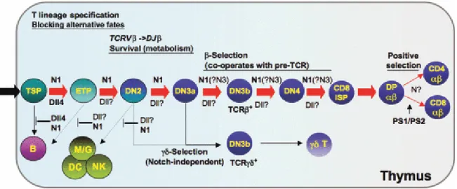
Notch signalling intensity is carefully regulated throughout T cell development and differentiation (Figure 4). Notch signalling is maintained a very low level in primitive hematopoietic progenitors located in the bone marrow. In the bone marrow Notch signalling is inhibited by the transcriptional repressor Leukemia/lymphoma-Related Factor
Markers Type of Cell Lineage Day of measurement (relative to the first day in culture) Lin–Sca1+c-cithigh+
CD24lo/+CD25–CD44+
ETPs (LSK-like) B and T-cells Day 0, 5 and 12
c-kit+ CD25+ DC44+ DN2 T-cells Day 5
c-kit- CD25+ DC44– DN3 T-cells Day 5 and 12
CD4+ CD8+ DP T-cells Day 12
CD4+ CD8- TCRβ+/ CD4- CD8+ TCRβ+
SP T-cells Day 12
B220+ CD19- pre-pro-B B-cells Day 5 B220+ CD19+ pro-B to
mature B-cells
B-cells Day 12
CD11b+ Monocytes Monocytes Days 5 and 12 NK1.1+ Natural killer
cells
Natural killer cells
Days 5 and 12
Table 1: In vitro lymphopoiesis of HMCs by OP-9 co-culture and flow cytometry markers used to identify the different lineages
Figure 4: Notch signalling in T- and B-cell development. In the thymus, Notch1 (N1) in thymus-seeding progenitors (TSPs), which are most commonly lymphoid primed multipotent progenitors (LMPPs), interacts with Delta-like 4 (Dll4) on thymic epithelium to suppress their B-cell potential and promote T-lineage specification. N1 also suppresses myeloid (M/G), dendritic cell (DC), and NK-cell fates at the early T-cell precursor (ETP) and double-negative 2 (DN2) stages, but the Notch ligand required for this suppression in
vivo has not been identified. N1 also regulates survival prior to β-selection, and then
cooperates with pre-T-cell receptor (TCR) signalling to promote survival and proliferation during the DN3 to double positive (DP) transition. Figure adapted from Stanley, P. and
(LRF)[219]. Knockout of LRF in hematopoietic progenitor bone marrow cells in mice was embryonic lethal due to severe anaemia. When these bone marrow LRF-negative cells were expanded in a co-culture system, the cells were no longer able differentiate into B cell, however they retain their ability to differentiate into T cell[219]. In a bone marrow repopulation assays, LRF-negative progenitor cells were unable to repopulate B cell linage cells in the peripheral blood, while the T cell population was relative normal compared to wild-type. Abnormally, lost of LRF in the bone marrow progenitors lead to extrathymic development of T cells in the bone marrow[219]. These experiments demonstrated that LRF acts in a cell-autonomous manner on hematopoietic progenitors, without altering Notch ligand expression in the bone marrow microenvironment[219].
The exact role Notch signalling in DN1-DN2 transition is ambiguous, however Notch signalling is required for the maintenance of CD25 expression in DN2 and DN3 cells and the survival of DN2, DN3 and DN4 cells[220, 221]. Notch signalling is maintained in mice until the β selection or pre-TCR checkpoint (Figure 4). Though Notch signalling is required for transition from DN4 to CD4+CD8+ double-positive (DP) cells, it is rapidly downregulated afterwards by pre-TCR signalling mechanisms[222, 223]. There are different subtypes of the TCR receptors. Mature T cells that select for the TCRαβ recognise peptidic antigens bound to major histocompatibility complex (MHC) proteins[224] and selection of which peptidic antigens the T cell will recognise occurs prior to leaving the thymus. Antigen selection for mature TCRγδ T cells occurs in the peripheral blood oppose as in the thymus. These TCRγδ T cells can be triggered to produce cytokines in their naïve state and do not require the MHC molecules for antigen selection[225]. In mice, selection for αβ over γδ T cell lineage is dependent on Notch signalling[203, 226]. The opposite is true in humans. High levels of Notch activation favour the γδ T cell lineage over the αβ T cell lineage[227].
The role of Notch signalling in the selection of mature signal positive (SP) CD4+ or CD8+ cells is controversial with gain- and loss-of-function experiments being at odds with one another. When the constitutive active form of Notch (NICD) was expressed in mice, decreased CD4+ and increased CD8+ SP thymocyte was observed[228]. However, when endogenous Notch signalling was inhibited in thymocytes, little or no effect on CD4+ and CD8+ SP development was seen[228]. Modulation of the downstream Notch effectors, such
![Figure 2: Clathrin (A) and Caveolin (B) mediated vesicles. A) Diagram of the assembly and abscission of clathrin coated pits taken from Ungewickell & Hinrichsen, 2007[49]](https://thumb-eu.123doks.com/thumbv2/123doknet/2167449.9952/24.918.175.823.194.618/clathrin-caveolin-mediated-vesicles-diagram-abscission-ungewickell-hinrichsen.webp)