MEMOIRE PRESENTE A
L'UNIVERSITÉ DU QUÉBEC À CHICOUTIMI
COMME EXIGENCE PARTIELLE
DE LA MAÎTRISE EN RESSOURCES RENOUVELABLES
Par:
AZADEH EFTEKHARI
B.Sc. PATHOLOGIE VÉGÉTALE
CHEMICAL AND BIOLOGICAL STUDY ON
CONSTITUENTS OF POPULUS TREMULOIDES BUDS
ÉTUDE CHIMIQUE ET ACTIVITÉS BIOLOGIQUES DES
CONSTITUANTS DES BOURGEONS DU POPULUS
TREMULOIDES
bibliothèque
Paul-Emile-Bouletj
Mise en garde/Advice
Afin de rendre accessible au plus
grand nombre le résultat des
travaux de recherche menés par ses
étudiants gradués et dans l'esprit des
règles qui régissent le dépôt et la
diffusion des mémoires et thèses
produits dans cette Institution,
l'Université du Québec à
Chicoutimi (UQAC) est fière de
rendre accessible une version
complète et gratuite de cette œuvre.
Motivated by a desire to make the
results of its graduate students'
research accessible to all, and in
accordance with the rules
governing the acceptation and
diffusion of dissertations and
theses in this Institution, the
Université du Québec à
Chicoutimi (UQAC) is proud to
make a complete version of this
work available at no cost to the
reader.
L'auteur conserve néanmoins la
propriété du droit d'auteur qui
protège ce mémoire ou cette thèse.
Ni le mémoire ou la thèse ni des
extraits substantiels de ceux-ci ne
peuvent être imprimés ou autrement
reproduits sans son autorisation.
The author retains ownership of the
copyright of this dissertation or
thesis. Neither the dissertation or
thesis, nor substantial extracts from
it, may be printed or otherwise
reproduced without the author's
permission.
Nowadays, a large number of compounds derived from plants are used in modern medicine and a majority of them are inspired by traditional applications. About 60% of anticancer drugs and 75% of compounds for infectious diseases are either natural products or their derivatives. However, it is estimated that only bioactive molecules of 5 to 15% of more than 250'000 plants species on earth have been investigated. Particularly, few scientific studies have been conducted on bioactive compounds from the medicinal plants of the boreal forest, wide-spread in Quebec and Canada. In recent years, LASEVE laboratory has evaluated the potential pharmacology of many plants and it was apparent that the buds of Populus tremuloides would represent an interesting subject of study in chemistry and pharmacology. In North America, leaf-buds of P. tremuloides were used in traditional medicine to treat numerous diseases. The aim of this research project was to isolate and characterize the polyphenolic constituents in buds of P. tremuloides and to evaluate the biological activities of the purified fractions and pure compounds. Phytochemical investigations of the EtOH extract of P. tremuloides leaf-buds led to the isolation of 24 phenolic compounds. Among them, one compound is a new molecule and was not reported in literature before; two compounds were not reported before for Salicaceae family; two compounds were not reported before for Populus genus; and ten compounds were identified for the first time in P. tremuloides. Three of the identified compounds have exhibited a moderate to strong cytotoxicity (5-20 uM) towards some of cellular tested lines (WS1, DLD-1, A-549). Anti-inflammatory activity of five phenolic compounds having a glycerol section has been evaluated using LPS-stimulated RAW 264.7 macrophages. Only one of the tested compounds significantly inhibited nitric oxide production (IC50: 6.5 ± 0.4 uM), without cytotoxicity. Finally, none of the phenolic compounds isolated in the present work have exhibited a significant activity against
RESUME
De nos jours, un grand nombre de composés d'origine végétale sont utilisés dans la médecine moderne. Plusieurs d'entre eux ont été développés en s'inspirant des informations de la médecine traditionnelle. Environ 60 % des anticancéreux et 75 % des antimicrobiens de l'arsenal thérapeutique actuel sont des produits naturels ou des dérivés. Seulement 5-15 % des 250 000 plantes ont été étudiées afin d'en évaluer le potentiel pharmacologique. Les plantes de la forêt boréale québécoise ont été très peu étudiées au niveau de leur composition chimique et de leur potentiel pharmacologique. Au cours des dernières années, le laboratoire LASEVE a évalué le potentiel pharmacologique de plusieurs plantes et il en est ressorti que les bourgeons du Populus tremuloides représentaient un sujet d'étude intéressant au niveau chimique et pharmacologique. De plus, cette espèce a été utilisée en médecine traditionnelle pour traiter de nombreuses maladies. L'objectif de ce projet était d'isoler et de caractériser les composés phénoliques majoritaires des bourgeons de Populus tremuloides et d'en évaluer l'activité biologique (anticancéreuse, anti-inflammatoire et antibactérienne). Les travaux effectués sur l'extrait éthanolique des bourgeons de Populus tremuloides ont mené à l'identification de 24 composés phénoliques. Un de ces composés n'a jamais été rapporté auparavant dans la littérature scientifique et deux autres n'ont jamais été signalés dans la famille des
Salicacées. De plus, dix de ces composés sont identifiés pour la première fois dans le Populus tremuloides. Trois des composés identifiés ont démontré une cytotoxicité de forte
à modérée (5-20 uM) envers certaines des lignées cellulaires testées (WS1, DLD-1, A-549). L'activité anti-inflammatoire de cinq composés phénoliques constitués d'une section glycérol a été évaluée sur des macrophages de souris RAW 264.7 stimulé avec le LPS (lipopolysaccharide). Un seul des composés testés a significativement inhibé, sans cytotoxicité, la production d'oxyde nitrique avec une IC50 de 6.5 ± 0.4 uM. Finalement, aucun des composés phénoliques isolés dans ces travaux n'a démontré une activité importante contre Staphylococcus aureus et Escherichia coli.
ACKNOWLEDGMENTS
At first, I would like to express my gratitude to the director of my program, Professor André Pichette, for his patience and gentle supervision. It was great to have the privilege to work with him and to learn from his expertise in the past two years. Special thanks to my co-director, Professor Jean Legault, for his support and guidance. I am very grateful for all of his time and attention during this research.
Special thanks to Dr. Vakhtang Mshvildadze and Mr. Serge Lavoie, for their kind help throughout this study and to my colleagues with whom I shared these years of research in LASEVE laboratory.
I am pleased to have participated in the master program offered by the Université du Québec à Chicoutimi, where I had the chance to develop my skills and find forward-looking friends.
Last, but not least, I am also most grateful to my husband, Alireza, for his never-ending support and understanding in all the situations and to my parents for unconditional support and encouragement.
ABSTRACT ii RÉSUMÉ iii ACKNOWLEDGMENTS iv TABLE OF CONTENTS v LIST OF FIGURES vii LIST OF TABLES viii I. INTRODUCTION 1 1.1 General Introduction 2 1.2 Research objectives 3 II. LITERATURE REVIEW: Populus tremuloides Michx 5 2.1 Principle Characteristics of Populus tremuloides 6 2.2 Applications by Amerindians 6 2.3 Chemistry of tne genus Populus 7 2.4 Populus tremuloides Michx 9 III. SCIENTIFIC PAPER: 14 Cytotoxic phenolic compounds in leaf-buds of Populus tremuloides 14 Résumé 17 Abstract 18 Introduction 19 Experimental 19 General 19 Plant material 21 Extraction and Isolation 21
(IS, 2^-l-[4-Q-i?-coumaroyl-/?-D-glucopyranosyloxy]cyclohexanediol (1) 23
Acid hydrolysis of (1) 23 Cell lines and culture conditions 23 Cytotoxicity assay 24
Antibacterial assays 24 Results and discussion 25 Concluding remarks 33 Acknowledgement 34 References 34 IV. RESULTS AND DISSCUSSTIQN: Other results related to phytochemical study of
Populus tremuloides 38
4.1 Isolation and purification of phenolic compounds 39 4.2 Identifiation of purified phenolic compounds 39 4.2.1 Identification of compound 20 40 4.2.2 Identification of compound 21 41 4.2.3 Identification of compound 22 43 4.2.4 Identification of compound 23 45 4.2.5 Identification of compound 24 46 4.3 Evaluation of cytotoxicity against human cell lines 48 4.4 Evaluation of the anti-inflammatory activity of isolated compounds on LPS activated RAW 264.7 macrophages 50 4.5 Evaluation of antibacterial activity of compounds isolated from Populus tremuloides.
52 V. CONCLUDING REMARKS AND FUTURE DIRECTIONS 54 VI. REFERENCES 57 APPENDIX 64
LIST OF FIGURES
Figure 1 : (a) Distribution of P. tremuloides in North America, (b) trees of P.tremuloides, (c) leaves of P.tremuloides [13, 19] 6 Figure 2: Bud of Populus tremuloides 9 Figure 3: Molecular structure of some compounds described by Fernandez et al [50] 10 Figure 4: Triterpenes constituents from the heartwood of P. tremuloides [51] 12 Figure 5: Five phenolic compounds from buds of P. tremuloides 40 Figure 6: !H !H COSY, HMQC and HMBC correlations (21) 42 Figure 7: *H 'H COSY, HMQC and HMBC correlations (24) 46
LIST OF TABLES
Table 1 : GC-MS identification of the P. tremuloides pulp and paper constituents [1] 11 Table 2: Components of buds ofP. tremuloides [29] 13 Table 3: 'H and 13C NMR spectroscopic data for compound 20 in methanol-d4 41 Table 4: *H and 13C NMR spectroscopic data for compound 21 in methanol-d4 42 Table 5: 'H and 13C NMR spectroscopic data for compound 22 in methanol-d4 44 Table 6: !H and 13C NMR spectroscopic data for compound 23 in methanol-d4 45 Table 7: *H and 13C NMR spectroscopic data for compound 24 in methanol-d4 47 Table 8: In vitro cytotoxicity results of phenolic compounds 49 Table 9: NO Inhibition induced by phenolic compounds on LPS-activated RAW 264.7
macrophages 52 Table 10: Antibacterial activity of phenolic compounds against S. aureus and E. coli 53
The use of plants as medicines has a long history. Written proofs of man's ingenuity in using plants for treatment of various diseases are present by ancient Chinese, Indians, and North Africans [1]. These plants were initially used without modification, then as concentrated extracts to increase their effectiveness or as dried form to keep them for longer times with less degradation. In the 1900s, people learned how to isolate active compounds from medicinal plants.
Even today, natural products are important sources of drugs and almost 60% to 75% of the world's population use plant-based medicines for initial pharmaceutical cure [2]. Between 1981 and 2002, it is estimated that almost 50 % of new chemical entities authorized to initiate clinical studies were natural products or related natural products [3]. Moreover, natural products exhibiting various biological activities usually possess a complex and diverse structure leading to strong challenges in organic synthesis [3, 4], About 60% of anticancer drugs and 75% of compounds for infectious diseases are either natural products or their derivatives [5, 6]. It is commonly accepted that less than 15 % of the plants were studied regarding their biological activities [7, 8].
In Canada, one of the most important natural resources is the boreal forest. The latter plays an important role in the traditional medicine of Canadian First Nations communities [9]. However, real effectiveness of these potential drugs is rarely studied in scientific ways. Few biophamiaceutical studies were undertaken on plant species of the boreal forest of Canada [9, 10]. Based on the knowledge acquired by First Nations communities, LASEVE
Following this result, a large effort of this laboratory has been devoted to research on potential pharmaceutical contents in biomass of the boreal forest.
This project is inspired by previous research work performed on other Populus species
{Populus balsamiferd). The present research is focused on chemical and biological
properties of bud constituents of the most widely distributed species of Populus in boreal forest, named Populus tremuloides.
1.2 Research objectives
The main goals of this research project are:
• Isolation and characterization of polyphenolic constituents from the buds of P.
tremuloides.
• . Investigation of crude extract, purified fractions and pure compounds for antibacterial, anti-inflammatory and cytotoxic activities (in vitro).
• Identification of the active compound(s) through bioassay-guided fractionation. • Study of the relationship between the chemical structures and the biological
activities.
This thesis is divided into five chapters. The first chapter is dedicated to the thesis outlines. Chapter II covers literature reviews about botany, applications and phytochemistry of the genus Populus and the species of Populus tremuloides. Chapter III presents a
scientific paper dealing with Cytotoxic polyphenols in leaf-buds of P. tremuloides. This article was accepted in Canadian Journal of Chemistry. Chapter IV describes the scientific work related to anti-inflammatory phenolic compounds from buds of Populus tremuloides and finally in Chapter V, conclusions and future directions are covered.
Populus tremuloides (Quaking aspen), from Salicaceae family is a decidous tree native
to cooler areas of North America. It is a tall tree, usually 20 to 25 meters high at maturity, with a trunk diameter comprised between 20 to 80 cm [12, 13]. As shown in figure 1, this tree has a geographical distribution from Alaska to the east coast of Canada and is present down south in scattered populations (New Mexico state). However, It is absent from the central and south-eastern plains. It grows in humid calcium-rich soils.
(a) (bf (c)
Figure 1: (a) Distribution of P. tremuloides in North America, (b) trees of'P. tremuloides, (c) leaves of P. tremuloides [13, 19].
2.2 Applications by Amerindians
Quaking aspen has been used as herbal medicine for centuries. First Nations communities widely used this species especially for its antiseptic and analgesic properties for wounds healing and against skin complaints and disorders [9]. Now, it has similar applications in modern herbalism.
Its bark had applications in the treatment of rheumatism, arthritis, gout, lower back pains, urinary complaints, digestive and liver disorders, debility, and anorexia [14-16]. It also could reduce fevers and relieve the pain of menstrual cramps [16]. Infusion of inner bark was a remedy for coughs as well as a treatment for stomach pains, colds and fever [16-17].
Leaf-buds had applications as a nasal salve for children and adults to fight coughs, colds and irritated nostrils [9, 18]. They were also used against chronic bronchitis and rheumatism and externally for treating superficial wounds, external haemorrhoids, frostbite, sunburn and as an ointment for myalgia [19].
2.3 Chemistry of tne genus Populus
Having fast growth rates, the genus Populus attracted many economical interests, such as in the wood pulp industry and motivated longstanding investigations about its chemistry [20]. The genus tree family contains phenolic glycosides which are important secondary metabolites regarding plant resistance to both insect and mammalian herbivores [21, 22]. Finally, chemical principles from Populus species are known to have various biological activities such as fungicidal, antioxidant, antitumor, antiseptic and antiviral properties [23-25].
Several classes of natural products such as polyphenols, terpenoids, fatty acids, aliphatic alcohols and hydrocarbons are found in the genus Populus [26-29]. Due to difficulty in identification of Saiicaceae species only based on morphological identification, secondary
(Populus spp) is related to a complex mixture of compounds in their bud exudates which
include benzoic and phenolic acids and their esters, fiavanoid aglycones, hydrocarbons and terpenoids [30, 31]. The presence of some phenolic compounds can be used as a 'fingerprint' to identify poplar species [32] since many phenolic components of buds exudates of European, Asian, and North American poplar species have been studied in recent years [33-38]. The analysis of poplar bud exudates have shown a high degree of complexity. The typical poplars of a particular section have a bud exudate composition which is characteristic of that section. For example, section Leuce, subsection Albidae, e.g
P. alba L., exude only hydrocarbons [39] whereas those of subsection Trepidae, e.g. P. tremuloides Michx., secrete a phenolic exudate which includes only the flavanones
produced by the first steps of flavonoid metabolism, such as naringenin (S,7,4'-trihydroxyflavonone) and its methyl ethers [29]. By contrast poplars of section Aigeiros, such as P.deltoides Marsh [40] and P.nigra L. [41], and of section Tacamahacha, such as
P.balsamifera L. [42], produce a more complex bud exudate, which contains primarily
flavonoids resulting from the further metabolism of naringenin. Phenolic compounds are also exuded onto the leaf surfaces of some poplars, such as P. deltoïdes [43] and P. nigra [44]. The bud of P. tremuloides is shown in figure 2.
Propolis, a resinous hive product collected by honeybees from parts of plants, especially buds and exudates of Populus species, has been used as folk medicines about 300 years before Christ (BC). Different biological activities, such as anticancer, antioxidant, anti-inflammatory, antibiotic and antifungal effects have been reported for propolis and its
prevent diseases such as inflammation, heart disease, diabetes and even cancer. Recently there is a renewed interest in the composition of propolis and related poplars [45-48]. Recently, it has been proven that the phenolic content of northern propolis is mainly inherited from Populus spp., a very attractive tree for honey bees of Canada. Thus, considering the promising biologically-active substances in propolis inheritated from poplars of section Leuce, subsection Trepidae, its plant source which is P. tremuloides is worth further investigation [49].
Figure 2: Bud of Populus tremuloides.
2.4 Populus tremuloides Michx
While P. tremuloides has a rather prominent importance as a model system for ecological and environmental research by the forest industries, few phytochemical or pharmacological studies were conducted so far on P. tremuloides. Fernandez et al. has identify 44 compounds (12 of them are shown in figure 3) such as phenolic compounds,
fatty acids, steroids, triterpenes and lipids in processing of P. tremuloides Michx. for pulp and paper, with a rapid gas chromatography-mass spectrometry (GC-MS) method, summarized in table 1 [50].
Stignnast-5-en-3p-ol O!eanen-3(3-ol Tirucalle-7-,24-diene-3p-ol
Cyc!oart-24-en-3p-ol 12-Ursen-3|3-ol 24-Methylcydoart-24,(24')-en-3p-o!
RO
Citrostadienol 20(29)-Lupen-3p-yl R a-Tocopherol
0
OH O OH
5,4'-Dihydroxy-7methoxyfiavanone 3-(4-Hydroxypheny!)-2-propenoicacid Hexadecanoic acid
Table 1: GC-MS identification of the P. tremuloides pulp and paper constituents [1]. Compound family
Monoaryl phenolics
Fatty acids
Flavonoids
Sterols / triterpene alcohols
Steryl / triterpene esters
Lipids Name of compounds benzoic acid 1 -Etliyi-4-hydroxybenzene 3-(2-Hydroxyphenyl)-2-propenoic acid 2-hydroxybenzyl alcohol 4-Hydroxy-2-methyiacetophenone 4-Hydroxybenzoic acid
3-(4-Hydroxyphenyl) propanoic acid 3-(4-Hydroxy-3-methoxyphenyl)-2-propen-l-ol 3-(4-Hydroxy-3-methoxyphenyi)-2-propenal 3~(4-Hydroxyphenyl)-2-propenoic acid 4-(4-Hydroxy-3-methoxyphenyl)-2-propenoicacid 4-(3-Hydroxy-l-propenyl)-2,6-dimethoxyphenol 3-(4-Hydroxy-3,5-dimethoxyphenyl)-2-propen-lal 9-oxononanoic acid Hexadecanoic acid (Z,Z)-9,12-Octadecadienoic acid Octadecadienoic acid Eicosanoic acid Docosanoic acid Tetracosanoic acid ] -Docosanol 4 ' ,5 -Dihydroxy-7-methoxyflavanone 4',5,7-Trihydroxyflavanone 3,5,7 -Trihydroxy-4 ' -methoxy flavone 3,4',5,7-Tetrahydroxyflavone Stigmast-5-en-3P-oi Oleanen-3P-ol Tirucalla-7,24-diene-3p-ol Cycloart-24-en-3p-ol 12-Ursen-3|3-ol 24-Methylcycloart-24,(24)-en-3|3-ol Citrostadeniol Stigmast-5-en-3P-yl acetate Tirucalla-7,24-diene-3p-yl hexadecanoate 12-Oleanen-3j}-y] hexadecanoate 20(29)-Lupen-3 P-yl hexadecanoate 12-Ursen-3p-y! hexadecanoate Tirucalla-7,24-diene-3p-yl octadecanoate 12-Oleanen-3p-y! octadecanoate 20(29)-Lupen-3P-yl octadecanoate 12-Ursen-3P-yl octadecanoate Tiraculla-7,24-diene-3p-yl eicosanoate a-tocophérol
The extractives of unsaturated alcohol fraction from P. tremuloides heartwood have been studied by Abramovitich and Micetich [51] and their work resulted in the identification of triterpenes and sterols, as shown in figure 4.
AcO AcO'
a-amyrin acetate p-amyrin acetate a-amyrenonol
AcO'
lupeol acetate
AcO'
24-methylenecycloartanol butyrospermol acetate
Figure 4: Triterpenes constituents from the heartwood of P. tremuloides [51].
Pearl et al. [52] showed that leaves of P. tremuloides provide a good source of the 6-monobenzoate of salicin, populin, while the bark of the same species provides a good source of the 2-monobenzoate of salicin, tremuloidin [52, 53]. Lindroth et al. [54] identified phenolic glycosides components, such as salicin, salicortin, tremuloidin, tremulacin from the crude extract of P. tremuloides leaves which exhibited differential toxicity against the insects such as Papilio glaucus subspecies.
Phytochemical investigation by English et al. [29] with GC-MS on buds of P.
tremuloides revealed phenolic compounds, flavonoids and hydrocarbons listed in table 2.
Table 2: Components of buds of P. tremuloides [29].
Compound family Phenolic compounds Flavonoides Hydrocarbones (straight chain) Hydrocarbon alcohol Name of compounds Benzyl benzoate p-Coumaric acid Ferulyl alcohol Ferulic acid Caffeic acid Benzyl j?-coumarate Ferulyl benzoate Benzyl ferulate Benzyl caffeate Isosakuranetin Isosakuranetin chalcone Sakuranetin Sakuranetin chalcone Kaempferol methyl ether Tricosane
Pentacosane Heptacosane Nonacosane 1 -Hexacosanol
Chapter 111
SCIENTIFIC PAPER:
Cytotoxic phenolic compounds in leaf-buds of
Populus tremuioides.
Graphical abstract
Bioassay-guided fractionation and separation of the EtOH extract of Populus
tremuloides leaf-buds resulted in the isolation of a new compound.
(lS,2S)-l-[4-O-(E)-coumaroyl-/?-D-glucopyranosyloxy]cyclohexane-diol 1, and 18 known products. In vitro cyto toxic and antibiotic activities were evaluated for 18 of them.
Cytotoxic phenolic compounds in leaf-buds of Populus
tremuloides.
André Pichette*, Azadeh Eftekhari, Patricia Georges, Serge Lavoie, Vakhtang
Mshvildadze and Jean Legault.
Chaire de recherche sur les agents anticancéreux d'origine naturelle, Département des sciences fondamentales, Université du Québec à Chicoutimi, Québec, Canada, G7H 2B1.
* Corresponding author
Université du Québec à Chicoutimi Département des sciences fondamentales Laboratoire LASEVE
555, boulevard de l'Université Chicoutimi, Québec, Canada G7H2B1
Phone: +1 (418) 545-5011 ext. 5081
Fax:+1 (418)545-5012
Résumé
Les enquêtes phytochirnique menés sur l'extrait éthanolique des feuilles-bourgeons du
P. tremuloides ont conduit à l'isolement de 19 composés phénolique. Parmi eux, on y
trouve est une nouvelle molécule, (IS, 2S) -1 - [4-O-E-coumaroyl p-D-glucopyranosyloxy] cyclohexanediol. La structure de cette dernière a été déterminée par spectroscopie (RMN et MS) et par des méthodes chimiques. Les autres Dix-huit composés isolés ont été testés pour l'activité cytotoxique en comparaison avec celle des lignées cancéreuses des poumons (A549) et celle du côlon (DLD-1) présent dans les cellules humaines affectés. L'activité antibactérienne a également été évaluée contre VEscherichia coli et le Staphylococcus
Abstract
Phytochemical investigations of the EtOH extract of Populus tremuloides leaf-buds led to the isolation of 19 phenolic compounds. Among them, (IS, 25)-l-[4-O-i?-coumaroyl-/5-D-glucopyranosyloxy]cyclohexanediol was reported for the first time and its structure was determined by spectroscopic (NMR and MS) and chemical methods. Seventeen of isolated compounds were tested for their cytotoxicity against lung carcinoma (A549) and colorectal adenocarcinoma (DLD-1) human cell lines. Antibacterial activity was also evaluated against Escherichia coli and Staphylococcus aureus.
Keywords
Introduction
The buds exudates of many plant species of Populus genus are known as raw material processed by bees into propolis (1). The latter product has been widely used in popular medicine as antibacterial (2-4), anti-inflammatory (5), antioxidant (6-7) and cytostatic treatments (8). The biological activity of propolis samples is mainly due to phenolic compounds like flavonoids, aromatic acids and diterpenic acids (9-10) which are the principal constituents of the buds of Populus species (11-13). Recently, studies in the northern-type propolis showed a potential source of biologically-active substances in P.
tremuloides (14) which are widely spread across North America (15). In spite of its use as
ointment by Amerindian traditional medicine to treat numerous diseases such as coughs, colds and irritated nostrils (16), few studies were carried out on the medicinal applications. In the present study, the isolation and structure elucidation of a new phenolic compound from Populus tremuloides Michaux, along with 18 known products are described.
Experimental
General
Optical rotations were measured with an automatic polarimeter Rudolph Research Analytical Autopol IV. FTIR spectra were recorded with a Perkin-Elmer SpectrumOne. High resolution electrospray ionization mass spectrum was conducted in positive mode with an Applied Biosystems/MDS Sciex QSTARXL QqTOF MS system. The ID and 2D NMR spectra ( ' H -!H COSY, HSQC and HMBC) were performed using an Avance 400
Bruker spectrometer equipped with a 5 mm QNP-probe. Chemical shifts were expressed in 8 (ppm) units relative to TMS as an internal standard and coupling constants were given in Hertz. Preparative HPLC was performed on an Agilent 1100 liquid chromatography system, equipped with a solvent delivery system, an autosampler and a UV-MWD detector. Samples were eluted in an Intertsil prep-ODS column C18 (20 x 250 mm; 10 um) at room temperature with a flow rate of 10 mL min'1. The GC-MS analysis were performed with an instrument (Agilent Technologies 6890N) fitted with a mass selective detector (Agilent Technologies 5973), a split-splitless injection port and an apolar capillary column DB-5MS (30 m x 0.25 mm x 0.25 um).
Analytical thin-layer chromatography was performed with silica gel 60 F254, 0.25 mm pre-coated TLC plates (Silicycle, Québec, Canada). Flash column chromatographies were performed on silica gel (40-63 um with indicator F254, Silicycle, Québec, Canada) and on Ci8 reversed phase silica gel (carbon 11 %, 40-69 urn, Silicycle, Québec, Canada). Polyamide CC-6 was purchased from Macherey-Nagel (Germany) and Diaion HP-20 from Supelco. Detection of the phenolic compounds was carried out by spraying TLC plates with polyethylene glycol (NP/PEG) reagent followed by heating at 110 °C and detected by UV absorption at 254 and 365 nm. TLC identification of monosaccharides were performed with CHaClr-MeOH—I^O (50:25:5) solvent system. The compounds were visualized by spraying an orthophosphoric acid solution of naphtoresorcinol 5 % in EtOH, followed by heating at 110 °C.
The commercial samples used for biological tests, namely prunin (3), kaempferide (13) and trans-ferulic acid (19), were purchased from Indofine Chemical Company (USA). Rhamnocitrin (10) was purchased from Apin Chemicals Ltd (UK).
Plant materia!
Leaf-buds of Populus tremuloides Michaux were collected in the boreal forest to the south of Chicoutimi, Québec, Canada, in April 2006. Samples were identified by Patrick Nadeau (Département des sciences fondamentales, Université du Québec à Chicoutimi). A voucher specimen (QFA-A540466) was deposited at the Herbarium Louis-Marie of Université Laval, Québec, Canada.
Extraction and Isolation
The buds of Populus tremuloides (1 kg) were exhaustively extracted with EtOH (3 L, 60 °C, 3 times, 2 h each time) followed by EtOH-H2O (7:2). The extracts were filtered and pooled. After evaporation of EtOH in vacuo, the aqueous phase was extracted successively with hexane (500 mL x 5) and saturated M-BUOH with H2O (500 mL x 5). The n-BuOH phase was decanted and evaporated in vacuo. The residue (80 g) was fractionated using an open Diaion® column eluted with H2O-MeOH with 30 %, 50 % and 80 % of MeOH. Three fractions were obtained: A (6.46 g), B (7.24 g) and C (58.96 g).
Fraction C was purified on silica gel CC, eluted with CHCl3-MeOH gradient (60:1—>5:1, v/v) and three fractions were obtained. Fr. Cl (16.73 g) was subjected to silica gel using a gradient of CHCl3-MeOH (90:1-^60:1, v/v) as eluent. Subfr. Cl.l (384 mg), obtained from CHCl3-MeOH (90:1), was separated on silica gel CC with CHCl3-MeOH (80:1) as eluent,
to give three fractions. Subfr. C1.1A was purified by preparative HPLC with a gradient elution of MeOH-H2O (50:50—»85:15, v/v) yielding compounds 15 (249 mg), 16 (5 mg) and 17 (41 mg). Subfr. C1.1B (2.36 g) was applied successively on a silica gel CC and a reversed-phase CC using gradients MeOH-EbO (50:50—>-70:30, v/v) as eluent to give 11 (160 mg). Compound 9 (3 mg), 12 (18 mg) and a mixture of 10 and 13 (27 mg) were isolated after a silica gel CC eluted with CHCl3-MeOH (90:1) and a preparative HPLC (isocratic CH3CN-H2O 40:60) of Fr. C1.2. Compound 18 (50 mg) was obtained from fraction C1.3 (294 mg) after repeated silica gel CC (CHCI3-MeOH 75:1) and Polyamid flash column (MeOH-B^O, 50:50-^75:25). Fr. C2 was chromatographed on silica gel CC with a gradient elution of CHCl3-MeOH (75:1 —>15:1, v/v) to give eight fractions. Subfr. C2.7 (558 mg) was separated by preparative HPLC using an isocratic mobile phase of CH3CN-H2O-HCOOH (40:60:1) to afford 2 (19 mg) and 14 (3 mg).
Fr. C3 (5.4 g) was purified on silica gel using a gradient of CHCl3-MeOH (25:1—>7:1) for elution to give five subfractions. C3.2 was separated on silica gel CC with CHCl3-MeOH (20:1) giving 6 (289 mg). Some purifications on different silica gel CC of subfr. C3.2.1 permitted to obtain 19 (2 mg). Subfr. C3.4 (418 mg) was separated by preparative HPLC using an isocratic mobile phase of CH3CN-H2O (30:70) to afford compounds 1 (20 mg), 3 (8 mg), 4 (14 mg) and 8 (37 mg). Subfr. C3.5 (482 mg) was separated by HPLC using a gradient of MeOH-H2O (10:90 -* 100:0) to give 5 (21 mg) and 7 (35 mg).
(1 S, 2S)-1 -[4-O-E-coumaroy!-|3-D-g!ucopyranosyloxy3cycIohexanedio!
(1)
White amorphous powder; [a]D25 -35.3° (c 1.0, MeOH); IR (neat) vmax 3328, 2935, 1696, 1602, 1160, 1080, 1024, 982 and 833 cm"1; *H and 13C NMR spectroscopic data, see Table 2; HR-ESI-MS m/z 447.16225 [M+Na]+ (calcd for C2iH28O9Na, 447.16310).
Acid hydrolysis of (1)
Compound 1 was dissolved in HC1 10 % and heated at 110 °C for 4 h. The resulting hydrolysate was extracted with CHCI3. The organic phase was dried (MgSCH) and the solvent evaporated under reduced pressure. The presence of />-coumaric acid was confirmed with standard sample on TLC (CHCl3-MeOH 10:1 as eluent and developing with NP/PEG reagent). The presence of cyclohexane-1,2-dioI in the organic phase (aD = + 3.4) was confirmed with a GC-MS analysis: Injector temperature 250 °C; ionization voltage, 70 eV (EI-MS); column temperature, 40°C for the initial 2 min followed by an increase of 15 °C min"1 up to 350 °C; carrier gas, He; column flow rate, 1 ml.min'1. Cyclohexane-1,2-diol was detected at Rt 7.55 min. The aqueous phase was neutralized with N,N-dioctylmethylamine (10 % in CHCI3) and the solvents were evaporated under reduced pressure. The residue contained the monosaccharide D-glucose (aD= + 24.8).
Cell lines and culture conditions
Lung carcinoma (A549), colorectal adenocarcinoma (DLD-1) and normal skin fibroblast (WS1) human cell lines were obtained from the American Type Culture Collection (ATCC). All cell lines were cultured in minimum essential medium containing Earle's salts and L-glutamine (Mediatech Cellgro, VA), to which were added 10 % fetal bovine serum
(Hyclone), vitamins (IX), penicillin (100 I.U. mL"1) and streptomycin (100 \xg mL*1), essential amino acids (IX) and sodium pyruvate (IX) (Mediatech Cellgro, VA). Cells were kept at 37 °C in a humidified environment containing 5 % CO2. Antibacterial activity was tested on Escherichia coli ATCC 25922 and Staphylococcus aureus ATCC 25923E.
Cytotoxicity assay
Exponentially growing cells were plated in 96-well microplates (BD Falcon) at a density of 5 x 10³ cells per well in 100 fiL of culture medium (DMEM with 10 % S VF) and were allowed to adhere for 24 h before treatment. Increasing concentrations of each compound in MeOH or DMSO were then added (100 uL per well) and the cells were incubated for 48 h. The final concentration of MeOH or DMSO in the culture medium was maintained at 0.25 % (v/v) to avoid solvent toxicity. Cytotoxicity was assessed using resazurin (17) on an automated 96-well Fluoroskan Ascent Fl™ plate reader (Labsystems) using excitation and emission wavelengths of 530 and 590 nm, respectively. Fluorescence was proportional to the cellular metabolic activity in each well. Survival percentage was defined as the fluorescence in experimental wells compared to that in control wells after subtraction of blank values. Each experiment was carried out thrice in triplicate. IC50 results were expressed as means ± standard deviation.
Antibacterial assays
Antibacterial activity was evaluated using the microdilution method (42) but with some modifications: exponentially growing bacteria were plated in 96-well flat bottom microplates (BD Flacon) at a density of 5 * 103 gram-negative E. coli (ATCC 25922) or
40 x 103 gram-positive S. aureus (ATCC 25923) per well in 100 uL nutrient broth (Difco). The concentration of ethanol in the culture medium was maintained at 0.25 % (v/v) to avoid solvent toxicity. Fifty microliters of 4 % resazurin was added to each well and the microplates were incubated for 6 h at 37 °C. Fluorescence was measured after 6 h on an automated 96-well Fluoroskan Ascent Fl™ plate reader (Labsystems) using excitation and emission wavelengths of 530 nm and 590 nm respectively.
Results and discussion
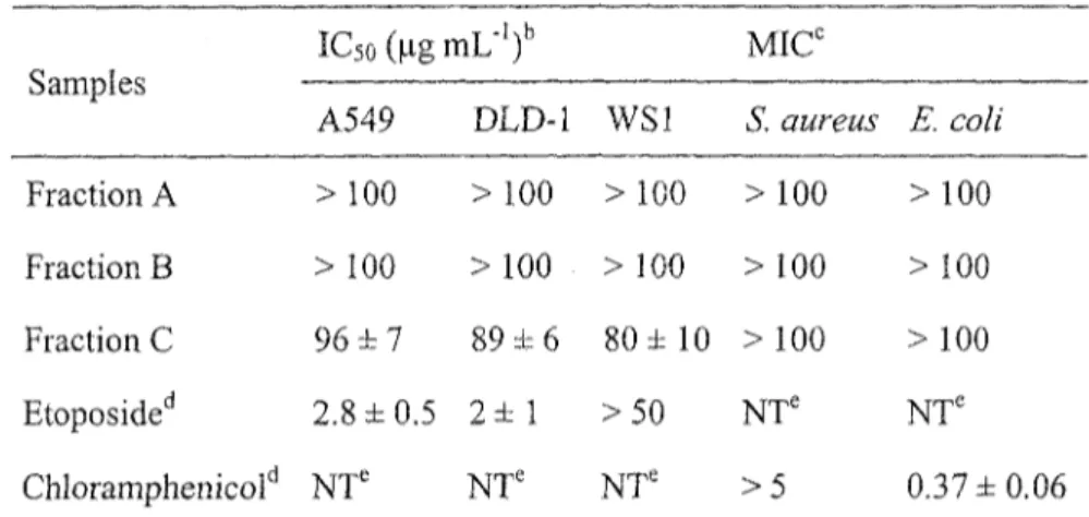
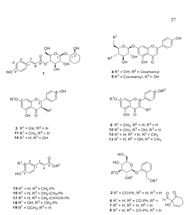
The leaf buds of Populus tremuloides were extracted with EtOH and EtOH-EbO under reflux. After evaporation of EtOH in vacuo, the aqueous phase was successively partitioned with hexane and «-BuQH. The M-BUOH soluble extract was purified on an open Diaion® column with a gradient of decreasing polarity and three fractions were obtained. Each fraction was investigated for in vitro cytotoxic and antibacterial biological activities. Cytotoxic activity evaluations were carried out on human lung cancer (A549), human colorectal cancer (DLD-1) and normal skin fibroblasts (WSl) using the resazurin reduction test as previously described in the literature (17). Antibacterial activity was evaluated against Escherichia coli and Staphylococcus aureus. The results (Table 1), show that the last fraction C was found to exert a weak cytotoxic activity against A549 (IC50, 96 ± 7 ug mL"1) and DLD-1 (IC50, 89 ± 6 ug rnL'1), but was inactive toward bacterial cell lines. Thus, bioassay-guided fractionation of fraction C was undertaken with a combination of different chromatographic techniques leading to the isolation of a new compound 1 together with 18 known compounds (figure 1): chaenomeloidin (2) (18), prunin (3) (19), echinaticin (4) (20),
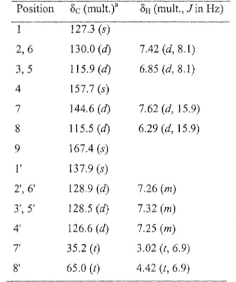
echinacin (5) (21), tremulacin (6) (22), salicine (7) (23), tremuloidin (8) (24), genkwanin (9) (25), rhamnocitrin (10) (26), sakuranetin (11) (27), acacetin (12) (28), kaempferide (13) (29), aromadendrin (14) (30), phenylmethyl coumarate (15) (31), phenethyl p-coumàrate (16) (32), cinnamyl coumarate (17) (33), phenylmethyl caffeate (18) (34) and trans-ferulic acid (19) (35). Known compounds were identified by comparison of their spectroscopic data with the values found in the literature. NMR spectroscopic data for phenethyl p-coumarate (16), which was also isolated from buds of P. tremuloides, were not available. Therefore, complete *H and 13C NMR spectroscopic data for 16 are also displayed.
Table 1: In vitro cytotoxicity and antibiotic results of the Diaion® column's fractions21
Samples Fraction A Fraction B Fraction C Etoposided Chloramphenicold IC5o(ngmL-')b A549 > 100 > 100 96 ± 7 2.8 ±0.5 NT' DLD-1 > 100 > 100 89 ± 6 2 ± 1 NT' WS1 > 100 > 100 80 ± 10 >50 NT' MICC S. aurens > 100 > 100 >100 NT' > 5 E. coli >100 > 100 > 100 NTe 0.37 ±0.06 a Mean values (± standard deviation) for triplicate assays.
b Concentration of extract that caused 50 % inhibition of cell proliferation. c Minimum concentration of extract that resulted in inhibition of visible growth
Positive control. ' Not tested.
HO 4" OH O 3 R1 = Glc;R2 = H 11 R1 = C H3; R2= H 14 R1 = H;R2 = OH 4 R1 = OH; R2 = Coumaroyl 5 R1 = Coumaroyl; R2= OH O RJ OH O 9 R1 = CH3; R2 = H;R3 = H 10 R1 = CH3; R2 = OH; R3 = H 12R1 = H; R2 = H;R3 = CH3 13R1= H; R2 =OH; R3 = CH3 R V 3 HO 4 9 OR2 15R1 = H ; R2 = CH2-Ph 16 R1 = H; R2 = CH2-CH2-Ph 17R1 = H;R2 = CH2~CH=CH-Ph 18R1 = OH; R2= CH2-Ph 19R1 =OCH3;R2= H 2 R1 = CO-Ph; R2 = H;R3 = H O Q 6 R1 = H; R2 = CO-Ph; R 7 R1 = H; R2 = H; R3 = H 8 R1 = H; R2 = CO-Ph; R3 = H HO
Figure 1 : Polyphenoîs from Populus tremuloides
The molecular formula (C21H28O9) of 1, a white amorphous powder, was determined from its HR-ESI-MS spectrum (positive ion mode) on the basis of a quasimolecular ion peak at mlz 447.1623 [M+Na]+ (calcd 447.1631). Infrared absorption bands at 3328, 1602, 1160, 982 and 833 cm"1 suggested the presence of hydroxyl groups, aromatic system and an
ester carbonyl groups. The 13C NMR spectrum (Table 2) displayed 19 carbon signals separated by DEPT spectrum into five méthylènes, seven aliphatic oxymethines, four unsaturated methines and three quaternary carbons (one for an ester carbonyl). Among them, six resonances could be assigned to a sugar moiety. The 'H NMR spectrum confirmed the presence of a hexose moiety with anomeric proton at S H 4 . 4 5 (1H, d, J - 7.8 Hz). The *H NMR spectrum also shows the presence of two /rans-olefmic protons at 8H 7.66 (1H, d, J= 15.9 Hz) and SH 6.37 (1H, d, J= 15.9 Hz) and a 1,4-disubstituted aromatic rings with two protons at SH 7.47 (2H, d, J= 8.6 Hz) and 6.81 (2H, d, J = 8.6 Hz). HMBC correlations at SH 7.47 (H-2", H-6") and 5C 147.3 (H-7"), 5H 6.81 (H-3", H-5") and 8C 161.5 (C-4") and §H 7.66 (H-7") and 8C 168.6 (C-9") suggested the presence of a coumaroyl moiety. Analysis of the COSY, HSQC and HMBC spectra led to the identification of a third aglycone sytem: the cyclohexane-l,2-diol. HMBC correlation between the methine proton at 8H 4.87 (H-41) and the carbonyl group at 8c 168.6 (C-9") suggested the linkage between the glucose and the coumaroyl moiety. Finally, the correlation between SH 4.45 (H-l') and Sc 79.4 (C-l), indicated the linkage site of the glucose moiety to the aglycone.
Acidic hydrolysis of 1 and TLC analysis of the aqueous phase afforded identification of glucose as the sugar component. Absolute configuration of the glucose as D was determined by optical rotations in comparison with authentic standard. The presence of p-coumaric acid in the organic phase was confirmed by TLC in comparison with authentic standard. Cyclohexane-l,2-diol was also detected in the organic phase using GC-MS and NMR analysis (36). The absolute configuration of cyclohexane-l,2-diol could be determined directly from the organic phase since the other aglycon part, namely p-coumaric acid, is
optically inactive. The organic phase showed positive value in optical activity measurement meaning that (15,25)-cyclohexane-l,2-diol has been isolated (37). The structure of 1 was thus confirmed as (15, 2,S)-l-[4-O-is-coumaroyl-/?-D-glucopyranosyloxy]cyclohexanediol.
Table 2: 'H (400 MHz) and 13C NMR (100 MHz) spectroscopic data for compound 1 in methanol-d4. Position 1
2
3 4 56
1'2'
3' 4' 5' 6' 1" 2", 6" 3", 5" 4" 7" 8" 9" Sc (mult.)a 79.4 (d) 71.0 (d)31.5(0
22.4 (0
23.1 (t) 27.6 (0 102.2 (d) 75.1 (d) 75.6 (d) 72.5 \d) 76.1 (d) 62.4 (0 127.2 (s) 131.3 (d)U6.9(d)
161.5(5) 147.3 (d) 114.8 (J) 168.6(5) 5H (mult.,/in Hz) 3.87 (m) 3.85 (m) 1.79 (m) 1.55 (m) 1.65 (m) 1.34 (m) 1.71 (m) 1.31 (m) 1.82 (m) 1.63 (m) 4.45, (d, 7.8) 3.35 (<W, P.3, 7.8) 3.65 (t 9.3) 4.87 (m) 3.53 (m) 3.62 (cfJ, 15.0, 5.4) 3.54 (m) 7.47 (d, 8.6) 6.81 (rf, 8.6) 7.66(415.9) 6.37 (d, 15.9)Compound 16 has been identified by many authors but surprisingly, no complete NMR assignation was given (38). Therefore, complete *H and BC characterisation was accomplished using !H, 13C and 2D spectra (Table 3). First, the same/j-coumaroyl moieties as in 1 was identified with SH at 6.29 (1H, d, J= 15.9 Hz, 8), 6.85 (2H, d, J= 8.1 Hz, H-3 and H-5), 7.42 (2H, d, J = 8.1 Hz, H-2 and H-6) and 7.62 (1H, d, J= 15.9 Hz, H-7) and Sc at 115.5 (C-8), 115.9 (C-3 and C-5), 127.3 (C-l), 130.0 (C-2 and C-6), 144.6 (C-7), 157.7 (C-4) and 167.4 (C-9).
Table 3: 'H (400 MHz) and 13C NMR (100 MHz) spectroscopic data for compound 16 in methanol-d4. Position 1 2,6 3,5 4 7 8 9
r
2', 6' 3', 5' 41 7 8' ôc (mult.)a 127.3 (s) 130.0 (d)115.9(0
157.7 (s) 144.6 (d) 115.5 (d) 167.4 (s) 137.9 (s) 128.9 (d) 128.5 (d) 126.6(rf) 35.2 (0 65.0 (t) SH (mult., J i n HZ) 7.42 (d, 8.1) 6.85 (d, 8.1) 7.62 (d, 15.9) 6.29(0 15.9) 7.26 (m) 7.32 (m) 7.25 (m) 3.02 (t, 6.9) 4.42 (t, 6.9) Multiplicities were deduced from DEPT experimentsTable 4: In vitro cytotoxicity results of isolated compounds (l-19)a Compounds 1 2 3 4 5 6 7 8 9 10 11 12 13 14 15 16 17 18 19 Etoposided A549 > 100 NTC > 100 NTC > 100 > 100 >100 81 ± 3 9 ± 3 31 ±2 > 100 27 ± 3 60 ±10 > 100 19±2 >100 > 100 45.8 ±0.9 > 100 2.8 ±0.5 IC5oOiM±SD)b DLD-1 >100 NTC > 100 NTC > 100 > 100 >100 > 100 9.2 ± 0.9 37 ± 3 > 100 23 ± 6 >100 >100 19.2 ±0.9 > 100 >100 39 ± 3 > 100 2 ± 1 WS1 >100 NTC > 100 NTC 42 ± 4 >100 >100 >100 5.8 ±0.3 87 ± 3 >100 20 ± 2 42 ± 5 > 100 26 ± 3 >100 > 100 51±7 >100 >50 a Mean values (± standard deviation) for triplicate assays.
b Concentration that caused 50% inhibition of cell proliferation. c Not tested.
Additionally, five overlapped *H NMR signals between SH 7.20-7.30 along with two méthylène triplets at 3.02 (2H, t, J= 6.9 Hz, H-71) and 4.42 (2H, t, J= 6.9 Hz, H-8') were attributed to a phenethyl moiety. The HMBC correlation between H-8' and C-9 confirmed the link between the phenethyl and the/>-coumaroyl groups.
Compounds 10 and 13 were isolated as a mixture. Separation of each constituent was not performed due to their low amounts. Therefore, careful examination of NMR spectra (!H. l3C, DEPT and HSQC) and comparison with literature allowed the identification of these components as rhamnocitrin (10) (29) and kaempferide (13) (26). Moreover, the biological results were obtained using commercial pure products. Because of low isolated yields of compounds 3 and 19, those compounds were also tested from commercial materials.
All isolated compounds, except compounds 2 and 4, were evaluated using resazurin reduction test for their cytotoxicity against human lung cancer (A549), human colorectal cancer (DLD-1) and normal skin fibroblasts (WS1) (17). Results presented in Table 4 are expressed as the concentration of product inhibiting cell growth by 50 % (IC50). Etoposide was used as positive control with IC50 of 2.8 and 2.0 uM against A549 and DLD-1 cell lines, respectively. The phenolic compounds were regarded as active when the IC50 was smaller than 100 uM (39). The compound 9 was found the most active with IC50 ranging from 5.8 to 9.2 uM. Moreover, compounds 10, 12 and 15 were moderately active against cancer cells with IC50 ranging from 19 to 37 uM. In contrast to compounds 9, 12 and 15, the compound 10 was significantly selective toward cancer cells with IC50 of 31 uM for A549 and 37 uM for DLD-1 in comparison to 87 uM for normal cells, WS1. Although the
cytotoxicity of compounds 9 and 12 were known on A549 cells (40-41), the activity on human colorectal adenocarcinoma DLD-1 was never reported. Finally, compounds 13 and 18 were found weakly cytotoxic and all the other compounds tested were inactive. As far as structure-activity relationships are concerned, these in vitro results suggest that the addition of a double bond in C-2 position in molecule 9, with regard to the compound 11, increases the cytotoxic activity. Similarly, the presence of a methoxy group in C-7 position and a hydroxyl in C-4' position in the flavone 9, seem to have a beneficial effect on the cytotoxic activity in comparison with the acacetin (12), where the inversion of these groups reduces the activity. On the other hand, the presence of hydroxyl group in R2 of compounds 10 and 13 is detrimental for the activity in comparison with compounds 9 and 12, respectively. In the case of compounds 15-19, only molecules bearing a benzyl groups exhibited cytotoxicities (15 and 18). Moreover, hydroxyl group in R1 of compound 18 reduces significantly the cytotoxicity in comparison to 15. All compounds were also evaluated for their antibacterial activities against S. aureus and E. coli but no significant activity was observed.
Concluding remarks
In conclusion, the structure of a new compound 1 was described and 19 compounds were identified from P. tremuloides. Among them, compounds 2 and 5 were reported for the first time in Populus genus and compounds 3. 4, 9, 10, 12, 16 and 17 for the first time in
Populus tremuloides. Compound 9 was found the most cytotoxic against lung carcinoma
compound 10 was selective toward both cancer cell lines in comparison to normal cells. Finally, all compounds tested do not possess antibacterial activity.
Acknowledgement
The financial support of Fonds Québécois de la Recherche sur la Nature et les Technologies (FQRNT, fonds forestiers 02) is gratefully acknowledged. The authors wish to thank Carole Grenon for the HPLC separations, Catherine Dussault and Maxime Lebrun for biological assays. Finally, the authors also thank Dr Richard Menini for his corrections and helpful comments on this manuscript
References
(1) Marcucci, M. C. Apidologie 1995, 26, 83. doi: 10.1051/apido: 19950202.
(2) Kujumgiev, A.; Tsvetkova, I.; Serkedjieva, Y.; Bankova, V.; Christov, R.; Popov, S. J.
Ethnopharmacol. 1999, 64, 235. doi: 10.1016/S0378-8741(98)00131-7.
(3) Nieva Moreno, M. I.; Isla, M. I.; Cudmani, N. G.; Vattuone, M. A.; Sampietro, A. R. J.
Ethnopharmacol. 1999, 68, 97. doi: 10.1016/S0378-8741(99)00051-3.
(4) Sforcin, J. M.; Fernandes Jr, A.; Lopes, C. A. M.; Bankova, V.; Funari, S. R. C. J.
Ethnopharmacol. 2000, 73, 243. doi: 10.1016/S0378-8741(00)00320-2.
(5) Miyataka, H.; Nishiki, M.; Matsumoto, H.; Fujimoto, T.; Matsuka, M.; Satoh, T. Biol.
Pharm. Bull. 1997, 20, 496. url: http://ci.nii.ac.jp/naid/110003639087/en/.
(6) Orhan, H.; Marol, S.; Hepsen, I. F.; Sahin, G. Toxicology 1999,139, 219. doi: 10.1016/S0300-483X(99)00128-6.
(7) Volpert, R.; Elstner, E. F. Z. Naturforsch., C: J. Biosci. 1993, 48, 851.
(8) Banskota, A. H.; Tezuka, Y.; Prasain, J. K.; Matsushige, K.; Saiki, I.; Kadota, S. /. Nat.
(9) Popova, M.; Bankova, V.; Butovska, D.; Petkov, V.; Nikolova-Damyanova, B.; Sabatini, A. G.; Marcazzan, G. L.; Bogdanov, S. Phytochem. Anal. 2004,15, 235. doi: 10.1002.pca.777.
(10) Silici, S.; Kutluca, S. J. Ethnopharmacol. 2005, 99, 69. doi: 10.1016/j.jep.2005.01.046. (11) English, S.; Greenaway, W.; Whatley, F. R. Can. J. Bot. 1991, 69, 2291. doi:
10.1139/b91-288.
(12) Greenaway, W.; May, J.; Whatley, F. R. J. Chromatogr. A 1989,472, 393. doi: 10.1016/S0021-9673(00)94139-6.
(13) Hashimoto, T.; Tori, M.; Asakawa, Y.; Wollenweber, E. Z Naturforsch., C: J. Biosci. 1988, 43, 470.
(14) Christov, R.; Trusheva, B.; Popova, M.; Bankova, V.; Bertrand, M. Nat. Prod. Res.
2005,19, 673. doi: 10.1080/14786410512331328159.
(15) Burns, R. M.; Honkala, B. H. Silvics of North America 1990. United States Department of Agriculture, Forest Service, Washington, DC. url:
www.na.fs.fed.us/spfo/pubs/silvics_manual/volume_2/populus/tremuloides.htm. (16) Moerman, D. E., Native american ethnobotany. 1998, Timber Press Inc.
(17) O'Brien, J.; Wilson, I.; Orton, T.; Pognan, F. Eur. J. Biochem. 2000, 267, 5421. doi: 10.1046/J.1432-1327.2000.01606.X.
(18) Mizuno, M.; Kato, M.; Misu, C ; linuma, M ; Tanaka, T. J. Nat. Prod. 1991, 54, 1447. doi: 10.1021/np50077a042.
(19) Lewinsohn, E.; Berman, E.; Mazur, Y.; Gressel, J. Phytochemistry 1986, 25, 2531. doi: 10.1016/S0031-9422(00)84502-l.
(20) Singh, K. N.; Pandey, V. B.; Banerjee, S.; Bohlmann, F.; Keinan, E. Chem. Ind. 1986, 713.
(21) Guvenalp, Z.; Ôzbek, H.; Unsalar, T.; Kazaz, C ; Demirezer, L. Ô. Turk. J. Chem. 2006, 30, 391. url: http://journals.tubitak.gov.tr/chem/issues/kim-06-30-3/kim-30-3-13-0511-5.pdf.
(22) Rasmussen, B.; Nkurunziza, A. J.; Witt, M.; Oketch-Rabah, H. A.; Jaroszewski, J. W.; Staerk, D. /. Nat. Prod. 2006, 69, 1300. doi: 10.1021/np060204e.
(23) Otsuka, H.; Yamasaki, K.; Yamauchi, T. Phytochemistry 1989, 28, 3197. doi: 10.1016/0031-9422(89)80306-1.
(24) Ishikawa, T.; Nishigaya, K.; Takami, K.; Uchikoshi, H.; Chen, I.-S.; Tsai, I.-L. J. Nat.
Prod. 2004, 67, 659. doi: 10.1021/np034052o.
(25) Zahir, A.; Jossang, A.; Bodo, B.; Provost, J.; Cosson, J.-P.; Sevenet, T. J. Nat. Prod. 1996, 59, 701. doi: 10.1021/np960336f.
(26) Sakakibara, ML; Difeo Jr, D.; Nakatani, N.; Timmermann, B.; Mabry, T J.
Phytochemistry 1976, 75, 727. doi: 10.1016/S0031-9422(00)94430-3
(27) Perry, N.B.; Foster, L.M. Planta Med. 1994, 60, 491. doi: 10.1055/s-2006-959549. (28) Tian, F.; McLaughlin, J.L. Pharm. Biol. 2000, 38, 229. doi:
10.1076/1388-0209(200007)3 8:3 ; 1 -S;FT229.
(29) Majumder, P.L.; Chattopadhyay, A. J. Indian Chem. Soc. 1985, 62, 616.
(30) Mânez, S.; Paya, M.; Terencio, C ; Villar, A. Planta Med. 1988,187. doi: 10.1055/s-2006-962396.
(31) El-Batta, A.; Jiang, C ; Zhao, W.; Anness, R.; Cooksy, A.L.; Bergdahl, M. J. Org.
Chem. 2007, 72, 5244. doi: 10.1021/jo070665k.
(32) English, S.; Greenaway, W.; Whatley, F.R. Phytochemistry 1992, 31, 1255. doi: 10.1016/0031 -9422(92)80272-G.
(33) Mali, R.S.; Papalkar, A.S. J. Chem. Res. 2001, S433. doi: 10.3184/030823401103168370.
(34) Yamauchi, R.; Kato, K.; Oida, S.; Kanaeda, J.; Ueno, Y. Biosci., Biotechnol,
Biochem. 1992, 56, 1321. url:
http://www.joumalarchive.jst.go.jp/jnlpdf.php?cdjournal=bbbl992&cdvol=56&noissue =8&startpage=1321&lang=en&from=jnlabstract
(35) Kelley, C.J.; Harruff, R.C.; Carmack, M. J. Org. Chem. 1976, 41, 449. doi: 10.1021/jo00865a007.
(36) Pouchert, C. J., 1983. The Aldrich Library of NMR Spectra. Edition II. p. 160. (37) Porwoll, J., 2007. Handbook of Fine Chemicals. Aldrich Chemical Co., Milwaukee,
http://www.sigmaaldrich.com/catalog/ProductDetail.do?lang=en&N4=421804|ALDRIC H&N5=SEARCH_C0NCAT_PN0|BRAND_KEY&F=SPEC
(38) Lee, Y.-T.; Don, M.-J.; Hung, P.-S.; Shen, Y.-C; Lo, Y.-S.; Chang, K.-W.; Chen, C-F.; Ho, L.-K. Cancer Lett 2005, 223, 19. doi: 10.1016/j.canlet.2004.09.048.
(39) Boyd, M. R. In Anticancer Drug Development Guide: Preclinical Screening, Clinical
Trials, and Approval; Teicher, B. A., Ed.; Humana Press: Totowa, NJ, 2004, pp. 41-62.
(40) Nagaoka, T.; Banskota, A.H.; Tezuka, Y.; Saiki, I.; Kadota, S. Bioorg. Med. Chem.
2002,10, 3351. doi: 10.1016/S0968-0896(02)00138-4.
(41) Ryu, S.Y.; Kim, J.O.; Choi, S.U. Planta Med. 1997, 63, 384. doi: 10.1055/s-2006-957714.
(42) Banfi, E.; Scialino, G.; Monti-Bragadin, C. J. Antimicrob. Chemother. 2003, 52, 796. doi: 10.1Q93/jac/dkg439.
Chapter IV
RESULTS AND DISCUSSION:
Other results related to phytochemicai study of
This chapter is devoted to the detailing of another five purified molecules found in this species which are not mentioned in chapter III. Thus, other processes of isolation and characterisation of interesting fractions will be discussed here. In addition, the performed biologica! assays which were not presented so far will be explained in the next sections.
4.1 Isolation and purification of phenolic compounds
Fraction C2 (12.5 g) was chromatographed on silica gel column chromatography (CC) using gradient CHCla-MeOH (75:1 —• 15:1, v/v) as eluent to give pure 22 (630 mg). Subfraction C2.3 was further separated on silica gel CC using CHCb-MeOH gradient (40:1 —> 25:1) as eluent to yield compound 21 (16 mg). Subfraction C2.7 was purified by preparative HPLC using an isocratic mobile phase of CH3CN-H2O (40:60) to afford pures
20 (11 mg) and 24 (11 mg). Fraction C3 (10.37 g) was separated on silica gel CC using
CHCls-MeOH gradient (15:1 —» 7:1, v/v) as eluent to give 5 fractions. Then, subfraction C3.2 (1.19 g) was separated into four fractions by passage over silica gel, eluted with CHCl3-MeOH (20:1) and pure 23 (11 mg) was obtained by preparative HPLC, gradient MeOH-H2O (50:50-^70:30, v/v).
4.2 Identifiation of purified phenolic compounds
The phenolic compounds presented in figure 5 (20-24) have been identified mainly by their!H and 13C NMR spectral data and their molecular formula. These compounds are newly identified products in the P.tremuloides and compounds 20 and 24 were never reported for family of Salicaceae.
20 R-,,R3, R5 = O H ; R2, R4 = H 21 R-, = OAC; R2 .R, =H; R3, R5 = OH 22 R1 = OAC; R2 = H; R3,R4, R5 = OH 23 R-, = OAC; R2 , R3, R4,R5 = OH
Figure 5: Five phenolic compounds from buds of P. tremuloides.
4.2.1 Identification of compound 20
The *H and 13C NMR spectra of compound 20 showed the presence of two equivalent p-coumarate moieties as one doublet at 8H 7.44 (2H, d, J=8.6 Hz) and 6.80 (IE, d, 7=8.6 Hz) for aromatic rings and 5H 7.66 (1H, d, J= 15.9 Hz) and 6.35 (1H, d, J= 15.9 Hz) for alkene function (confirmed by 13C NMR spectrum) of trans geometry. Remaining signals 5H 4.28-4.16 (5H, m) in the *H NMR and 5C 66.2 (t) and 68.4 (d) in the 13C NMR spectra suggested the presence of a 1,3-0-di-trans-p-coumaroylglycerol. Spectroscopic data of compounds 20 were in agreement with the values reported in the literature [55].
Table 3: 'H and ' C NMR spectroscopic data for compound 20 in methanol-d4. Position 1 2
r
2' 3' 4' 7' 8' 9' 5c (mult.) 66.2 (t) 68.4 (d) 126.9 (s) 131.1 (d) 116.7 (d) 161.1 (s) 146.9 (d) 114.7 (d) 168.9 (s) SH (mult., J i n Hz) 4.28 (br d, 5.2) 4.16 (quint, 5.2) 7.44 (d, 8.6) 6.80 (d, 8.6) 7.66 (d, 15.9) 6.35 (d, 15.9}4.2.2 Identification of compound 21
The *H and !3C NMR spectra of compound 21 indicated the same structure as pure 20 (l,3-O-Di-trans-/?-coumaroylglycerol) excepted for a difference in the triglycerol moiety. These spectra data (see table 4) confirmed the presence of an acetyl group; 5H 2.08 (3H, s), §c 20.9 (COCH3), 172 (COCH3), at C-2 of 21 instead of hydroxyl group as in 20. This was ascertained by *H *H COSY, HMQC and HMBC correlations designed by arrows in figure 6. This molecule is named as 2-acetyl-l,3-dicoumaroylglycerol (Lasiocarpin A) and the spectroscopic data of compound 21 were in agreement with the values reported in the literature [56].
o
Figure 6: 'H 'H COSY, HMQC and HMBC correlations (21).
Table 4: H and C NMR spectroscopic data for compound 21 in methanol-d4. Position 1 2
r
2' 3' 4' 7' 8' 9' Ac CO Me Sc (mult.)a 63.5 (t) 71.0 (d) 127.1 (s) 131.4 (d) 116.9 (d) 161.5 (s) 147.3 (d) 114.5 (d) 168.7 (s) -172.0 (s) 20.9 (q)5H (mult., J in Hz) 8H (mult., Jin Hz) 4.47 (dd, 12.1,4.1) 4.35 (dd, 12.1, 5.9) 5.37 (m) -7.45 (d, 8.7) 6.79 (d, 8.7) -7.63 (d, 15.9) 6.34 (d, 15.9) -2.08 (s)
4.2.3 Identification of compound 22
The 'H and 13C NMR spectra of compound 22, with [a] D 25 3.16° (c = 0.5, MeOH), were found to be similar to those of 21, except for a difference in one of the benzene rings with three protons as one singulet at C-2" SH 7.04 (1H, s), one doublet at C-5" 5H 6.77 (2H, d, J= 8.4 Hz), and two other as one doublet at C-6" 6H 6.95 (2H, d, J= 8.3 Hz). These spectral data (see table 5) indicated the presence of a hydroxyl group at C-3" of 22 instead of two p-coumaroyl groups in 21. Additionally, the presence of two double bound protons signals at 8H 7.63 (1H, d, J= 15.9 Hz), 5H 6.34 (1H, d, J= 15.9 Hz) and 5H 7.57 (1H, d, J= 15.9 Hz), §H 6.27 (1H, d, /= 15.9 Hz) demonstrated that 2-acetyl-l-caffoyl-3-coumaroylglycerol (lasiocarpin B) was an asymmetrical triglycéride with acetic, p-hydroxycinnamic and 3,4-dihydroxy cinnamic acids (figure 6). The spectroscopic data of compound 22 were in agreement with the values reported in the literature [56].
Table 5: !H and 13C NMR spectroscopic data for compound 22 in methanol-d4. Position 5c (mult.)a SH (mult., / i n Hz) ÔH (mult., J in Hz)
1 2
r
2' 3 ' 4' 7' 8' 9' 1" 2" 3" 4" 5" 6" 7" 8" 9" Ac CO Me 63.5 CH2 71.1 CH 127.1 (s) 131.4 (d) 116.9 (d) 161.5 (s) 147.3 (d) 114.5 (d) 168.7 (s) 127.6 (s) 115.2 (d) 146.9 (s) 149.8 (s) 116.5 (d) 123.2 (d) 147.7 (d) 114.4 (d) 168.7 (s) -172.0 (s) 20.9 (q) 4.47 (ddd, 11.9,4.0, 5.37 (m) -7.45 (d, 8.7) 6.79 (d, 8.7) -7.63 (d, 15.9) 6.34 (d, 15.9) -7.04 (d, 2.1) -6.77 (d, 8.4) 6.95 (dd, 8.3,2.1) 7.57 (d, 15.9) 6.27 (d, 15.9) -2.08 (s)4,2.4 Identification of compound 23
The 'H and 13C NMR spectra of compound 23 (see table 6) were similar to those obtained for 22. However, two olefmic protons at SH 7.57 (1H, d, J= 15.9 Hz) and at 5H 6.27 (1H, d, J- 15.9 Hz) as well as the presence of aromatic rings with three protons as mentioned above for 22 confirmed a symmetric glycerol ester having an acetyl group at C-2 and two caffeoyl groups at C-l and C-3 as 2-acetyl-l,3-dicaffoylglycerol (Lasiocarpin C) (see figure 5). The spectroscopic data of compound 23 were in agreement with the values reported in the literature [56].
Table 6: *H and 13C NMR spectroscopic data for compound 23 in methanol-d4. Position 1 2
r
2' 3' 4' 5' 6' T 8' 9' Ac CO Me 5c (mult.)a 63.5 (t) 71.1 (d) 127.6 (s) 115.3 (d) 146.9 (s) 149.8 (s) 116.5 (d) 123.2 (d) 147.7 (d) 114.4 (d) 168.7 (s) -172.0 (s) 20.9 (d) 8H (mult., J i n Hz) 4.47 (dd, 12.0, 4.0) 5.37 (m) -7.04 (d, 1.8) -6.77 (d, 8.3) 6.95 (dd, 8.3, 1.8) 7.57 (d, 15.9) 6.27 (d, 15.9) -2.08 (s) §H (mult, / i n Hz) 4.34 (dd, 12.0, 5.8)-4.2.5 Identification of compound 24
The presence of two non-equivalent p-coumarate moieties was detected from the !H NMR spectrum of compound 24. Additionally, this spectrum showed the presence of a 1,2-diacylglycerol moiety 5H 5.24 (IH, m), 8H 4.49 (1H, dd, J = 11.8 and 3.8 Hz) and 4.35 (1H, dd, J = 11.8 and 6.5 Hz) for acylated méthylène protons and §H 3.79 (2H, m) for hydroxymethyl protons (see table 7). Based on the above spectral data, the structure of 24 was determined as 1,2-dicoumaroylglycerol. This was confirmed by i3C NMR, 'H 'H COSY, HMQC and HMBC correlations (see figure 7) and the spectroscopic data of compound 24 were in agreement with the values reported in the literature [56].
Table 7: 'H and 13C NMR spectroscopic data for compound 24 in methanol-d4. Position 1 2 3 1' 2' 3' 4' T 8' 9' 1" 2" 3" 4" 7" 8" 9" 8C (mult.)a 63.8 (t) 73.7 (d) 61.7 (t) 127.2 (s) 131.3 (d) 116.9 (d) 161.4 (s) 147.1 (d) 114.7 (d) 168.9 (s) 127.1 (s) 131.3 (d) 116.9 (d) 161.4 (s) 147.1 (d) 115.0 (d) 168.7 (s) 8H (mult., J in Hz) 8H (mult., J in Hz) 4.49 (dd, 11.8, 3.8) 4.35 (dd, 11.8, 6.5) 5.24 (m) 3.79 (brd, 5.1) -7.44 (d, 8.4) 7.04 (d, 1.8) 6.78 (d, 8.4) -7.62 (d, 15.9) 6.33 (d, 15.9) -7.46 (d, 8.4) 6.79 (d, 8.4) -7.66 (d, 15.9) 6.37 (d, 15.9)
-The next section presents the evaluation of anticancer, inflammatory and anti-bacterial activities of isolated compounds from Populus tremuloides.
4.3 Evaluation of cytotoxicity against human cell lines
Cytotoxicity of compounds 20-24 have been evaluated against human lung cancer (A549), human colorectal cancer (DLD-1) and normal skin fibroblasts (WS1) using resazurin assay as described by O'brien et al. [57]. Resazurin is a non-fluorescent dye reduced to fluorescent resorufin by living cells. The fluorescent is proportional to the quantity of cells. Consequently, this assay allows to determine inhibition of cell growth and cytotoxicity. Resazurin is easy to use as homogeneous assay substrate. It has a fast and robust read-out potential and usability for a wide range of cell types. On the other hand, there are possibilities of interaction with test reagents, spontaneous conversion to fluorescent resafurin and second step conversion of fluorescent resafurin to non-fluorescent metabolites. These disadvantages may cause conflicts in data interpretation [58]. The cytotoxicity was also assessed using Hoescht assay as described by Rago et al. [59], which measure cell DNA quantity, were also carried out to confirm the results of the resazurin tests. Hoechst stains are part of a family of fluorescent stains for labelling DNA It provides a linear relationship between fluorescence and DNA content over a broad range of DNA. This reaction is DNA-specific and does not work with other cellular components such as RNA, protein, or carbohydrate. This enables the rapid and accurate measurement of cell number involving minimal processing time, making this assay well suited for cell proliferation studies [60]. The results presented in table 8 are expressed as the concentration of compound inhibiting cell growth by 50 % (IC50). The etoposide was used as positive control, exhibited IC50 of 3.4 ± 0.1, 27 ± 5 and 34 ± 4 uM against A549, DLD-Î and WS1
respectively for the resazurin test as well as 2.8 ± 0.5, 2 ± 1 and > 50 uM for Hoechst assay.
Compound 23, never previously reported for anticancer activity, exhibited a significant cytoxicity against A549 and DLD-1 with IC50 of 9 and 20 uM respectively for the resazurin tests. Hoechst assays IC50 values were 9 and 12.8 uM. However, compound 23 was not selective towards cancer cell lines. IC50 values of compound 21 indicated that this molecule was weakly active on A549 and DLD-1 using both cytotoxic assays, whereas compounds 20, 22 and 24 were found to be inactive (IC5o> 100 uM) against all cell lines tested.
Table 8: In vitro cytotoxicity results of phenolic compounds.
Compounds 20 21 22 23 24 Etopsidee A-549b > 100 49 ± 6 >100 9 ± 1 >100 3.4 ±0.1
Resazurin
DLD-1C >100 75 ± 5 >100 20 ± 1 > 100 27 ± 5 IC50(uM)a WSld > 100 60 ± 3 >100 10 ± 1 >100 34 ± 4 A-549 81 ± 2 32 ± 4 45 ± 2 9 ± 1 > 100 2.8 ±0.5Hoechst
DLD-1
>100
42 ± 5
43 ±6
12.8 ± 1 > 100 2 ± 1 WS1 >100 55 ± 4 77 ± 2 14 ± 7 >100 >50 a IC50 value of extract which was defined as a concentration that caused 50% inhibition of cell proliferation.b Human lung carcinoma
c Human colorectal adenocarcinoma d Human normal skin fibroblasts e positive control.
4.4 Evaluation of the anti-inflammatory activity of isolated
compounds on LPS activated RAW264.7 macrophages
Exponentially growing cells were plated in 24-well microplates (BD Falcon) at a densisty of 2 x 10⁵ cells per well in 400 ul of culure medium and were allowed to adhere overnight. Cells were then treated or not with positive control N(G)-nitro~L-arginine methyl ester (L-NAME), or growing concentrations of pure compounds dissolved in the appropriate solvents, and incubated at 37°C, 5% CO2 for 24h. The final concentration of solvent in the culture medium was maintained at 0.5% (v/v) to avoid solvent toxicity. Cells were then stimulated with 10 ug.ml"1 lipopolysaccharide (LPS). After 24h, cell-free supernatants were collected and stored at -80°C until NO determination using the Greiss reaction [61] with 1% sulphanilamide and 50 ul of 0.1% N-1-naphtylethylenediamine dihydrochloride in 2.5% H3PO4 at room temperature for 20 min. Absorbance at 540 nm was then measured using an automated 96-well Varioskan Ascent plate reader (Thermo Electron) and the presence of nitrite was quantified by comparison with an NaNO2 standard curve. All the samples were tested at the highest concentration which presents no cytotoxicity. Anti-inflammatory activity was expressed as the concentration of drug inhibiting nitric oxide overproduction by 50% (IC5Q).
The anti-inflammatory activity of phenolic triglycéride component 21 has been reported to be beneficial in the treatment of chronic inflammatory diseases [62] which prompted us to examine the activity of similar compounds. In the inflammation process, nitric oxide (NO) is related to inflammatory reaction and is produced from L-arginine by the inducible
NO synthase (iNOS) in certain cells activated by various pro-inflammatory agents like lipopolysaccharide (LPS). Although NO acts as a host defense, it contributes to tissue injury in inflammatory diseases [63]. Thus, effective inhibition of NO gathering by inflammatory stimuli represents a beneficial therapeutic strategy.
Anti-inflammatory activity was evaluated using NO inhibition on LPS-activated RAW 264.7 macrophages. NO released from cells is detected and quantified. L-NAME, a NO synthase inhibitor, prevented the formation of NO in LPS-stimulated RAW 264.7 macrophages [64] and as used as positive control. Resazurin test was also made to ensure that the reduced production of NO is not related to cytotoxicity. The resazurin assay was performed with growing concentrations of the extracts ranging from 0 to 40 uM.
The result presented in table 9 show that the compounds 21, 23, and 24 were found cytotoxic against RAW 264.7 macrophages. Therefore, it is not possible to determine IC50 for these compounds. In contrast the compound 20 and 22 are not cytotoxic for macrophages. The compound 20 weakly inhibit NO overexpression induced by LPS with an IC50 of 62 uM. Interestingly, the compound 22 demonstrates a strong inhibition of NO production with an ICsoof 6.5 uM. In comparison, the IC50 of positive control L-NAME is about 1 mM.
Table 9: NO Inhibition induced by phenolic compounds on LPS-activated RAW 264.7 macrophages. Compounds 20 21 22 23 24 IC50(fiM)a 62 ± 30b NDC 6.5 ±0.4 NDC NDC
a IC50 value of extract which was defined as a concentration that caused 50% inhibition of cell proliferation.
b Data represent the mean ± standard deviation of three independent experiments. c ND: not determined due to macrophage cytotoxicity.
4.5 Evaluation of antibacterial activity of compounds isolated
from Populus tremuloides.
The antibacterial activity of compounds 20-23 was tested against two bacteria strains including gram-positive Staphylococcus aureus (ATCC 25923) and gram-negative
Escherichia coli (ATCC 25922). The S. aureus and E. coli bacteria are implicated in
various pathologies and often responsible for infections contracted in hospitals (nosocomial infections) [65, 66]. The results are expressed as IC50 (uM) (table 10). Chloramphenicol was used as positive control in this antibacterial assay with IC50 of >5 uM for S.aureus and 0.37 ± 0.06 uM for E.coli. The results presented in table 10 show that compound 20 and 23 was inactive against S.aureus whereas compound 21 and 22 was found weakly active with
IC5o of 34 and 59 uM, respectively. Interestingly, the compound 22 and 23 demonstrated significant antibacterial activity against E.coli with IC50 of 25 and 16 uM, respectively.
Table 10: Antibacterial activity of phenolic compounds against S. aureus and E. coli.
Compounds — 20 21 22 23 Chloramphenicold S.aureus Inactivec 34 ± 4 b 5 9 ± 5b Inactivec > 5b o(pM)a E.coli
Inactive
cInactive
c2 5 ± 2
b16±3
b0.37±0.06
ba IC50: concentration of compound inhibiting fifty percent of bacterial proliferation. b Data represent the mean ± standard deviation of three independent experiments. c Pure compound was considered inactive when IC50 >100 uM.
d
![Figure 3: Molecular structure of some compounds described by Fernandez et al [50].](https://thumb-eu.123doks.com/thumbv2/123doknet/7742040.251458/19.948.169.786.386.983/figure-molecular-structure-compounds-described-fernandez-et-al.webp)
![Table 1: GC-MS identification of the P. tremuloides pulp and paper constituents [1].](https://thumb-eu.123doks.com/thumbv2/123doknet/7742040.251458/20.939.227.742.269.1081/table-gc-ms-identification-tremuloides-pulp-paper-constituents.webp)
![Figure 4: Triterpenes constituents from the heartwood of P. tremuloides [51].](https://thumb-eu.123doks.com/thumbv2/123doknet/7742040.251458/21.943.181.757.359.699/figure-triterpenes-constituents-heartwood-p-tremuloides.webp)
![Table 2: Components of buds of P. tremuloides [29].](https://thumb-eu.123doks.com/thumbv2/123doknet/7742040.251458/22.946.241.708.372.903/table-components-buds-p-tremuloides.webp)