Université de Montréal
Mechanisms underlying activation of neural stem cells in
the adult central nervous system
par
Catherine-Alexandra Grégoire
Département de pathologie et biologie cellulaire Faculté de médecine
Thèse présentée à la Faculté de médecine
en vue de l’obtention du grade de Philosophiae Doctor (Ph.D) en Pathologie et biologie cellulaire
option système nerveux
Avril, 2016
Université de Montréal
Cette thèse intitulée:
Mechanisms underlying activation of neural stem cells in
the adult central nervous system
Présentée par
Catherine-Alexandra Grégoire
a été évaluée par un jury composé des personnes suivantes :
Dre. Nicole Leclerc, Présidente-rapporteure Dr. Karl Fernandes, Directeur de recherche
Dr. Michel Cayouette, Membre du jury Dre. Diane Lagace, Examinatrice externe Dre. Naglaa Shoukry, Représentante du doyen
Résumé
À la fin du 19e siècle, Dr. Ramón y Cajal, un pionnier scientifique, a découvert les éléments cellulaires individuels, appelés neurones, composant le système nerveux. Il a également remarqué la complexité de ce système et a mentionné l’impossibilité de ces nouveaux neurones à être intégrés dans le système nerveux adulte. Une de ses citations reconnues : “Dans les centres adultes, les chemins nerveux sont fixes, terminés, immuables. Tout doit mourir, rien ne peut être régénérer” est représentative du dogme de l’époque (Ramón y Cajal 1928). D’importantes études effectuées dans les années 1960-1970 suggèrent un point de vue différent. Il a été démontré que les nouveaux neurones peuvent être générés à l’âge adulte, mais cette découverte a créé un scepticisme omniprésent au sein de la communauté scientifique. Il a fallu 30 ans pour que le concept de neurogenèse adulte soit largement accepté. Cette découverte, en plus de nombreuses avancées techniques, a ouvert la porte à de nouvelles cibles thérapeutiques potentielles pour les maladies neurodégénératives. Les cellules souches neurales (CSNs) adultes résident principalement dans deux niches du cerveau : la zone sous-ventriculaire des ventricules latéraux et le gyrus dentelé de l’hippocampe. En condition physiologique, le niveau de neurogenèse est relativement élevé dans la zone sous-ventriculaire contrairement à l’hippocampe où certaines étapes sont limitantes. En revanche, la moelle épinière est plutôt définie comme un environnement en quiescence.
Une des principales questions qui a été soulevée suite à ces découvertes est : comment peut-on activer les CSNs adultes afin d’augmenter les niveaux de neurogenèse ? Dans l’hippocampe, la capacité de l’environnement enrichi (incluant la stimulation cognitive, l’exercice et les interactions sociales) à promouvoir la neurogenèse hippocampale a déjà été démontrée. La plasticité de cette région est importante, car elle peut jouer un rôle clé dans la récupération de déficits au niveau de la mémoire et l’apprentissage. Dans la moelle épinière, des études effectuées in vitro ont démontré que les cellules épendymaires situées autour du canal central ont des capacités d’auto-renouvellement et de multipotence (neurones, astrocytes, oligodendrocytes). Il est intéressant de noter qu’in vivo, suite à une lésion de la moelle épinière, les cellules épendymaires sont activées, peuvent s’auto-renouveller, mais peuvent seulement
donner naissance à des cellules de type gliale (astrocytes et oligodendrocytes). Cette nouvelle fonction post-lésion démontre que la plasticité est encore possible dans un environnement en quiescence et peut être exploité afin de développer des stratégies de réparation endogènes dans la moelle épinière.
Les CSNs adultes jouent un rôle important dans le maintien des fonctions physiologiques du cerveau sain et dans la réparation neuronale suite à une lésion. Cependant, il y a peu de données sur les mécanismes qui permettent l'activation des CSNs en quiescence permettant de maintenir ces fonctions. L'objectif général est d'élucider les mécanismes sous-jacents à l'activation des CSNs dans le système nerveux central adulte. Pour répondre à cet objectif, nous avons mis en place deux approches complémentaires chez les souris adultes : 1) L'activation des CSNs hippocampales par l'environnement enrichi (EE) et 2) l'activation des CSNs de la moelle épinière par la neuroinflammation suite à une lésion. De plus, 3) afin d’obtenir plus d’information sur les mécanismes moléculaires de ces modèles, nous utiliserons des approches transcriptomiques afin d’ouvrir de nouvelles perspectives.
Le premier projet consiste à établir de nouveaux mécanismes cellulaires et moléculaires à travers lesquels l’environnement enrichi module la plasticité du cerveau adulte. Nous avons tout d’abord évalué la contribution de chacune des composantes de l’environnement enrichi à la neurogenèse hippocampale (Chapitre II). L’exercice volontaire promeut la neurogenèse, tandis que le contexte social augmente l’activation neuronale. Par la suite, nous avons déterminé l’effet de ces composantes sur les performances comportementales et sur le transcriptome à l’aide d’un labyrinthe radial à huit bras afin d’évaluer la mémoire spatiale et un test de reconnaissante d’objets nouveaux ainsi qu’un RNA-Seq, respectivement (Chapitre III). Les coureurs ont démontré une mémoire spatiale de rappel à court-terme plus forte, tandis que les souris exposées aux interactions sociales ont eu une plus grande flexibilité cognitive à abandonner leurs anciens souvenirs. Étonnamment, l’analyse du RNA-Seq a permis d’identifier des différences claires dans l’expression des transcripts entre les coureurs de courte et longue distance, en plus des souris sociales (dans l’environnement complexe).
Le second projet consiste à découvrir comment les cellules épendymaires acquièrent les propriétés des CSNs in vitro ou la multipotence suite aux lésions in vivo (Chapitre IV). Une analyse du RNA-Seq a révélé que le transforming growth factor-β1 (TGF-β1) agit comme un régulateur, en amont des changements significatifs suite à une lésion de la moelle épinière. Nous avons alors confirmé la présence de cette cytokine suite à la lésion et caractérisé son rôle sur la prolifération, différentiation, et survie des cellules initiatrices de neurosphères de la moelle épinière. Nos résultats suggèrent que TGF-β1 régule l’acquisition et l’expression des propriétés de cellules souches sur les cellules épendymaires provenant de la moelle épinière.
Mots-clés : Cellules souches neurales, neurogenèse adulte, hippocampe, moelle épinière, environnement enrichi, exercice, comportement, neuroinflammation, transforming growth factor-β1, cellules initiatrices de neurosphères.
Abstract
At the end of the 19th century, Dr. Ramón y Cajal, a scientific pioneer, discovered that the nervous system was composed of individual cellular elements, later called neurons. He also noticed the complexity of this system and mentioned the impossibility of new neurons to be integrated into the adult nervous system. One of his famous quotes: “In adult centers the nerve paths are something fixed, ended, immutable. Everything may die, nothing may be regenerated” is representative of the prevalent dogma at the time (Ramón y Cajal 1928). Key studies conducted in the 1960-1970s suggested a different point of view. It was demonstrated that new neurons could be born during adulthood, but this discovery created an omnipresent skepticism in the scientific community. It took 30 years for the concept of adult neurogenesis to become widely accepted. This discovery, along with more advanced techniques, opened doors to potential therapeutic avenues for neurodegenerative diseases. Adult neural stem cells (NSCs) reside mainly in two niches in the brain: the subventricular zone of the lateral ventricles and the dentate gyrus of the hippocampus. Under normal conditions, neurogenesis level is relatively high in the SVZ whereas some steps are rate-limiting in the hippocampus. In contrast, the spinal cord is rather defined as a quiescent environment.
One of the main questions that arose from these discoveries is: how do you activate adult NSCs in order to increase neurogenesis levels? In the hippocampus, environmental enrichment (including cognitive stimulation, exercise and social interactions) has been shown to promote hippocampal neurogenesis. The plasticity potential of this region is important as it could play a crucial role in rescuing learning and memory deficits. In the spinal cord, studies conducted in vitro demonstrated that ependymal cells found around the central canal have self-renewal and multipotency capacities (neurons, astrocytes, oligodendrocytes). Interestingly, it turns out that in vivo, following a spinal cord lesion, ependymal cells become activated, can self-replicate, but can only give rise to glia cell fate (astrocytes and oligodendrocytes). This new post-injury function shows that plasticity can still occur in a quiescent environment and could be exploited to develop endogenous spinal cord repair strategies.
As mentioned above, NSCs play important roles in normal brain function and neural repair following injury. However, little information is known about how a quiescent NSC becomes activated in order to perform these functions. The general objective of this project was to investigate the mechanisms underlying activation of neural stem cells in the adult central nervous system. My specific aims were to address this question using adult mice in two complementary models: 1) activation of hippocampal NSCs by environmental enrichment, and 2) activation of spinal cord NSCs by injury-induced neuroinflammation. Moreover, 3) to gain new insights into the molecular mechanisms of these models, we will perform transcriptomics studies to open new lines of investigation.
The first project is expected to provide us with new insights into the basic cellular and molecular mechanisms through which environmental enrichment modulates adult brain plasticity. We first evaluated the contribution of individual environmental enrichment components to hippocampal neurogenesis (Chapter II). Voluntary exercise promotes neurogenesis, whereas a social context increases neuronal activation. We then determined the effect of these components on behavioural performances and transcriptome using an eight-arm radial maze to assess spatial memory, novel object recognition, and RNA-Seq, respectively (Chapter III). Runners show stronger spatial short-term memory recall, whereas mice exposed to social interactions had a better cognitive flexibility to abandon old memory. Surprisingly, RNA-Seq analysis indicated clear differences in the expression of modified transcripts between low runners and high runners, as well as for social interacting mice (within the complex environment).
The second project consists of discovering how ependymal cells acquire NSC properties in vitro or multipotentiality following lesions in vivo. A RNA-Seq analysis revealed that the transforming growth factor-β1 (TGF-β1) acts as an upstream regulator of significant changes following spinal cord injury (Chapter IV). We therefore confirmed the presence of this cytokine after lesion and investigated its role on proliferation, differentiation, and survival of neurosphere-initiating cells from the spinal cord. Our results suggest that TGF-β1 regulates the acquisition and expression of stem cell properties of spinal cord-derived ependymal cells.
Keywords: Neural stem cells, adult neurogenesis, hippocampus, spinal cord, environmental enrichment, exercise, behaviour, neuroinflammation, transforming growth factor-β1, neurosphere-initiating cells.
Table of contents
Résumé ... i
Abstract ... iv
Table of contents ... vii
List of tables ... xiii
List of figures ... xiv
List of acronyms and abbreviations ... xvii
Dedication ... xx
Acknowledgments ... xxi
Chapter I – Introduction ... 1
I.1. General overview of nervous system development ... 2
I.1.1. Common origin of the brain and spinal cord ... 2
I.1.2. Regional specification of the neural tube ... 3
I.1.3. Embryonic neural stem cells ... 4
I.1.3.1. Definition of neural stem cell ... 4
I.1.3.2. Cell expansion ... 5
I.1.3.3. Neurogenesis ... 5
I.1.3.4. Gliogenesis ... 6
I.2. Adult neural stem cells ... 8
I.2.1. Origin of adult NSCs ... 8
I.2.2. Adult neural stem cell niche ... 9
I.2.3. Brain ... 10
I.2.4. Subventricular zone of the lateral ventricles ... 11
I.2.5. Subgranular zone of the dentate gyrus ... 13
I.2.6. Hypothalamus ... 17
I.2.7. Spinal cord ... 18
I.2.8. Regenerative capacities throughout evolution ... 19
I.3.1. Subventricular zone of the lateral ventricle and olfactory bulb ... 21
I.3.1.1. SVZ/OB functions ... 21
I.3.1.2. Significance in human SVZ/OB ... 22
I.3.2. Dentate gyrus of the hippocampus ... 23
I.3.2.1. Hippocampus anatomy ... 23
I.3.2.2. Functions of the hippocampus ... 24
I.3.2.3. Hippocampal neurogenesis and memory ... 26
I.3.2.4. Hippocampal neurogenesis, mood and psychiatric disorders ... 28
I.3.2.5. Significance in human hippocampus ... 29
I.3.2.6. Neurogenesis and information encoding ... 30
I.3.2.6.1. Pattern separation ... 32
I.3.2.6.2. Memory resolution ... 34
I.3.2.6.3. Memory forgetting ... 36
I.4. Activation of adult neural stem cells by physiological events ... 38
I.4.1. Hippocampal NSCs stimulated by environmental enrichment ... 38
I.4.1.1. Environmental enrichment ... 38
I.4.1.2. Physical activity ... 40
I.4.1.3. Social context ... 41
I.4.1.4. Stress ... 41
I.4.1.5. Environmental complexity ... 42
I.4.1.6. Hippocampal neurogenesis lineage and markers ... 42
I.4.2. Spinal cord ependymal cells (NSCs) stimulated by spinal cord injury ... 45
I.4.2.1. Impact of SCI on ependymal cells ... 45
I.4.2.2. SCI-induced changes in the spinal cord niche ... 46
I.4.2.3. Potential role of inflammation in ependymal cell (NSC) activation ... 46
I.4.3. Mediators of NSC activation ... 48
I.5. Next generation sequencing ... 51
I.6. Rationale for studies ... 52
I.6.1. Components of environmental enrichment (study 1) ... 52
I.6.2. Exercise and social context – Learning and memory (study 2) ... 53
Chapter II. Untangling the influences of voluntary running, environmental complexity, social
housing and stress on adult hippocampal neurogenesis ... 56
II.1. Article context ... 57
II.2. Authors’ contributions ... 57
II.3. Abstract ... 59
II.4. Introduction ... 60
II.5. Materials and methods ... 62
II.5.1. Housing conditions and Experimental groups: Alternating EE paradigm ... 62
II.5.2. Housing conditions: Strain comparison, Handling effect, and Wheel versus Disc paradigms ... 63
II.5.3. Tissue Preparation ... 64
II.5.4. Immunohistochemistry ... 64
II.5.5. Corticosterone assay ... 65
II.5.6. Cell quantifications ... 65
II.5.7. Statistical analyses ... 66
II.6. Results ... 68
II.6.1. Design of the Alternating EE paradigm ... 68
II.6.2. Running, but not the Complex environment, stimulates hippocampal neurogenesis ... 69
II.6.3. The Complex environment, but not running, increases depolarization-associated c-fos expression ... 70
II.6.4. The Complex environment reduces plasma corticosterone ... 72
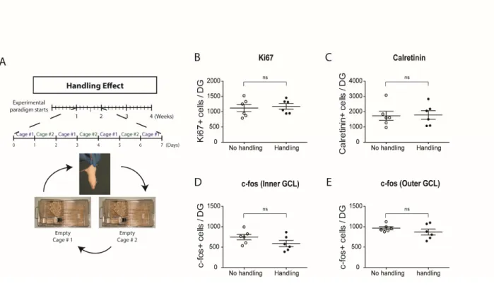
II.6.5. Absence of neurogenic effects of the Complex environment is not due to daily handling or type of running apparatus ... 73
II.7. Discussion ... 75
II.7.1. Running ... 75
II.7.2. Environmental complexity ... 76
II.7.3. Stress ... 78
II.7.4. Social context ... 79
II.8. Summary ... 81
II.9. Figures and figure legends ... 83
Chapter III. Behavioural studies and transcriptomics following exposure to running and social interactions ... 94
III.1. Chapter context ... 95
III.2. Author’s contributions ... 95
III.3. Introduction ... 97
III.4. Materials and methods ... 99
III.4.1. Mice ... 99
III.4.2. Housing conditions and Experimental groups ... 99
III.4.3. Tissue fixation and processing ... 99
III.4.4. Immunohistochemistry ... 100
III.4.5. Cell quantifications ... 100
III.4.6. Conditioned place preference ... 101
III.4.7. Delayed non-matching to place radial arm maze ... 101
III.4.8. Novel object recognition ... 102
III.4.9. RNA-Seq ... 103
III.4.10. Statistical analyses ... 104
III.5. Results ... 105
III.5.1. Running mice and socially-enriched mice show learning and memory differences ... 105
III.5.2. Design of the transcriptomics paradigm ... 107
III.5.3. Validation of RNA-Seq results ... 107
III.5.4. Low and high runners have distinct genetic changes ... 108
III.5.5. Genetic changes induced by the complex environment ... 109
III.5.6. Running and CE induce distinct genetic changes in the dentate gyrus niche ... 110
III.6. Discussion ... 111
III.7. Future directions ... 114
III.8. Figures and legends ... 115
IV.1. Chapter context ... 139
IV.2. Author’s contributions ... 139
IV.3. Introduction ... 141
IV.4. Materials and methods ... 142
IV.4.1. Mice ... 142
IV.4.2. Spinal cord injury ... 142
IV.4.3. Tissue fixation and processing ... 143
IV.4.4. Intracerebroventricular (ICV) osmotic pumps ... 143
IV.4.5. Culture experiments ... 143
IV.4.6. In situ hybridization ... 146
IV.4.7. Immunohistochemistry ... 146
IV.4.8. Western blotting ... 147
IV.4.9. RNA-Seq ... 147
IV.4.10. Statistical Analyses ... 148
IV.5. Results ... 149
IV.5.1. RNA-Seq transcriptomic profiling of ependymal cells following SCI ... 149
IV.5.2. Bio-informatics analyses of injured ependymal cells ... 150
IV.5.3. Identification of TGF-β as a potential upstream regulator of ependymal responses to SCI ... 150
IV.5.4. Exogenous TGF-β1 infusion reduces NIC numbers but increases ependymal proliferation within the intact spinal cord ... 150
IV.5.5. NIC expansion: TGF-β1 treatment in vitro suppresses growth factor-mediated formation of primary neurospheres ... 152
IV.5.6. TGF-β1 acts directly on neural precursors and promotes reversible quiescence of NICs ... 153
IV.5.7. Downstream effects: TGF-β promotes proliferation and astrocytic differentiation of NIC-derived progenitors ... 154
IV.6. Discussion ... 155
IV.7. Future directions ... 158
IV.8. Figures and legends ... 160
V.1. Discussion ... 179
V.1.1. Activation of hippocampal NSCs by environmental enrichment ... 179
V.1.1.1. Impact of individual components of EE ... 179
V.1.1.2. Behavioural impact of exercise and social interactions ... 181
V.1.2. Activation of spinal cord NSCs by injury-induced neuroinflammation ... 183
V.1.2.1. TGF-β1 impact on ependymal cells ... 184
V.1.3. Insights from both models ... 185
V.2. Perspectives ... 187
V.2.1. Mouse model – possibilities and limitations ... 187
V.2.2. Follow-up experiments ... 188
V.2.2.1. EE project ... 188
V.2.2.2. Spinal cord project ... 189
V.2.3. Other avenues ... 190
V.2.3.1. Reward system ... 190
V.2.3.2. Exercise pill ... 191
V.2.3.3. Proliferating ependymal cells versus NIC ... 192
V.3. Conclusions ... 193
VI. Bibliography ... i
VII. Appendix: Endogenous neural stem cell responses to stroke and spinal cord injury (review) ... i
List of tables
Table III.1. Top 25 of up-regulated genes in Low Runners ... 120
Table III.2. Top 25 of down-regulated genes in Low Runners ... 121
Table III.3. Top 25 of up-regulated genes in Exercise ... 122
Table III.4. Top 25 of down-regulated genes in Exercise ... 123
Table III.5. Top 25 of up-regulated genes in High Runners ... 124
Table III.6. Top 25 of down-regulated genes in High Runners ... 125
Table III.7. Top 25 of up-regulated genes in Complex Environment ... 126
Table III.8. Top 25 of down-regulated genes in Complex Environment ... 127
Table IV.1. Top 25 of up-regulated genes ... 162
Table IV.2. Top 25 down-regulated genes ... 163
Table IV.3. Top 10 upstream regulators (growth factors) ... 165
List of figures
Figure I.1. Early human brain development ... 4
Figure I.2. Evolution of neuroepithelial cells ... 6
Figure I.3. Subventricular zone neurogenesis lineage ... 12
Figure I.4. Hippocampal neurogenesis lineage ... 14
Figure I.5. Neural stem cell behaviour ... 16
Figure I.6. Central canal ependymal cell behaviour under normal conditions ... 19
Figure I.7. Hippocampal formation circuitry ... 24
Figure I.8. Dorsoventral axis projections of the hippocampus ... 26
Figure I.9. Circuitry properties of newborn neurons ... 31
Figure I.10. Pattern separation in the dentate gyrus ... 32
Figure I.11. Memory resolution ... 35
Figure I.12. Environmental enrichment ... 39
Figure I.13. Hippocampal neurogenesis lineage and markers ... 44
Figure I.14. Central canal ependymal cell behaviour under injury conditions ... 45
Figure II.1. The Alternating EE paradigm ... 83
Figure II.2. Effects of Alternating EE on the main stages of dentate gyrus neurogenesis ... 85
Figure II.3. Effects of Alternating EE on depolarization-associated c-fos expression ... 87
Figure II.4. Plasma corticosterone concentrations are reduced in the Complex environment . 89 Figure II.5. Daily handling does not affect basal neurogenesis in the Alternating EE paradigm ... 90
Figure II.6. Comparison of the effects of running wheels and running discs on adult neurogenesis ... 91
Figure II.7. Summary of long-term effects of individual EE variables on hippocampal
neurogenesis, c-fos expression and corticosterone levels ... 92
Figure III.1. Behavioural tests ... 116
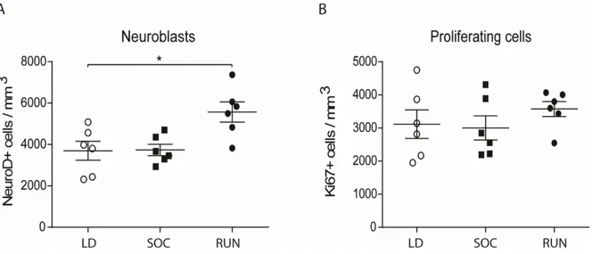
Figure III.2. Proliferating cells and neuroblasts numbers after eight weeks ... 117
Figure III.3. RNA-Seq experimental design ... 118
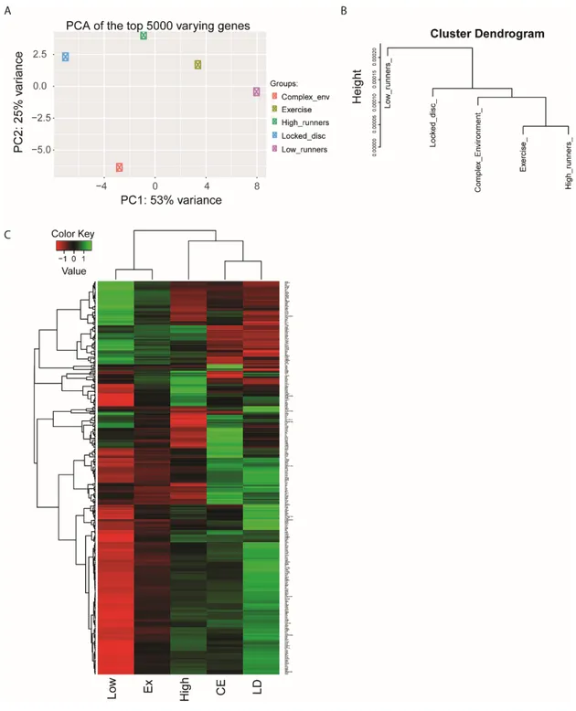
Figure III.4. RNA-Seq overview ... 119
Figure III.5. Signaling pathways of Low and High runners ... 128
Figure III.6. Biological Processes of Low and High Runners ... 129
Figure III.7. Cellular Components for Low and High Runners ... 130
Figure III.8. Molecular Functions for Low and High Runners ... 131
Figure III.9. Enrichr analysis for genes unique to High Runners ... 132
Figure III.10. Enrichr analysis for genes unique to Low Runners ... 133
Figure III.11. Genes modified in CE ... 134
Figure III.12. Modified genes comparison between Low and High Runners and CE ... 135
Figure III.13. Enrichr analysis for genes unique to runners ... 136
Figure III.14. Enrichr analysis for genes unique to CE ... 137
Figure IV.1. RNA-Seq analysis ... 160
Figure IV.2. GO enrichment analysis ... 164
Figure IV.3. Spinal cord contusion in adult mice induces strong expression of TGF-β1 mRNA ... 166
Figure IV.4. NIC recruitment and proliferation ... 167
Figure IV.5. Effect of TGF-β1 on NIC expansion ... 169
Figure IV.6. Effect of TGF-β1 on purified cultures ... 171 Figure IV.7. Neurosphere growth inhibition is TGF-β1-specific, not due to indirect effects 172
Figure IV.8. Self-renewal assay ... 173
Figure IV.9. TGF-β1 promotes proliferation and astrocytic differentiation in vitro ... 174
Figure IV.10. Summary ... 176
List of acronyms and abbreviations
AP: Alkaline phosphataseAPC: Antigen presenting cell
Ara-C: Cytosine-β-D-arabinofuranoside BDNF: Brain-derived neurotrophic factor BMP: Bone morphogenetic protein BNST: Bed nucleus of the stria terminals BrdU: Bromodeoxyuridine
CA: Cornu ammonis CE: Complex environment CNS: Central nervous system CPP: Conditioned place preference CSF: Cerebrospinal fluid
DCX: Doublecortin DG: Dentate gyrus
DNMP: Delayed nonmatching to place EE: Environmental enrichment
EGF: Epidermal growth factor eNSC: Embryonic neural stem cell EZ: Ependymal zone
FACS: Fluorescence-activated cell sorting FGF-2: Fibroblast growth factor-2
GABA: γ-aminobutyric acid GCL: Granular cell layer
GFAP: Glial fibrillary acidic protein GLAST: Glutamate transporter HPP: Hippocampus
ICM: Inner cell mass IEG: Immediate early gene
IL-1β: Interleukin-1beta IL-6: Interleukin-6
IPC: Intermediate progenitor cell KCC2: K+-Cl- co-transporter LIF: Leukemia inhibitor factor LPS: Lipopolysaccharide
MAM: Methylazoxymethanol acetate MAPK: Mitogen-activated protein kinase ML: Molecular layer
mPFC: Medial prefrontal cortex MTL: Medial temporal lobe MWM: Morris water maze NIC: Neurosphere-initiating cells NKCC1: Na+-K+-2Cl- co-transporter NOR: Novel object recognition NPY: Neuropeptide Y
NSC: Neural stem cell nACC: Nucleus accumbens OB: Olfactory bulb
OCT4: Octamer-binding transcription factor 4 PV: Parvalbumin
RAM: Radial arm maze RGC: Radial glial cell RGL: Radial glia-like cell RMS: Rostral migratory stream RSP: Retrosplenial area¸ SCI: Spinal cord injury SEZ: Subependymal zone SGZ: Subgranular zone SHH: Sonic hedgehog SST: Somatostatin
SVZ: Subventricular zone
TGF-β1: Τransforming growth factor-beta 1 TK: Thymidine kinase
TNF-α: Tumor necrosis factor- alpha VEGF: Vascular endothelial growth factor VTA: Ventral tegmental area
Dedication
I would like to dedicate my thesis dissertation
To my family,
in appreciation of their unconditional support throughout the years
To my friends,
without whom I wouldn’t have gone this far
And to the love of my life,
who was there for me during the most challenging stretch.
Acknowledgments
This was quite a journey! Six year and a half went by since I have started as an intern in the Fernandes laboratory back in January 2010. I am taking the opportunity to thank my Ph.D supervisor, Dr. Karl Fernandes, for giving me the chance to be part of his laboratory and for providing useful advice throughout the years. I also want to say a special thanks to my committee members, Dr. Diane Lagace, Dr. Graziella DiCristo, Dr. Lionel Carmant, and Dr. Laurent Descarries for their insight and guidance, as well as my thesis committee members Dr. Nicole Leclerc, Dr. Michel Cayouette, and Dr. Naglaa Shoukry.
In May 2010, I decided to pursue my project to complete a two-year master’s degree, which transformed into a six-year Ph.D degree by taking advantage of the fast-tracking process from M.Sc to Ph.D. I was glad to continue the environmental enrichment project that was started from scratch by a great scientist, Matthew Bednarczyk, who unfortunately past away in late 2014 (RIP). I keep wonderful memories from my first two years in the laboratory as I was surrounded by two amazing people who created a cheerful environment where it was a pleasure to come in to work: Stef Beaudoin and Greg Paliouras you will never be forgotten! I also had the chance to work with our wonderful technician, Anne Aumont, who is always there for the students and who created an organized lab easy to work in. I will always remember all the stories we shared when we commuted together on the train.
In late 2013, my motivation went down as I found myself at a status quo. I wanted to extend my knowledge and potential with new techniques, which was unfortunately not possible with my current project. I am grateful to my colleague, Laura Hamilton, for encouraging me at the time to continue and keep up the good work. In parallel, Karl was there to address my concerns and we finally agreed on a solution of adding the spinal cord project to my thesis, as no one was currently working on it at that time. In early 2014, I therefore had the chance to start performing in vitro experiments in our new lab at the CRCHUM. It was quite challenging to have 2 major branches for a total of 4 projects over the years, but I can say that I am proud of
my accomplishments considering all the challenges I had to face in both my professional and personal lives over these six years and a half.
I would also like to thank my colleagues Loïc Cochard (AKA Lolo), Chuck Levros (AKA Chuck maybe), and Brianna Goldenstein (AKA Bri) for their friendship and support, especially in the most challenging years that were these last two. Furthermore, the CRCHUM environment gave the opportunity to students to hang out more together and wonderful friendships emerged. Sacha (Lissouba), Vi, Arnaud, Maxime, Marc-André, Kessen, Camille (UdeM), Yoko, Nico, Jorge, Juliette, and Lamia, I will always remember our animated discussions and nights out having fun. To my friends outside the lab who kept me going by their admiration for my work and for always being honest and supportive, I will always be thankful to you for being my dearest friends for so many years: Sam, Cyn, Pré, the “Winnerslicious”, Rémi, and Shirley. To those I forgot to mention, please be tolerant, I know that you are also important, but one side effect of writing this dissertation is amnesia (not to be taken seriously).
Finally, the last but not the least, my family and my boyfriend, for their unconditional support for everything I have undertaken. Maman, papa et Charles, je vous serai à jamais reconnaissante pour tout ce dont vous m’avez donné et appris. Ludo, tu es une source d’inspiration et de motivation. Je vous aime !
I.1. General overview of nervous system development
The central nervous system (CNS) is composed of the brain and the spinal cord. Understanding how these structures developed is important in order to study their similarities and differences in how they regulate stem cells. The goal of this section is to briefly introduce a few concepts such as the role of embryonic cells forming the CNS, the stem cells and their importance in potential regeneration.
I.1.1. Common origin of the brain and spinal cord
Mammalian development starts from a fertilized egg, known as a zygote. This zygote goes through multiple cleavages: a 16-cell morula stage, a 32-cell stage, and a 64-cell blastocyst stage, composed of two clear layers (Gilbert 2006). The first layer of the blastocyst consists of external cells named the trophoblast that will form the embryonic portion of the placenta. The second layer is made of cells found within the trophoblast and is defined as the inner cell mass (ICM). The ICM is known to be pluripotent because it can create the entire embryo and related structures, except the trophoblast (Gage 2000; Gilbert 2006). The ICM gives rise to the hypoblast and the epiblast. These two tissues eventually develop into three well-defined layers: the endoderm, mesoderm and ectoderm in a well-defined structure called a gastrula following gastrulation (Gilbert 2006).
During later embryonic development, the ectoderm differentiates in three components: the epidermis, neural crest, and neural tube (Gilbert 2006). The neural tube will be described in greater detail as it forms the brain and spinal cord, the two regions of interest in this dissertation. Approximately 50% of the ectoderm is predetermined to become neural ectoderm, or the neural plate (Gilbert 2006). The remaining cells are thought to become the epidermis of the skin, mainly due to bone morphogenetic protein (BMP) signalling (Kandel et al. 2000). The neural plate will become the neural tube by a two-step process known as neurulation. Primary neurulation consists mainly of proliferating cells that form neural folds and invagination to create the neural groove (Gilbert 2006). Eventually, the folds will fuse to form the anterior
portion of the neural tube. Secondary neurulation is the creation of the posterior portion of the neural tube by merging mesenchyme cells, which are multipotent stromal cells (Gilbert 2006).
I.1.2. Regional specification of the neural tube
The anterior portion of the neural tube starts differentiating before the secondary neurulation finishes to form five main regions of the brain (Gilbert 2006; Kandel et al. 2000) (Fig.I.1). Three primary brain vesicles are created: the prosencephalon (forebrain (1)), mesencephalon (midbrain (2)), and rhomencephalon (hindbrain (3)). The prosencephalon is subdivided into the telencephalon ((1a) transforming into the cerebral hemispheres, including the olfactory bulb and hippocampal regions) and the diencephalon ((1b) transforming into the optic vesicles, thalamic, and hypothalamic regions). The mesencephalon will become the cerebral aqueduct, which contains the cerebrospinal fluid (CSF). Finally, the rhomencephalon subdivides into the metencephalon ((3a) transforming into the cerebellum and pons) and the myelencephalon ((3b) transforming into the medulla). It is from this caudal region of the neural tube, corresponding to the sixth major region of the CNS, which forms the spinal cord (Gilbert 2006; Kandel et al. 2000).
Figure I.1. Early human brain development
During development, the neural tube first divides into three primary vesicles (1. Prosencephalon (Forebrain), 2. Mesencephalon (Midbrain), and 3. Rhombencephalon (Hindbrain), then into five secondary vesicles (1a. Telencephalon, 1b. Diencephalon, 3a. Metencephalon, 3b. Myelencephalon, and 2. Mesencephalon stays the same). Each region has their adult derivative with specific functions. (Inspired from (Gilbert 2006), Fig. 12.9, and created by C-A. Grégoire)
A pattern is also established by a posterior-anterior gradient of retinoid acid that leads to the expression of Hox genes in the posterior region. These genes are responsible for the formation of the hindbrain and spinal cord (Gilbert 2006; Kempermann 2011). Other gradients are also present in the brain during development, including Pax6 (higher anterior pole concentrations) and Emx2 (higher posterior pole concentrations). These opposing gradients are important for positional identity (Bishop et al. 2002; Kempermann 2011).
I.1.3. Embryonic neural stem cells
The brain and spinal cord develop from differentiation of the neural tube. This process starts by the presence of embryonic neural stem cells (eNSCs) in the blastocyst.
I.1.3.1. Definition of neural stem cell
The eNSCs, present in the ICM have the capacity to differentiate into any types of tissues, including CNS tissue. They are therefore defined as pluripotent (Kempermann 2011). This pluripotency comes from the expression of several genes such as Oct4, Nanog, and sox2. The eNSCs located in the germinal neuroepithelium first become neuroepithelial precursor cells, then radial glia or neural stem cells (NSCs). NSCs can self-renew and divide asymmetrically to give rise to an intermediate progeny that can generate any tissue of the CNS (Gage 2000; Kempermann 2011).
I.1.3.2. Cell expansion
Cell expansion involves many cell divisions and thickening of the neural tube (Gilbert 2006; Kempermann 2011; Kriegstein and Alvarez-Buylla 2009). Nuclei of neuroepithelial cells are found at different heights in the lumen of the neural tube during development. During the S phase of the cell cycle, the nuclei move from the apical surface towards the pial surface of the neural tube, but return to the apical surface to perform mitosis. This phenomenon is called interkinetic nuclear migration. Neuroepithelial cells are first dividing symmetrically. Then, once the epithelium has thickened, neuroepithelial cells will elongate to become radial glial cells (RGCs, Fig. I.2). At this moment, RGCs start expressing astrocytic markers such as the glutamate transporter (GLAST), brain lipid-binding protein, Tenascin C, and in some species, glial fibrillary acidic protein (GFAP). RGCs divide asymmetrically to give rise to other RGCs or intermediate progenitor cells (IPCs). IPCs will then divide symmetrically to form identical daughter cells (Gilbert 2006; Kempermann 2011; Kriegstein and Alvarez-Buylla 2009).
I.1.3.3. Neurogenesis
Once cells are ready to form neurons, cell-division plane changes and one of the two daughter cells will detach, migrate, and differentiate (Gilbert 2006). IPCs migrate from the subventricular zone (SVZ) to the ventricular zone, a region next to the germinal neuroepithelium. There, they will form a second layer called the mantle or intermediate zone. In this layer, they differentiate into neurons and glia. This region is defined as the gray matter since it is where cell bodies reside. The neurons project axons away from the ventricular zone to create the marginal zone. This zone contains few cells but has many myelinated axons and therefore comprises the white matter (Gilbert 2006). Cajal-Retzius cells secreting reelin, a stop-and-go signal for migration, are found in this zone (Feng et al. 2007; Sanes et al. 2012). These three main regions: ventricular, intermediate, and marginal, are well retained in the spinal cord, whereas they are reorganized in the cerebral cortex. A new layer, called the neocortex, is formed at the outer side of the brain located between the marginal and intermediate zones. This cortical plate will eventually form the six layers of the cerebral cortex following an inside-out pattern.
Migrating neurons (i.e., neuroblasts) will use a radial glia cell process as a scaffold to migrate up the layers of cells (Gilbert 2006) (Fig. I.2).
I.1.3.4. Gliogenesis
At the end of cortical development, RGCs are no longer needed for scaffolding migration. Therefore, they detach from the ventricle, lose their radial process, and adapt an astrocytic morphology (Kriegstein and Alvarez-Buylla 2009). These astrocytes will keep their regenerative capacities in specific regions of the brain (Fig. I.2). For a long time, scientists believed regeneration was impossible in the adult brain. However, some of the adult mammalian brain and spinal cord cells maintain regenerative, stem cell properties (see section below).
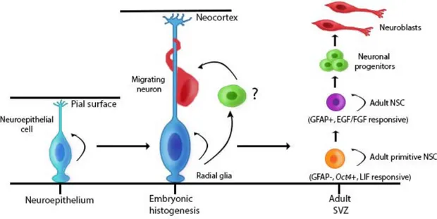
Figure I.2. Evolution of neuroepithelial cells
During development, neuroepithelial cells divide symmetrically and will elongate to become radial glia cells. The cells then behave as scaffolders for migrating neurons reaching the
neocortex, and divide asymmetrically. In adults, a rare population of primitive neural stem cells (GFAP- and leukemia inhibitor factor (LIF) responsive) give rise to GFAP+ neural stem cells. GFAP+ neural stem cells self-renew and give rise to neural progenitors that will mature into neuroblasts. (SVZ: Subventricular zone. Inspired from (Alvarez-Buylla et al. 2001) and (Sachewsky et al. 2014), and created by C-A. Grégoire and Loïc Cochard)
I.2. Adult neural stem cells
Adult NSCs are multipotent cells able to self-renew in the nervous system (Gage 2000). They are also referred to as primary progenitor cells or precursors. In specific regions of the brain, they are able to divide asymmetrically to give rise to three different types of cells in the following order: neurons (the functional component of the nervous system), astrocytes and oligodendrocytes (known as glia cells) (Kriegstein and Alvarez-Buylla 2009). In vitro, this multipotency is maintained in spinal cord-derived cultures (Weiss et al. 1996), but is lost in vivo as only glia-restricted progeny are produced (Horner et al. 2000; Martens et al. 2002). The regions of the hippocampus and spinal cord will be discussed in greater detail but first, we will review the origin and microenvironment of NSCs located within the hippocampus and spinal cord.
I.2.1. Origin of adult NSCs
Recent studies have investigated the origin of the adult NSCs present in the SVZ, dentate gyrus (DG), and spinal cord (Fuentealba et al. 2015; Li et al. 2013; Yu et al. 2013). One study used in utero delivery of retroviruses and bromodeoxyuridine (BrdU, a thymidine analogue that incorporates into DNA during DNA synthesis) to show that the majority of pre-B1 cells present in the SVZ were produced by embryonic progenitors dividing between E13.5 and E15.5 (Fuentealba et al. 2015). A lineage-tracing method with a barcoded retroviral library was used to determine the clonal relationships among cells. This experiment showed that there is a link between progenitors producing olfactory bulb (OB) interneurons after birth and those producing other neurons in the forebrain. However, this relationship eventually disappears in mid-fetal development, suggesting a lineage divergence (Fuentealba et al. 2015). The adult NSCs of the DG were demonstrated to originate from the ventral hippocampus (HPP) (Li et al. 2013). This region is known to be a source of sonic hedgehog (SHH), which is important for the maintenance of the adult NSC population. A continuous stream consisting of Gli1-responsive cells (SHH effector) was observed from the ventricular zone of the ventral HPP to the ventral granular cell layer (GCL) of the DG. They concluded, using several genetic approaches, that embryonic NSCs from the ventral HPP contributed to create the postnatal subgranular zone (SGZ)
throughout the longitudinal axis of the HPP (Li et al. 2013). Another study, demonstrated the necessity of SHH for postnatal ependymal zone (EZ) cell formation (Yu et al. 2013). During late development, it was shown that adult ependymal cells, found around the central canal, are derived from two distinct progenitor domains: p2 and pMN from the ventral ventricular zone. They also observed a severe disruption in the formation of the EZ when embryonic SHH signaling was absent (Yu et al. 2013).
I.2.2. Adult neural stem cell niche
The NSC niche is a microenvironment composed of multiple cell types (such as astrocytes and ependymal cells), secreted molecules, extracellular matrix, and blood vessels (endothelial cells). The NSCs are therefore exposed to a variety of signals and cell-cell interactions (Riquelme et al. 2008). In adults, only specific regions maintain the capacity to produce new neurons, hence suggesting that adult NSC niches are environments supporting self-renewal and multipotency through signaling. However, the right balance between proliferation and differentiation signals is a priority to avoid tumor development or exhaustion of the NSC pool. Moreover, vasculature is an important factor as angiogenesis may have an impact on neurogenesis via hormones and cytokines (Riquelme et al. 2008). For example, vascular endothelial growth factor (VEGF) is responsible for angiogenesis and has been shown to influence neurogenesis, especially stimulating progenitor’s proliferation (Jin et al. 2002). Furthermore, transplantation experiments showed the importance of external cues present in the microenvironment on stem cell fate. The spinal cord is defined as a non-neurogenic region, but fibroblast growth factor-2 (FGF-2)-responsive adult spinal cord-derived cells differentiated into neurons when transplanted and integrated into the GCL of the DG (Shihabuddin et al. 2000). Another experiment isolated neuronal precursors (predisposed to become interneurons) from the SVZ via magnetic activated cell sorting and transplanted them into the striatum (Seidenfaden et al. 2006). However, once implanted into the SVZ niche, these cells underwent glial differentiation. This example, once again, demonstrates the importance of the niche.
I.2.3. Brain
Adult neurogenesis is the postnatal formation by precursor cells of mature neurons (Kempermann et al. 2004; Ming and Song 2005). It was believed for a long time that neurogenesis in mammals was a development-restricted phenomenon. However, in the mammalian brain, adult neurogenesis occurs in a few areas including two main regions: the SVZ of the lateral ventricles and the DG of the HPP (Ihrie and Alvarez-Buylla 2011; Kempermann et al. 1998; Ming and Song 2005). Research conducted in 1965 shed light on the existence of postnatal neurogenesis, specifically in the DG of the HPP. Altman and Das first injected thymidine-H3 in rats to label cell nuclei where DNA was synthesized in order to identify proliferating cells (Altman and Das 1965). In this study, they demonstrated that the number of cells undergoing proliferation decreases with age (up to 8 month-old), while the number of differentiated cells (corresponding to granule cells) increases (Altman and Das 1965). In 1977, newly formed neurons were also labeled with thymidine-H3 and analyzed by electron microscopy, following a 30-day chase period in both the rodent DG and OB (Kaplan and Hinds 1977). This research showed the survival capacity of these newly-born neurons. Moreover, important research conducted in songbirds gave additional validity to the concept of adult neurogenesis in other vertebrates (Alvarez-Buylla and Kirn 1997; Alvarez-Buylla et al. 1988; Goldman and Nottebohm 1983). Unfortunately, widespread skepticism about adult neurogenesis delayed more scientific inquiries into its existence. Nonetheless, additional studies in most vertebrates and mammals confirmed the presence of stem cells in the SVZ and DG in vivo, and led to their isolation (in rodents) in vitro (Gage 2000; Lindsey and Tropepe 2006; Palmer et al. 1997; Reynolds and Weiss 1992). Finally, experiments using post-mortem brains from patients who received intravenous injections of BrdU showed that adult neurogenesis also occurs in the adult human brain (Curtis et al. 2007; Eriksson et al. 1998). Recent studies used stable 14C present in genomic DNA and compared it to 14C in the atmosphere (from nuclear bomb tests during the Cold War) to determine cell population date of birth (Ernst and Frisen 2015; Spalding et al. 2005). Using this method, they noticed that the age of human OB neurons was almost the same as the tested individuals, suggesting low turnover levels below 1% in this region. However, the role of OB between rodents and humans may differ (more details in section I.3.1.2) (Bergmann et al. 2012). Nonetheless, this technique allowed the discovery that
hippocampal neurogenesis occurs throughout life in humans at a rate similar to middle-aged mice (Spalding et al. 2013). These discoveries increased interest in adult neural stem cells as they could be perceived as potential therapeutic tools for neurodegenerative diseases and injuries.
I.2.4. Subventricular zone of the lateral ventricles
The SVZ, the main neurogenic niche, is comprised of several cell types. Ependymal cells, known as type E cells, are present along the lateral ventricle (next to the SVZ) forming a pinwheel (Mirzadeh et al. 2008). E cells are responsible for the production and circulation of a small amount of cerebrospinal fluid (Abrous et al. 2005; Doetsch et al. 1999a; Mirzadeh et al. 2008). Slowly dividing (quiescent) astrocytes, defined as type B1 cells, are present at the center of the ependymal pinwheels. B cells extend a basal process to the blood vessels and an apical cilium towards the cerebrospinal fluid (Kriegstein and Alvarez-Buylla 2009; Mirzadeh et al. 2008) (Fig. I.3). The B1 cells can be subdivided into activated and quiescent NSCs (Codega et al. 2014; Lim and Alvarez-Buylla 2016). Codega and colleagues isolated quiescent (GFAP+/CD133+) and activated (GFAP+/CD133+/EGFR+/Nestin+) NSC populations. After in vivo transplantation, both cells were neurogenic but behaved with different kinetics (quiescent NSCs (qNSCs) showing delayed kinetics). qNSCs rarely gave rise to neurospheres, in contrast to activated NSCs (aNSCs) that were enriched in colony-forming cells. Importantly, qNSCs could become activated in cultures with expression of Nestin and EGFR, suggesting that qNSCs and aNSCs can intervert between both states (Codega et al. 2014). Regeneration experiments conducted by Doetsch and colleagues in CD1 mice helped to first identify the NSCs in the SVZ niche. Cytosine-β-D-arabinofuranoside (Ara-C) was infused into the lateral ventricle via an osmotic pump for six days to eliminate dividing cells. At the end of the infusion, they observed the disappearance of all transit-amplifying cells (C cells) and proliferating neuroblasts (A cells, Fig. I.3), which are clustered migrating cells that go through the rostral migratory stream (RMS) to reach the OB and mature into interneurons. Only B cells and ependymal cells remained, suggesting they are potential NSCs (Doetsch et al. 1999b). This group further showed that only the astrocytes were labeled with a proliferating marker after Ara-C treatment, suggesting these cells, and not ependymal cells, repopulate the SVZ niche. After 36 hours of Ara-C treatment
cessation, C cells reappeared. Four days after cessation, A cells were also observed. Additionally, retrovirus and in vitro cultures experiments both confirmed that A and C cells were derived from B cells, and not from ependymal cells (Doetsch et al. 1999a). From these findings, B ependymal cells were defined as NSCs in the SVZ. Recently, Sachewsky and colleagues showed the existence of a rare population, called adult primitive NSCs (Sachewsky et al. 2014). These primitive cells are activated by the leukemia inhibitor factor (LIF) and demonstrated stem cell properties as they express octomer-binding transcription factor 4 (Oct4) and integrate into the ICM of blastocysts. In presence of LIF, these cells generated self-renewing and multipotent neurosphere colonies that gave rise to GFAP+ NSCs in vitro. Moreover, following in vivo depletion of GFAP+ NSCs, Oct4+ NSCs were able to repopulate the SVZ, suggesting that primitive NSCs are found upstream of GFAP+ NSCs (Fig. I.2) (Reeve et al. 2016; Sachewsky et al. 2014).
Figure I.3. Subventricular zone neurogenesis lineage
This figure represents the subventricular zone niche comprised of the ependymal and subependymal zones. Ependymal cells contact the cerebrospinal fluid while they line the ventricle. Activated B cells (radial glia-like cells), are in contact with blood vessels and divide asymmetrically to self-renew and give rise to C cells (transit-amplifying cells). C cells divide quickly, mature into neuroblasts, and migrate through the rostral migratory stream to the
olfactory bulbs. (CSF: Cerebrospinal fluid, EZ: ependymal zone, SEZ: Subependymal zone, SVZ: Subventricular zone. Inspired by (Bond et al. 2015), Created by C-A. Grégoire and Loïc Cochard).
I.2.5. Subgranular zone of the dentate gyrus
Adult neurogenesis also occurs in the SGZ of the DG, located in the HPP (Gage 2000). The DG is comprised of three layers: the molecular layer, the GCL, and the polymorphic cell also known as the hilus (Fig. I.4) (Anderson et al. 2007). In this niche, the cell lineage starts with slowly-dividing radial-glia like cells (RGL), known as type 1, whose endfeet touch the vasculature. These cells express GFAP, Nestin, and Sox2, a SRY transcription factor. They extend their radial process through the GCL to reach the molecular layer (Bonaguidi et al. 2012; Kempermann et al. 2015b; Seri et al. 2004). RGLs divide slowly in an asymmetric manner to produce intermediate progenitor cells (type 2a and 2b, or D cells). A proportion of these progenitors correspond to the Sox2+/Ki67+ proliferating cell population lacking radial processes, described as non-radial precursors (Suh et al. 2007). Most of these progenitors also express Tbr2 (a transcription factor) and are committed to a neuronal fate (Hodge et al. 2008). These progenitors divide quickly to give rise to neuroblasts (type 3), and express markers such as doublecortin (DCX, a microtubule-associated protein), and NeuroD (a basic helix-loop-helix transcription factor). Immature neurons will then become post-mitotic, mature into glutamatergic granule cells, and functionally integrate the circuitry (Bonaguidi et al. 2012; Kriegstein and Alvarez-Buylla 2009; van Praag et al. 2002) (Fig. I.4). These newly-born neurons form functional synapses with CA3 pyramidal cells at two weeks, which stabilize at four weeks (Gu et al. 2012). Only a small portion of these neurons become integrated as the majority of adult generated neurons die within the first four days after birth (Sierra et al. 2010). In order to identify the NSCs of the DG, Seri and colleagues used thymidine-H3 and GFAP labeling to look at the ultrastructure of proliferating cells (Seri et al. 2001). GFAP+ cells, described as B cells (or type 1 cells) in the SVZ, have an astrocytic morphology in contrast to GFAP-negative cells (rapidly dividing progenitors, SVZ D cells or type 2 cells) (Seri et al. 2001). Similarly to the Doetsch experiments (Doetsch et al. 1999a; Doetsch et al. 1999b), Seri and colleagues infused Ara-C and gave procarbazol (anti-mitotic drug) in drinking water for
seven days to eliminate proliferating cells. Two days following treatment, 91% of the cells left in the SGZ were type B cells. On day four, progenitor cells reappeared, and their numbers reached control levels by day 15 following treatment. They also introduced an avian leucosis virus including an alkaline phosphatase (AP) gene to label proliferating infecting cells and track their progeny. The maturation of GFAP+/AP+ SGZ astrocytes was studied. It was confirmed that these astrocytes behaved like NSCs and gave rise to neurons (Seri et al. 2001). For simplicity, the term RGL will be used throughout the dissertation to describe neural precursors behaving like stem cells in the DG.
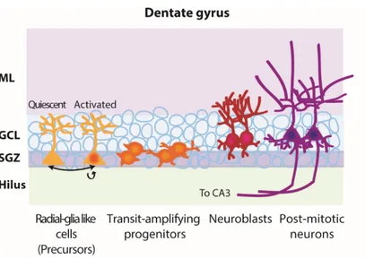
Figure I.4. Hippocampal neurogenesis lineage
The dentate gyrus niche is comprised of several layers: the molecular layer, the granular cell layer, the subgranular zone and the hilus. Hippocampal neurogenesis starts in the subgranular zone where quiescent precursors become activated and divide asymmetrically to give rise to another precursor and quickly dividing progenitors. These progenitors will then migrate a short distance to reach the granular cell layer and mature into neuroblasts. Neuroblasts will become post-mitotic and integrate the circuitry by receiving entorhinal cortex inputs in the molecular layer and projecting their axons towards the CA3 region of the hippocampus. (GCL: Granular cell layer, ML: Molecular layer, SGZ: Subgranular zone. Created by C-A. Grégoire and Laura Hamilton.).
Recently, different DG neurogenic lineage models were introduced to the field and caused controversy (Bond et al. 2015) (Fig. I.5). The first model is called the “disposable stem cell” model (Encinas et al. 2011). Quiescence is known to be one way to conserve the stem cell pool and limit their replication to avoid mutations in several tissues (Li and Clevers 2010). However, in this model, activated NSCs are believed to leave the stem cell pool, convert to the astrocytic fate, and not return to their quiescent state. This correlates with the observations made in aged mice where NSCs are lost and neurogenesis diminishes (Encinas et al. 2011). Encinas and colleagues confirmed progenitors were coming from RGLs following asymmetric division using a transgenic mouse model. They then used a BrdU paradigm in Nestin-CFPnuc reporter mice, which allowed them to label proliferating cells shortly after the pulse period, and long-retaining cells (RGLs) after a chase period. For a period of seven days, BrdU+ RGL numbers stayed constant, but dropped to zero after 10-15 days. In parallel, the number of BrdU+ astrocytes climbed and reached the initial number of RGLs by day 10, suggesting that these astrocytes could emerge from the activated RGLs. Moreover, they observed that activated RGLs were going through three rounds of division before exiting the cell cycle and becoming astrocytes (Encinas et al. 2011). Most tools, including the transgenic mice used in the previous study, label cell populations (Dhaliwal and Lagace 2011). However, an in vivo clonal analysis approach allowed tracing of individual nestin+GFAP+ RGL precursors (Bonaguidi et al. 2011). The inducible, sparse-labeling (low concentration of tamoxifen) model nestin-CreERT2; Z/EG (lacZ and GFP) was used to understand RGLs’ behaviour. The vast majority of RGLs are quiescent, but once they become activated they go through three modes of division: 1) symmetrical (shown for the first time) to expand the RGL pool (to a lower extent), 2) asymmetrical to give rise to a progenitor cell that will become a neuron and maintain the RGL pool, and 3) asymmetrical to give rise to an astroglia and maintain the RGL pool. After division, the activated RGL returns to quiescence. Bonaguidi and colleagues confirmed their clonal analysis results with the mosaic-analysis with double markers reporter that allows to label daughter cells and their progeny (Bonaguidi et al. 2011). Another model suggested RGL heterogeneity (DeCarolis et al. 2013). DeCarolis and colleagues used two inducible transgenic mouse lines to demonstrate the validity of this model: Nestin-CreERT2/R26R:YFP and GLAST::CreERT2/R26R:YFP. Currently, there is no unique markers for hippocampal RGLs, therefore they used Nestin (labels both RGLs and progenitors) and GLAST (labels both
astrocytes and RGLs). A week following Ara-C treatment, an increase in dividing GLAST-YFP+ cells, but not Nestin-GLAST-YFP+ cells was observed. This suggests that GLAST-GLAST-YFP+ cells contribute to the recovery of the RGL population. Consistent with this finding, following a 14-day exercise paradigm, only GLAST-YFP+ cells increased in numbers, even if proliferation was amplified in both lines (DeCarolis et al. 2013).
Figure I.5. Neural stem cell behaviour
Neural stem cells may have different behaviour based on different models. One model suggests that activated radial-glia like cells divide symmetrically. Another model suggests asymmetric division of activated radial-glia like cells that give rise to another radial-glia like cell and a progenitor that will differentiate into different cell types such as astrocytes, neurons and oligodendrocytes. Lastly, another model suggests a disposable model where a quiescent radial-glia like cell will leave the stem cell pool and convert into an astrocyte. (RGL: Radial-radial-glia like cells. Inspired by (Bond et al. 2015). Created by C-A. Grégoire and Loïc Cochard).
I.2.6. Hypothalamus
Another brain region that gained popularity over the last decade as a potential neurogenic niche is the hypothalamus (Goodman and Hajihosseini 2015). The hypothalamus plays an important role in the secretion of essential hormones implicated in temperature regulation, food intake, sex drive, and sleep-wake cycle besides other physiologic functions (Kandel et al. 2000). The main focus of research on the hypothalamus has been on energy balance with several articles published on this topic. Two major neurons are found in the arcuate nucleus (adjacent to the third ventricle): neuropeptide Y (NPY) and pro-opiomelanocortin-expressing neurons, known as orexigenic and anorexigenic, respectively (Kokoeva et al. 2005; Pierce and Xu 2010). In 2004, progenitor cells from the hypothalamus were first isolated in cultures (Markakis et al. 2004). Then, central infusion of the ciliary neurotrophic factor into the lateral ventricle mouse brains, known to reduce body weight, induced proliferation of newborn BrdU+ cells. 42 days following infusion, 43% of the proliferating cells expressed neuronal markers (Kokoeva et al. 2005). Another study was conducted to confirm these previous results (Kokoeva et al. 2007). Three days following infusion, half of the BrdU+ cells also expressed Ki67, and infusion directly in the third ventricle led to increased proliferation throughout the hypothalamic parenchyma, but none around the lateral ventricle (Kokoeva et al. 2007). Moreover, a study shed light on the possibility that inhibition of de novo proliferation could lead to less food intake, therefore suggesting a link between hypothalamic new cell proliferation and energy balance (Pierce and Xu 2010).
In the past few years, researchers have been investigating the role of hypothalamic tanycytes that showed stem cell-like properties (Chaker et al. 2016; Goodman and Hajihosseini 2015; Lee et al. 2012; Robins et al. 2013). Four subtypes of tanycytes exist: α1, α2, both found dorsally, β1, and β2, found ventrically (Rodriguez et al. 2005). In 2012, β2-tanycytes were first identified as the neurogenic radial glia-like nonciliated ependymal cells in postnatal and pre-adult mice (Lee et al. 2012). Li and colleagues demonstrated that cells from pre-adult hypothalami could give rise to self-renewing and multipotent neurospheres in vitro. Moreover, in vivo, they showed that the majority of sox2+ adult stem cells were present in the mediobasal region of the hypothalamus, and not in the lateral third-ventricle wall where tanycytes are found (Li et al.
2012). It was later specified, using a lineage-tracing approach in adult GLAST::CreERT2 mice,
that α-tanycytes are the self-renewing and multipotent cells (astrocytes and neurons) in vivo, and give rise to neurospheres in vitro (Robins et al. 2013). Recently, Chaker and colleagues demonstrated for the first time that hypothalamic neurogenesis persists through aging (Chaker et al. 2016).
I.2.7. Spinal cord
In the spinal cord, ciliated cells involved in the propulsion of the cerebrospinal fluid, known as ependymal cells, seem to maintain a regenerative capacity. Located around the central canal of the rodent spinal cord, ependymal cells behave like stem cells as they demonstrate self-renewal and multipotency capacities in vitro (Johansson et al. 1999; Martens et al. 2002; Meletis et al. 2008; Weiss et al. 1996) (Fig. I.6). However, in vivo, BrdU-retaining ependymal cells show a glial-restricted differentiation into astrocytes and oligodendrocytes (Horner et al. 2000), hence lacking neurogenic capacity (Martens et al. 2002). In the field, this discrepancy between the in vitro and in vivo data contributes to define ependymal cells simply by their name instead of referring to them as stem cells. In the naive spinal cord, proliferating ependymal cell number is very limited and shows a dorso-ventral gradient with greater density at the dorsal part of the central canal. Proliferating cells are often found in doublets and associated with blood vessels. Vimentin+ ependymal cells might also express Nestin+ or GFAP+ processes elongating dorsally in the grey matter (Hamilton et al. 2009). In this niche, ependymal cells are mostly quiescent under normal conditions and divide symmetrically, suggesting a maintenance proliferation mechanism for this precise pool of ciliated cells (Barnabe-Heider et al. 2010; Hamilton et al. 2009; Meletis et al. 2008) (Fig. I.6). In the adult human spinal cord, the presence of NSC-like cells has been demonstrated for the first time in 2008 (Dromard et al. 2008), and their self-renewing capacity in an adherent monolayer culture was further shown in a more recent article (Mothe et al. 2011).The human spinal cord-derived neurospheres showed NSC and proliferation marker expression (Sox2, Nestin, and Ki67). They have the capacity to differentiate into astrocytes and neurons, but have limited self-renewal capacity. The ependymal layer organization in the human spinal cord is different than the one found in rodents. In humans, the central canal is often occluded and the ependymal layer is disorganized, frequently showing
rosettes or microcanals. The human central canal is surrounded by a hypocellular region containing high levels of GFAP+ and Nestin+ cells localized in the ventral part of the central canal. As in rodents, the human ependymal layer maintains immature features during adulthood. For instance, it expresses Nestin and Sox2, and proliferates at a low rate under naive conditions (Hugnot and Franzen 2011). In summary, the existence of self-replicating ependymal cells has been demonstrated in the adult human spinal cord, suggesting potential to develop endogenous spinal cord repair strategies that could target these cells (Dromard et al. 2008; Mothe et al. 2011).
Figure I.6. Central canal ependymal cell behaviour under normal conditions
Under normal conditions, ependymal cells surrounding the central canal of the spinal cord will slowly self-renew as this niche is mostly quiescent in vivo giving rise to other ependymal cells. In contrast, in vitro, neurospheres can be grown from dissociated spinal cord-derived cells and these cells have multipotent capacities leading to neuron, astrocyte and oligodendrocyte differentiation (Created by C-A. Grégoire, Loïc Cochard, and Brianna Goldenstein).
I.2.8. Regenerative capacities throughout evolution
Throughout evolution, regenerative abilities have varied among species (Bonfanti and Peretto 2011; Ferreira et al. 2012). The distinction between regeneration and repair is important,
as repair is imperfect regeneration because it does not normally restore both the structure and function (Bonfanti 2011). Invertebrates conserve their ability to regenerate whole body parts, and vertebrates, such as fish, amphibians, and reptiles, maintain indeterminate growth throughout their lives (Lee-Liu et al. 2013; Tanaka and Ferretti 2009). In these species, embryonic radial glia cells are still present, and maintain the capacity of producing neurons and glial cells throughout adulthood. Moreover, in newts, proliferation and neurogenic regions (or hot spots) are not necessarily the ones demonstrating regenerative capacities. In response to injury, a de novo generation occurs by the reactivation of quiescent ependymoglial GFAP+ cells that will re-establish zones of regenerative proliferation and neurogenesis (Kirkham et al. 2014). In urodele amphibians, these ependymoglial cells transiently lose their GFAP expression and start expressing NSC markers such as Nestin. Then, they participate in the repair of the neuroepithelial tube to eventually rebuild the spinal cord (Walder et al. 2003). Newts and salamanders can regenerate their limbs throughout life, whereas frogs and toads can do so only during development (Bonfanti 2011). More recently, a group suggested that Sox2+ neural precursor cells in Xenopus tadpoles are involved in the regeneration process (Gaete et al. 2012). They observed a significant increase in Sox2 labeling four days after amputation of the tail, and 20% to 60% of them were proliferating (BrdU+). An increase in Sox2 mRNA levels was also observed as soon as one day following injury when compared to uncut tail controls. Moreover, they used a Sox2 dominant negative construct, mediating its translocation to the nucleus, and only 46% of tadpoles maintained their tail regeneration capacities (Gaete et al. 2012). These results show the importance of NSCs in the regenerative process. However, in mammals, these radial glial cells only exist during embryonic development and transform into astrocytes in the adult nervous system (Voigt 1989). Thus, mammals lose the ability to regenerate injured tissue. In adult birds, these radial cells are still present but restricted to the walls of the lateral ventricle (Alvarez-Buylla et al. 1990). In the SVZ of the lateral ventricles, NSCs are activated following stroke, and produce new cells that respond to the injury, eventually forming a glial scar (Gregoire et al. 2015; Zhang et al. 2008). A similar process is observed in the spinal cord following lesion, where ependymal cells produce astrocytes implicated in glial scar formation (Barnabe-Heider et al. 2010; Gregoire et al. 2015). One theory is that regenerative capacities over evolution would be negatively correlated with the development of the immune system as it gets more specialized, leading to a loss in regeneration (Bonfanti 2011).
I.3. Functional significance of adult NSCs
Adult neurogenesis is an interesting field due to therapeutic avenues that can emerge from understanding its functional importance. The physical location of NSCs can also determine their functions. The NSCs located in the SVZ and the DG will be discussed in further detail in this section.
I.3.1. Subventricular zone of the lateral ventricle and olfactory bulb
I.3.1.1. SVZ/OB functions
In the rodent brain, the OB plays an important role in olfaction as this sense is well developed to survive in their natural habitat (Lazarini and Lledo 2011). The SVZ of the lateral ventricle is the starting location of neurogenesis. The final location is achieved once neuroblasts migrate along the RMS to the OB and mature into either of two local interneuron types (periglomerular or granule cells). Only half of newly-born neurons will successfully integrate the OB circuitry. The OB acts as a relay station that processes and refines sensory inputs triggered by environmental cues. The information is then transmitted to the primary and accessory olfactory cortex via mitral and tufted principal neurons (Kempermann 2011; Lazarini and Lledo 2011; Sakamoto et al. 2014).
The functional significance in olfaction remains elusive even though several neurogenesis ablation studies demonstrated the importance of newly-born neurons in the maintenance of OB circuitry, olfactory memory formation, and odorant discrimination (Breton-Provencher et al. 2009; Moreno et al. 2009; Valley et al. 2009). Other functions are linked to the SVZ germinal niche. In vivo, Menn and colleagues showed that B cells are able to differentiate into migrating non-myelinating and myelinating oligodendrocytes through Olig2+ type C progenitors (Menn et al. 2006). Under normal conditions, the oligodendrocyte/neuron ratio is low, but following a demyelinating lesion in the corpus callosum, the number of oligodendrocytes increased by fourfold. This result suggests that B cells have the capacity to