third degree). The patient with a third degree was not treated for the LS during the first pregnancy, and there was some delayed healing. There were no issues with her second delivery when LS was quiescent. No patients developed LS in obstetric scars.
In a series of 40 patients studied for the effect of the oral con-traceptive on LS, four became pregnant and reported complete remission of disease during pregnancy.3A study of 33 pregnan-cies in 29 patients with vulval LS reported 27 spontaneous vagi-nal births, two instrumental deliveries and 4 LSCS. Only one non-compliant patient required a section due to LS-related scar-ring.4One patient developed LS in a perineal scar. Two women with newly diagnosed LS and who delivered by normal sponta-neous vaginal delivery without problems have been reported.5
The proportion of women with an intact perineum at delivery was 9.6% in nulliparae and 31.2% in multiparae.6In our popula-tion with LS, 10/21 nulliparae had tears and 9/21 had epi-siotomies; therefore, there was no increased incidence of perineal tears and episiotomies compared to the general popula-tion. No patients reported sexual dysfunction during follow-up of up to 4 years in many cases.
There is conflicting literature on the requirements for TCS during pregnancy and by extrapolation the severity of LS during pregnancy. There was no significant change in the TCS require-ments in one study,4but in others the effect was variable.7,8In our cohort, 55% patients had the same TCS requirements during pregnancy but 45% decreased their use suggesting improvement in their LS.
In conclusion, LS does not worsen during pregnancy and can improve. When the disease is well controlled, there is no contra-indication to vaginal delivery and women can be reassured about this.
D. Trokoudes, F.M. Lewis*
St John's Institute of Dermatology, Guy's & St Thomas’ Hospital, London, UK *Correspondence: F.M. Lewis. E-mail: fi[email protected]
References
1 Goldstein AT, Marinoff SC, Christopher K et al. Prevalence of vulvar lichen sclerosus in a general gynecology practice. J Reprod Med 2005;50: 477–480.
2 Lee A, Bradford J, Fischer G. Long-term management of adult vulvar lichen sclerosus: a prospective cohort study of 507 women. JAMA Derma-tol 2015;151: 1061–1067.
3 Gunthert AR, Faber M, Knappe G et al. Early onset vulvar lichen sclerosus and pre-menopausal women and oral contraceptives. Eur J Obstet Gynecol Reprod Biol 2008;137: 56–60.
4 Nguyen Y, Bradford J, Fischer G. Lichen sclerosus in pregnancy: a review of 33 cases. Aust N Z J Obstet Gynaecol 2018;58: 686–689.
5 Haefner HK, Pearlman MD, Barclay ML et al. Lichen sclerosus in preg-nancy: presentation of two cases. J Low Genit Tract Dis 1999;3: 260–263. 6 Smith LA, Price N, Simonite V et al. Incidence of and risk factors for
per-ineal trauma: a prospective observational study. BMC Pregnancy Childbirth 2013;13: 59.
7 Dalziel KL. Effect of lichen sclerosus on sexual function and parturition. J Reprod Med 1995;40: 351–354.
8 Helm KF, Gibson LE, Muller SA. Lichen sclerosus et atrophicus in children and young adults. Pediatr Dermatol 1991;8: 97–101.
DOI: 10.1111/jdv.15788
X-linked hypohidrotic
ectodermal dysplasia: clinical
and molecular genetic analysis
of a large Russian family with a
synonymous p.Ser267
=
(c.801A
>G) splice site mutation
Editor
X-linked hypohidrotic ectodermal dysplasia (XLHED) is the most common form of ectodermal dysplasias.1Mutations in the EDA gene are the cause of XLHED.2This study describes a large Russian family with XLHED (Fig. 1).
The proband’s examination revealed a large forehead with prominent supraorbital ridges and forehead bumps, wide cheek-bones, small saddle-shaped nose with hypoplastic alae nasi, nar-row and short maxillary regions, slightly deformed ears. The hair, eyebrows and eylashes were light-coloured, sparse and brit-tle. The skin was dry and pale. However, according to the mother, the proband had good heat tolerance. Examination revealed hypoplastic nipples, but nail structure was normal. The proband was susceptible to respiratory infections. Intraoral examination showed complete adontia. The mouth mucosa was pale, with noticeable hypersalivation. The maxilla and mandi-bula were hypoplastic with underdeveloped alveolar processes. X-ray orthopantomography revealed four rudimentary baby teeth and two rudimentary permanent central maxilla incisors.
The proband’s EDA gene coding sequence and exon–intron junction analysis by Sanger sequencing revealed a synonymous p. Ser267= (c.801A>G) variant in exon 7. This variant was not present in the dbSNP or the Genome Aggregation Database but was described in HGMD (CS1614178) by Wohlfart et al.3The c.801A>G variant segregates with the disease: it was detected in all affected males in hemizygous state, in females– in heterozy-gous state and was not detected in a healthy blood relative (Fig. 1). The probability of the simultaneous inheritance of the XLHED and the revealed EDA gene variant in this family being a coincidence is about 0.01%.
In silico Human Splicing Finder and ESE finder analysis revealed that c.801A>G variant could break the site of an exonic splicing enhancer (ESE) and create a new exonic splicing silencer (ESS), resulting in pre-mRNA splicing alteration. However, three other splicing tools – BDGP, SplicePort and NetGene2 –
© 2019 European Academy of Dermatology and Venereology
JEADV2019, 33, e437–e495
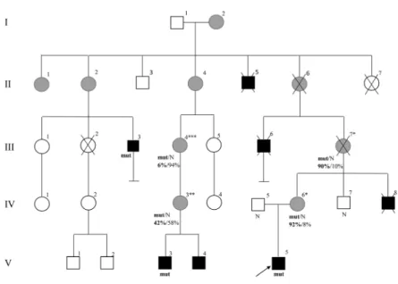
Figure 2 Functional pathogenicity evidence of the c.801A>G variant in the EDA gene. (a) RT-PCR analysis of EDA mRNA structure revealed that the c.801A>G variant leads to exon 7 skipping (Δ7). On the top, the scheme of analysed EDA locus is depicted. PAGE elec-trophoregram showsΔ7 EDA mRNA isoform in both patients’ samples (III.3, V.5), wild-type (WT) and Δ7 isoforms in the mother’s sample (IV.6) and WT isoform in the father’s sample (IV.5). Sanger sequences of corresponding PCR products are shown below. (b) Results of minigene assay. The scheme of minigene cloning and analysis is depicted above. Agarose gel electrophoregram of RT-PCR fragments generated from minigene-spliced RNA of wild-type (WT) and mutant (MUT) constructions are depicted below. The empty rectangle repre-sents the artificial exon created by a cryptic splice acceptor site of EDA intron 7 and cryptic splice donor site of the vector intron. Analysis revealed that mutant construction produces only transcripts with exon 7 sipping, while wild-type construction shows its inclusion. Figure 1 Pedigree of a Russian family with ectodermal hypohidrotic dysplasia. Squares and circles represent males and females, respectively. The black symbol indicates the affected member; the grey symbol, the female carriers; and the open symbols, the unaf-fected members. The patient above the arrow is the proband of this family. mut– p.Ser267= (c.801A>G) variant in the EDA gene. N – cor-responds with EDA reference sequence. Numbers under the genotype of female carriers indicate the percentage of X chromosome inactivation: 92%– of the mutant X chromosome, 8% – of the intact X chromosome (e.g. proband’s mother IV.6). The obtained data cor-relate with the severity of the clinicalfindings seen in female carriers. *mild clinical manifestations: slight dryness of the skin. **moderate clinical manifestations: micro and hypodontia, dry skin.***severe clinical manifestations: hypotrichosis, hypodontia, hypohidrosis, dry skin with atopic eczema, poor mammary gland development, difficulty breastfeeding, increased susceptibility to infections.
© 2019 European Academy of Dermatology and Venereology
JEADV2019, 33, e437–e495
did not predict any splicing changes. To validate the influence of the synonymous variant c.801A>G on EDA pre-mRNA splicing, we performed reverse transcription with nested PCR analysis of total RNA obtained from patients’ peripheral blood mononu-clear cells (Fig. 2a). RT-PCR analysis revealed the shorter iso-form of EDA mRNA with exon 7 skipping (Δ7) in the patients’ samples, while the unaffected father had only wild-type (WT) isoform and the mother had both isoforms. To additionally prove the pathogenic role of the investigated variant, we per-formed a minigene splicing assay (Fig. 2b).4
The result confirmed that the EDA c.801A>G variant leads to exon 7 skipping. The absence of the exon 7 in the structure of mature EDA mRNA leads to a frame shift resulting in a trun-cated non-functional EDA protein p.(D265Gfs7*), lacking part of the conserved motifs in the TNF-related domain and the whole cysteine-rich C-terminal domain.5According to variant interpretation criteria,6the c.801A>G variant should be consid-ered pathogenic: PVS1, PS3, PM1, PM2, PP3 and PP5. Thus, an independent comprehensive analysis of the synonymous p.Ser267= (c.801A>G) EDA gene variant’s clinical significance in a large Russian family showed its critical splicing effect on ectodysplasin-A synthesis.
X-linked hypohidrotic ectodermal dysplasia shows incom-plete penetrance in females. The probability of recognizing a female carrier via clinical examination is evaluated to be 60– 70%.5 The incomplete penetrance in female carriers can be caused by unequal X chromosome inactivation. The unequal X chromosome inactivation pattern analysis was performed for female carriers.7It is worth noting that two females in the studied family are confirmed as asymptomatic mutation carri-ers while other female relatives have variable HED signs (Fig. 1). The obtained data confirm the theoretical suggestions about XLHED pathogenic mechanisms in female carriers: there is an explicit correlation between the disease severity and the non-random X chromosome inactivation level in the studied family.
T.B. Milovidova,* O.A. Schagina, M.V. Freire, N.A. Demina, A.Y. Filatova, M.Y. Skoblov, A.A. Stepanova, A.L. Chuhrova, A.V. Polyakov
Federal State Budgetary Scientific Institution “Research Centre for Medical Genetics”, Moscow, Russia *Correspondence: T.B. Milovidova. E-mail: [email protected] The research was carried out within the state assignment of FASO Russia.
References
1 Dall’Oca S, Ceppi E, Pompa G, Polimeni A. X-linked hypohidrotic ecto-dermal dysplasia: a ten-year case report and clinical considerations. Eur J Paediatr Dent 2008;4(Suppl): 14–18.
2 Savasta S, Carlone G, Castagnoli R et al. X-linked hypohidrotic ectodermal dysplasia: new features and a novel EDA gene mutation. Cytogenet Genome Res 2017;152: 111–116.
3 Wohlfart S, Hammersen J, Schneider H. Mutational spectrum in 101 patients with hypohidrotic ectodermal dysplasia and breakpoint mapping
in independent cases ofrare genomic rearrangements. J Hum Genet 2016; 61: 891–897.
4 Filatova AY, Vasilyeva TA, Marakhonov AV, Voskresenskaya AA, Zinch-enko RA, Skoblov MY. Functional reassessment of PAX6 single nucleotide variants by in vitro splicing assay. Eur J Hum Genet 2019;27: 488–493. 5 Vincent MC, Biancalana V, Ginisty D, Mandel JL, Calvas P. Mutational
spectrum of the ED1 gene in X-linked hypohidrotic ectodermal dysplasia. Eur J Hum Genet 2001;9: 355–363.
6 Richards S, Aziz N, Bale S et al. Standards and guidelines for the interpre-tation of sequence variants: a joint consensus recommendation of the American College of Medical Genetics and Genomics and the Association for Molecular Pathology. Genet Med 2015;17: 405–424.
7 Bolduc V, Chagnon P, Provost S et al. No evidence that skewing of X chro-mosome inactivation patterns is transmitted to offspring in humans. J Clin Invest 2008;118: 333–341.
DOI: 10.1111/jdv.15798
Incidence of cutaneous adverse
events after exposure to
tenofovir
–emtricitabine in
HIV-uninfected vs HIV-infected
patients: pharmacovigilance
within a large Midwestern U.S.
patient population from the
Research on Adverse Drug
events And Reports program
Editor
Tenofovir disoproxil fumarate (TDF) combined with emtric-itabine (FTC) for HIV pre-exposure prophylaxis (PrEP; FDA approval 07/2012) has been shown to dramatically decrease HIV acquisition rates in high-risk populations.1–4 TDF-FTC is also used as part of multidrug combination antiretroviral (cART) regimens for treatment of HIV(+) patients (FDA approval 08/ 2004). When TDF-FTC is used as part of cART, cutaneous adverse events (cAEs), including serious cAEs, have been well-reported.5,6 However, no cAEs were reported in premarketing trials for those exposed to PrEP.1–3,7The aim of this study is to determine the incidence of cAEs for TDF-FTC in HIV( ) per-sons compared to HIV(+) perper-sons receiving TDF-FTC as a com-ponent of cART.
Using RADAR methodology,8retrospective data were extracted (2001—2017) from a medical record database for a large urban, Midwestern U.S. patient population of>6 million patients for persons prescribed TDF-FTC for HIV prevention (ICD9: V01, 79; ICD10: Z20.6), and compared to those with HIV/AIDS infection (ICD9: V08, 042; ICD10: Z21, B20-24) who were
© 2019 European Academy of Dermatology and Venereology
JEADV2019, 33, e437–e495