increased release? Metabolic Brain Disease, 17(4), p.251–261.
I
NCREASED
E
XTRACELLULAR
B
RAIN
G
LUTAMATE IN
A
CUTE
L
IVER
F
AILURE
:
D
ECREASED
U
PTAKE OR
I
NCREASED
R
ELEASE
?
Christopher Rose
1Department of Cellular Neurosciences, Max-Delbr¨uck Center for Molecular Medicine, Robert-R¨ossle Str. 10, Berlin 13122, Germany. E-mail: c.rose@mdc-berlin.de
ABSTRACT
Glutamatergic dysfunction has been suggested to play an important role in the pathogenesis of hepatic encephalopathy (HE) in acute liver failure (ALF). Increased extracellular brain
glutamate concentrations have consistently been described in different experimental animal models of ALF and in patients with increased intracranial pressure due to ALF. High brain ammonia levels remain the leading candidate in the pathogenesis of HE in ALF and studies have demonstrated a correlation between ammonia and increased concentrations of extracellular brain glutamate both clinically and in experimental animal models of ALF. Inhibition of glutamate uptake or increased glutamate release from neurons and/or astrocytes could cause an increase in extracellular glutamate. This review analyses the effect of ammonia on glutamate release from (and uptake into) both neurons and astrocytes and how these pathophysiological mechanisms may be involved in the pathogenesis of HE in ALF.
Key words: Acute liver failure; ammonia; glutamate; astrocytes; hepatic encephalopathy.
INTRODUCTION
A
CUTEL
IVERF
AILUREAcute liver failure (ALF) resulting from viral infections or toxic liver injury is a life-threatening condition. Hepatic encephalopathy (HE) and brain edema are serious neurological complications of ALF, which must be treated promptly, ideally by a liver transplantation. HE in ALF progresses through altered mental status to stupor and coma within hours or days. Seizures and hyperexcitability are not uncommon. Mortality rates are high in ALF and death most frequently results from brainstem herniation due to increased intracranial pressure as a result of cytotoxic brain edema. The pathophysiologic mechanisms underlying the precise cause of HE and brain edema in ALF remain largely unknown. However, ammonia remains the leading candidate in the pathogenesis of ALF.
A
MMONIA INALF
Hyperammonemia is a consistent finding in experimental animal models of ALF resulting from hepatectomy, hepatic devascularization, or toxic liver injury, and in all cases, severe HE and brain edema are observed. A positive correlation has been reported between arterial ammonia concentrations and the appearance of brainstem herniation in patients with ALF (Clemmensen et al., 1999). In experimental animal models of ALF, brain ammonia levels may reach
concentrations as high as 5 mM (Swain et al., 1992a), concentrations known to result in deleterious effects, by both direct and indirect mechanisms, on cerebral metabolism and neurotransmission. The importance of ammonia in ALF is emphasized and demonstrated in studies where ammonia-lowering strategies were shown to be beneficial in the prevention of brain edema and severe HE in rats with ALF (Cordoba et al., 1999; Rose et al., 1999, 2000). In the present review article, “ammonia” will be used to represent the sum of ammonium ions (NH4) and ammonia (NH3) unless a distinction is made.
THE
GLUTAMATE
SYSTEM
G
LUTAMATER
ELEASE ANDU
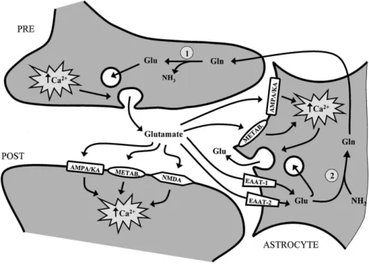
PTAKEGlutamate is the principle excitatory neurotransmitter in the brain. Figure 1 demon-strates a simplified schematic representation of the steps that occur at the glutamate synapse. Glutamate is synthesized in the presynaptic nerve terminal via the enzyme glutaminase and stored in synaptic vesicles, which eventually dock with the presynaptic neuronal membrane and release glutamate into the synaptic cleft by a calcium-dependent mechanism. Released glutamate binds to and activates receptors (ionotropic and metabotropic) both on the postsynaptic neuron and the neighboring astrocyte. Situated on the postsynaptic neuron are ionotropic receptors: N-methyl-D-aspartate (NMDA), a-amino-3-hydro-methyl-4-isoxasole-propionic acid (AMPA) and
kainate, all ion channel-gated receptors. Also on the postsynaptic neuron are metabotropic receptors, which are not ion-gated receptors but are instead coupled to calcium dependent second messenger systems. On the neighboring astrocyte, AMPA, kainate, and metabotropic receptors are found. Released glutamate is removed from the extracellular space by high-affinity Na +-dependent transporters located both on astrocytes and neurons. GLT-1 and
GLAST, more commonly referred to as, EAAT-2 and EAAT-1 respectively, are located on astrocytes. EAAT-3 is found on neurons, away from the presynaptic cleft and is, therefore, thought to play a minor role in glutamate clearance from the synaptic cleft. Glutamate uptake by astrocytes is an important pathway since ammonia removal in brain relies entirely from the synthesis of glutamine by the enzyme glutamine synthetase, found uniquely in astrocytes. The resulting glutamine produced is retransported to the presynaptic nerve terminal as the immediate precursor of the neurotransmitter glutamate, completing the glutamate–glutamine cycle.
increased release? Metabolic Brain Disease, 17(4), p.251–261.
I
NCREASEDE
XTRACELLULARB
RAING
LUTAMATE INALF:
E
FFECT OFA
MMONIAIn recent years, the results of several studies have suggested that alterations in glutamatergic synaptic regulation are implicated in the pathogenesis of ALF. Using the technique in vivo cerebral microdialysis, several reports have consistently described increased extra-cellular concentrations of brain glutamate in different models of experimental ALF (Bosman et al., 1992; de Knegt et al., 1994; Hilgier et al., 1999; Michalak et al., 1996). It has been suggested that ammonia (directly or indirectly) is causing this increase in extracellular brain glutamate concentrations. A positive correlation has been reported between extracellular brain concentrations of glutamate and arterial ammonia concentrations in acute (ischemic) liver failure in the rat (Michalak et al., 1996). Furthermore, using mild hypothermia as a treatment in rats with ALF, extracellular brain glutamate concentrations were normalized together with a lowering of brain (cerebrospinal fluid) ammonia (Rose et al., 2000).
Fig. 1. Keys steps involved at the glutamatergic synapse. Glutamate is released from the presynap-tic nerve
terminal (PRE) into the synapse and stimulates glutamate receptors on the postsynaptic neuron (POST) or the neighboring astrocyte. Removal of glutamate occurs by high affinity astro-cytic glutamate transporters. Intracellular astrocytic glutamate stimulates the formation of glutamine by glutamine synthetase 2 and subsequently the elimination of ammonia. Astrocytic glutamine is shuttled to neurons to reform glutamate by the enzyme glutaminase 1 , completing the glutamate– glutamine cycle. Furthermore, stimulation of glutamate receptors on astrocytes results in an increase in intracellular calcium and subsequently a release of glutamate, possibly acting as a neuronal signal.
G
LUTAMATERGICD
YSFUNCTION INALF
Several studies have demonstrated glutamate receptor changes in brain of animals with ALF. [3H]-kainate binding sites were significantly reduced in cerebral cortical preparations from galactosamine-treated rabbits with severe encephalopathy (Ferenci et al., 1984). Similar findings of a significant loss of [ 3H]-kainate binding sites were subsequently reported in frontal cortex in ischemic liver failure in the rat (Michalak and Butterworth, 1997). In contrast, densities of neuronally localized NMDA binding sites are unchanged in ischemic liver failure in both rabbits (de Knegt et al., 1993) and rats (Michalak et al., 1996). Fur-thermore, a selective loss of AMPA receptor binding sites was found in the brains of rats with ischemic liver failure (Michalak and Butterworth, 1997). This selective loss of AMPA sites could be the consequence of exposure of the brains of these animals to increased ammonia concentrations generated in ALF. In support of this possibility, previous studies have demonstrated a selective depression of AMPA currents by exposure of hippocampal pyramidal neurons to 3 mM ammonia (Fan and Szerb, 1993). In a separate series of electro-physiological studies, millimolar concentrations of ammonia led to a reduction in the degree of depolarization of cerebral cortical preparations by AMPA (Moroni et al., 1995), suggesting that ammonia has the capacity to modify the structural or functional characteristics of the AMPA receptor.
Therefore, increased extracellular glutamate in the brains of animals with HE in ALF, along with a concomitant loss of AMPA/kainate receptors could result in a relative increase of NMDA receptor-mediated transmission. In support of this theory, the noncompetitive NMDA receptor antagonist, memantine, administered to rats with liver ischemia resulted in significant improvement in clinical grading and less slowing of electroencephalogram activity (Vogels et al., 1997). These interesting findings support the notion that NMDA receptor activity is one of the characteristics of HE in experimental ALF. Furthermore, NMDA receptor activation has previously been shown to be increased in acute
hyperammonemia in the absence of liver failure, and the NMDA receptor antagonist MK-801 was found to afford significant protection in animals administered a lethal injection of ammonia (Marcaida et al., 1991).
POSSIBLE
REASONS
FOR
INCREASED
EXTRACELLULAR
BRAIN
GLUTAMATE
IN
ALF
G
LUTAMATER
ELEASE FROMN
EURONSAt physiological pH, more than 98% of ammonia is in NH4+ form, however NH3 enters cells
more readily. The ammonium ion (NH4+) has an ionic radius very similar to K+ and has
been suggested to enter cells through K+ channels. Therefore adding NH
4+ is equivalent to
increasing [K+]
e, which subsequently decreases the membrane potential which in turn can
increased release? Metabolic Brain Disease, 17(4), p.251–261.
demonstrated to lower the membrane potential without altering membrane resistance. This suggests that metabolic factors rather than ion-conductance changes underlie the depolarization. A probable mechanism for the ammonium ion-induced depolarization is the loss of intracellular K+ and subsequently an increase in extracellular K+ during
exposure to NH4+, which by reducing the equilibrium potential of K+ produces
de-polarization without altering membrane conductance. However, ammonium ions decrease synaptic transmission mediated by hyperpolarizing Cl--dependent inhibitory postsynaptic
potentials, an effect which results from the inactivation of the extrusion of Cl- from
neurons (Raabe, 1989). Furthermore, ammonium ions depress excitatory synaptic trans-mission in neurons (Szerb and Butterworth, 1992). It has been suggested that this depression of excitatory transmission by ammonium ions could be due to less glutamate available presynaptically for release. Glutaminase, found in neurons, produces glutamate plus ammonia from glutamine and subsequently ammonia has shown to inhibit
glutaminase (Bradford et al., 1989). In addition, brain glutamate concentrations are reduced in rats with either acute (ischemic) liver failure or thioacetamide-induced liver damage (Swain et al., 1992b). Furthermore, postsynaptically, ammonia reduces the effectiveness of released glutamate (Fan et al., 1990) through AMPA receptors. Hamberger et al. demonstrated the inhibitory effect of ammonia on glutamate release as 3–5 mM NH4Cl reduced potassium evoked glutamate release from hippocampal slices (Hamberger
et al., 1979, 1982). However, reviewing Fig. 1 from Hamburger et al. (1982) upon initial ammonia application (3 mM), an increase in glutamate release is demonstrated when compared with controls. Therefore, pretreatment with ammonia may reduce potassium stimulated glutamate release. However, glutamate is released in hippocampal slices upon an acute application of ammonia. Overall, ammonia inhibits glutamate synaptic
transmission in neurons by (1) inhibiting current gated AMPA receptors postsynaptically and (2) depleting synaptically released glutamate from neurons by inhibiting the
glutamate-producing enzyme, glutaminase.
I
NHIBITION OFG
LUTAMATEU
PTAKEA great deal of evidence suggests that the increased extracellular brain glutamate in ALF results from an inhibition of glutamate uptake. Exposure of rat hippocampal slices to blood extracts from patients with varying severity of HE resulted in inhibition of D-aspartate uptake and the
relative potency of inhibition was found to correlate with ammonia concentrations of the extracts (Schmidt et al., 1990). Decreased glutamate uptake was described in synaptosomal preparations from rats with experimental ALF (Oppong et al., 1995). Furthermore, exposure of primary cultured astrocytes to millimolar concentrations of ammonia also resulted in a significant reduction in uptake of [ 3H]-D-aspartate (Bender and Norenberg, 1996). In extracts
from cerebral cortex of rats in coma stages of encephalopa-thy following hepatic devascularization, a significant loss of GLT-1 (EAAT-2) protein and gene expression was observed (Knecht et al., 1997). Similar findings were found in the brains of mice with
thioacetamide-induced ALF (Norenberg et al., 1997). Recently, it has been shown that ammonia in concentrations equivalent to those reported in brain of ALF (5 mM), when added to cultured rat cortical astrocytes for 7 days, results in a comparative loss of glutamate uptake and in
addition loss of glutamate transporter GLAST (EAAT-1) protein and mRNA (Chan et al., 2000). Interestingly, EAAT-2 knockout mice manifest brain edema (Tanaka et al., 1997). Moreover, decreased astrocytic uptake of glutamate could limit ammonia detoxification by brain by limiting the availability of the substrate for glutamine synthetase. In ALF, the temporal development of brain edema is undefined and it is un-certain whether increased extracellular brain glutamate is a cause or an effect of astrocytic swelling. However, it has been
demonstrated that a significant increase in extracellular glutamate arises before the development of brain edema (Michalak et al., 1996). It is ironic that glutamate uptake is inhibited in astrocytes during high toxic ammonia concentrations when glutamate is an important and necessary precursor for glutamine synthetase—the only ammonia removal system in the brain. Recently, Mort et al. (2001) demonstrated that an acute application of ammonia potentiates glutamate uptake in glial cells isolated from salamander retina,
suggesting glutamate uptake inhibition may be a later phenomenon in the pathogenesis of HE and brain edema in ALF.
G
LUTAMATER
ELEASE FROMA
STROCYTESGlutamate can be released from astrocytes by three different mechanisms: two by independent (reverse glutamate transporter and swelling induced) and one by calcium-dependent mechanisms (vesicular release). The role of ammonia in each of these glutamate release pathways is reviewed hereafter.
C
ALCIUM-I
NDEPENDENTG
LUTAMATER
ELEASEAstrocytic Cell Swelling in ALF. As mentioned previously, brain edema leading to increased
intracranial pressure is a fatal consequence of ALF. It has been suggested that ammonia plays an important role in the pathogenesis of brain edema (astrocytic swelling) in ALF. Increased brain water content has been described in both normal (Takahashi et al., 1991) and in portacaval shunted (Blei et al., 1994) rats infused with ammonia as well as in rats with liver devascularization (Rose et al., 1999). Similar results have been described in vitro following the exposure of cultured astrocytes to ammonia (Norenberg et al., 1991). Furthermore, prevention of brain edema in rats with ALF has been shown with ammonia-lowering strategies (Cordoba et al., 1999; Rose et al., 1999, 2000). Unrelated to ammonia, swelling-induced glutamate release was demonstrated in cortical astrocytes exposed to a hypo-osmotic medium (Kimelberg et al., 1990). Ammonia-induced astrocytic swelling has been suggested to result in glutamine accumulation in astrocytes (by glutamine synthetase), possibly because of the inhibition of glutaminase (Bradford et al., 1989). However, an increase in extracellular brain glutamate could also lead to glutamate-induced astroglial swelling (Hansson et al., 1997).
Reversed Glutamate Transport. It has been demonstrated that when insufficient energy is
available to regulate the membrane potential, glutamate transporters reverse, that is, glutamate transported out of the astrocyte instead of being transported in (Szatkowski et al., 1990). A fall in ATP levels leads to an inhibition of the Na+/K+ pump and therefore a rundown of the
transmembrane gradients for [K+], [Na+], and voltage. To re-establish the ion and voltage
increased release? Metabolic Brain Disease, 17(4), p.251–261.
stoichiometry as forward uptake (i.e., 2 Na+ and 1 glutamate anion come out of the cell and 1 K+
is transported into the cell). An increase in energy demand occurs upon ammonia application by stimulating glycolysis through activation of phosphofructokinase (Sugden and Newsholme, 1975), and therefore possibly resulting in the stimulation of cerebral glucose utilization in acutely hyperammonemic animals (Hawkins et al., 1973). However, applications of NMR spectroscopy studies in rats with hyperammonemia due to ALF could not demonstrate any significant loss of ATP (Bates et al., 1989). Recently, much evidence has emerged suggesting an inhibition or slow down of energy metabolism in ALF. Ammonia chloride (5 mM) when applied to cultured astrocytes stimulates lactate production and lactate dehydrogenase activity (Bélanger et al., 2001). Similar results have been found in CSF of rats at precoma and coma stages with ALF (Chatauret et al., 2001). Clinically, using in vivo microdialysis techniques, extracellular lactate was found to increase in association with increased intracranial pressure in patients with ALF (Tofteng et al., 2001). Increase in lactate production indirectly suggests that anaerobic pathways may be stimulated to compensate for a decreased pyruvate oxidation (possibly because of the ammonia inhibition on the enzyme α-ketoglutarate dehydrogenase in the tricarboxylic acid cycle (Lai and Cooper, 1986)). However, acute ammonia exposure did not reverse glutamate transporters but increased glutamate uptake into astrocytes (Mort et al., 2001), suggesting that glutamate transporter reversal is not the cause of the initial increase in extracellular glutamate.
Calcium-Dependent Glutamate Release The role of astrocytes in synaptic transmission is
an important new concept for nor-mal brain function. It was originally thought that astrocytes were unresponsive to excita-tory neurotransmitters and only neurons could respond with depolarization and excitation. However, in a recent review (Vesce et al., 2001), the active role of astrocytes in synaptic transmission and its importance in neuron-glia signaling is described. Glutamate released from neurons can bind to its respective receptors (AMPA/kainate and metabotropic) on the astrocyte and trigger [Ca2+]i elevation. Interestingly, astrocytes possess
vesicular proteins (e.g. SNARE complex) which are essential in vesicular neurotransmitter release (Araque et al., 2000). This raises the strong possibility that astrocytes can release glutamate by Ca2+-dependent mechanisms. It was subsequently demonstrated that glutamate
binding to its receptors on astrocytes triggers a [Ca2+]
i response and stimulates glutamate
release. However, glutamate release is not specific to glutamate binding. Other studies have shown glutamate can be released upon triggering [Ca2+]
i with bradykinin (Papura et al., 1994),
KCl (Nicholls and Sihra, 1986), or more recently ATP (Innocenti et al., 2000; Jeremic et al., 2001). Furthermore, Pasti et al. (2001) demonstrated that increased cytosolic calcium regulates exocytotic release of glutamate. More specifically, it was further demonstrated that Ca2+
-dependent release of glutamate was mediated by prostaglandins because cyclooxy-genase inhibitors potently inhibited glutamate release (Bezzi et al., 1998). Lipoxygenase and epoxygenase inhibitors were ineffective and furthermore cyclooxygenase metabolites (prostaglandins) PGD2, PGE2, and PGF2a, all induced rapid glutamate release with PGE2
being the most effective.
Ammonium chloride at concentrations of 5, 10, and 20 mM has been shown to de-polarize cultured rat cortical astrocytes and that the sustained depolarization induced by ammonia
depended on changes in intracellular ion concentrations rather than changes in ion conductances. Also, ammonia at 5 mM stimulates an increase in [Ca2+]
i in cultured astrocytes,
using calcium-imaging techniques (Rose and Kettenmann, unpublished data). Calcium can increase intracellularly by entering the cell from the extracellular space through Ca2+
voltage-gated channels and/or specific pumps and transporters or can be released from internal stores. Ammonia depolarizes astrocytes and at very high concentrations can open voltage-gated calcium channels when the membrane potential is raised sufficiently high. In addition, ammonia releases calcium from IP3-sensitive intracellular calcium stores in microglia (Minelli et al., 2000). This has raised the possibility that ammonia can stimulate glutamate release from astrocytes in a Ca2+-dependent manner.
SUMMARY
Increased extracellular concentrations of brain glutamate in ALF can result from an in-crease in glutamate release and/or a decrease in glutamate removal (uptake) both by neurons and/or astrocytes. Glutamate can be released from neurons and astrocytes by cell swelling induced mechanisms, reversal of glutamate transporters, and/or calcium-dependent mechanisms. In neurons, an increase in glutamate release is unlikely to occur because ammonia inhibits glutaminase reducing the amount of glutamate available for synaptic release. Fur-thermore, cytotoxic edema in ALF develops in astrocytes and not neurons, eliminating the possibility of swelling-induced release of glutamate from neurons. An inhibition of glu-tamate removal by the high affinity transporters on astrocytes would result in an increase in extracellular brain glutamate concentrations. However, it has been demonstrated in vitro that acute application of ammonia potentiates glutamate uptake into astrocytes (Mort et al., 2001). Ammonia-induced astrocytic swelling may potentially stimulate glutamate release from astrocytes; however, this would suggest that increased extracellular brain glutamate is the result and not a cause of astrocytic swelling. Interestingly, increased extracellular brain glutamate concentrations precede the onset of brain edema in rats with ALF due to hepatic devascularization (Michalak et al., 1996). Reversal of glutamate transporters occurs when high-energy phosphates are depleted. Although ATP levels have been found to be unchanged in ALF, increased lactate production has been demonstrated, suggesting an inhibition of glucose oxidation but this appears to arise late in the development of HE in ALF. Astrocytes play an important role in synaptic transmission and under normal physiological conditions are capable of releasing glutamate in a calcium-dependent manner.
There is now increasing evidence that ammonia can stimulate [Ca2+]
i leading to stimulation
increased release? Metabolic Brain Disease, 17(4), p.251–261.
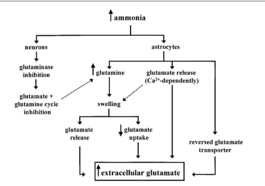
Figure 2. Diagram demonstrating the possible mechanisms involved in the development of HE and brain edema in ALF.
occur during the development of HE and brain edema in ALF. Ammonia enters neurons and inhibits glutaminase resulting in (1) less glutamate produced and available for release and (2) disruption of the glutamate–glutamine cycle between astrocytes and neurons. Ammonia also inhibits AMPA receptor activation but does not affect NMDA receptors. Overall, ammonia decreases glutamate release from neurons by inhibiting synaptic transmission and decreasing intracellular glutamate. Ammonia also enters astrocytes and (1) is detoxified by glutamine synthetase producing glutamine and (2) stimulates glutamate release in a calcium-dependent manner leading to increased extracellular glutamate. With inhibition of the glutamate–glutamine cycle, glutamine remains “trapped” in the astrocyte resulting in intracellular hypertonicity and cytotoxic swelling. Deregulation of the release of glutamate from astrocytes could also be a factor involved in astrocytic swelling. Once the astrocyte is swollen, glutamate uptake is inhibited to decrease the ion uptake preventing further swelling. Furthermore, inhibition of glutamate uptake and swelling-induced release of glutamate may add to the already increased extracellular
concentrations of glutamate. Because NMDA receptors are not affected by ammonia as are the AMPA/kainate receptors, this may explain the seizures and hyperexcitability, not uncommonly seen in patients with ALF.
ACKNOWLEDGMENTS
Christopher Rose is a recipient of the Alexander von Humboldt fellowship and wishes to thank them for their support.
REFERENCES
Araque, A., Li, N., Doyle, R.T., and Haydon, P. (2000). Snare protein-dependent glutamate release from astrocytes.J. Neurosci. 20:666–673.
Bates, T.E., Williams, S.R., Kauppinen, R.A., and Godian, D.G. (1989). Observation of cerebral
metabolites in an animal model of acute liver failure in vivo: 1H and 31P nuclear magnetic resonance study. J. Neurochem. 53:102–110.
Belanger, M., Chan, H., Hazell, A.S., and Butterworth, R.F. (2001). Increased lactate dehydrogenase (LDH) expression and activity in cultured astrocytes exposed to ammonia. J. Neurochem. 78(Suppl. 1):25.
Bender, A.S., and Norenberg, M.D. (1996). Effects of ammonia on L-glutamate uptake in cultured astrocytes.Neurochem. Res. 21:567–573.
Bezzi, P., Carmignoto, G., Pasti, L., Vesce, S., Rossi, D., Rizzini, B.L., et al. (1998). Prostaglandins stimulate calcium-dependent glutamate release in astrocytes. Nature 391:281–285.
Blei, A.T., Olafsson, S.M., Therrien, G., and Butterworth, R.F. (1994). Ammonia-induced cerebral edema and intracranial hypertension in rats after portacaval anastomosis. Hepatology 19:1431– 1444.
Bosman, D.K., Deutz, N.E.P., Maas, M.A.W., van Eijk, H.M.H., Smit, J.J.H., de Hann, J.G., et al. (1992). Amino acid release from cerebral cortex in experimental acute liver failure, studied by in vivo cerebral cortex microdialysis. J. Neurochem. 59:591–599.
Bradford, H.F., Ward, H.K., and Foley, P. (1989). Glutaminase inhibition and release of neurtransmitter glutamate from synaptosomes. Brain Res. 476:29–34.
Chan, H., Hazell, A.S., Desjardins, P., and Butterworth, R.F. (2000). Effects of ammonia on glutamate transporter (GLAST) protein and mRNA in cultured rat cortical astrocytes. Neurochem. Int. 37:243– 248.
Chatauret, N., Rose, C., Therrien, G., and Butterworth, R.F. (2001). Mild hypothermia prevents cerebral edema and CSF lactate accumulation in acute liver failure. Metab. Brain Dis. 16:95–102. Clemmensen, J.O., Larsen, F.S., Kondrup, J., Hansen, B.A., and Ott, P. (1999). Cerebral herniation in patients with acute liver failure is correlated with arterial ammonia concentration. Hepatology 29:648–653.
Cordoba, J., Crespin, J., Gottstein, J., and Blei, A.T. (1999). Mild hypothermia modifies ammonia-induced brain edema in rats after portacaval anastomosis. Gastroenterology 116:686–693.
increased release? Metabolic Brain Disease, 17(4), p.251–261.
de Knegt, R.J., Kornhuber, J., Schalm, S.W., Rusche, K., Riederer, P., and Tan, J. (1993). Binding of the lig-and [3H]MK-801 to the MK-801 binding site of the N-methyl-D-aspartate receptor during experimental encephalopathy from acute liver failure and from acute hyperammonemia in the rabbit. Metab. Brain Dis. 8:81–94.
de Knegt, R.J., Schalm, S.W., van der Rijt, C.C.D., Fekkes, D., Dalm, E., and Hekking-Weyma, I. (1994). Extra-cellular brain glutamate during acute liver failure and during acute hyperammonemia simulating acute liver failure: An experimental study based on in vivo brain dialysis. J. Hepatol. 20:19–26.
Fan, P., Lavoie, J., L´e, N.L.O., Szerb, J.C., and Butterworth, R.F. (1990). Neurochemical and
electrophysiological studies on the inhibitory effect of ammonium ions on synaptic transmission in slices of rat hippocampus: Evidence for a postsynaptic action. Neuroscience 37:327–334.
Fan, P., and Szerb, J.C. (1993). Effects of ammonium ions on synaptic transmission and on responses to quisqualate and N-methyl-D-aspartate in hippocampal CA1 pyramidal neurons in vitro. Brain Res. 632:225–231.
Ferenci, P., Pappas, S.C., Munson, P.J., Henson, K., and Jones, E.A. (1984). Changes in the status of neurotrans-mitter receptors in a rabbit model of hepatic encephalopathy. Hepatology 4:186–191. Hamberger, A., Hedquist, B., and Nystrom, B. (1979). Ammonium ion inhibition of evoked release of endogenous glutamate from hippocampal slices. J. Neurochem. 33:1295–1302.
Hamberger, A., Lindroth, P., and Nystrom, B. (1982). Regulation of glutamate biosynthesis and release in vitro by low levels of ammonium ions. Brain Res. 237:339–350.
Hansson, E., Blomstrand, F., Khabiti, S., Olsson, T., and R¨onnback, L. (1997). Glutamate induced astroglial swelling-methods and mechanisms. Acta Neurochir. 70:148–151.
Hawkins, R.A., Miller, A.L., Nielsen, R.C., and Veech, R.L. (1973). The acute action of ammonia on rat brain metabolism in vivo. Biochem. J. 134:1001–1008.
Hilgier, W., Zielinska, M., Borkowska, H.D., Gadamski, R., Walski, M., Oja, S.S., et al. (1999). Changes in the extracellular profiles of neuroactive amino acids in the rat striatum at the symptomatic stage of hepatic failure.J. Neurosci. Res. 56:76–84.
Innocenti, B., Parpura, V., and Haydon, P.G. (2000). Imaging extracellular waves of glutamate during calcium signalling in cultured astrocytes. J. Neurosci. 20:1800–1808.
Jeremic, A., Jeftinija, K., Stevanovic, J., Glavaski, A., and Jeftinija, S. (2001). ATP stimulates calcium-dependent glutamate release from cultured astrocytes. J. Neurochem. 77:664–675.
Kimelberg, H.K., Goderie, S.K., Higman, S., Pang, S., and Waniewski, R.A. (1990). Swelling-induced release of glutamate, aspartate and taurine from astrocyte culture. J. Neurosci. 10:1583–1591. Knecht, K., Michalak, A., Rose, C., Rothstein, J.D., and Butterworth, R.F. (1997). Decreased glutamate transporter (GLT-1) expression in frontal cortex of rats with acute liver failure. Neurosci. Lett. 229:201–203.
Lai, J.C.K., and Cooper, A.J.L. (1986). Brain Æ-ketoglutarate dehydrogenase complex: Kinetic properties, regional distribution and effects of inhibitors. J. Neurochem. 47:1376–1386. Marcaida, G., Felipo, V., Hermenegildo, C., Minana, M.-D., and Grisolia, S. (1992). Acute ammonia toxicity is mediated by the NMDA type of glutamate receptors. FEBS Lett. 296:67–68.
Michalak, A., and Butterworth, R.F. (1997). Selective loss of binding sites for the non-NMDA
(glutamate) receptor ligands [3H]-kainate and (S)-[3H]-5-fluorowillardiine in the brains of rats with acute liver failure. Hepatology 25:631–635.
Michalak, A., Rose, C., Butterworth, J., and Butterworth, R.F. (1996). Neuroactive amino acides and gluta-mate (NMDA) receptors in frontal cortex of rats with experimental acute liver failure. Hepatology 24:908– 914.
Minelli, A., Lyons, S., Nolte, C., Verkhratsky, A., and Kettenmann, H. (2000). Ammonium triggers calcium elevation in cultured mouse astroglial cells by initiating Ca2C release from
thapsigargin-densitive intracellular stores. Eur. J. Physiol. 439:370–377.
Moroni, F., Mannaioni, G., Cherici, G., Leonardi, P., Carla, V., and Lombardi, G. (1995). Excitatory amino acid neurotransmission and ammonia toxicity. In: Capocaccia, L., Merli, M., and Riggio, O. (eds.), Advances in Hepatic Encephalopathy and Metabolic Nitrogen Exchange, CRC Press, Boca Raton, FL, Chap. 22, pp. 130–139.
Mort, D., Marcaggi, P., Grant, J., and Attwell, D. (2001). Effect of acute exposure to ammonia on glutamate transport in glial cells isolated from the salamander retina. J. Neurophysiol. 86:836–844. Nicholls, D.G., and Sihra, T.S. (1986). Synaptosomes possess an exocytotic pool of glutamate. Nature 321:772– 773.
Norenberg, M.D., Baker, L., Norenberg, L.O.B., Blicharska, J., Bruce-Gregario, H.H., and Neary, J.T. (1991). Ammonia-induced astrocyte swelling in primary culture. Neurochem. Res. 16:833–836. Norenberg, M.D., Huo, Z., Neary, J.T., and Roig-Cantesano, A. (1997). The glial glutamate transporter in hyperam-monemia and hepatic encephalopathy: Relation to energy metabolism and glutamatergic neurotransmission. Glia 21:124–133.
Oppong, K.N.W., Bartlett, K., Record, C.O., and Al Mardini, H. (1995). Synaptosomal glutamate transport in thioacetamide-induced hepatic encephalopathy. Hepatology 22:553–558.Papura, V., Basarsky, T.A., Liu, F., Jeftinija, K., Jeftinija, S., and Haydon, P.G. (1994). Glutamate-mediated astrocyte-neuron signalling. Nature 369:744–749.
Pasti, L., Zonta, M., Pozzan, T., Vicini, S., and Carmignoto, G. (2001). Cytosolic calcium oscillations in astrocytes may regulate exocytotic release of glutamate. J. Neurosci. 21:477–484.
Raabe, W. (1989). Ammonium decreases excitatory synaptic transmission in cat spinal cord in vivo. J. Neurophysiol. 62:1461–1473.
Rose, C., Michalak, A., Rama Rao, K.V., Quack, G., Kircheis, G., and Butterworth, R.F. (1999). L-ornithine-L-aspartate lowers plasma and cerebrospinal fluid ammonia and prevents brain edema in rats with acute liver failure. Hepatology 30:636–640.
increased release? Metabolic Brain Disease, 17(4), p.251–261.
Rose, C., Michalak, A., Rambaldi, A., Chatauret, N., Pannunzio, M., and Butterworth, R.F. (2000). Mild hypother-mia delays the onset of coma and prevents brain edema and extracellular brain glutamate accumulation in rats with acute liver failure. Hepatology 31:872–877.
Schmidt, W., Wolf, G., Grungreiff, K., Meier, M., and Reum, T. (1990). Hepatic encephalopathy influences high-affinity uptake of transmitter glutamate and aspartate into the hippocampal formation. Metab. Brain Dis. 5:19–31.
Sugden, P.H., and Newsholme, E.A. (1975). The effects of ammonium, inorganic phosphate and potassium ions on the activity of phosphofructokinases from muscle and nervous tissues of vertebrates and invertebrates.Biochem. J. 150:113–122.
Swain, M., Butterworth, R.F., and Blei, A.T. (1992a). Ammonia and related amino acids in the pathogenesis of brain edema in acute liver failure in rats. Hepatology 15:449–453.
Swain, M., Bergeron, M., Audet, R., Blei, A.T., and Butterworth, R.F. (1992b). Monitoring of
neurotransmitter amino acids by means of indwelling cisterna magna catheter: A comparison of two rodent models of fulminant hepatic failure. Hepatology 16:1028–1035.
Szatkowski, M., Barbour, B., and Attwell, D. (1990). Nonvesicular release of glutamate from glial cells by reversed electrogenicglutamate uptake. Nature 348:443–446.
Szerb, J.C., and Butterworth, R.F. (1992). Effect of ammonium ions on synaptic transmission in the mammalian nervous system. Prog. Neurobiol. 39:135–153.
Takahashi, H., Koehler, R.C., Brusilow, S.W., and Traystman, R.J. (1991). Inhibition of brain glutamine accumu-lation prevents cerebral edema in hyperammonemic rats. Am. J. Physiol. 281:H825–H829. Tanaka, K., Watase, K., Manabe, T., Yamada, K., Watanabe, M., Takahashi, K., et al. (1997). Epilepsy and exacerbation of brain injury in mice lacking the glutamate transporter GLT-1. Science 276:1699–1702.
Tofteng, F., Jorgensen, L., Ansen, B.A., Ott, P., Kondrup, J., and Larsen, F. (2001). Cerebral microdialysis of glutamate and lactate in patients with fulminant hepatic failure. Hepatology 34:658A. (Abstract No. 1943.)
Vesce, S., Bezzi, P., and Volterra, A. (2001). Synaptic transmission with the glia. News Physiol. Sci. 16:178–184. Vogels, B.A.P., Maas, M.A.W., Daalhuisen, J., Quack, G., and Chamuleau, R.A.F.M. (1997). Memantine, a non-competitive NMDA-receptor antagonist improves hyperammonemia-induced encephalopathy and acute hep-atic encephalopathy in rats. Hepatology 25:820–827.