Université de Montréal
The impact of early life adversity on cortical structure in adolescent twins followed since birth
par
Elmira Ismaylova
Département des Sciences Biomédicales Faculté de Médecine
Mémoire présenté à la Faculté des Études Supérieures en vue de l’obtention du grade de Maître en Sciences (MSc.)
en Sciences Biomédicales option Psychiatrie
Juillet, 2013
Université de Montréal Faculté des Études Supérieures
Ce mémoire intitulé:
The impact of early life adversity on cortical structure in adolescent twins followed since birth
présenté par Elmira Ismaylova
a été évalué par un jury composé de personnes suivantes :
Patricia Conrod, Ph.D. président-rapporteur Linda Booij, Ph.D. directrice de recherche Adrianna Mendrek, Ph.D. membre du jury
Résumé
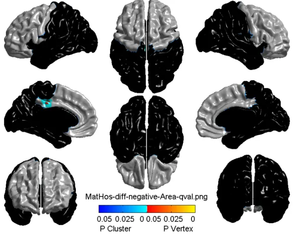
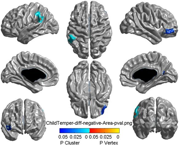
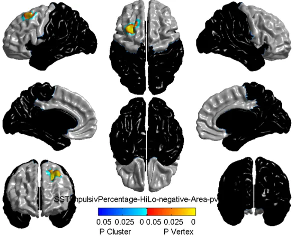
Des études animales ont montré que l’exposition du fœtus à l’adversité affecte le développement cérébral et la régulation d’émotions plus tard. Cette régulation serait reliée aux changements structurels cérébraux, particulièrement au circuit fronto-limbique. Cependant, ces résultats n’ont pas été entièrement répliqués chez l’humain. Le but de cette étude était de tester si l'adversité précoce conduit à des altérations structurelles des régions (orbitofrontal, préfrontal, cingulaire) fronto-limbiques, identifiées comme régions-clés dans la (de)régulation d’émotions. Les mesures principales de l’adversité étaient un poids léger à la naissance et l’hostilité maternelle puisqu’ils étaient parmi les plus prédictifs des résultats développementaux et comportementaux chez l’humain. Les mesures secondaires, incluant le tempérament difficile d’enfant et l’impulsivité en adolescence, étaient utilisées du à leur lien avec le développement cérébral et émotionnel. Les participants étaient des jumeaux identiques, membres de l’Étude des Jumeaux Nouveau-nés du Québec (ÉJNQ, N = 650 paires) suivis depuis 5 mois à 15 ans, leur âge actuel. Ceci a permis de mieux contrôler le facteur génétique et ainsi mieux isoler les effets d’environnement. Trente-sept paires ont été recrutées. La structure cérébrale de chacun, obtenue avec l’imagerie par résonance magnétique, a été analysée avec la régression linéaire. Le poids à la naissance n’a eu aucun effet. L’hostilité maternelle a prédit une réduction de l’aire du gyrus cingulaire postérieur. Tempérament difficile a prédit une réduction de l’aire du cortex orbitofrontal. L’impulsivité était associée avec l’aire et volume du cortex préfrontal réduits. Ces résultats soulignent l’importance des interventions précoces afin d’empêcher des altérations menant à la psychopathologie.
Abstract
Animal studies have shown that fetal exposure to adversity affects brain development and emotion regulation later on. Emotion regulation would be related to a structural change in the brain, particularly the fronto-limbic circuitry. However, results have not been entirely confirmed in humans. The purpose of the present study was to test whether early adversity leads to structural changes in the fronto-limbic (prefrontal, orbitofrontal and cingulate) regions, previously identified as key areas in emotion (dys)regulation. Main measures of adversity were low birth weight and maternal hostility because these were among the best predictors of developmental and behavioral outcomes in humans. Secondary measures, including difficult child temperament and adolescent impulsivity, were used because of their link with brain and emotion development. Participants were identical twins part of Quebec Study of Newborn Twins (QSNT, N = 650 pairs), followed from 5 months to 15 years, their current age. Using identical twins allowed to better control the genetic factors and, thus, to better isolate the specific effects of early environment. Thirty-seven pairs have been recruited. Each twin’s brain structure was assessed with magnetic resonance imaging and analyzed using linear regression. Birth weight showed no effect on brain structure. Maternal hostility predicted a reduction in cortical area of posterior cingulate gyrus. Difficult child temperament predicted a reduction in cortical area of orbitofrontal cortex. Impulsivity was associated with smaller cortical area and volume in prefrontal cortex. These results highlight the importance of the early interventions in order to prevent the alterations leading to development of psychopathology.
Keywords: adversity, brain, structure, development
Table des matières
IDENTIFICATION DU JURY……….ii
RÉSUMÉ EN FRANÇAIS………...iii
ABSTRACT………...iv
TABLE DES MATIÈRES……….v
LISTE DES TABLEAUX………vii
LISTE DES FIGURES………...viii
LISTE DES SIGLES...ix
DÉDICACE...x
REMERCIEMENTS………xi
CHAPTER 1: LITERATURE REVIEW……….1
1.1 Introduction………...………...1
1.2 Role of early environment in brain development ……….2
1.3 Serotonin and brain development………...2
1.4 Neural basis of emotion regulation……….3
1.5 Serotonin and neural basis of emotion regulation...5
1.6 Animal and human models………..6
1.7 Effects of environmental adversity on brain……….8
1.7.1 Brain function………..8
1.7.2 Brain structure………...9
1.7.3 Brain chemistry……….10
1.8 Epigenetics………...11
1.8.1 Genome versus epigenome………11
1.8.2 Histone acetylation...12
1.8.3 DNA methylation………...12
1.9 Epigenetics and mental health………..14
1.10 Overall aim and rationale of the study………...16
1.11 Primary aim and hypothesis………...19
CHAPTER 2: METHODS………..21 2.1 Participants.………..……….………21 2.1.1 QSNT cohort………...21 2.2 Current sample………..22 2.2.1 Inclusion criteria………22 2.2.2 Exclusion criteria………...23 2.3 Procedure………23 2.3.1 Phone pre-screening………..23 2.3.2 Screening………...23 2.3.3 Questionnaires...24 2.3.4 Computerized task………...24 2.3.5 Brain imaging………25 2.3.6 Statistics……….25 2.3.6.1 Cortical measures………...25 2.3.6.2 Adversity measures………26 2.3.6.3 Within-pair analysis………...28 CHAPTER 3: RESULTS………...…… 29
3.1 Characteristics of the sample………29
3.2 Primary hypothesis………31
3.2.1 Birth weight………...31
3.2.2 Maternal hostility………..31
3.3 Secondary hypothesis……….32
3.3.1 Difficult child temperament………..32
3.3.2 Aggression and hyperactivity………33
3.3.3 Impulsivity……….33
CHAPTER 4: DISCUSSION………. 36
4.1 Limitations and Forces………...……...39
4.1.1 Limitations………...………..39
4.1.2 Forces………40
4.2 Implications and future directions………...41
Liste des tableaux
3.I Adversity characteristics table………..29
3.II Behavior characteristics table……….29
3.III Results of whole-brain level analysis………30
Liste des figures
3.1 Cortical surface area related to maternal hostility………...32
3.2 Cortical surface area related to difficult child temperament………33
3.3 Cortical surface area related to impulsivity……….34
Liste des sigles
5-HT Serotonin
5-HTT Serotonin transporter ACC Anterior Cingulate Cortex
ADHD Attention Deficit Hyperactivity Disorder ATD Acute Tryptophan Depletion
CpG Cytosine-phosphate-Guanine
dlPFC dorsolateral Prefrontal Cortex
fMRI functional Magnetic Resonance Imaging
Kiddie-SADS Kiddie Schedule for Affective Disorders and Schizophrenia
LG Licking and Grooming
MRI Magnetic Resonance Imaging
OFC Orbitofrontal Cortex
PET Positron Emission Tomography PCC Posterior Cingulate Cortex
PFC Prefrontal Cortex
PTSD Post-Traumatic Stress Disorder ROI Region Of Interest
SST Stop Signal Task
VLBW Very Low Birth Weight vlPFC ventrolateral Prefrontal Cortex vmPFC ventromedial Prefrontal Cortex
À ma merveilleuse mère qui m’a élevée avec l’amour et le soutien inconditionnel.
Remerciements
Cette année a été haute en défis et en accomplissements. En rétrospective, il m’est évident qu’elle est le fruit de belles collaborations. Je vous remercie tous d’avoir été présents, d’avoir cru en ce projet et en mes capacités.
Je remercie, d’abord et surtout, ma directrice de recherche, Linda Booij, qui m’a accompagnée et orientée tout au long de ce projet.
Je remercie Kevin F. Casey pour toute l’énergie et la patience dont il a fait preuve en m’aidant avec l’analyse des données d’imagerie cérébrale.
Je remercie également Melissa Lévesque pour son soutien et ses conseils tout au long de ma maîtrise.
Je remercie aussi Bruce Pike et son équipe de l’Institut Neurologique de Montréal, car sans leur aide ce projet n’aurait pas été réalisable.
Je remercie sincèrement les trente-sept familles de jumeaux qui ont accepté de participer à cette recherche, ainsi que Marie-Pier, Michèle-Andrée, Floor et Miriam qui ont contribué au bon fonctionnement du projet.
Enfin, je remercie toute ma famille. Je remercie particulièrement mes parents de leurs encouragements, de leur support et de leur amour inconditionnel.
Chapter 1
Literature Review
1.1 IntroductionA great philosopher, Jean-Paul Sartre, once said that psychology can be entirely explained with two simple words: “childhood decides” [1]. However, there is now more and more evidence that behavioral and emotional development has already some roots in gestation and/or in the first few weeks following birth, as a consequence of brain development happening in utero and soon after birth. The question that immediately comes to mind is by what means do the in utero and early postnatal experiences shape brain development? This is where the notion of plasticity intervenes. Simply put, the developmental plasticity is an organism’s ability to adapt to the environment during early life and to implement long-lasting changes in the physiology [2, 3]. Thanks to this plasticity, environmental signals interact with the genetic blueprint to create developmental trajectories in the central nervous system, which in turn regulates the perception and consequent responses to the environment [4]. Even though organisms can adapt to the surrounding environment across the lifespan, the in utero and early postnatal phases are critical stages during which the environment calibrates the neural circuitry through various mechanisms [5].
The aim of the present thesis is to study the impact of the in utero and early postnatal environment on the brain circuitry in adolescence. In order to grasp how the early environment shapes the brain, I will first describe the role of early environment on brain development and potential underlying factors such the functioning of the serotonin (5-HT) system. Next, I will discuss the neural basis of the emotion and link it with the 5-HT system. Also, I will go over the existing evidence of the effects of environmental adversity on brain function, structure and chemistry as well as the underlying epigenetic mechanisms. After this introduction chapter, I will present the empirical study I conducted as part of my thesis.
1.2 Role of early environmental factors on brain development, and potential underlying neurochemical factors
The in utero and early postnatal periods are crucial for shaping the brain circuitry because that is when the brain is highly developing. Although the brain continues to develop in childhood and adolescence [6, 7], adversity occurring during the vulnerable in utero and early postnatal periods can alter the developmental process of the brain, including regions of the fronto-limbic system such as the hippocampus, amygdala and prefrontal cortex (PFC) [8, 9]. Taking into account that the development rate varies across these brain regions, it could be expected that an adverse events will have the most significant impact on the region currently undergoing its major development [6].
Although many neurotransmitters play a role in brain development [9], one of the most important organism’s neurotransmitters of the brain development process is 5-HT, which is known to modulate several psychological and physiological processes, such as mood, sleep and appetite [10]. Even though 5-HT-sensitive neurons are scattered throughout the entire brain, the majority is located in the raphe neurons, the afferent projections of which lead to the cerebellum, limbic system and the basal ganglia [11]. It is in those raphe neurons that the brain 5-HT synthesis takes place and those serotonergic neurons are one of the earliest to emerge in the developing living organism.
A more detailed developmental trajectory of 5-HT system in a living organism will be described in the following paragraph.
1.3 Serotonin and brain development
In fact, in rats, first 5-HT innervated raphe neurons are generated at only 12 days of gestation and the mature patterns, in terms of density and innervation of 5-HT fibers, are already reached at the end of the third postnatal week [11]. A similar pattern can be observed in the human brain, where the first 5-HT neurons become evident by five weeks of gestation [12]. The 5-HT levels continue to increase throughout the first two to five years of life and, then, gradually decrease until adult levels around the age of 14 years [13]. Since 5-HT
regulates the brain maturation, its amount growing into a key brain area becomes crucial for further development. Indeed, once 5-HT terminals have arrived into a target area, the effects of 5-HT on local developmental processes, including neurogenesis, neuronal removal, neuronal differentiation, axon myelination, as well as synaptic remodeling and maintenance, become apparent [14-19]. For instance, removal of 5-HT during very early fetal development has been shown to cause a long-lasting reduction in the number of neurons in adult rat brain, specifically in hippocampus and frontal cortex [17]. It becomes clear that 5-HT is not just a mere neurotransmitter, but that it is also decisive for the brain development [20-22]. Furthermore, 5-HT has been shown to be involved in individual’s self-control and emotional regulation [23]. Moreover, altered 5-HT neurotransmission has been shown to be involved in many psychiatric disorders [24]. In other words, alterations in 5-HT levels might result in individual’s faulty emotion regulation.
1.4 Neural basis of emotion regulation
Emotion regulation is generally defined by various processes by which we modify the experience and expression of our emotions in response to environmental cues [25]. This ability is imperative to sustain mental health, and impairments in emotion regulation are observed in a range of psychopathologies [26-30]. Various studies have examined the functional integrity of neural circuitry supporting abnormal emotion processing, which is a key feature of various mental illnesses. The most common procedures to examine the underlying neural mechanisms of emotion regulation in humans would consist of administering an emotion-processing task with stimuli of emotional content to subjects with (a vulnerability to) a disorder characterized by emotion dysregulation as well as to healthy controls during a magnetic resonance imaging (MRI) session and, then, comparing the neural responses to these stimuli in both groups. The general resulting patterns indicate altered activity in the frontal cortex and amygdala in response to emerging fearful, sad and angry stimuli (e.g. facial expressions) [31, 32] and, interestingly, an altered connectivity between the amygdala and PFC in the subjects with (vulnerability to) emotion dysregulation [27, 31, 33-36]. These region-specific activities are indicative of the fronto-limbic regulatory role in emotion processing. Besides, the anatomical data in monkeys has supported the notion that PFC activity modulates the amygdala, by
showing direct connections between the amygdala and PFC regions [37, 38]. In humans, PFC regions have also been linked to the down-regulation of amygdala activity [39, 40]. Additional human studies have found inverse correlations between activity in the amygdala and ventromedial prefrontal cortex (vmPFC) during emotion regulation and have demonstrated that the vmPFC serves as a mediator between dorsolateral PFC and the amygdala [41, 42]. Moreover, compared to healthy controls, subjects with disorders had significantly decreased volume of parahippocampal gyrus and orbitofrontal cortical (OFC) regions [43]. All these data suggest that the functional and structural alterations in the fronto-limbic circuitry leads to an altered emotion perception and further processing, thus increasing individual’s vulnerability to various psychopathologies [44].
The processes underlying emotion perception were found to be dependent upon the functioning of i) the ventral system, including amygdala, insula, ventral regions of anterior cingulate cortex (ACC) [45] and PFC, which is mostly involved in identification of the emotion significance of a stimulus, production of an affective state in response to that stimulus and autonomic regulation of emotion responses and of ii) the dorsal system, including hippocampus, as well as dorsal regions of PFC and ACC, which is mainly involved in the regulation of the affective state and subsequent behavior of the individual [46]. Studies that took a closer look at all those interacting areas found that, in fact, amygdala is part of an extended neural network. More precisely, it has rich connections with vmPFC [38, 47, 48]and hippocampus [49, 50]. The vmPFC modulates the activity of the amygdala through the descending projections, via afferents leading to the amygdala cells that inhibit its own activity. Moreover, the amygdala and hippocampus co-modulate each other such that the amygdala influences hippocampally-mediated memory formation, while the hippocampus influences amygdala responses when emotional stimuli are encountered [51]. Specifically, in new environmental contexts, the hippocampus inhibits the vmPFC, which releases the amygdala from vmPFC inhibition [52]. Those three structures seem to coordinate together during learning and regulation of emotion and might be, thus, vulnerable altogether when responding emotionally to the stress-induced changes in the organism.
Considering the abundant presence of 5-HT receptors and innervations in amygdala [11], OFC and ACC [53], these brain regions might be very sensitive to 5-HT alterations [54-74]. In the following paragraphs, the impact of 5-HT alteration, specifically 5-HT synthesis and transportation, on brain function and structure will be discussed.
1.5 Serotonin and the neural basis of emotion regulation
A lot of scientific evidence of the role of 5-HT in emotion processing comes from acute tryptophan depletion (ATD) studies (a method to study experimentally the effects of low 5-HT on brain and behavior [24, 75] , that have been shown to modulate the connectivity between the amygdala and two prefrontal regions, namely ventral ACC and ventrolateral prefrontal cortex (vlPFC), when processing emotional faces on the screen [76]. In other words, 5-HT depletion significantly affected the functioning of the cortico-limbic circuitry by reducing the emotion processing of emotional faces within PFC-amygdala pathways.
Other evidence for the role of 5-HT in the neural regulation of emotions comes from genetic knockout studies in animals and from molecular imaging studies in humans. Most of these studies focused on the 5-HT transporter (5-HTT). The 5-HTT can be found in the median and dorsal raphe nuclei, cerebral cortex, as well as certain hippocampal areas [77]. The major role of 5-HTT consists in the reuptake of 5-HT from the extracellular space, necessary for modulation of the strength, duration and subsequent 5-HT neurotransmitter release [78]. For instance, the 5-HTT knockout mice have been found to have functional deficits in the somatosensory cortex [79-81]. Another genetic knockout study found a significant reduction in cortical thickness in 5-HTT knockout mice, compared with controls [82].
The findings of multiple molecular imaging studies, on the other hand, have indicated amygdala hyper-reactivity [54-64, 71, 72, 83] upon exposure to emotional stimuli, as well as a reduced functional connectivity between the amygdala and the perigenual cingulate cortex in adults with 5-HTT gene polymorphism [54]. Moreover, a decreased grey matter volume in fronto-limbic structures, including ACC, amygdala, hippocampus and the cingulate cortex, has also been observed [54, 84, 85]. These results suggest that alterations in 5-HT system may
alter the brain structure and function of the fronto-limbic network, in turn representing a vulnerability factor for affective disorders.
Hence, the latter studies support the role of 5-HT genes in brain development, as well as emotion regulation. But what about the impact of the early environment on neural regulation of emotions and 5-HT function? Before going into that, it is essential to understand that manipulating environments experimentally is very challenging when one deals with humans.
1.6 Animal and human models of studying the impact of the environment on brain and behavior
Indeed, even the most accurately designed experiments involving humans are very rarely able to control all aspects of physical and social environment, despite the careful assignment of specific subjects to specific experimental conditions. Ultimately, the differences between subjects become clouded, and sometimes entirely masked, by interfering experiences that differ among subjects both within and between treatment groups [86].
Research with animals, on the other hand, has allowed the experimenters to systematically manipulate the environment and subsequent experiences would be rigorously controlled throughout the entire period of investigation [86]. That is why for decades, researchers have employed animal models to explore the behavioral and physiological effects of early life adversity.
Among the most frequently used early-life stress animal models, particularly in rodents, are interventions in mother-pup interaction time periods [87]. For instance, the central characteristic of the early handling paradigm [88] is a daily physical manipulation of the litter, where the pups are separated from the mother for a short period of time, i.e. maximum of 15 minutes. It has been shown that this procedure, which is carried out during the first three weeks of life, stimulates maternal care behavior towards the offspring [89] and elicits acute neuroendocrine responses from the pups [90]. While the early handling stimulates maternal care, repeated maternal separations of the dam from the litter are meant to reduce the amount
of maternal care for the pups, thereby promoting emotional and physical neglect [91]. Another well-known early life stress measure consists in an impoverishment of the postnatal environment [92-95]. In this model, the mothers are provided with reduced bedding material from the first two postnatal weeks of their litter. This manipulation results in an inconsistent maternal care, in turn leading to a higher stress exposure during the first year of the offspring’s life.
Another laboratory paradigm that comes most closely to human work involves separation paradigms applied in monkeys. This has involved separating infants from their mothers at birth, hand-rearing them in a nursery for the first month, and then rearing them with same age peers until 6 months of age, after which they would be moved into larger groups that also contain mother-reared same age mates and sometimes older adults [96]. Both peer-reared and mother-reared youngsters would, then, continue to live in these mixed social groups at least until puberty. While 6-months old youngsters would be scattered in the cage, their behaviour and other relevant outcome measures would be monitored for four consecutive weeks [97].
Obviously such studies are more difficult to do in humans. Most the studies in humans that looked at the impact of in utero or early postnatal experiences are cross-sectional and retrospective. In a longitudinal study design, prospective measures are prevailing over the retrospective measures because the former looks for forward for the outcome and relates this to earlier adverse or protection factors whereas the latter looks backwards by examining the exposure to various factors in relation to an outcome established in the beginning of the study and this is bound to have confounding factors. Hence, when studying the impact of adversity, prospective studies could shed more light on cause and effect. Furthermore, since the brain, behavioral and emotional development is partly under the influence of genetic factors, it is ideal to be able to control for genetic factors when studying the specific impact of the environment. Only a longitudinal monozygotic twin design following individuals as early as possible (e.g. since birth, or ideally since time of gestation) enables the researchers to distinguish environmental from confounding genetic effects since, unlike singletons, monozygotic twins are assumed to share one hundred percent of their genetic background
[98], rendering the entire design ideal for the investigation of the impact of early adverse environment on subsequent brain and behavioral alterations.
Now comes the time to disentangle the impact of the early environment on different neuronal levels, including brain function, structure and chemistry, by applying some of the research paradigms described above. The literature reviewed further below depicts the impact on each of these components in animals and in humans.
1.7 Effects of environmental adversity on the brain 1.7.1 Brain function
In male rat pups, maternal separation during neonatal period has been associated with various functional differences, most notably the atypical decrease of the activity of the hippocampus functioning [89] and a greater amygdala response to stress, even once the rodents have reached the adulthood [99]. Obviously, such controlled experiments are impossible to do so in humans. However, a design that comes somewhat close to such maternal separation paradigms are studies conducted in orphans. Indeed, studying the emotional development of children who had been institutionalized during infancy and, then, subsequently removed from this environment allows us to ask questions regarding the long-term correlates of early adversity in a human sample. For instance, in a functional magnetic resonance imaging (fMRI) study, 10 years-old children, who have been in an orphanage care in their childhood, exhibited a heightened activity of the amygdala in response to impulsivity task, comparatively to children who have never been institutionalized [100, 101]. In another fMRI study, children aged from 8 to 18 years, with a history of emotional neglect and lack of maternal care, showed significantly greater amygdala activity in response to emotion-processing task [102]. It becomes clear that lack of a stable caregiver is a stressor for the animal and human infants. Taken together, these findings suggest that early adverse caregiving is followed by differences in brain activity, particularly in limbic regions, that can persist into childhood.
Rats, which were repeatedly separated from their mothers early in life, exhibited growth of basolateral amygdala dendrites, as well as shrinkage of the PFC and of the hippocampus in CA3 and dentate gyrus [103]. Moreover, the rodents reared under those high-stress conditions have been found to have fewer number of cells that differentiated into neurons in dentate gyrus of the hippocampus [104]. Analogously, in macaques reared solely by peers, this early stress has been shown to affect brain development, including the reduction of hippocampal volume and of the corpus callosum area [105].
Those results are in line with the results of structural MRI studies conducted in humans. Indeed, early child abuse and maltreatment have been associated with smaller hippocampus [106-110], smaller orbitofrontal [103] and a smaller corpus callosum volumes [108]. Furthermore, children with history of in utero exposure to various neurotoxins, such as methamphetamines or alcohol, exhibited smaller hippocampus and putamen volumes [111], as well as reductions in corpus callosum and overall brain volume [112-117]. Therefore, an association was revealed between perinatal adversity and brain morphology. Other structural MRI studies have found a significantly reduced callosal area in very preterm infants (VPT) born in less than 30 weeks of pregnancy [118]. Additionally, the volume of tracts deriving from the corpus callosum in VPT infants was reduced [119]. This decline in interhemispheric fiber tracts passing through the corpus callosum might be the sign of altered interhemispheric communication. Indeed, the disruption of corpus callosum’s development might have implications for its associated brain structures such as the fornix, septum, cingulate cortex and the hippocampus [120].
The alterations of the brain structure have been found not only on the volume level but also on the level of cortical thickness. Cortical thickness has been typically defined as the shortest line from the cortical surface to the grey and white matter boundary [121]. The average thickness of the cerebral cortex is between 2 and 5 mm [122-124]. Slight variations of the cortical mantle were shown to be dependent of the brain region [125] and age [124]. Moreover, cortical morphology has been known to vary in affective disorders characterized by emotion dysregulation, including schizophrenia [123] and anxiety disorders [126, 127], as well as in
healthy individuals with impulsive personality trait [128]. Information on thickness variation can, thus, be of interest for the understanding of clinical and healthy behaviors.
A number of studies have shown that cortical thickness was found to be significantly impacted by in utero and early postnatal environmental factors. For instance, orbitofrontal, middle frontal and parahippocampal cortices were found to be thinner in adolescents having been exposed to maternal smoking in utero, as compared with non-exposed individuals [129]. Also, individuals born with a very low birth weight exhibited cortical thinning in parahippocampal and temporal medial gyri [130, 131]. Cortical thickness as well as smoking behaviors are however also partly under genetic control [132, 133], and thus the unique contribution of environmental factors cannot be determined in these studies.
1.7.3 Brain chemistry
Although the early environment could affect many neurotransmitters, and all of whom interact, given the role of 5-HT in emotion regulation and brain development (see above) the impact of the in utero and early postnatal environment on 5-HT are of particular interest. For instance, animal research has shown that early maternal separation in monkeys and rodents alters 5-HT neurotransmission in the frontal cortex [134, 135]. Moreover, it has been shown that 5-HT levels in a developing animal can be altered by viral infections [136], malnutrition [86, 137], social enrichment or isolation [138, 139], hypoxia [69], in utero exposure to neurotoxins [140] and maternal consumption of drugs such as cocaine [141, 142], nicotine [143] and alcohol [70, 144].
One human study tested whether perinatal adversity factors had a long-term impact on brain 5-HT neurotransmission in adulthood [145]. Basically, twenty-six 27-year old males underwent a positron emission tomography (PET) scan with the tracer alpha-[¹¹C] methyl-L-tryptophan, as an index of 5-HT synthesis capacity. Measures of in utero and early postnatal adversity were derived from the medical records. The results indicated that lower birth weight, maternal smoking during pregnancy and physical distress at birth, predicted lower brain 5-HT synthesis in adulthood in the medial OFC and hippocampus, whereas the childhood and later life
adversity did not affect brain 5-HT synthesis. These findings suggest that fronto-limbic 5-HT pathways are vulnerable to environmental challenges during the period when they undergo the most crucial neurodevelopmental changes [145].
Given the impact of early environmental factors on brain 5-HT, the following question arises: how do those early environmental stressors instigate changes in the brain 5-HT system? The answer to this question can take various forms. Indeed, there are numerous ways to alter 5-HT function in the brain, including direct lesions [146] or via changes in intracellular [147] or transcriptional factors [148]. Nonetheless, one of the underlying mechanisms by which the 5-HT alterations occur is via environmentally-induced stable changes in genetic expression [149], that are most probably caused by epigenetic mechanisms.
1.8 Epigenetics
1.8.1 Genome versus epigenome
Simply put, the genomic sequence, also called DNA sequence, is identical throughout the body and lifespan [150]. Epigenomes, on the other hand, are tissue-specific and drive distinct genome expression programs [151]. In other words, genome defines organism’s genetic information, whereas the epigenome determines for those genes “to be or not to be” expressed. Apart from controlling the gene expression, epigenetic mechanisms are useful for fine-tuning the gene expression repertoire in response to environmental cues, therefore adding plasticity to the hard-coded genome [150]. In other words “genetics proposes and epigenetic disposes” [152]. Genome cannot possibly operate independently of its environmental context [153]. That is why the development should be viewed as an active process of adaptation occurring as a function of the continuous dialog between the genome and its environment [153]. Here is where the integration of epigenetics occurs in order to examine those genome-environment interactive processes. Two well-known epigenetic mechanisms, histone acetylation and DNA methylation, will be described below.
Before getting into explanation of this epigenetic mechanism, it is essential to grasp the general structure of the DNA sequence. DNA is organized into units referred as nucleosomes, each of which contains about 150 base pairs which are wrapped around the core region of histone proteins [154]. Those histones and DNA put together are referred to as chromatin. The positively-charged histones and the negatively-charged DNA are tightly bound to each other, resulting in a closed chromatin configuration [154]. This restrictive configuration blocks the transcription factor binding and is, thus, associated with a limited gene expression. Histone modification is required to unblock the access for the transcription factor binding to DNA regulatory sites and, therefore, to activate the gene expression. Basically, series of enzymes bind to the histone tails, which are chains of amino acids extending outside the nucleosome, and modify the local chemical properties of specific amino acids along the histone tails [155-157]. For instance, the enzyme called histone acetyltransferase transfers an acetyl group (COCH3+) onto specific sites of histone tails. The addition of the acetyl group reduces the positive charge of the histones and, hence, loosens up the histone-DNA configuration. This, in turn, opens the chromatin and facilitates the access of transcription factor binding to DNA sites, activating the gene expression. However, among the modifications of histone, the DNA methylation has been the most studied with regard to understanding early life experiences and their underlying neurobiological aspects [150].
1.8.3 DNA methylation
DNA methylation is a covalent modification of the DNA molecule itself by enzymatic addition of a methyl group (CH3+) onto the cytosine ring residing in a CpG (cytosine-phosphate-guanine) dinucleotide [136]. In the beginning, an enzyme called cytosine-5-DNA-methyltansferase recognizes the appropriate regulatory site of the genome. Then, the ferment covalently binds with the genome at the regulatory site, literally twists the cytosine out of the genome sequence and adds the methyl group onto the cytosine. Afterwards, the enzyme pulls out, while the methylated cytosine is going back to its initial place. This CpG methylation is closely associated with suppression of transcription and long-term gene silencing, since it inhibits the DNA transcription factors binding to their recognition elements in the gene [154].
Moreover, DNA methylation pattern is shaped and fashioned during the perinatal period, when it is highly vulnerable to environmental exposures. Indeed, early restriction of certain dietary components, such as folic acid and vitamin B12 during gestation, has been shown to affect the DNA methylation patterns in sheep [158]. Moreover, individuals exposed to famine in the perinatal period had exhibited the altered DNA methylation patterns compared to their siblings six decades later [159]. This advances the possibility that DNA methylation plasticity might play a role in the programming of the genome regarding its adaptive responses to changing environment early in life and perhaps throughout life [160].
Following the idea that DNA methylation and chromatin state are in a dynamic equilibrium even in adult neurons, it should be possible to revert the epigenetic programming in the other direction toward increased methylation, leading to a reversal of the maternal genetic programming that is being passed onto the offspring. In two distinct studies [161, 162], adult rats, with high maternal Licking Grooming (LG) or low maternal LG rearing history, were infused with methionine, a donor of the methyl group in the organism. The animals were assessed in an unfamiliar open-field arena, and the gene expression of each animal was evaluated. The results showed that methionine treatment reversed behavioural response to stress, as well as the epigenetic programming of the hippocampus promoter. Simply put, normally, offspring of low LG mothers expressed lower levels of hippocampus promoter gene expression and spent less time exploring the unfamiliar inner field than did offspring of high LG mothers [163]. This reduced gene expression and an anxious behavior imply that hippocampal gene expression may play a role in the development of anxiety-mediated behavior. However, the methionine-treated offspring of low LG displayed behavior similar to that of offspring of high LG. Conversely, methionine-treated offspring of high LG mothers exhibited the anxious behavior of low LG mother’s offspring. These results suggest that although early-life experience has a stable effect on the hippocampal gene expression, the latter is still potentially reversible later on life.
Indeed, in rodents, it is possible to reset gene expression programs, carved early in life, by maternal care styles [164] and by exposure to pharmacological agents known to affect the DNA methylation machinery, such as methionine [160]. DNA methylation is, thus, a partly reversible biological signal, suggesting that a hard-coded genome is not the final answer to our question. The complex nature of DNA methylation renders it an ideal template for establishing sustaining gene effects controlling brain function and behavior from early development to adulthood [150].
A fostering study is a good concept to show it [165]. Basically, rat pups are cross-fostered within six hours of birth to mothers of the same phenotype (pups from high or low LG mothers to other high or low LG mothers, respectively) or alternative maternal phenotype (pups from high or low LG mothers to low or high LG mothers, respectively) and then, tested in the pacing chamber with a focus on the sexual behavior rating. The analysis revealed that biological low LG-reared offspring fostered onto low LG mothers exhibited a higher sexual receptivity rating than did animals in any other group, whereas the low LG-reared offspring fostered onto high LG mothers showed a significant decrease in sexual receptivity. These data indicate that early inborn experience is not definitive and that it can be changed or reversed with certain subsequent environmental exposures, such as modification in maternal rearing style.
Moreover, when it comes to epigenetics, counting how many times a day you hug your child acquires a special meaning. Recent data suggests that early adverse psychosocial exposures, such as poor maternal care, impact the epigenome, resulting in differences in epigenetic program and, consequently, in mental and behavioral developmental differences later on [166]. Thus, certain behavioral and mental pathologies might be a consequence of early life exposures that alter epigenetic programming [167].
1.9 Epigenetics and mental health
Indeed, an argument supporting epigenetic application in psychiatric research is its involvement in development of diseases. Various studies have indicated a genetic influence in
all psychiatric diseases, with heritability sometimes reaching 80% [168]. However, in spite of more than two decades of genetic psychiatric research and a clear refinement of molecular techniques, no gene has consistently been identified with any psychiatric disorder. This is where the term “missing heritability” comes in play. It refers to the discrepancy between high epidemiological heritability estimates and the proportion of phenotypic variation actually explained by DNA sequence differences [169]. One of the multiple theories explaining this discrepancy implies that the heritability estimates might be inflated by epigenetic modifications [170].
Indeed, recent studies provided more evidence that differences in vulnerability to various mental disorders might be associated with individual variation in DNA methylation. For instance, a distinct study provides an example where the polymorphism in hippocampus promoter gene, linked to the reduced gene expression [171], has been associated with the early-onset schizophrenia [172, 173]. Simply put, in normal brain, hippocampus promoter regions are unmethylated, allowing active gene expression. However, in schizophrenia, brain over-expression of DNA methyltransferase increases gene promoter methylation, leading to a reduction in gene expression. This lack in expression of genes, playing a key role in inhibitory neurotransmission, might contribute to the breakdown of the normal synchronized activity of brain circuitry, in particular fronto-limbic network, which is thought to be part of the disorder process [174].
Similarly, the role of epigenetics has been illustrated in depression and anxiety disorders. For instance, adult rats born to low LG mothers displayed increased anxiety and a reduced gene expression of the hippocampus promoter region [175]. In humans, the exposure to maternal depressive symptoms during pregnancy was associated with reduced 5-HTT gene expression in infants [176, 177].
Analogously to the case of depression, methylation of the 5-HTT gene has been shown to play an important role in risk for Post-Traumatic Stress Disorder (PTSD). To be more precise, the effect of cumulative traumatic burden on risk for PTSD was shown to be modified by
methylation levels at the 5-HTT locus [178]. In other words, individuals exposed to a greater number of traumatic events were at higher risk of PTSD, but only at lower 5-HTT methylation levels. However, at higher methylation levels, these same individuals are protected against this disorder. Considering that exposure to potential traumatic events varies with different environmental factors, such as living in inner city or in suburbs [179], these PTSD-related epigenetic differences provide further evidence for the role of epigenetic processes as mediators of environmental context on mental health.
The entire data reveals that in addition to DNA sequence, epigenetic modifications of DNA contribute to complex phenotypes, resulting sometimes in mental disorders
Therefore, defining the genes as some static hard drive, on which individual’s information gets stored, is not quite correct. It becomes clear that genes, as complex polymorphic molecular nanomachines, are sensitive to any changes in the individual’s environment and lifestyle. Therefore, we should not only talk about the hard-coded bits of information stored on the DNA, but rather about the dynamic components of our body that can be altered under certain circumstances. Specifically, genetic polymorphisms, as well as environmental factors such as malnutrition, infections, drugs of abuse, chemical exposures and psychosocial factors can all alter epigenetic marks resulting in inherited developmental diseases [136, 180].
In a nutshell, current literature studying gene x environment interaction in relation to mental health highlights the importance of the interaction of genes and early environment, as their interplay seems to alter brain circuitry, in turn leading to affective and behavioral disorders.
1.10 Overall study rationale and aim
In order to elucidate the impact of the early environment on brain development and the role of 5-HT system in this process, our laboratory is carrying out a longitudinal five-year-long project investigating the impact of perinatal adversity on the adolescent brain, in particular the brain regions known to be affected by 5-HT system alterations such as prefrontal
and cingulate cortices, and the underlying epigenetic mechanisms, in particular DNA methylation.
This study is being conducted in a longitudinal cohort of monozygotic twins followed since birth, allowing a control for genetic factor as these twins are assumed to share 100% of their genetic pool. Aside the fact that monozygotic twins share the same genes, they grow up sharing several factors in their in utero and postnatal environment, such as in utero exposure to maternal smoking and maternal depression. Yet, it does not mean that they share all possible environmental experiences: some experiences are unique to each twin (i.e., non-shared), reflected in e.g. birth weight discordance. Focusing on the within-pair difference of non-shared environmental stress factors offers a unique tool to better isolate each twin’s adverse experiences and psychobehavioral development.
For the purpose of the present thesis, I will focus on the impact of non-shared early environment on brain structure. Given the extensive evidence of fronto-limbic circuitry being affected by the shared early environment and the involvement of fronto-limbic regions in the emotion and behavior regulation, I will specifically focus on the cortical structure, namely cortical area, volume and thickness, in the following regions of interest (ROI): PFC, anterior cingulate (ACC) and posterior cingulate cortex (PCC) as well as amygdala.
The majority of previous imaging studies are cross-sectional and few studies are done in the cohort samples. My study, on the other hand, will apply imaging method in a longitudinal birth cohort. My study will generate knowledge in humans on the consequences of early adversity-induced changes on brain structure as a marker for psychopathology arising later in life. Moreover, I am working with monozygotic twin population with a particular focus on the non-shared early environment. This is possibly one of the most powerful ways available to examine the specific role of early adverse influences on brain development and behavior. The results may contribute to a better understanding of early environmental targets to foster brain development, allowing the identification of people at risk and, thereby, guiding future designs of interventions.
In terms of non-shared early environment, I will focus on the following factors: birth weight and maternal hostile behaviors at the age of 5 months. The choice of these variables is based on literature showing a putative link with brain development. Specifically, numerous studies have demonstrated that low birth weight, defined by a birth weight less than 2.5 kg, is associated with various disorders characterized by emotion dysregulation, such as attention deficit hyperactivity disorder (ADHD) [181], aggression, and hyperactivity-impulsivity [182] in childhood and adolescence. Furthermore, the low birth weight has been associated with brain structure alterations, namely the reductions in the cortical area of corpus callosum [183]. By looking at within-pair differences in twins, one can better isolate the effect of each early adverse factor on the individual brain and behavior.
With regard to parenting style, hostile-reactive parenting [184] has been shown to be negatively associated with emotional and brain development. For instance, hostile parental discipline in the first year following birth has been shown to predict reactive aggression in childhood [185]. Furthermore, compared to high maternal aggressiveness, low maternal aggressiveness has been shown to be a protective factor as it predicted larger bilateral hippocampal volumes in adolescence [186].
The association between early adverse factors and various problematic behaviors, arising in childhood and in adolescence, becomes evident. Therefore, I will also examine the impact of difficult temperament at the age of 18 months, aggression and hyperactivity between 6 and 10 years of age as well impulsivity at 15 years of age because those behaviors have often been found to emerge as the consequence of the early environment.
Indeed, prenatal exposure to increased maternal body mass index and maternal stress has been found to predict difficult child temperament during the first years of life [187, 188]. Moreover, scientific evidence has revealed that lower cortical thickness in the left OFC in adolescents has been associated with difficult temperament during the first year following birth [189], indicating that early temperament has great implications in the long-term architecture of the cerebral cortex.
Physical aggression from first to sixth grade has been linked to higher maternal harshness and to lower level of maternal sensitivity earlier in life [190]. Certain forms of aggressive behavior at 8 years of age, namely oppositional defiant and conduct disorders, were associated with the cortical thinning of prefrontal, cingulate and insular cortices, indicating that aggression during childhood years might lead to alteration in brain structure [191].
On the other hand, childhood hyperactive behavior has been linked to the tobacco smoke exposure before birth [192]. Furthermore, hyperactivity at the age of 10 years was associated with cortical thinning in the medial and dorsolareal prefrontal cortex (dlPFC) [193].
Similarly, prenatal exposure to tobacco as well as lower quality of early child care has been shown to predict the risk for developing impulsivity in adolescence [194, 195]. Furthermore, impulsive behavior at 13 to 15 years of age has been linked to cortical thinning of right dlPFC [195].
1.11 Primary aim and hypothesis
My primary aim is to examine the impact of non-shared early life adversity, namely birth weight, maternal hostility at the age of 5 months, on the brain structure in the adolescent twins. My hypothesis is that within a twin pair, higher levels of early life adversity, in other words lower birth weight and more experiences of maternal hostility, will be correlated with the lower cortical area, volume and thickness of fronto-limbic circuitry (PFC, ACC, PCC, amygdala).
1.12 Secondary aim and hypothesis
My secondary aim is to examine the impact of problematic behaviors, namely difficult child temperament at the age of 18 months, average aggression and hyperactivity from 6 to 10 years of age, and impulsivity at the age of 15 years, on brain structure. My hypothesis is that within a twin pair, higher levels of temperament difficulty as well as higher levels of aggression, hyperactivity and impulsivity, will be correlated with lower cortical area, volume and thickness of fronto-limbic circuitry (PFC, ACC, PCC, amygdala).
In the following chapter, I will go over the methods in more details, covering the cohort of participants, the brain imaging and the analyses used for my study. Then, I will present my results and end with a discussion.
Chapter 2
Methods
2.1 ParticipantsParticipants were drawn from the Quebec Study of Newborn Twins (QSNT), a representative sample of 650 twin pairs born in province of Quebec between April 1st 1995 and December 31st 1998.
2.1.1 QSNT cohort
The main objective of QSNT was to document individual differences in the cognitive, behavioral and social-emotional aspects of developmental health across childhood, their early bio-social determinants, as well as their putative role in later social-emotional adjustment, academic and health outcomes. Almost 600 families of twins (359 dizygotic, 238 monozygotic twin pairs) were initially assessed when the twins were 5 months of age. These twins and their family were then followed regularly. In other words, QSNT is an ongoing prospective longitudinal cohort, therefore composed of multiple follow-up studies each focusing on various measures. One of the measures taken in this cohort was cortisol in order to estimate the genetic and environmental contributions to daytime cortisol secretion in infant twins and to investigate whether these contributions were a function of familial adversity. Results revealed that genetic factors might shape cortisol activity and lead to stress-related pathologies only in twins with high levels of early familial adversity, characterized by maternal smoking during pregnancy during pregnancy, low birth weight, maternal hostile-reactive behaviour, low family income and low maternal education [196]. These twins and their families were then followed regularly. Indeed, at the age of 1.5 years, the gene-environment interplay was further explored and the results indicated that in conditions of high familial adversity, both shared and unique environment factors, but not genetic factors, accounted for the cortisol variance in twins [197]. Furthermore, a broad range of physiological, cognitive, behavioral and health phenotypes were documented longitudinally through multi-informant and multi-method measurements. For instance, twins’ peer difficulties were assessed in kindergarten through
multiple informants, including teachers and parents, in order to examine genetic and environmental contributions to peer difficulties during early school years. Findings revealed that genetic factors accounted for a strong part of both early and stable peer difficulties, indicating the need to intervene early and to target peer context to prevent those arising difficulties [198]. Moreover, functional neuroimaging performed in a subsample of the cohort at age of 8 years in order to measure the neural regulation of sadness showed no genetic effects for any brain area, while environmental factors entirely accounted for individual variation in brain activation related to sadness [199]. Results of another study demonstrated that genetic and environmental influences each appear to be crucial to adolescent sleep problems [200]. Many other measures and analyses have been conducted in this cohort. Given the detailed longitudinal assessments in the twins since early on, this makes this cohort uniquely-suited for the study of the role of the early years and gene-environment interaction in development.
2.2 Current Sample
As of today, the assessment of the (now) 15-year-old twins is currently ongoing. Almost yearly home interviews have been conducted. The monozygotic twins were subdivided in three waves according to the year of birth: first wave consisted of those born in 1995-1996 period, second wave was composed of those born in 1996-1997 period and the twins born in 1997-1998 period were assigned to the third wave. Out of three waves, two have completed the data collection at age of 15 and turned 16. These twins are the ones included in this study. Based on the following inclusion and exclusion criteria, we recruited 37 twin pairs from the QSNT cohort.
2.2.1 Inclusion criteria
To be eligible for the participation in the actual study, fist of all, the participant had to be a member of the QSNT cohort. Finally, the participant and his primary caregiver had to be willing to sign the corresponding consent forms.
2.2.2 Exclusion criteria
Any participant who was diagnosed with a mood disorder or substance abuse disorder during the clinical interview, described in details later on, was excluded, as these disorders were likely to confound fMRI results. For the same reasons, any participant having reported any medical or neurological illness, including congenital abnormalities, seizures, heart disease and cancer, during the phone pre-screening, described in details further below, was also excluded. Moreover, use of medication likely to affect brain function was also an exclusion criterion. Finally, general MRI-exclusion criteria included claustrophobia, presence of braces (which could distort the actual image) and metals in the body that are not MRI compatible, such as cardiac pacemaker, foreign metallic objects. Answers on the MRI questionnaire given by the participant during the phone pre-screening were verified by the parents and checked by the MRI technician.
2.3 Procedure
2.3.1 Phone pre-screening
The screening occurred in two stages. First, parents and the twins were called to introduce them to the study. When they indicated that they would be interested to participate, a second call was made during which participants were screened for the exclusion criteria, including use of medication, presence of metal in the body and history of major physical and psychological issues. In addition, participants were screened for the presence of depression and use of recreational drugs. For the former, each participant was asked if he ever felt sad or down, as well as the frequency, the duration and the persistence of those sad feelings. For the latter, each participant was asked if he has ever tried recreational drugs, drunken alcohol or smoked cigarettes, as well as frequency, amount and type of drugs that have been taken.
2.3.2 Screening
Following the telephone pre-screening interview, potential eligible individuals were invited to Montreal Neurological Institute for a session. There, all participants and their
parents were asked to sign an informed consent form, a brief interview was done to screen participants for current behavioral problems, involving the interactive program Dominic Adolescent and the clinical interview Kiddie-SADS (Schedule for Affective Disorders and Schizophrenia). The former is a 15-minute long program designed to screen a broad spectrum of behavioral disorders in children and adolescents [201]. Moreover, Dominic pinpointed the potential problematic areas, such as signs of abuse disorder or depressive symptoms, thereby preceding a more detailed and specific examination of the diagnosis criteria using the clinical interview Kiddie-SADS. Both these instruments permitted to screen in great detail for the exclusion criteria, namely mood disorders and substance abuse, which could have a pronounced effect on fMRI data.
2.3.3 Questionnaires
Furthermore, pubertal status was assessed by asking the participants to fill out the Pubertal Development scale [202] . This scale consisted of five items, namely body hair, voice change, skin change, growth spurt and facial hair, on a four-point scale (no development, development barely begin, development definitely underway or development already complete). Then, participants were asked to fill out the self-report questionnaires, such as the (iv) assessment of personality (Eysenck Junior Personality Questionnaire [203]). It consisted of 97 items rated on a four-point scale and involved various personality dimensions, including harm avoidance, novelty-seeking and reward dependence. The personality questionnaire was intended to rule out the confound factors such as anxiety- and impulsivity-related temperaments. Next, (v) participants were asked to fill out the Perceived Stress Scale. This last self-report questionnaire of the study was administered in order to measure how unpredictable and overloaded individuals appraise their life stress [204].
2.3.4 Computerized task
Upon the completion of the questionnaires, a computerized Stop Signal task (SST) [205] was administered to the twins outside the scanner to measure current impulsivity. It lasted roughly 15 minutes and it was used to assess subtle distinctions in impulse regulation
across individuals. During this task, participants viewed an arrow in the centre of the screen pointing to the left or to the right and they were asked to indicate its orientation as quickly as possible. Each trial began with a 500 ms-long fixation cross, followed by a 2000 ms-long arrow. For 25% of trials, participants were asked to inhibit their response when the arrow was preceded by a “beep” sound. If the participant made an error by pressing the arrow despite the preceding “beep” sound, it would count as a failure to inhibit the unwanted gesture and would, thereby, represent the impulsivity. This computerized task was administered in order to assess impulsivity and possibly link this data to neural responses to emotional stimuli [206, 207].
2.3.5 Brain imaging
Finally, all participants were scanned in a 3 Tesla Siemens TIM Trio scanner. For fMRI, 36 functional whole-brain images (multi-slice gradient echo EPI with 4 mm isotropic resolution and TR/TE = 3s/30 ms) were be acquired using a 32 channel head coil. The subjects underwent an anatomical scan (8 minutes), Diffusion Tensor Imaging (6 minutes) and two functional scans. The first scan was a resting state scan (6 minutes) with no task presentation to assess the brain’s resting state neural network; while the second functional scan (8 minutes) consisted of an emotional face processing task. Overall, the fMRI session was approximately 40 minutes long. My thesis focused on the anatomical scan.
2.3.6 Statistics
2.3.6.1 Cortical measures
Cortical surface area reflects the width of cellular columns, while cortical thickness is related to the density of cells in a column [208]. Even though cortical volume is the product of cortical surface area and thickness, it is by no means certain that an alteration of one component of cortical morphology entails an alteration in all others. Dissociations between morphometric cortical properties, such as reduced cortical surface area but intact cortical thickness, have been reported in the few studies of neuropsychiatric disorders that took in account all the composite dimensions of the cortex [209, 210]. A distinct in vivo neuroimaging
study of healthy young adults revealed that while cortical volume, thickness, surface area are organized as networks, these networks have quite distinct organizational properties [211].
The anatomical scan was used for volumetric brain morphometry, in which it assesses the contrast density of grey matter in the brain and extraction of cortical thickness, volume, and area, allowing cortical morphometry to be explored while taking the convoluted nature of the cortical surface into account. Volumetric brain morphometry and analysis of cortical thickness, volume, and area was accomplished using the SurfStat toolbox for Matlab. Simply put, in case of volumetric brain morphometry, the anatomical data is segmented into grey matter, white matter and cerebrospinal fluid components. After smoothing these segmented grey matter images, the signal represents the contrast weight in each voxel, which is thought to be related to the function of those regions of the brain, and which can be regressed against main variables of interest representing early adversity, namely birth weight, maternal hostility and child temperament, as well as secondary behavioural variables of interest, including aggression and hyperactivity during childhood as well as impulsivity score during Stop Signal Task. The analysis of cortical morphometry works by defining a triangular mesh on the white-grey border determined during the previous segmenting step, and expanding that mesh outwards until the grey-cerebrospinal fluid border is encountered. Thus, every point on the mesh has value that represents the thickness of the cortex at this point. Since this mesh has a fixed number of intersection points in every brain, each point has a fixed amount of the brain’s surface area attributable to it (relative to a standard model), and by convoluting the thickness measure and the area measure, a volume measure can also be attributed to each point on the cortical surface.
2.3.6.1 Adversity and behavioral measures
Birth weight was derived from medical records. When the twins were 5 months old, their mothers were asked to complete a questionnaire on maternal parenting behaviors. The hostile maternal parenting symbolized a tendency to respond in a negative and restrictive manner to the child with a 7-item scale: (1) I have been angry with my infant when he was
particularly fussy; (2) I have raised my voice or shouted at my infant when he was particularly fussy; (3) when my infant cries, he gets on my nerves; (4) I have spanked my infant when he was particularly fussy; (5) I have lost my temper when my infant was particularly fussy; (6) I have left my infant alone in his bedroom when he was particularly fussy; and (7) I have shaken my infant when he was particularly fussy. The answers ranged from (0) not at all what I did or think to (10) exactly what I did or think. The hostile parenting scale had a good internal reliability above the 0.70 level [212].
When the twins were 18 months old, mothers were asked to fill out a 7-item difficult temperament scale of the Infants Characteristics Questionnaire, ranging from (1) easy temperament to (7) very difficult temperament. The internal consistency was acceptable (α = 0.84) [212].
Between the ages 6 and 10, mothers were asked to rate twins’ aggressive behaviors using 7-item questionnaire from the Social Behavior Questionnaire. The list of seven 7-items included “Fights with other children”, “Hits, kicks, bites others”, “Fights back when provoked by another child” and “Says mean things behind someone’s back”. Answers ranged from (0) do not apply, to (1) apply sometimes and (2) apply often. Internal consistency was acceptable (α = 0.87) [212].
Also between the ages 6 and 10, mothers were asked to rate twin’s hyperactive–impulsive behaviors, using a 5-item computerized questionnaire on the twin’s typical behavior, derived from the Social Behavior Questionnaire. Specifically, the mother indicated to what extent the child “is restless or hyperactive”, “fidgets constantly”, “acts before thinking”, “has difficulty awaiting turn in games”, and “has difficulty staying calm to do things”. All items were assessed on a 3-point Likert-type scale ranging from (0) never, to (1) sometimes and (2) often. This instrument has been shown to possess good criterion validity and high inter-rater and test-retest reliabilities in both normal and clinical samples. Furthermore, the scale yielded good internal consistency (α=0.90) [213].
At 15 years of age, the commission error percent obtained during the performance during the Stop Signal Task represented impulsivity scores on a scale of 100%. In other words, the more twins would press the arrow when they were not supposed to, the higher the commission error percent would get and the higher the impulsivity score would be.
2.3.6.3 Within-pair analysis
To perform the within-pair analyses, first each twin was classified as high or low on adversity measures (difficult child temperament at 18 months of age, maternal hostility at 5 months of age, impulsivity at 15 years of age, birth weight as well as aggression and hyperactivity average from 6 to 10 years of age). Then, all the adversity values of the twin classified as “high” were subtracted from corresponding values of the twin classified as “low”, and the differences calculated in this way gave the within-pair differences. The within-pair differences in structural measures (cortical area, volume and thickness) were obtained in the same way. In order to examine the within-pair association between adversity and cortical structure, within-pair differences in each adversity measure were regressed against within-pair differences in each cortical structure. This linear regression was applied until all possible configurations of within-pair differences (e.g., birth weight x cortical area, birth weight x cortical volume, birth weight x cortical thickness, maternal hostility x cortical area, maternal hostility x cortical volume, maternal hostility x cortical thickness etc.) were reached for each adversity measure. The analyses was conducted at two scales 1) at all vertices over the cortical mantle (whole-brain level analysis), 2) and within an a priori selected region of interest comprising the frontal and limbic lobes (frontal-level analysis). By using difference scores within MZ twin pairs, the impact of specific environmental factors can be investigated [98].
Chapter 3
Results
3.1 Characteristics of the sampleThe descriptive statistics, namely adversity and behavior characteristics, were based on 37 monozygotic twin pairs (respectively, Table 3.I and Table 3.II). Approximately half of the investigated sample was female (20 F for female twin pairs versus 17 M for male twin pairs). The characteristics of the sample (mean, standard deviation, minimum and maximum values) are representative of twins, as the birth weight and gestational age range corresponds to the one of a healthy twin population [214, 215]. However, maternal hostility is quite high compared to that of a typical twin population (mean = 0.06) [212], whereas average aggression and hyperactivity are lower than in a typical healthy twin population (respectively, mean = 0.11 [212], and mean = 0.66 [213]).
Table 3.I Adversity characteristics
Characteristics of the investigated sample
Characteristics N Mean Std. Deviation Minimum Maximum
Gender 74 34 M/ 40 F - - - Gestational age (weeks) 70 36.6 1.8 33 40
Birth weight (kg) 74 2.5 0.6 1 3.7 Maternal hostility (5 months) 61 1.5 1.4 0 4.8
Table 3.II Behavior characteristics
Characteristics of the investigated sample
Characteristics N Mean Std. Deviation Minimum Maximum
Difficult child temperament (18 months) 70 2.8 1.4 1 7 Average aggression (6-10 years) 70 0.7 0.5 0 42 Average hyperactivity (6-10 years) 66 0.3 0.4 0 1.7 Impulsivity (error % in Stop Signal Task) 74 6.3 4.7 0 26.3
The results of whole-brain level and frontal-level analyses are presented respectively in Table 3.III and Table 3.IV, with the help of the t-value, k-value, representing number of significant adjacent vertices in the brain, and the p-value after adjustment for multiple comparisons. The