HAL Id: hal-03023691
https://hal.archives-ouvertes.fr/hal-03023691
Submitted on 25 Nov 2020HAL is a multi-disciplinary open access archive for the deposit and dissemination of sci-entific research documents, whether they are pub-lished or not. The documents may come from teaching and research institutions in France or abroad, or from public or private research centers.
L’archive ouverte pluridisciplinaire HAL, est destinée au dépôt et à la diffusion de documents scientifiques de niveau recherche, publiés ou non, émanant des établissements d’enseignement et de recherche français ou étrangers, des laboratoires publics ou privés.
bioimaging, principle and perspectives
Bruno Viana, Cyrille Richard, Victor Castaing, Estelle Glais, Morgane
Pellerin, Jianhua Liu, Corinne Chanéac
To cite this version:
Bruno Viana, Cyrille Richard, Victor Castaing, Estelle Glais, Morgane Pellerin, et al.. NIR-persistent luminescent nanoparticles for bioimaging, principle and perspectives. Near Infrared Emitting Nanopar-ticles for Bioimaging Applications, 2020, �10.1007/978-3-030-32036-2_8�. �hal-03023691�
imaging, principle and perspectives
Bruno Vianaa, Cyrille Richardb, Victor Castainga, Estelle Glaisa,c, Morgane Pellerina,c, Jianhua Liub, Corinne Chanéacc
a Chimie ParisTech, PSL University, CNRS UMR 8247, Institut de Recherche de Chimie Paris, 75005 Paris, France.
b Unité de Technologies Chimiques et Biologiques pour la Santé, CNRS UMR 8258; Inserm U 1022; Université Paris Descartes, Sorbonne Paris Cité, Faculté des
Sciences Pharmaceutiques et Biologiques, 75006 Paris, France. C: Laboratoire de Chimie de la Matière Condensée de Paris, CNRS UMR 7574
Sorbonne Université, 75005 Paris France
Abstract
The development of nanoparticles for NIR imaging and diagnostics is an area of considerable interest. Among the different imaging modalities, op-tics emerged has an interesting technique since it is a non-invasive, cheap imaging technique allowing real time imaging. In-vitro, this technique is very useful, however in-vivo fluorescence imaging suffers from suboptimal signal-to-noise ratio, which is caused by the strong tissue autofluorescence under constant external excitation. To address this limitation, novel types of optical nanoprobes are actually being developed in the deep red/near infra-red (NIR) range and among them, persistent luminescence nanoparticles (PLNPs), with long lasting near-infrared luminescence capability. These NPs allow optical imaging to be performed in an excitation-free and conse-quently autofluorescence-free manner. This chapter will first introduce the physical phenomenon associated to the long luminescence delay of such na-noprobes, from minutes to hours after ceasing the excitation, and will then highlight the tools used in physico-chemistry laboratories to characterize these nanoparticles with a focus on the ZnGa2O4 nanoparticles which are widely studied over the world. Then their biocompatibility will be men-tioned and finally the evaluation in term of new advances for in-vivo bioim-aging theranostics nanoprobes will be presented. We will conclude this chapter by envisioning perspectives for such nanomaterials.
Keywords : Persistent luminescence, nanoparticles, deep-red and NIR, im-aging, therapy and theranostics.
Introduction
Since the discovery of SrAl2O4:Eu2+,Dy3+ long persistent lumines-cence (PersL) by Matsuzawa et al. more than 20 years ago 1, many persistent phosphors have been developed and studied. In the past decade, visible per-sistent phosphors based on sulfides, aluminates, gallates, and silicates hosts doped with various active ions have been developed 2.Several compounds with sufficiently strong and long-lasting (> 10 h) persistent luminescence properties in the green and blue ranges have already been commercialized and widely used in various applications, such as security and emergency route signs, dials and displays. More recently, persistent phosphors in the deep red emission range at nanometric scale have been proposed for in-vivo bio-imaging 3. Optical imaging is highly complementary to other imaging methods, such as X-rays or magnetic resonance imaging, in particular due to its potential for data acquisition at high speeds. It allows the visualization of dynamic biological processes, events related to physiology and disease progression 4. For that purpose, fluorescent probes enable the study of bio-logical processes in great detail 5. Among these probes, semiconductor quan-tum dots (QDs) exhibiting fluorescence optical properties have emerged as a class of nanoparticles for bioimaging and diagnostics. The possibility to detect and diagnose cancer or other human diseases at earlier stages than with current imaging methods caused a drastic increase of interest in nano-imaging technology. A non-invasive very cheap nano-imaging technique, which is comfortable, portable, highly sensitive and that allows real-time imaging is still to be developed. The field of biomedical optics has matured rapidly during the last ten years, and is expected to continue its maturation in the next years. It provides a rapid, immediate (real-time dynamics) and cheap method for diagnosis. However, in spite of these great advantages, biomed-ical optics is limited because photons are scattered and absorbed by the tis-sues. The penetration depth of photons inside a tissue depends strongly on the type of tissue 6, but most importantly, it depends on the wavelength (λ) of the photons used. Scattering drastically decreases when λ increases in the so-call tissue transparency window 7 (see in figure 1 that mainly the red and near-infrared photons can go through living tissues). In-vivo imaging of ex-ogenous fluorescent probes that target diseased tissues has also shown prom-ising results in clinical settings, such as the early detection of breast cancer, the outlining of tumor margins during surgery and endoscopic diagnosis of cancer micrometastasis. However, the method is limited by tissue attenua-tion (scattering and absorpattenua-tion of the excitaattenua-tion or the emission light) and by tissue autofluorescence 8. To minimize tissue attenuation effects, re-searchers have been concentrated on near-infrared (NIR) fluorophores that are excited and emit in the spectral window between wavelengths of 650–
950 nm. However, tissue autofluorescence still produces a substantial back-ground signal in this spectral range that severely limits the quality of images, especially when very low concentrations of the fluorescent probe accumu-late at the target site 9.
Persistent luminescence phosphors are materials able to store optical energy and release it gradually by photon emission. This particular property is linked to the presence of trapping levels located in the forbidden gap of the material (see figure 1) 2, 10. Optical excitation of these materials leads to pho-toluminescence (PL) but can also induce charge trapping 11. The trapped charges (electrons, holes) can then slowly be released following thermal stimulation, leading to their recombination at emitting centers followed by light emission for a long time (typically from minutes to hours 1, 12) after stopping the irradiation. For persistent luminescence, the trap depth should be smaller than 1 eV to enable charging and discharging at room temperature 2.
It appeared that metal transition cations such as Mn2+ or Cr3+, or rare earths elements are good dopants for in-vivo optical bio-imaging based on persis-tent luminescence in the deep red/NIR range 3 but others compounds listed below have recently demonstrated a lot of interest. Please notice that ZnGa2O4:Cr3+ (ZGO) is one of the most interesting long lasting phosphor in the gallate family. It shows deep red emission at 696 nm originating from the 2E to 4A
2 transition of Cr3+ ions 13. A focus on this compound and its mains characteristics are presented in the following part of the chapter.
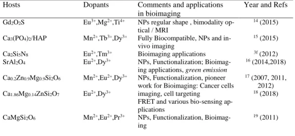
Table 1: Main materials used with metal transition (MT) cations and RE cations for red/ near infrared emission and applications in bioimaging.
Hosts Dopants Comments and applications
in bioimaging
Year and Refs
Gd2O2S Eu3+,Mg2+,Ti4+ NPs regular shape , bimodality
op-tical / MRI
14 (2015)
Ca3(PO4)2/HAP Mn2+,Tb3+,Dy3+ Fully Biocompatible, NPs and
in-vivo imaging
15 (2015)
Ca2Si5N8 Eu2+,Tm3+ Bioimaging applications 3f (2012)
SrAl2O4 Eu2+,Dy3+ NPs, Functionalization;
Bioimag-ing applications, green emission
16 (2014,2018) Ca0.2Zn0.9Mg0.9Si2O6 Ca1.86Mg0.14ZnSi2O7 Mn2+,Eu2+,Dy3+ Eu2+,Dy3+ NPs, Functionalization, pioneer work for Bioimaging: Cancer cells imaging, cell targeting
FRET and various bio-sensing ap-plications
17 (2007, 2011,
2012)
18 (2018)
CaMgSi2O6 Mn2+,Eu2+,Pr3+ NPs, Functionalization,
Bioimag-ing
MAlO3 (M=La, Gd) Mn4+/Ge4+ Bioimaging in pork tissue 11 (2014)
GdAlO3 Mn4+,Ge4+@Au
Sm3+, Cr3+
Trimodality imaging
Optical and magnetic dual mode imaging
20 (2016) 21 (2018)
ZnGa2O4 Cr3+ NPs, Functionalization,
Bioimag-ing (cancer cells imagBioimag-ing, cell tar-geting, cytotoxicity, visible light stimulation NIR Photostimulation, X-rays activation
Oral administration & breast can-cer imaging Toxicology analysis Protobiotic analysis 3e (2014) 22 (2014) 23 (2017),(2018) 24 (2018) 25 (2017) 26 (2017)
ZnGa2O4 in hollow cavity Cr3+ Photodynamic therapies 27 (2018)
ZnGa2O4 Cr3+, Gd3+ NPs, Functionalization,
Bimodal-ity Optical/NMR imaging
28 (2015) ZnGa2O4/SiO2 ZnGa2O4/Fe2O3 ZGOCS@MSNs@Gd2O3 Cr3+ Cr3+ Cr3+
NPs, core-shell structure, drug de-livery
Cell labelling and magnetic vec-torization
Multimodal nanoprobes
29 (2014) 30 (2018) 31 (2017)
Zn1.1Ga1.8Ge0.1O4/SiO2 Cr3+,Eu3+ NPs, core-shell structure, drug
de-livery 32 (2015) Zn3Ga2Ge2O10 Zn1.1Ga1.8Ge0.1O4@SiO2 Zn1.25Ga1.5Ge0.25O4 Zn1.1Ga1.8Ge0.1O4 Cr3+ Cr3+ Cr3+, Yb3+, Er3+ Cr3+
Imaging of pork tissue, Photostim-ulation, cytotoxicity
bioimaging and drug delivery Metastasis Tracking and Chemo-photodynamic Therapy Nanothermometry 33 (2014) 34 (2018) 35 (2018) 36 (2017) Zn3Ga2Sn1O10 Cr3+ Imaging of goldfish 37 (2014) Zn2.94Ga1.96Ge2O10 Zn3Ga2Ge2O10 Cr3+,Pr3+ Cr3+ NPs, Functionalization Recognition of Breast Cancer Cells
38 (2013) 39 (2015)
Zn3Ga2GeO8 Cr3+,Yb3+,Er3+ Upconversion 40 (2014)
LiGa5O8 Cr3+ NPs, Functionalization,
Bioimag-ing, Visible light stimulation, pho-tostimulation 41 (2013,2014) Ca3Ga2Ge3O12 mSiO2@Gd3Ga5O12 Cr3+,Yb3+,Tm3+ Pr3+,Yb3+ Cr3+, Nd3+
NIR Stimulation, Upconversion In-vivo imaging
multimodal imaging and cancer therapy
42 (2014) 43 (2017) 44 (2018)
Sr2SnO4 Nd3+ Finger image 45 (2014)
SiO2/CaMgSi2O6
YAGG (garnet)
Eu2+, Pr3+, Mn2+
Er3+, Cr3+
Bioimaging, intraperitoneal injec-tion
Photostimulation imaging of pork tissue
Imaging in the second biological window
46 (2014) 47 (2018)
NaYF4 + SrAl2O4 Yb3+, Tm3+,
Eu2+, Dy3+
Upconversion & photodynamic therapy
48 (2018)
Visualization of abdominal in-flammation
50 (2018)
La3Ga5GeO14
@SiO2@Van
Cr3+, Zn2+ bioimaging-guided in-vivo & drug
delivery
51 (2018)
CaTiO3 Pr3+, Yb3+, Tm3+ Upconverting and guided
photo-thermal therapy 52 (2017) ZnSn2O4 Sr3Sn2O7 Cr3+, Eu3+ Nd3+
Cellular and deep tissue imaging Second window imaging
53 (2017) 54 (2017)
Persistent luminescence phenomenon, whereby luminescent materials keep emitting light after the excitation has stopped, has intrigued people since several centuries. As this property occurs in inorganic materials when the excitation has been stopped, this is sometimes compared to the biolumines-cence mechanism observed in some animals (such as fireflies and jelly-fishes). Others fluorescent probes in the NIR range as reported in this book are the semiconductor quantum dots (QDs) or upconversion nanoparticles (UCNPs).
Main characteristics of the persistent luminescence materials Long persistent luminescence is controlled by the slow liberation of trapped charge carriers at room temperature by a simple thermal de-excitation pro-cess. The luminescence can last for several minutes to hours after removal of the excitation source (typically several minutes of ultraviolet light) 1, 55. In persistent luminescence materials, energy can be stored by traps/defects that are charged under irradiation. Emergency signage that can be used in case of electricity failure is one of the main applications of these persistent phosphors, but others applications were proposed such as watch dials, when radioactive elements were fully forbidden more than twenty years ago, dec-orative objects and toys and more recently for bio-imaging as described in this review paper.
Fig. 1. left: Schematic of the energy levels and traps involved in the persistent
lu-minescence mechanisms. Right: Partial transparency of living tissues in deep red range (see light through girl left hand and figure 11 (c)), extracted from Saint Jo-seph charpentier, G. de La Tour, 1643, Musée du Louvres, Paris, France
For the materials concerned with the persistent luminescence properties, there is in the literature plenty of compounds tested with various successes. Rare earth and metal transition cations have been widely used as dopant for
persistent luminescence materials and in last years. More than two hundreds combinations of host materials and activator ions have been depicted. The reader could see recent reviews and book chapter on the topic 2, 56. For bio-imaging and in-vivo application, as seen in figure 1, the past five years have witnessed several major advances to establish deep red/near-infrared emit-ting persistent luminescence nanomaterials (called in the paper PLNPs as acronym for Persistent Luminescence NanoParticles) as a novel, approach for real-time used in-vivo in small animal.
The main requirements that the materials must fulfil for this application are the following: (i) compounds prepared as nanoparticles (PLNPs with size < 100 nm) and even ultrasmall nanoparticles ( < 10 nm) (ii) intensive emission extending in the deep red toward the near infrared range in the first or second windows corresponding to the tissue transparency 57, (iii) efficient function-alization, (iv) stability in aqueous solution (v) persistent emission over hours. These points are the reasons why applications using deep red emitting persistent phosphors for bio-applications were barely investigated at the first step of the development of the persistent phosphors in 1996 and the investi-gation started in 2007 after our pioneer paper 17a. Several materials were further developed for imaging applications (see figure 1 and review papers 56). Notice that at first all the researches were focused in the deep-red range (high transmission range of the living tissue, see also figure 1) where the Si detector also reaches its maximal sensitivity. More recently researchers ex-tend the range to the second and third biological windows (BW) even if the results are still very preliminary in that 1000 nm-1350 nm spectral range (BW-2) and above 1500 nm (BW-3) as presented in the following part of the chapter. In the development of the new phosphors with long persistent luminescence for bio-applications, one should keep in mind that various synthesis strategies are used for small size preparation of PLNPs with good crystallinity but also with the optimum of defects at the origin of the persis-tent luminescence. Hydrothermal, solvothermal, sol-gel, co-precipitation and microwaves methods have been widely used. In addition, effective sur-face functionalization strategies along with the bio-compatibility issues re-quired attention to make them useful for in-vivo bioimaging.
Persistent luminescence mechanisms
The physics behind the phenomenon is however not that simple and inten-sive research has been carried out the last years. For the trapping processes, which is the primary very important step, two kind of traps are envisioned: (i) Intrinsic defects and optimization of the traps by composition variation and thermal annealing. Intrinsic defects have been well identified in lot of materials.10a, 58 In garnet hosts, antisite defects for instance are very likely,
they shorten some Y-O bonds and create shallow electron traps close to the conduction band. Antisite defects in the vicinity of Cr3+ ions have been pro-posed to be involved in the persistent luminescence mechanism of the zinc-gallate spinel host 13, 59. One should also notice that thermal annealing under reducing atmosphere is often used to create or enhance intrinsic traps such as oxygen vacancies, which correspond to electron traps and can be respon-sible for the persistent luminescence 60.
(ii) Optimization of the traps by co-doping. Co-doping is the second strategy that has been intensively tested to enhance the persistent luminescence. Co-doping with one or two lanthanides cations are most of the time used to enhance the property. For instance, Dy3+ was used as electrons trap in the first hybrid enstatite/diopside silicate for the proof of concept of the in-vivo bioimaging application with PLNPs 17a. Moreover it appeared that Pr3+ was indeed better when only the diopside bandgap was considered 11. The varia-tion of traps depths strongly depends on the couple host/dopant and corre-sponds to a so-call bandgap engineering as introduced in previous works 61, with the prediction of the energy level diagram 62. Such bandgap engineering was proposed in various hosts used for persistent luminescence but was also tested with success in others Photonic applications such as scintillation and lighting. It is then currently possible to vary the position of the conduction band and the traps depth by varying the composition playing with cationic and anionic substitutions or a careful choice of the doping cation for an op-timal couple composition/dopant.
To go in deeper understanding, knowledge of the energy formation of de-fects, defects stability and defects energy position are required, through band structure and defects calculations 63. The persistent luminescence mecha-nism has been characterized by lots of different spectroscopies including optical, EPR to control the origin of defects, 64 X-rays spectroscopies 65 to give insights to the mechanism, thermoluminescence 19 and photoconduc-tivity 66 to measure the traps depth 10a, 67. In the case of electron traps and hole traps 68, the stored charges can be released by various processes as ex-pressed in the following part such as thermal 69, optical 70 or other physical stimulations 71, resulting in stimulated emissions from the active recombi-nation centers.
ZnGa2O4:Cr nanoparticules for persistent luminescence applications
If metal transition doped ZnGa2O4 nanoparticles can be obtained by differ-ent techniques, our activity recdiffer-ently focus on the hydrothermal synthesis as-sisted by microwave heating in order to obtain ultrasmall nanoparticles with persistent luminescence properties. To avoid sintering and keep the nano-metric size during the following thermal treatment, the nanoparticles are
embedded in a silica layer using a sol-gel chemistry way. A mixture of TEOS:EtOH 1:4 is added to a basic solution of nanoparticles. Doped with trivalent chromium for deep red /NIR emission, the obtained ZnGa2O4:Cr3+,Bi3+@SiO2 nanoparticles are then calcined in air during 2 h at 1000 °C. The nanoparticles are monodisperse with a sub-10 nm size 72, To better characterize the persistent luminescence properties of the ZnGa2O4:Cr3+ nanocrystals, we also recently proposed with colleagues from Orleans, France 73 to prepare these nanocrystals embedded in a glass phase in the so-called ZGO nano glass-ceramics. The TEM picture of both nano-objects are presented in figure 2. The nanoparticles embedded in glassy ma-trix present an average diameter about 16 nm while after hydrothermal syn-thesis assisted by microwave, diameter nanoparticles smaller than 10 nm can be obtained (see figure 2). The nanoparticles are spherical and well dis-persed. In both cases after crystallization at 900 °C and thermal treatment at 1000 °C respectively, the obtained nano-objects are well crystallized in the pure spinel phase and present persistent luminescence properties.
Fig 2. TEM pictures of a) ZnGa2O4:Cr3+ (ZGO) nanoparticles embedded in glass
matrix (TC = 900 °C) b) ZGO nanoparticles after hydrothermal synthesis assisted by microwave heating, associated distribution diagram, adapted from 74,73.
Cr3+-doped ZGO nanoparticles photoluminescence and persistent lumines-cence spectra are presented figure 3. Several contributions of the chromium emission are well identified: the R line corresponding to the zero phonon line. Stokes and anti-Stokes phonon side bands (S-PSB and AS-PSB respec-tively) were also recognizable. The N2 line is attributed to the emission of a Cr3+ ion in the vicinity to an antisite defect 13. The persistent luminescent emission spectra is mostly dominated by the N2 line, indicating that these cations, spatially closed to the electron traps are partly responsible of per-sistent luminescence (see following part). Figure 3 shows the perper-sistent lu-minescence decay after 2 min of UV irradiation of ZGO:Cr3+ nanoparticles embedded in glass. With these ultrasmall nanoparticles, it is possible to de-tect the signal more than 2 hours after swithing off the UV lamp.
Fig. 3: Left, (top) Emission spectra of photoluminescence and (bottom) persistent
luminescence at room temperature, adapted from 13. Right: Persistent
lumines-cence decay of ZnGa2O4:Cr3+ nanoparticules (within more than 2 hours) adapted
from 73 .
As experimentally determined as well as calculated 75, 75b, in such nano-materials, antisites defects around the Cr3+ cations are needed to get persis-tent luminescence properties. NMR and EPR magnetic spectroscopies can
gives some insights on the disorder, and on the surrounding of the dopant (Cr3+) or the host (Ga3+) while thermoluminescence give insights on the per-sistent luminescence capability 73. In the particular case of ZnGa
2O4:Cr3+ and derivatives, electron paramagnetic resonance (EPR) is a technique of choice to learn about Cr3+ environments. Indeed, there is a splitting of the 4A
2 chromium level into two degenerated states (one ms = ± 3/2 and one ms = ± ½) due to the crystal field distortion and the spin orbit coupling. An application of a magnetic field can raise the degeneracy of these levels. De-pending on the Cr3+ vicinity, the splitting of these levels may be different leading to various EPR signals. Several EPR studies have been performed on ZnGa2O4:Cr3+, resulting in a better understanding of the local material structure and the possibility to distinguish several Cr3+ environments using simulations. First, the simulations on powder phosphors have shown that five different Cr3+surroundings may exist in ZnGa
2O4:Cr3+ material 75a. The results also strengthened the assumption about the role of antisite defects on persistent luminescence properties. Indeed, it has been demonstrated that the charge trapping in ZnGa2O4:Cr3+ may be related to two antisite defects in the Cr3+ neighborhood (Cr-γ, Cr-δ, Cr-ε). We recently demonstrated that simulations using these five Cr3+ also perfectly fits our nano-crystals em-bedded in glass matrix 73 (see figure 4). An important EPR result in this system is the possible quantification of chromium environment. This quan-tification enables the dependence on synthesis parameters. Indeed, we can notice a global increase of undistorted Cr3+ environment when T
cryst in-creases revealing a better symmetry around Cr3+ in agreement with the im-provement of crystalline quality. However, the persistent luminescence properties of lower crystallization temperature materials are poor, this may be due to the presence of parasitic defects due to poor crystallinity leading to quenching processes at the expense of the radiative processes.
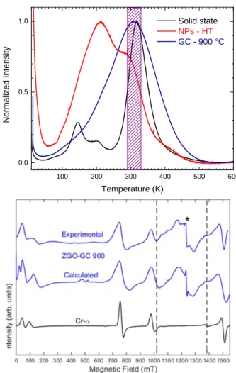
Thermoluminescence (TL) or thermally stimulated luminescence (TSL) is the most commonly used technique to demystify the trap number and their depths. This is a required characterization technique prior to persistent lu-minescence compounds developemnt. At first, in the thermolulu-minescence experiments, traps of the the compounds mustbe filled by optical (from X-rays to NIR photons) or others stimulations. Notice that mechanolumines-cence 76 is also recently an important field of investigation. As mentioned above, in a second step thermal energy or others excitation processes, can release these traps. During the TL experiments, the sample is linearly heated, from 10 K to 600 K in our facility. When the thermal energy brought by the heater is sufficient i. e. in the order of the energy difference between a re-combination center and the trap, charges start to be de-trapped. After being released, charges can either be trapped again in the same or others traps in
the vicinity or recombine on the emitted centre. In this latest process, ther-moluminescence occurs and can be recorded. The result of the experiment, as presented in figure 4, shows luminescent intensity as a function of the temperature. The number and position of the glow curve peaks give insights on the traps depths and distributions. Notice that the trap depth can also be estimated from the thermoluminescence profile. Regarding to the glow curve shape, several equations can link the trap depth to the peak tempera-ture 77, 78. In case of broader bands, which appears to be often the case for PLNPs, a simple estimation can be made using 79 :
𝐸 = 𝑇𝑚 500
Where E is the trap depth and Tm is the temperature of the peak maximum.
For the persistent luminescence properties at room temperature, the release should occur at room temperature, in case of in-vivo applications it has to be close to the living tissues temperature. This means that materials should pre-sent thermoluminescence peaks centred at ca. 310 K to be suitable for this application. Another option is to design materials with several trap depths. One kind of traps corresponding to room temperature and deeper traps, that cannot be thermally emptied at room temperature. In this case, a possible in-vivo re-activation of the persistent luminescence via trap redistribution using optical stimulation (using NIR light) can be envisionned.
The thermoluminescence results for ZnGa2O4:Cr3+ are displayed on figure 4 for several particles size and various shapes. The bulk material shows a rel-atively sharp thermoluminescence peak, labelled as the main peak, centred at 318 K, making it suitable for in-vivo imaging based on persistent lumi-nescence. It also shows two shallow traps, weaker in intensity around 145 K and 200 K. Going from bulk material elaborated via solid state method to nanoparticles elaborated by hydrothermal synthesis assisted by microwave heating, the thermoluminescence glow curve becomes quite different. In-deed, the corresponding profile presents a broad signal that seems to be com-posed of two major contributions, one around 210 K
Fig. 4: Left : Thermoluminescence glow curves with different sizes and shapes and
right: Simulation and experimental EPR spectra for the ZGO:Cr, adapted from 73.
and one around 300 K. The first one can be related to the shallow traps whereas the second can be related to the previous main peak. As the surface over volume ratio increases when the size is reduced, surface defects may have a major contribution in nanoparticles. With that in mind it has been assumed that the shallow traps can be related to surface defects. The ther-moluminescence glow curve of ZnGa2O4:Cr3+ glass ceramics elaborated at 900 °C, presented on the same figure (see Fig. 4), presents a broad main peak centred at ca. 310 K. The two shallow traps can be observed as humps enlarging the main peak at around 145 K and 205 K. In the glass ceramics samples, the surface over volume ratio is very high because of the nanoscale of the crystals. Still, the surface defects quantity may be very low comparing to nanoparticles elaborated via microwave assisted hydrothermal method as the nanocrystals are here embedded in a glassy matrix. This can explain the relatively low response of shallow traps, as it is assumed that they are related to surface defects. However, the main response peak appears broader in glass ceramics samples compared to bulk material. This has previously been explained by the crystal quality of the material.
Biocompatibility
To be used in-vivo, the nanoprobes should have the lowest toxicity. The main matrices presented above are either NPs incorporated into silica (it can also be mesoporous silica as seen below) or oxide-based NPs. Since they are designed for the sustained release of drugs, the tissue compatibility required a careful investigation. This was done by several groups (see for instance 80). No pathological abnormality could be observed in both gross and micro-scopic histological examinations of various tissues including heart, liver, spleen, lung and kidney up to one month after vein injection into mice, sug-gesting that the mesoporous silica PLNPs had not caused significant tissue toxicity and inflammation even they had not completely be degraded.
Cr3+ close to 2 antisites Cr clusters Cr3+ non-distorted Cr3+ close to 1 antisite
It was confirmed that neither gross nor histopathological abnormalities could be observed in the major organs of mice, such as in liver, spleen, kid-neys, heart, intestines, stomach, muscles or lungs 81. Furthermore, at a high vein-injection dose of 50 mg.kg-1 per day, the acute toxicity of PLNPs was almost negligible compared with the blank control according to the moni-toring of the body-weight change, the visible and/or palpable dermal infec-tion, the presence of ascites, the grooming or the impaired mobility. At an extremely high dose of 1200 mg.kg-1, no mouse survived after intraperito-neal or intravenous injection of MCM-41-type or SBA-15-type mesoporous silica nanoparticles (MSNs), however the subcutaneous injection did not cause death at an equally high administration dose and doses of up to 200 mg.kg-1, which was high enough for drug loading and delivery owing to their high drug loading capacities, was always safe by both intraperitoneal and intravenous injection 82.
Concerning persistent luminescence nanoparticles by themselves, two arti-cles have been recently published concerning biocompatibility and toxicity assays in-vivo. The first one was reported in 2017 by Ramirez-Garcia et al. In this study, mice were injected with a single intravenous administration of either hydroxylated or PEGylated persistent luminescence nanoparticles at different concentrations, from 1 to 8 mg per mice and a set of standard tests were carried out 1 day, 1 month and even 6 months after the administration
25. High concentrations of hydroxylated nanoparticles generate structural
al-terations at histology level, endoplasmic reticulum damage and oxidative stress in liver, as well as rising in white blood cells counts. A mechanism involving the endoplasmic reticulum damage could be responsible of the observed injuries in case of ZGO-OH. On the contrary, no toxicological ef-fects related to PEGylated nanoprobes treatment were noted during the in-vivo experiments, denoting the protective effect of PEG-functionalization and thereby, their potential as biocompatible in-vivo diagnostic probes. In 2018, Zhang and co-workers reported a 60 days in-vivo study of ZGO de-rivatives (Zn1.1Ga1.8Sn0.1O4:Cr3+) compound. In this work, healthy Balb/c mice were intravenously injected with ZGO derivative at a dose of 10 mg/kg. ZGO pre-irradiated with a 254 nm UV lamp was also set as one of the factors to evaluate the possible effect of NIR persistent luminescence of ZGO in-vivo. No signs of apparent weakness, spontaneous animal death and significant body weight gain or loss were observed within 60 days. It was found that all of the parameters were in the normal reference range. In the haematological analysis, various serum biochemical parameters were meas-ured with particular attention paid to liver and kidney function. Indicators of kidney function, including creatinine and urea nitrogen, were also within normal ranges and were similar to these of control mice. These results show
no obvious injury to the liver and kidney with ZGO exposure in mice, even at long exposure times. Based on long-term in-vivo biodistribution studies, the major organs from mice were sliced for hematoxylin and eosin staining and histological examination to determine whether or not ZGO exposure caused tissue damage, inflammation or lesions. The structures of the organs exhibited hardly any difference from the control group. No apparent histo-pathological abnormalities or lesions were found in any of the experimental groups. All of these data suggested that no significant toxicity was induced by ZGO injection, even up to 60 days.
Excitation capabilities and long term in-vivo imaging.
Though intended for diagnosis applications in living animals, persistent lu-minescence nanoparticles suffer from severe limitations. First, as the release of photons is thermally activated, accumulations of the signal (from seconds to minutes) are required during recording and materials with intensive per-sistent luminescence on one side and low noise detector on the other side should be used. The second point concerns the excitation mechanisms of the PLNPs but recent improvements have been made as described in this part of this chapter. Indeed the first generation of PLNPs has to be excited ex-vivo by UV light prior to systemic administration preventing long-term imaging in living animal. Depending on the nanoparticle characteristics, slow accu-mulation of stealth nano-carriers within malignant stroma by the enhanced permeability and retention effect usually requires from 2 to 24 hours 83. This was far too long in relation to the emission from persistent luminescence nanoparticles, which hardly exceeds one hour in-vivo.
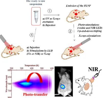
To overcome this major restriction, developments of new materials and of new modalities have been undertaken. First, efforts to optimized composi-tions and enhanced optical characteristics have been made. In that sense, compounds based on gallate and gallo-germanate spinels compounds have attracted large attention, due to their bright deep persistent luminescence when doped with trivalent chromium (Cr3+) after UV excitation in compar-ison with the previously used silicate nanoprobes. Then the second point was to excite the persistent luminescence material at lower energy in the biological window if possible, longer time after the injection. The new mo-dality has been proposed in different ways as seen in Figure 5.
Figure 5 summarizes the various approaches that can be followed for long term in-vivo imaging with persistent luminescent nanoparticles: A suspen-sion of PLNPs in a biological buffer can be pre-activated ex situ, then in-jected into the animal and placed under a photon-counting system to detect the emitted persistent luminescence signal (see step 1, Fig. 5). PLNPs can be activated or re-activated in situ (after the injection) through animal tissues (step 2, Fig. 5). This breakthrough for in-vivo optical imaging allows the most simple and convenient recovery of the persistent luminescence signal, whenever required. The efficiency is lower than for the persistent lumines-cence under UV excitation (step 1) but is indeed sufficient to be detected and to localize the probe in-vivo. Furthermore for some inorganic persistent luminescent NPs, when deeper traps are observed in the thermolumines-cence spectra such as in garnet and spinels hosts, deep-red persistent emis-sion can be stimulated by near infrared light, as the traps can be depopulated by low-energy light stimulation in the so-called photostimulated persistent luminescence (PSPL) 22, 70, 84. The photostimulation capability of several ma-terials is widely reported 85 and researchers have focused their attention on the Cr3+ doped samples. The photostimulation technique has been used over the years for UV dosimetry, as well as for dating geological and archaeolog-ical materials 86. One can adjust the depth of the traps responsible for the persistent luminescence and therefore to control carefully the composition. In that case, the release of the traps and thus the emission could be started at the convenience of the user using a red/near infrared LED for instance which will be in the best transparency range. The first preliminary carried out tests have shown the originality and feasibility of this new modality 87. The excitation energy for charge detrapping may vary from red to NIR lamps, LED or laser sources. The photostimulation or excitation by low-energy light can also be carried out by additional step, corresponding to wavelength conversion 40, 42,88, or possible charge through second order ef-fects under high excitation power 73, 89.
Fig 5: Top: different strategies to perform in-vivo optical imaging with
persistent luminescence nanoparticles (PLNP). (1) Excitation of a sus-pension of nanoparticles followed by tail vein injection and optical im-aging using a photon counting camera. (2) Once the in-vivo persistent luminescence signal has completely disappeared, some PLNP can give further persistent luminescence signal either by photo-or X-rays stimu-lations possibly giving n decay signals. (3) Some PLNP can be excited without preliminary excitation (with lower efficiency) in the animal body using visible, near infrared –for upconversion- or X-rays photons,
Bot-tom Traps could be emptied by a near infrared source (energy ~ 1eV)
photo-transfer to shallow traps leading to persistent luminescence. This demon-strated the photostimulation capability at convenience time. Within this experiment, one observed long-lasting phosphorescence under 977 nm light excitation 87, making in-vivo excitation of the spinels probes
envi-sioned and possible long term imaging applications.
However, in-vivo tracking is still hindered by its limited penetration in tissue even in the near infrared. An alternative way is to use NIR or X-ray as exci-tation source for imaging techniques. Indeed compared to the traditional ex-citation source, X-ray possesses competitive advantages of weaker scatter-ing and deeper penetration depth in tissues, as well as simplified image reconstruction for optical tomography. Developing X-ray rechargeable per-sistent luminescence nanoplatform, by combining the advantages of X-ray excitation and NIR persistent emission in the second biological window, will enable to localize tumors and achieve treatment simultaneously 90. This opens the door for achieving deeper tissue and higher sensitivity optical bi-oimaging with better spatial resolution. An example of such modality with ZGO:Cr PLNPS is presented in the following part of this chapter.
Strategies developed to perform long time imaging
The pioneer work using persistent luminescence nanoparticles (PLNPs) for optical in-vivo bioimaging was reported in 2007 by Scherman and co-work-ers 17a. As discussed in the previous section, to be used in-vivo, the probe should have a nanometric scale and should emit light in the transparency window (> 650 nm). In their work, the authors prepared a silicate doped with Eu2+, Dy3+ and Mn2+ and in-vivo imaging without any background was formed for 30 min to 1 hour without any in-vivo excitation, allowing per-forming in-vivo imaging without autofluorescence. Then after these pioneer works, lots of bio-applications and various modalities have been presented as seen Table 1, and some of them are detailed in the following part of the chapter. To overcome this major restriction and to allow longer time imag-ing, in 2014 Maldiney et al. changed the matrix and used zinc gallium oxide doped with Cr3+ 3e. As can be seen on Figure 6, contrary to the first silicate –
type generation, this material has several excitation peaks, among them one being at the limit of the tissue transparency window (Figure 6A) that can also sensitized the persistence luminescence. On nanosized ZnGa2O4:Cr, they observed a persistent luminescence decay signal after both UV and ac-tivation with visible orange/red light-emitting diodes (LEDs) (Figure 6B). A suspension of this material dispersed into 5% glucose was injected into mice, and for the first time the authors have shown that the probe could be activated in-vivo with orange/red light-emitting diodes. The authors have also shown that UV pre-excitation before LED re-activation is favourable
but not necessary (Figure 6C and 6H). In such conditions, there was no more time limit and the probe could be detected in-vivo whenever wanted through a simple excitation with the LEDs (Figure 6E-G).
Fig. 6. In-vivo imaging using in situ excitable PLNPs. (a-c) Optical
prop-erties of powder after either UV or visible excitation. (d-h) In-vivo excita-tion of un-funcexcita-tionalized PLNPs (e) or stealth ZGO in healthy (f) on tumor bearing mice (g), adapted from 3e.
It is of great importance to develop deep red/NIR PLNPs with rechargeable nature in-vivo by a deep tissue penetrating source (see figure 1, old painting showing only red light through living tissues). Recent years have witnessed the rapid development of utilizing innovative excitation sources for imaging techniques 91. For instance X-ray photon energies are able to stimulate lu-minescent centers in phosphors and then generate light. Furthermore, com-pared with the traditional excitation sources (UV/visible/NIR), X-ray pos-sesses competitive advantages of weaker scattering and deeper penetration depth in tissues and simplified image reconstruction for optical tomography 92. As a proof of concept, Xue et al selected ZnGa
2O4:Cr3+ PLNPs as models for X-ray-activated NIR persistent emission 23b. In-vivo NIR persistent lu-minescence bioimaging based on these X-PLNPs was demonstrated (see fig-ure 7). More importantly, these X-PLNPs were also repeatedly charged by X-rays at a deep location up to 20 mm. These results revealed that X-ray could act as a new excitation source for persistent renewable bioimaging with a deep penetration depth based on NIR PLNPs.
Fig. 7. (a) Principle of in-vivo imaging using X-ray excitation. (b)
Ap-plications in-vivo, adapted from 23band 93.
Multimodal imaging
Multimodal imaging has drawn much attention in biomedical applications because it provides more accurate, complete, and reliable information on diagnosis 94. Each imaging modality has its own advantages and disad-vantages regarding sensitivity, spatial/temporal resolution, and penetration depth. The NIR optical imaging possesses the advantage of high sensitivity
but limited spatial resolution. In contrast, MRI provides excellent spatial resolution and unlimited depth penetration but remains at an inherently low sensitivity 95. The combination of NIR emitting PLNPs and MRI contrast agent into one single nanoplatform would offer attractive synergistic ad-vantages in biomedical imaging with high sensitivity, good spatial resolu-tion, high signal over noise ratio, and no ionizing radiation.
Several strategies have been reported using gadolinium to have access to bimodal imaging PLNPs. In 2014, Yan and co-workers reported the surface functionalization of Zn1.1Ga1.8Ge0.1O4 with DTPA-Gd complexes to get bi-modal imaging agents, able to be detected both by optical imaging and as a positive T1 MRI contrast agent 96. In 2015, Maldiney et al. reported another method to prepare PLNPs-based bimodal imaging agent. In their strategy, instead of using the surface of the PLNPs to link DTPA-Gd complexes, the authors co-doped their NPs by incorporating various amount of Gd3+ ions at the beginning of the synthesis, by substituting Ga3+ ions 28. In such
condi-tions, a negative contrast was obtained, with a r2 value close to 60 mM-1.s -1. In 2017, Zou et al used a third strategy to prepare bimodal imaging agents. In their work, a novel core-shell structure multimodal imaging probe was prepared, based on the coverage of of mesoporous silica nanoparticles (MSNs) loaded ZnGa2O4:Cr3+, Sn4+ (ZGOCS@MSNs) by a Gd2O3 shell (∼1.5 nm) 31. Compared with previously reported Gd3+-based NIR persistent luminescence-based multimodal nanoprobes, the as-prepared nanoparticles enable surface available, no persistent intensity loss and only a slight size increase.
Instead of gadolinium, ultrasmall iron oxide nanoparticles (also called USPIO) are used in clinic as negative (T2) MRI contrast agents. In 2015, Teston et al reported the first synthesis of mesoporous nanohybrids (MPNHs) composed of a combination of PLNPs and USPIOs embedded into a mesoporous silica structure to get a bimodal imaging agent 97. In ad-dition of its bimodal imaging property, the authors showed that this nano-platform could be attracted by a magnet. They used this property to label and attract cells in-vivo 30.
Theranostics nanoprobes
During the past 10 years, it has been shown that diagnosis and therapy could be combined within a single multifunctional nanomaterial, known as theranostic nanoparticles 98, 3b. The ideal theranostic nanomaterial should possess several advantages 99: (i) the ability for highly selective
accumula-tion in the diseased tissue, (ii) the capability of delivering an effective ther-apeutic action selectively, and (iii) safety concerns and the material should undergo biodegradation into nontoxic byproducts. Such nanomaterials are one of the keys for detection and treatment of early stage cancer in the 21st century 100. Theranostic nanoparticles are still in the very early translational stages, with nearly all efforts devoted to preclinical studies and no clinical trials to date. In this section, we will highlight the recent use of PLNPs for the development of new theranostic nanoprobes.
Typically, mesoporous silica (mSiO2) can be used as a promising drug car-rier because of its stable mesoporous structure, high specific surface area and good biocompatibility 101. Using mesoporous silica alone as a drug car-rier it appears impossible to effectively locate a target site (i.e., tumor) and monitor in real time due to the lack of detectable signals. In 2014, Maldiney et al introduced a multifunctional nanoplatform based on persistent lumi-nescence nanoparticles for both in-vivo optical imaging and drug delivery 29.
Taking advantage of the well-known biocompatibility of mesoporous silica and non-toxic ZGO, the authors modelled a novel core-shell structure on the basis of a hybrid chromium-doped zinc gallate/mesoporous silica architec-ture specifically designed to allow both highly sensitive optical detection through living tissues and concomitant drug delivery. Employing doxorubi-cin as a drug model, the authors demonstrated that these mesoporous persis-tent luminescence nanophosphors can be successfully loaded with an anti-cancer agent, and subsequently initiate its progressive release in a pH-sensitive manner. The use of such doxorubicin-loaded theranostic agent is finally shown to induce acute cytotoxicity toward U87MG cells in vitro, preserved persistent luminescence properties, and allowed both sensitive and non-invasive localization of the carrier in-vivo.
In 2016, Zhang and co-workers developed another core-shell NPs composed of Gd2O2, ZGO and mSiO2 102. This multimodal nanoprobe could be de-tected both by MRI and optical imaging and was used to monitor it’s bio-distribution in healthy and tumor bearing mice after loading with doxorubi-cin (see Figure 8).
Fig. 8. Synthesis procedure of mesoporous ZGO, loading with Dox and in-vivo
application, adapted from 102.
A few photosensitizers (PSs), such as Porfimer sodium, have been approved by the United States Food and Drug Administration (FDA) to treat certain cancers. However, most of the available PSs for PDT can only be activated under the UV/visible light (<700 nm) with low tissue penetration. New strat-egies in order to use such molecules in better conditions are under investi-gations.
Photodynamic therapy (PDT) is a relatively new modality for cancer treat-ment 103. PDT consists of three essential components: light, oxygen, and a photosensitizer 104. Photosensitizers, often pharmacologically inactive with-out illumination, can be activated by light of a specific wavelength. This activation is followed by transfer of energy to nearby oxygen molecules to generate cytotoxic reactive oxygen species (ROS), most importantly singlet oxygen (1O
2). PDT is a relatively less invasive treatment modality, inducing low systematic toxicity and causing little intrinsic or acquired resistance. One primary downside of PDT, however, is its inability to treat tumors lo-cated deep under the skin due to the short penetration depth of light in tissues 105. This problem can be partially alleviated by advanced light-delivering technologies that allow for illumination of certain internal cavities, such as the bladder, prostate, lung, and oesophagus. Nevertheless, it is considered as challenging or impossible for conventional PDT to treat tumors of large volumes or multiple loci. High X-rays dose (>5.0 Gy) are needed for effi-cient cancer therapy, and this dose was comparable or even higher than the fraction dose used in clinical radiotherapy 106. Many researches have shown that high-dose X-ray irradiation inevitably causes damage to normal tissues 107.
In this regard, reducing X-ray dose is a major concern for deep-tissue PDT practical applications. Persistent luminescence nanoparticles (PLNPs) are promising luminescent materials that can store excitation energy and then slowly release the trapped charge carriers to emit persistent luminescence without continuous excitation. PLNPs alone have limited impact on cancer cells development 108. In 2015, a novel X-ray inducible photodynamic ther-apy (PDT) approach was reported that allows PDT to be regulated by X-rays. Upon X-ray irradiation, the integrated nanosystem, which corresponds to a core of a nanoscintillator SrAl2O4:Eu2+ (SAO) and a mesoporous silica coating loaded with photosensitizers (MW540) 93, converts X-ray photons to visible photons to activate the photosensitizers and cause efficient tumor shrinkage (see Figure 9).
In 2018, Yang and co-workers have shown that Zn (II) phthalocyanine tetrasulfonic acid (ZnPcS4) can be covalently linked to ZGO in order to fab-ricate PDT nanoplatform (ZGO:Cr/W–ZnPcS4). The persistent lumines-cence continously excites the coupled PS after the X-ray irradiation has been removed, resulting in significantly reduced X-ray dosage (≈0.18 Gy), mini-mizing the side effects of PDT treatment 23a.
Photothermal therapy with PLNPs
Among all the cancer treatments, near infrared (NIR 700 nm–1000 nm) laser induced photothermal therapy (PTT) is a non-invasive mode that can gener-ate heats at the tumors site 109. Compared with conventional therapies, it has attracted much interest in tumor therapy as a minimally invasive and non-injurious therapeutic program as well as large penetration into bio-tissues. Photothermal therapy agents are capable of converting light energy into heat inducing a rise in the local temperature beyond 42 °C and killing conse-quently cancer cells. As a FDA-approved drug, indocyanine green (ICG) is attractive for localized hyperthermia by absorbing near-infrared light (around 800 nm) even in deep tissue to generate heat. To further enhance the fluorescence stability and Enhanced Permeation and Retention effect (EPR) of ICG in-vivo, a variety of nanocarriers have been developed. There-fore, the ICG loading nanoparticle is a brand-new photothermal therapy agent which could be diffusely used in clinic for photothermal therapy. Nev-ertheless, the traditional ICG loading nanoparticles cannot be tracked when they circulate in the body, therefore it is out of the question to ensure an accurate irradiation with additional near infrared (700 nm–1000 nm) laser. Optical imaging-guided therapy has received great attention recently and become a fine choice of loading the ICG and tracking it for PTT 110.
Fig. 10. PTT with ICG loaded PLNPs. Adapted from 110.
Very recently in 2017, Chang and workers used ZGO NPs and ICG co-loaded into mesoporous silica nanoparticles (ICG@mZGO nanoparticles) for imaging-guided PTT 111. The ICG@mZGO nanoparticles consisted of two parts: i) The mesoporous SiO2/ZnGa2O4:Cr3+ (mZGO) near-infrared PLNPs were used as near-infrared emitting probe to track NPs in-vivo after excited by light. They found that mesoporous silica nanoparticles could be a fine template to synthesize ZGO phosphors in situ with good morphology
and expected size. Mesoporous silica nanoparticles could also load and pro-tect ICG, which had the ability to avoid particle aggregation effectively dur-ing the transportation; ii) Indocyanine green (ICG), a kind of tricarbocya-nine dye, which substantially absorbed the near-infrared light (NIR) and in result returned a photothermal response.
In a different approach, upconverting PLNPs and photo-absorbing agents ICG co-loaded into mSiO2 (UC-PLNPs and ICG co-loaded mSiO2) nano-particles were developed for upconverting and persistent luminescent imag-ing guided PTT 52. The UCPLNPs and ICG co-loaded mSiO
2 nanoparticles include three parts: i) the upconverting PLNPs, doped with Pr3+, Er3+ and Tm3+ chosen for upconverting and persistent luminescent imaging. These NPs could be excited by NIR light and emit upconverting luminescence, which can be used for in-vivo imaging when the persistent luminescence signal became weak; ii) the ICG (indocyanine green), a water soluble ani-onic tricarbocyanine dye which is the only NIR agent approved by FDA and which has been widely used clinically for PTT; iii) the mesoporous silica nanoparticles working as a three dimension hard template for loading and protecting both upconverting PLNPs and ICG 52.
Perspectives of the NIR-persistent luminescent nanoparticles for bio-imaging
Firstly, to move the persistent emission wavelength to the second and third biological windows (see Figure 11-C) and compared the pro and drawbacks of such strategy, different approaches are currently explored: i) Traps can be coupled to various rare earth cations (Yb3+, Nd3+, Er3+, Ho3+, Tm3+ or even Pr3+) well known in the case of NIR lasers (see Fig 11-(B)) 112. ii) Down shifting can also be obtained by coupling different rare earth cations or metal transition (MT) and rare earth cations. The so-call traps redistribution by energy transfer can downshift the persistent emission wavelengths (see Fig 11-(A)). Such down-conversion processes were proposed recently for garnet and perovskite hosts coupling metal transition cation (Cr3+) with rare earth cations to promote by persistent energy transfer the persistent luminescence in the deep red (from Cr3+) but also in the second or third biological windows (results were obtained with Yb3+, Nd3+, Er3+, Ho3+ ions) 47, 113. iii) Transition metal cations such as Ni2+ or Co2+ were also recently investigated 114 (see Fig 11-(D)) to cover the second biological window and these MT cations have also large potential. Indeed, recently, Qiu and co-workers reported a
strategy for tuning the emission bandwidth and intensity in persistent lumi-nescence phosphors by synthetizing Ni2+-doped Zn
1+ySnyGa2−x−2yO4 phos-phors, with an emission band peaking from 1270 to 1430 nm in the second near-infrared (NIR) window 115. Ueda 116 and Tanabe 117 and our groups also reported compositions able to be used in the second transparency window 114a.
Fig. 11. : (A) trap redistribution and persistent energy transfer in metal
transition (Cr) and rare earth doped garnet 47, (B)Trivalent rare earth
cations as efficient emitters for short wave infrared persistent lumines-cence 112, (C) Optical window in biological tissues; high: attenuation
co-efficient (log) as a function of the wavelength. Bottom: sensitivity of dif-ferent cameras (silicon (Si) cameras, indium gallium arsenide (InGaAs) or mercury cadmium telluride (HgCdTe)) 118 and (D): optical features
and persistent luminescence of ZnGa2O4:Ni and Zn(GeGa)2O4:Ni. Decay
profiles and scheme of excitation, adapted from 114a
Secondly, moving to the second and third near-infrared transparency win-dows (1000 nm - 1700 nm) allows further reducing of scattering, absorption and tissue autofluorescence effects. Moreover, if the quantum efficiency values, for Si, InGaAs (and HgCdTe) can be comparable, the acquisition time is also to be taken into account. For a CCD camera the dark current is of the order a few electron/pixel/hour and it is about 100 ke/pixel/s for In-GaAs. The readout noise has also to be taken into account (see values in Table 3). By the end, the signal is one to two orders of magnitude smaller for InGaAs detector (and HgCdTe) in the NIR range with respect to the Si detector in BW-1. Imaging platforms then require the use of powerful and cooled cameras.
Table 3: Characteristics of the NIR and deep-red sensors
1.5 micron NIR camera Visible camera
Material type InGaAs Si/CMOS Quantum efficiency 80 % 80 % Readout noise Dark current Pixel size about 30 e- 100 ke-/pixels/s About 15 micrometers about 1 e- < 0.5 e-/pixels/s About 4 micrometers
Then the balance between benefit and drawbacks for these two aspects re-lated to the second and third biological windows remains to be explored. In a decade, persistent luminescence has emerged as a useful property for the development of new optical imaging nanoprobes. Thanks to this property of
long lasting luminescence, imaging in the first transparency window (NIR-I) with high target to background ratio has been reported by several research groups (more than 40 research groups all over the world and more than 200 papers published in the last ten years). Beyond to their uses for imaging and therapy, new applications emerged. We can cite for instance their use for the development of nanothermometers. For this purpose, the idea is to use the luminescence emitted by nanoprobes to detect temperature variations at the nanoscale 11936.
Conclusions
Very recent and promising works on persistent luminescence nanoparticles concern the development of biocompatible nanoprobes for multimodal im-aging, theranostics and drug delivery, photodynamic and photothermal ther-apies, nanothermometry and hyperthermia etc. These PLNPs were even re-cently proposed as a guide during surgery operation to accurate delineation of hepatocellular carcinoma 120 .
Then, one of the next challenge will be: can these PLNPs be used at wave-lengths longer than 1000 nm ?. Indeed, optical imaging in the second near-infrared region (NIR-II, 1.0-1.7 μm) has recently attracted significant atten-tion owing to some advantages : if the ballistic light (without diffusion) and the so-call “snake” light with limited diffusion remain about the same, the diffuse light with all the information is lost can be very limited moving in that NIR range. Photons in the NIR-II region can provide high spatial reso-lution at deep tissue penetration depths owing to the reduced scattering of long-wavelength photons 121. Furthermore, imaging in the second near-in-frared region addresses several challenges simultaneously: minimal auto-fluorescence of biological tissue leading to increased sensitivity and signif-icantly reduced light attenuation from scattering and from absorption by the blood and other structures enables imaging with high spatiotemporal reso-lution and penetration depth 122. Consequently, large organisms such as a whole mouse may be rendered translucent when imaged in this area 123. The image reconstruction is also a very important work as was done for instance in the acousto-optic images 124.
Nevertheless there is still several others challenges for the scientific com-munity such as, the synthesis of long circulating “stealth” nanomaterials, the functionalization with targeting agents prerequisite for PLNPs later use. Others important requirements are to keep a high intensity at the nanoscale, and to well control the surface properties in order to stabilize the PLNPs in
different biological media. Then, with the nano-objects, imagination will be the only limit to the use and applications of these fascinating nanoparticles.
Acknowledgments
This work was supported by the French national research agency (ANR Natlurim and ANR PEPSI 14-CE08-0016-01), by a grant from the Pres Sor-bonne Paris-Cité (ODICEO project), by CNRS, by Biospace Lab, by the PRC Japon JSPS/French Bilateral grant (PRC1248) and by PHC Procore (33122PA). The authors acknowledge their collaborators: Q. le Masne de Chermont, T. Maldiney, E. Teston, G. Ramirez-Garcia, T. Lecuyer, J. Se-guin, D. Scherman, D. Gourier, A. Bessière, L. Binet, M. Allix, S. Sharma, S. Tanabe, F. Gazeau and L. Laumonier.
References
1. Matsuzawa, T.; Aoki, Y.; Takeuchi, N.; Murayama, Y., New long phosphorescent phosphor with high brightness, SrAl2O4:Eu2+,Dy3+. Journal of
the Electrochemical Society 1996, 143 (8), 2670-2673.
2. (a) Van den Eeckhout, K.; Smet, P. F.; Poelman, D., Persistent Luminescence in Eu2+-Doped Compounds: A Review. Materials 2010, 3 (4), 2536-2566; (b) Van den Eeckhout, K.; Poelman, D.; Smet, P. F., Persistent Luminescence in Non-Eu2+-Doped Compounds: A Review. Materials 2013, 6 (7), 2789-2818. 3. (a) Viana, B.; Sharma, S. K.; Gourier, D.; Maldiney, T.; Teston, E.; Scherman, D.; Richard, C., Long term in vivo imaging with Cr3+ doped spinel nanoparticles exhibiting persistent luminescence. Journal of Luminescence 2016,
170, 879-887; (b) Lécuyer, T.; Teston, E.; Ramirez-Garcia, G.; Maldiney, T.; Viana,
B.; Seguin, J.; Mignet, N.; Scherman, D.; Richard, C., Chemically engineered persistent luminescence nanoprobes for bioimaging. Theranostics 2016, 6 (13), 2488; (c) Jaque, D.; Richard, C.; Viana, B.; Soga, K.; Liu, X. G.; Sole, J. G., Inorganic nanoparticles for optical bioimaging. Advances in Optics and Photonics
2016, 8 (1), 1-103; (d) Sharma, S. K.; Gourier, D.; Viana, B.; Maldiney, T.; Teston,
E.; Scherman, D.; Richard, C., Persistent luminescence of AB(2)O(4):Cr3+ (A = Zn, Mg, B = Ga, Al) spinels: New biomarkers for in vivo imaging. Optical
Materials 2014, 36 (11), 1901-1906; (e) Maldiney, T.; Bessiere, A.; Seguin, J.;
Teston, E.; Sharma, S. K.; Viana, B.; Bos, A. J. J.; Dorenbos, P.; Bessodes, M.; Gourier, D.; Scherman, D.; Richard, C., The in vivo activation of persistent nanophosphors for optical imaging of vascularization, tumours and grafted cells.
Nature Materials 2014, 13 (4), 418-426; (f) Maldiney, T.; Sraiki, G.; Viana, B.;
Gourier, D.; Richard, C.; Scherman, D.; Bessodes, M.; Van den Eeckhout, K.; Poelman, D.; Smet, P. F., In vivo optical imaging with rare earth doped Ca2Si5N8
persistent luminescence nanoparticles. Optical Materials Express 2012, 2 (3), 261-268.
4. Hillman, E. M. C.; Amoozegar, C. B.; Wang, T.; McCaslin, A. F. H.; Bouchard, M. B.; Mansfield, J.; Levenson, R. M., In vivo optical imaging and dynamic contrast methods for biomedical research. Philosophical Transactions of
the Royal Society a-Mathematical Physical and Engineering Sciences 2011, 369
(1955), 4620-4643.
5. de Jong, M.; Essers, J.; van Weerden, W. M., Imaging preclinical tumour models: improving translational power. Nature Reviews Cancer 2014, 14 (7), 481-493.
6. Huang, Y.-Y., Hamblin, M., Chen, A.C.-H., 2009. Low-level laser therapy:
an emerging clinical paradigm. SPIE Newsroom.
http://dx.doi.org/10.1117/2.1200906.1669
7. S. J. Matcher, M. Cope, and D. T. Delpy, In vivo measurements of the wavelength dependence of tissue scattering coefficients between 760 and 900 nm measured with time-resolved spectroscopy, Applied Optics, 1997, 36, 386-396
8. H. Yukawa, M. Watanabe, N. Kaji, Y. Baba, Influence of
Autofluorescence Derived From Living Body on In Vivo Fluorescence Imaging Using Quantum Dots, Cell Medicine, 2015, 7, 75-82
9. Jaque, D.; Richard, C.; Viana, B.; Soga, K.; Liu, X.; Solé, J. G., Inorganic nanoparticles for optical bioimaging. Advances in Optics and Photonics 2016, 8 (1), 1-103.
10. (a) Holsa, J.; Aitasalo, T.; Jungner, H.; Lastusaari, M.; Niittykoski, J.; Spano, G., Role of defect states in persistent luminescence materials. Journal of
Alloys and Compounds 2004, 374 (1-2), 56-59; (b) Holsa, J.; Laamanen, T.;
Lastusaari, M.; Malkamaki, M.; Novak, P., Persistent luminescence - Quo vadis?
Journal of Luminescence 2009, 129 (12), 1606-1609.
11. Maldiney, T.; Bessière, A.; Seguin, J.; Teston, E.; Sharma, S. K.; Viana, B.; Bos, A. J.; Dorenbos, P.; Bessodes, M.; Gourier, D., The in vivo activation of persistent nanophosphors for optical imaging of vascularization, tumours and grafted cells. Nature materials 2014, 13 (4), 418-426.
12. (a) Aitasalo, T.; Deren, P.; Holsa, J.; Jungner, H.; Krupa, J. C.; Lastusaari, M.; Legendziewicz, J.; Niittykoski, J.; Strek, W., Persistent luminescence phenomena in materials doped with rare earth ions. Journal of Solid State Chemistry
2003, 171 (1-2), 114-122; (b) Dorenbos, P., Mechanism of persistent luminescence
in Eu2+ and Dy3+ codoped aluminate and silicate compounds. Journal of the
Electrochemical Society 2005, 152 (7), H107-H110.
13. Bessiere, A.; Jacquart, S.; Priolkar, K.; Lecointre, A.; Viana, B.; Gourier, D., ZnGa2O4:Cr3+: a new red long-lasting phosphor with high brightness. Optics
Express 2011, 19 (11), 10131-10137.
14. C. Rosticher, C. Chanéac, B. Viana, M. A. Fortin, J. Lagueux, L. Faucher, “Red Persistent luminescence and magnetic properties of nanomaterials for multimodal imaging” SPIE, Oxide-based Materials and Devices VI, OE108, in press (2015)
15. C. Rosticher, C. Chanéac, B. Viana, M. A. Fortin, J. Lagueux, L. Faucher, “Red Persistent luminescence and magnetic properties of nanomaterials for


![Fig. 3: Left, (top) Photoluminescence and (bottom) persistent luminescence spectrum at room temperature, adapted from Bessière et al [2011]](https://thumb-eu.123doks.com/thumbv2/123doknet/7766560.256058/39.892.226.559.292.789/left-photoluminescence-persistent-luminescence-spectrum-temperature-adapted-bessière.webp)