HAL Id: dumas-01781432
https://dumas.ccsd.cnrs.fr/dumas-01781432
Submitted on 7 Jun 2018
HAL is a multi-disciplinary open access
archive for the deposit and dissemination of sci-entific research documents, whether they are pub-lished or not. The documents may come from teaching and research institutions in France or abroad, or from public or private research centers.
L’archive ouverte pluridisciplinaire HAL, est destinée au dépôt et à la diffusion de documents scientifiques de niveau recherche, publiés ou non, émanant des établissements d’enseignement et de recherche français ou étrangers, des laboratoires publics ou privés.
Camille Conan
To cite this version:
Camille Conan. Apathy in depression : an arterial spin labeling study. Life Sciences [q-bio]. 2017. �dumas-01781432�
THÈSE D'EXERCICE / UNIVERSITÉ DE RENNES 1
sous le sceau de l’Université Bretagne Loire
Thèse en vue du
DIPLÔME D'ÉTAT DE DOCTEUR EN MÉDECINE
présentée par
Camille CONAN
Née le 28 mai 1987 à Paris
Apathie dans la
dépression : une
étude en arterial spin
labeling
Thèse soutenue à Rennes
le 2 octobre 2017
devant le jury composé de :
Dominique DRAPIER
Professeur – CHGR Rennes – Président du jury
Jean-Marie BATAIL
PH – CHGR Rennes – Directeur de thèse
Jean-Christophe FERRE
Professeur – CHU Pontchaillou – JugeGabriel ROBERT
PH MCU – CHGR Rennes - Juge
Carole DI MAGGIO
PH – CHGR Rennes - JugePROFESSEUR DES UNIVERSITES - PRATICIENS HOSPITALIERS
NOM Prénom AFFECTATION
ANNE-GALIBERT Marie
Dominique Biochimie et biologie moléculaire
BELAUD-ROTUREAU Marc-Antoine Histologie; embryologie et cytogénétique
BELLISSANT Eric Pharmacologie fondamentale; pharmacologie clinique;
addictologie
BELLOU Abdelouahab Thérapeutique; médecine d'urgence; addictologie
BELOEIL Hélène Anesthésiologie-réanimation, médecine d'urgence
BENDAVID Claude Biochimie et biologie moléculaire
BENSALAH Karim Urologie
BEUCHEE Alain Pédiatrie
BONAN Isabelle Médecine physique et de réadaptation fonctionnelles
BONNET Fabrice
Endocrinologie, diabète et maladies métaboliques; gynécologie
BOUDJEMA Karim Chirurgie générale
BOUGET Jacques
Professeur des universités en surnombre
Thérapeutique; médecine d'urgence; addictologie
BOUGUEN Guillaume Gastroentérologie; hépatologie; addictologie
BOURGUET Patrick
BRASSIER Gilles Neurochirurgie
BRETAGNE Jean-François Gastroentérologie; hépatologie; addictologie
BRISSOT Pierre
Professeur des Universités en surnombre
Gastroentérologie; hépatologie; addictologie
CARRE François Physiologie
CATROS Véronique Biologie cellulaire
CATTOIR Vincent Bactériologie-virologie; hygiène hospitalière
CHALES Gérard
Professeur des Universités Emérite Rhumatologie
CORBINEAU Hervé Chirurgie Thoracique et Cardio Vasculaire
CUGGIA Marc Biostatistiques, Informatique Médicale et technologies de communication
DARNAULT Pierre Anatomie
DAUBERT Jean-Claude
Professeur des Université Emérite Cardiologie
DAVID Véronique Biochimie et biologie moléculaire
DAYAN Jacques
Professeur des Université associé, à mi-temps
Pédopsychiatrie; addictologie
DE CREVOISIER Renaud Cancérologie; Radiothérapie
DECAUX Olivier Médecine Interne ; gériatrie et biologie du vieillissement;
addictologie
DESRUES Benoît Pneumologie; addictologie
DEUGNIER Yves
Professeur des Université en
DONAL Erwan Cardiologie
DRAPIER Dominique Psychiatrie d'adultes; addictologie
DUPUY Alain Dermato-vénéréologie
DUVAUFERRIER Régis Département de radiologie et d'imagerie médicale
ECOFFEY Claude Anesthésiologie-réanimation; médecine d'urgence
EDAN Gilles Neurologie
FERRE Jean Christophe Radiologie et imagerie Médecine
FEST Thierry Hématologie; transfusion
FLECHER Erwan Chirurgie thoracique et cardio-vasculaire
FREMOND Benjamin Chirurgie Infantile
GANDEMER Virginie Pédiatrie
GANDON Yves Radiologie et imagerie Médecine
GANGNEUX Jean-Pierre Parasitologie et mycologie
GARIN Etienne Biophysique et médecine nucléaire
GAUVRIT Jean-Yves Radiologie et imagerie Médecine
GODEY Benoit Oto-rhino-laryngologie
GUIGUEN Claude
Professeur des Universités Emérite Parasitologie et mycologie
GUILLÉ François Urologie
GUYADER Dominique Gastroentérologie; hépatologie; addictologie
HOUOT Roch Hématologie; transfusion
HUGÉ Sandrine
Professeur des Université associé Médecine générale
HUSSON Jean Louis
Professeur des Université Emérite Chirurgie orthopédique et traumatologique
JEGO Patrick Médecine Interne; gériatrie et biologie du vieillissement;
addictologie
JEGOUX Franck Oto-rhino-laryngologie
JOUNEAU Stéphane Pneumologie; addictologie
KAYAL Samer Bactériologie-virologie; hygiène hospitalière
KERBRAT Pierre Cancérologie; radiothérapie
LAMY DE LA CHAPELLE Thierry Hématologie; transfusion
LAVIOLLE Bruno Pharmacologie fondamentale; pharmacologie clinique;
addictologie
LAVOUE Vincent Gynécologie-obstétrique; gynécologie médicale
LE BRETON Hervé Département de Cardiologie et Maladies Vasculaires
LE GUEUT Maryannick Professeur des Université en surnombre
Médecine Légale et droit de la santé
LECLERCQ Christophe Cardiologie
LEDERLIN Mathieu Radiologie et imagerie Médecine
LEGUERRIER Alain
Professeur des Université en surnombre
Chirurgie thoracique et cardiovasculaire
LEJEUNE Florence Biophysique et médecine nucléaire
LEVEQUE Jean Gynécologie-obstétrique; gynécologie médicale
LIEVRE Astrid Gastroentérologie; hépatologie; addictologie
MABO Philippe Cardiologie
MENER Eric
Professeur associé des universités de MG
Médecine générale
MEUNIER Bernard Chirurgie digestive
MICHELET Christian Maladies Infectieuses; maladies tropicales
MOIRAND Romain Gastroentérologie; hépatologie; addictologie
MORANDI Xavier Anatomie
MOREL Vincent
Professeur associé Thérapeutique; médecine d'urgence; addicotologie
MORTEMOUSQUE Bruno Ophtalmologie
MOSSER Jean Biochimie et Biologie Moléculaire
MOURIAUX Frédéric Ophtalmologie
MYHIE Didier
Professeur associé des université de
ODENT Sylvie Génétique
OGER Emmanuel Pharmacologie Clinique
PARIS Christophe Médecine et santé au travail
PERDRIGER Aleth Rhumatologie
PLADYS Patrick Pédiatrie
POULAIN Patrice Département de Gynécologie-Obstétrique et Reproduction Humaine
RAVEL Célia Cytologie et Histologie - Pôle cellules et tissus
RIFFAUD Laurent Neurochirurgie
ROBERT-GANGNEUX Florence Laboratoire de parasitologie et mycologie
ROUSSEY Michel - (Professeur
émérite) Pédiatrie Génétique Médicale
SAINT-JALMES Hervé PRISM
SEGUIN Philippe Anesthésiologie et Réanimation Chirurgicale
Pôle anesthésie réanimation - SAMU
SEMANA Gilbert INSERM 4917
SIPROUDHIS Laurent Service des maladies de l'appareil digestif
SOMME Dominique Service de médecine gériatrique LA TAUVRAIS
TARTE Karin INSERM 4917
THOMAZEAU Hervé Chirurgie Orthopédique et Traumatologique
TORDJEMANN Sylvie née LUBART Pédopsychiatrie - Centre Médico-Psychologique – 154 rue de Chatillon Rennes
VERHOYE Jean-Philippe Département de chirurgie thoracique et cardio-vasculaire CCP
VERIN Marc Neurologie
VERGER Christian - (Professeur
émérite) Médecine et Santé au travail - Centre antipoison
VIEL Jean-François Epidémiologie, économie de la santé et prévention
VIGNEAU Cécile Service de néphrologie
VIOLAS Philippe Chirurgie Infantile - Pôle pédiatrique médico-chirurgical et génétique clinique
WATIER Eric Chirurgie Plastique, Reconstructrice et Esthétique ; Brûlologie
MAITRES DE CONFERENCES DES UNIVERSITES - PRATICIENS HOSPITALIERS
NOM Prénom AFFECTATION
ALLORY Emmanuel
Maître de conférence associé Médecine générale
AMIOT née BARUCH Laurence Hématologie
BARDOU-JACQUET Edouard Gastroentérologie ; Hépatologie
BEGUE Jean-Marc Physiologie Médicale
CABILLIC Florian Biologie cellulaire - Pôle cellules et tissus
CAUBETAlain Médecine et Santé au Travail
DAMERON Olivier (Maître de
Conférence) Laboratoire d'Informatique Médicale
DE TAYRAC Marie Biochimie et Biologie moléculaire
DEGEILH Brigitte Parasitologie et Mycologie
DUBOURG Christèle Biochimie et Biologie moléculaire
DUGAY Frédéric Histologie-Embryologie et Cytogénétique
EDELINE Julien Cancérologie ; Radiothérapie
GUILLET Benoit Département d'Hématologie Immunologie
HUGÉ Sandrine née LAFAYE
(professeur associé des universités de
Médecine Générale) Département de Médecine Générale
JAILLARD Sylvie Cytologie et Histologie
JOUNEAU Stéphane Pneumologie
LAVENU Audrey (Maître de Conférence)
Biostatistique
Laboratoire de pharmacologie
LE GALL François Département d'Anatomie et Cytologie Pathologiques
LE RUMEUR née FERRET Elisabeth Physiologie Médiale
MAHÉ Guillaume Service d'Imagerie médicale
MASSART née LE HERISSE
Catherine Biochimie générale et enzymologie
MENARD Cédric Immunologie
MENER Eric
(maître de conférences associé des universités de Médecine Générale à mi-temps)
Département de Médecine Générale
MILON née LE GUENJoëlle Anatomie Organogénèse
MOREAU Caroline Biochimie et biologie moléculaire
MOUSSOUNI Fouzia
(Maître de Conférence) INSERM U 49
MYHIE Didier
(maître de conférence associé des universités de
Médecine Générale)
Département de Médecine Générale
PANGAULT Céline Hématologie ; Transfusion
RENAUT Pierric
(maître de conférences associé des universités de médecine générale à mi-temps)
REYMANN Jean-Michel Pharmacologie
RIOU Françoise Département de Santé Publique
ROBERT Gabriel Psychiatrie d'adultes; addictologie
ROPARS Mickaël Anatomie Organogénèse
SAULEAU Paul Neurologie 5ème étage
TADIÉ Jean Marc Réanimation médicale, Médecine d'urgence
TATTEVIN-FABLET Françoise (maître de conférences associé des universités de Médecine Générale)
Département de Médecine Générale
THOMAS Patricia née AMÉ Micro Environnement et Cancer - Immunologie
TURLIN Bruno Département d'Anatomie et Cytologie Pathologiques
VERDIER-LORNE Marie clémence Pharmacologie
Remerciements
A Monsieur le Professeur Dominique DRAPIER
Je vous remercie pour votre enseignement et votre accompagnement tout au long de l’internat,
A Monsieur le Professeur Jean-Christophe FERRE
Merci d’avoir accepté de faire partie de mon jury et d’être présent pour la soutenance,
A Monsieur le Docteur Gabriel ROBERT
Merci de faire également partie de mon jury de thèse, merci pour les nombreuses séances de bibiographie que j’ai pu mettre largement à profit ces derniers mois,
A Mme le Docteur Carole DI MAGGIO
Je te remercie pour cette année de stage à Canguilhem que j’ai particulièrement apprécié, merci pour ta bienveillance, ton soutien,
A Monsieur le Docteur Jean-Marie BATAIL
Merci de m’avoir fait confiance en me proposant ce sujet, d’avoir été si présent et disponible pour m’initier à la recherche. Merci pour les nombreuses heures passées sur les lignes de code, pour tes inombrables relectures, pour ta motivation sans faille.
Dédicaces
A Isabelle, pour son implication,
Aux équipes et médecins qui m’ont accompagné tout au long de mon internat,
A mes collègues internes et surtout amis,
A ma famille pour leur soutien infaillible quelques soient mes choix,
Apathie dans la dépression :
une étude en arterial spin labeling
Apathy in depression :
an arterial spin labeling study
Table des matières
Glossaire 16 Résumé 17 Abstract 19 Préambule 21 1. Introduction 23 2. Methods 25 2.1. Patient population 25 2.2. Study design 25 2.2.1. Clinical assessment 25 2.2.2. Imaging protocol 26 2.3. Statistical analyses 282.3.1 Whole group analyses 28
2.3.2 Intergroup comparisons 28
3. Results 29
3.1 Imaging results 29
3.2 Clinical results 29
3.1.1 Whole group analyses 29
3.1.2 Intergroup comparisons 29 4. Discussion 30 4.1 General considerations 30 4.2 Clinical discussion 30 4.3 Perfusion patterns 30 5. Conclusion 33 Références 34 List of tables 38 List of figures 40
Glossaire
AAL : Automated Anatomical Labeling
AES : Apathy Evaluation Scale, clinician version BDI : Beck Depression Inventory
CBF : Cerebral Blood Flow, débit sanguin cérébral
DME : Depressive Mood Episode, épisode dépressif caractérisé DSC : Débit Sanguin Cérébral
DSM-IV : Manuel diagnostique et statistique des troubles mentaux - 4ème édition DSM-V : Manuel diagnostique et statistique
ERD : Echelle de Ralentissement Dépressif de Widlocher fMRI : Functional Magnetic Resonance Imaging
HamD : Hamilton Rating Scale for Depression
ISRS : Inhibiteur Selectif de Recapture de la Serotonine MADRS : Montgomery and Asberg Depressive Rating Scale MINI : Mini-International Neuropsychiatric Interview
MNI : Montreal Neurological Institute template ROI : Region Of Interest
SHAPS : Snaith Hamilton Pleasure Scale STAI-YA : State-Anxiety Inventory
STAI-YB : Trait-Anxiety Inventory
TMS : Transcranial Magnetic Stimulation YMRS : Young Mania Rating Scale
Résumé
Introduction
L’apathie est habituellement définie comme une diminution des comportements dirigés vers un but. Bien qu’elle soit observée chez environ 30 % des sujets déprimés, les mécanismes neuro-vasculaires sous-tendant l’apathie, notamment chez les patients souffrant de dépression, restent peu connus.
L’objectif principal de cette étude était de comparer la perfusion cérébrale des sujets déprimés apathiques aux déprimés non apathiques mesurée par Arterial Spin Labeling (ASL). L’ASL est une technique innovante d’IRM permettant de mesurer la perfusion cérébrale de manière quantitative et non-invasive en utilisant le sang artériel comme traceur endogène. L’objectif secondaire était l’étude du profil clinique des patients déprimés apathiques.
Méthode
L’étude a été menée à partir d’une cohorte de sujets présentant un épisode dépressif caractérisé selon les critères du DSM IV-TR, inclus entre novembre 2014 et juin 2016 à Rennes (www.clinicaltrial.gov; NCT02286024).
112 sujets déprimés ont été inclus, parmi lesquels 35 étaient apathiques (AES ≥ 42), 77 non apathiques (AES < 42). Chacun bénéficiait d’une évaluation clinique avec passation d’échelles, notamment d’apathie (AES), d’anxiété (STAI) et d’anhédonie (SHAPS) et pour 93 d’entre eux, d’une IRM cérébrale. Les données IRM incluaient une séquence anatomique 3D pondérée en T1 (3D-T1w) et une séquence d’ASL pseudo continue (pc ASL).
La 3D-T1w a été segmentée en tissus cérébraux tandis que la série ASL a été corrigée en mouvement puis recalée sur la 3D-T1w avant d’être quantifiée pour produire une carte de débit sanguin cérébral (DSC). Toutes les données anatomiques et de perfusion ont été spatialement normalisées sur l’atlas du MNI. Une analyse statistique en régions d’intérêt (ROIs) a enfin été menée pour comparer le DSC entre les groupes apathique et non apathique. Les ROIs concernées étaient définies à l’aide de l’atlas Freesurfer.
Résultats
Les sujets apathiques avaient un débit sanguin cérébral significativement plus élevé que les non apathiques au niveau du putamen gauche (p = 0,03), du noyau caudé gauche (p = 0,014), des noyaux accumbens gauche (p = 0,035) et droit (0,023), du cortex frontal supérieur gauche (p = 0,040) et droit (p = 0,041) et enfin, au niveau du cortex frontal moyen gauche (p = 0,022).
Sur le plan clinique, les déprimés apathiques étaient significativement moins anhédoniques.
Conclusion
Nous avons montré que les profils perfusionnels et cliniques des sujets déprimés apathiques et non apathiques diffèrent. Cette étude suggère l’existence d’anomalies affectant les régions clés impliquées dans la boucle dopaminergique meso-cortico-limbique du système de la récompense : le striatum dorsal, le striatum ventral et les cortex frontaux moyen et supérieur. Chaque région concernée semble sous-tendre une dimension de l’apathie avec respectivement une atteinte des dimensions comportementales, émotionnelles et cognitives. L’apathie semble donc être un biomarqueur intéressant pour caractériser différents phénotypes de dépression, avec des anomalies impliquant le réseau motivationnel pour les déprimés apathiques et des anomalies du réseau émotionnel pour les patients plus anhédoniques.
Mots-clés
Abstract
Introduction
Apathy is defined as a lack of goal-directed behavior. Although it is observed in about 30 % of depressed patients, neurovascular mechanisms underpinning apathy, especially in those patients, remain little-known.
The main objective of this study was to compare the cerebral perfusion of apathetic depressed patients with non-apathetic depressed patients by arterial spin labeling (ASL). ASL is a quantitative and non-invasive perfusion magnetic resonance imaging (MRI) technique that uses blood as an endogenous tracer to quantify tissue blood flow. The secondary objective was to study the clinical profile of apathetic depressed patients.
Methods
This study was conducted from a cohort of depressed patients meeting the DSM IV TR criteria, between November 2014 and June 2016 in Rennes, France (www.clinicaltrial.gov ; NCT02286024). 112 depressed patients were included, of whom 35 were apathetic (AES ≥ 42), 77 non-apathetic (AES < 42). Everyone got a clinical evaluation with scale screenings especially for apathy (AES), anxiety (STAI) and anhedonia (SHAPS) as well as a cerebral MRI for 93 of them. MRI data included a 3D T1-weighted anatomical sequence (3D-T1w) and a pseudo-continuous ASL sequence. The 3D-T1w was segmented into brain tissues while the ASL series were motion corrected and co-registered onto the 3D-T1w before quantification to CBF maps. Both anatomical and perfusion data were normalized to the MNI-space. An analysis was conducted on regions of interest (ROIs) to compare the CBF between apathetic and non-apathetic groups. ROIs were defined with Freesurfer atlas.
Results
Apathetic perfused more than non-apathetic in the left putamen (p = 0.03), the left caudate nuleus (p = 0,014), in the left (p = 0,035) and the right (p = 0,023) accumbens nucleus, in the left middle frontal cortex (p = 0,022), and in the left (p = 0,040) and right superior frontal cortex (p = 0,041).
From a clinical point of view, apathetic depressed patients were less anhedonic than non-apathetic ones.
Conclusions
We have shown that perfusional profiles of apathetic depressed patients and non-apathetic ones differ. This study suggests the existence of abnormalities affecting key regions involved
in meso-cortico-limbic dopaminergic loop of reward system : the dorsal striatum, the ventral striatum and the frontal gyrus. Each region seems underlying some key functions represented by respectively behavioral, emotional and cognitive dimensions of apathy. Apathy seems to be a useful biomarker to characterize phenotypes of depression, with abnormalities of the motivational network for apathetic patients instead of abnormalities of the emotional network for more anhedonic patients in order to better adjust the treatment of depression.
MeSH
Préambule
L’apathie est un syndrome clinique retrouvé dans de nombreuses pathologies neuropsychiatriques. Marin l’a défini dans les années 90 comme un déficit persistant de la motivation concernant trois domaines : le comportement (diminution des comportements dirigés vers un but), la cognition et les émotions (1).
Les critères actuels de diagnostic reposent sur les travaux de Robert et al. (2) où l’apathie est caractérisée comme une perte ou baisse de motivation comparativement à l'état antérieur ou au fonctionnement normal pour l'âge et le niveau culturel du patient. Deux des trois domaines définis par Marin doivent être impactés sur une durée d’au moins 4 semaines pour poser le diagnostic.
Initialement décrite dans la maladie d’Alzheimer, l’apathie est un syndrome transnosographique retrouvé dans diverses pathologies neurologiques (maladie d’Alzheimer, maladie de Parkinson, démences, accidents vasculaires cérébraux...) et psychiatriques, dont la dépression. C’est dans cette dernière affection qu’elle reste la plus méconnue, bien qu’elle concerne environ un tiers des patients.
Le but de notre étude a donc été de contribuer à définir l’apathie, par le biais de l’imagerie et de la clinique, dans le champ de la dépression. Notre hypothèse initiale était l’existence de deux sous types de dépression selon que les sujets soient apathiques ou non apathiques, existence sous tendue par une altération différente de circuits cérébraux (circuit de la récompense ou réseau émotionnel). Cette hypothèse s’appuie sur les travaux de Batail et al. (3) qui ont montré, sur un échantillon de 70 patients déprimés, que plus ils étaient apathiques, moins ils souffraient d’anhédonie et d’anxiété, suggérant une dichotomie entres les dimensions motivationnelles et affectives de la dépression.
La réalisation de notre étude fut possible grâce à l’analyse des données recueillies dans la cohorte Longidep, incluant des patients souffrant d’un épisode dépressif caractérisé ayant bénéficié d’évaluations cliniques standardisées et d’un bilan de neuro-imagerie incluant une IRM avec séquence d’ASL pseudo-continue. Le choix de cette technique d’imagerie fut porté devant sa rapidité et simplicité de réalisation puisqu’elle permet d’obtenir des images de perfusion cérébrale sans injection de produit de contraste.
Pour exploiter les données d’imagerie, nous avons tout d’abord cherché à définir les régions cérébrales pouvant être impliquées dans la physiopathologie de l’apathie en s’appuyant sur la littérature existante. Aucune étude ne s’est jusqu’alors intéressée à ce sujet dans la
dépression. En revanche, de nombreuses données sont disponibles en ce qui concerne l’apathie dans les pathologies neurologiques et en particulier dans les maladies de Parkinson et d’Alzheimer. Ainsi, les régions frontales (notamment le cortex orbito-frontal), pariétales, le cortex cingulaire antérieur et le striatum semblent principalement impliqués (4). Nous avons donc retenu les régions d’intérêt (ROIs) à étudier suivantes : cortex frontal supérieur, moyen et inférieur, cortex cingulaires antérieur et postérieur, striatum (putamen, noyaux caudé et accumbens), pallidum, thalamus ainsi que des régions davantage impliquées dans le réseau émotionnel au vu de notre hypothèse de départ (amygdale et insula).
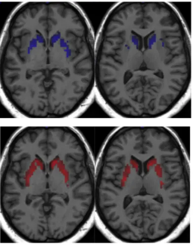
L’étape suivante fut celle du choix de la méthode de segmentation de ces ROIs. Une segmentation manuelle a rapidement été exclue en raison de sa complexité de réalisation et du grand nombre de sujets à analyser, au profit d’une segmentation automatique. La première série d’analyses que nous avons réalisée s’est faite avec le logiciel AAL, s’appuyant sur l’atlas anatomique de N. Tzourio-Mazoyer. L’étude des cartes de segmentation obtenues nous a permis de constater une segmentation imparfaite de certains noyaux gris centraux, notamment sur les parties les plus postérieures et inférieures des putamens et des noyaux caudés (figure 1). Nous avons également constaté de nombreuses données de perfusion aberrantes en raison d’une segmentation approximative au niveau des interfaces substance grise-liquide cérébro-spinal.
Ainsi, devant la segmentation peu satisfaisante du striatum, pourtant au centre de notre hypothèse de recherche de par son appartenance aux circuits de la motivation, et devant la fréquence élevée de données aberrantes, nous avons fait le choix de renouveler nos analyses avec le logiciel Freesurfer (5) dont la segmentation respectait davantage l’anatomie de nos régions d’intérêt.
La relecture attentive des cartes de perfusion des patients conservant des données de perfusion aberrantes pour plusieurs de leurs ROIs a permis de mettre en évidence des régions très artéfactées. Après comparaison de ces cartes de perfusion avec leurs séquences anatomiques en T1 3D, il s’est avéré que ces régions correspondaient à une perte de données secondaire à des artefacts d’origine dentaire (figure 2). Après discussion avec des experts de ces séquences, nous avons posé l’hypothèse que la présence de matériel dentaire pouvait perturber le marquage par onde radio-fréquence au niveau de la carotide interne, étape indispensable à la technique d’ASL. L’importance de cette perte de données était telle que nous avons dû exclure les patients concernés (figure 3).
L’article issu de ce travail est présenté ci-dessous.
1. Introduction
Apathy is now well recognized as an important behavioral syndrome in several neuropsychiatric disorders. Marin described apathy as a deficit of motivation, based on the concept of reduced goal-directed behavior associated to cognitive impairment or emotional distress (1).
Recently, Robert et al. (2) proposed revised criteria for apathy, used in Alzheimer’s disease and other neuropsychiatric disorders. In them, apathy is defined as a disorder of motivation (with reduced goal-directed behavior, reduced goal-directed cognitive activity and blunting of emotions) that persists at least four weeks (2).
There is presently wide acknowledgement that apathy is a transnosographic behavioral syndrome, found in various neurological disorders, such as Alzheimer’s disease, Parkinson’s disease and other dementias, traumatic brain injury and stroke, but also in psychiatric disorders including schizophrenia and major depressive disorder. Apathy is an important dimension to take account because of its consequences on the prognosis of patients. A poor functional prognosis is associated to apathy, with impairment of activities of daily living (6) and instrumental activities of daily living (7), as well as a cognitive impairment with executive dysfunction (7) and increased risk of conversion to dementia (8). Decreased response to treatment (4) and diminished quality of life (9) are also correlated with apathy.
Depression, as neuropsychiatric disorder, may meet the same characteristics and consequences of apathy for morbidity, but little is known about it. The prevalence range of apathy in depressed patients is large across studies, from 32 % to 94 % according to methods used and populations studied (10). Batail et al. (3) studied the clinical profile of apathetic depressed patients. From a cohort of 70 depressed patients, they have shown that apathy affected 30 % of them, which were characterized by a lower intensity of depression (W = 672, p = 0,044), were less anxious (W = 739, p = 0.004) and less anhedonic (W = 412, p = 0,004). The authors have then hypothesized that apathetic depressed patients, although they had motivation deficit, had intact capacity to feel pleasure. This suggests the existence of two subtypes of depression with very different clinical profiles, in terms of their emotional and motivational dimensions. Depressed patients suffering from apathy will probably not share the same neural deficits than non-apathetic ones and consequently can be seen as two different diseases.
The literature, in the field of addictology, highlights this hypothesis by proposing a distinction between deficits in the hedonic response to rewards or consummatory anhedonia « liking », associated with alteration in the subcortical network mediated by opioid system, and a diminished motivation to pursue them or motivational anhedonia « wanting », (11,12)
underlined by a dysfunction in the the meso-cortico-limbic dopamine system (11,13), which underlies goal-directed behavior (13).
Regarding apathy in Parkinson’s disease, SPECT studies are contradictory, demonstrating an inverse correlation between apathy and cerebral metabolism in the striatum, cerebellum, and prefrontal, temporal, parietal and limbic lobes or a positive correlation between apathy and prefrontal, temporal, parietal and limbic areas (14). In functional imaging, a decreased connectivity between the left striatal and frontal areas is noted (15), while T1 weighted imaging reported increased atrophy in the frontal and parietal lobes, insula and left nucleus accumbens in apathetic patients (14). In Alzheimer’s disease, most of studies about apathy involve the anterior cingulate cortex, the medial frontal cortex and some of them also involve the orbitofrontal cortex (16).
So, apathy seems to be related to a dysfunction in a large meso-cortico-limbic circuit including striatum, anterior cingulate, and inferior prefrontal gyrus (4,14,16–18). But to our knowledge, all these previous studies about pathophysiological mechanisms of apathy were performed on patients suffering from neurodegenerative diseases. Regarding apathy in major depressive disorder, no imaging data is available. Identifying perfusional specificities of apathy in depression would provide a better understanding of neurobiological mechanisms that support it to better adjust treatment.
In this study, we hypothesize that apathetic depressed patients will have different cerebral perfusion patterns than non-apathetic ones, particularly affecting key regions involved in emotional or reward processing. We aim to assess the neuroimaging correlates of apathy in depression, by arterial spin labeling (ASL). Indeed, ASL is a growing technique that is non-invasive (endogenous tracer), accurate and reproducible comparing to other perfusion imaging modalities, which allows an absolute quantification of cerebral blood flow (19).
2. Methods
2.1. Patient population
Patients included suffered from a MDE under DSM IV-TR criterion. Inclusion criteria were a MADRS score ≥ 16, with or without personal history of Mood Depressive Disorder (unipolar or bipolar subtype). Exclusion criteria included other Axis I disorders (except anxious comorbidities such as Post Traumatic Stress Disorder, Social Phobia, Generalized Anxiety Disorder, Panic Disorder) which were explored using the Mini-International Neuropsychiatric Interview (M.I.N.I.) (20). Patients with severe chronic physical illness were not included. Other exclusion criteria were potential safety contraindications for MRI (pacemakers, metal implants, pregnancy, and lactation), neurological problems or a history of significant head injury, and significant circulatory conditions that could affect cerebral circulation (i.e. non controlled hypertension).
2.2. Study design
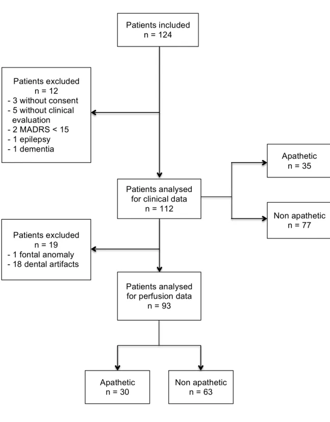
A prospective open cohort study was conducted. Depressed patients were recruited from the adult psychiatry department of Rennes, France. A complete description of the study had been given to the subjects, their written informed consent was obtained. The study was approved by an ethic committee and is registered in www.clinicaltrial.gov (NCT02286024). When recruited, patients underwent a structured clinical interview. After clinical assessment, patients had imaging protocol by a maximum of three days. Clinical data were anonymously retrieved in a notebook. Imaging data followed two different pathways, 1/ a routine care one, as usual and 2/ a research one where they were anonymously stored in an imaging data base (www.shanoir.org). All patients were recruited between November 2014 and February 2017. Initially 112 patients were included. Only 93 were analyzed in the imaging part of the study. See flow chart for more informations (Fig.1)
2.2.1. Clinical assessment
Patients were assessed by a single interview by a trained psychiatrist with following scales :
- Mini-International Neuropsychiatric Interview (M.I.N.I.) (20) - Apathy Evaluation Scale (AES) (21)
- Echelle de Ralentissement Dépressif de Widlocher (ERD) (23) - Montgomery and Äsberg Depression Rating Scale (MADRS) (24) - State Trait Anxiety Inventory (STAI) (25)
- Snaith Hamilton Pleasure Scale (SHAPS) (26)
Socio-demographic (age, gender, education) and disease characteristics (diagnosis, duration of disease, duration of episode, number of mood depressive episodes, antecedent of suicidal attempts/ eclectroconvulsive therapy/ transcranial magnetic stimulation, treatment resistance stage according to Thase and Rush’s classification) were retrieved.
2.2.2. Imaging protocol
2.2.2.1. Data acquisition
Patients were scanned on a 3T whole body Siemens MR scanner (Magnetom Verio, Siemens Healthcare, Erlangen, Germany) with a 32-channel head coil. Anatomical data included a high resolution 3D T1-weighted MPRAGE sequence (3D T1w) with the following imaging parameters : TR/TE/TI = 1900/2.26/900 ms, 256x256 mm2 FOV and 176 sagittal
slices, 1x1x1 mm3 resolution, parallel imaging GRAPPA2. Perfusion data were acquired using a pseudo-continuous ASL sequence with total scan time of approximately 4 minutes (27). The imaging parameters were : TR/TE = 4000/12 ms, flip angle 90°, matrix size 64x 64, labeling duration (LD)/post-labeling delay (PLD) = 1500/1500 ms, parallel imaging SENSE2. The labeling plane was placed 3 cm below the acquisition volume. Twenty axial slices were acquired sequentially from inferior to superior in the AC-PC plane, with 3,5 x 3,5 mm2 in-plane resolution, 5 mm slice thickness and 1 mm gap. Thirty repetitions, i.e., label/control pairs, (60 volumes), finally composed the ASL data series. Additionally, M0 images were acquired at the same dimension and resolution as equilibrium magnetization maps (TR = 10s, TE = 12ms).
2.2.2.2. Data pre-processing
Pre-processing of the image data was performed with an inhouse pipeline using MATLAB (v. R2014a, The MathWorks Inc.) and SPM8 toolbox (Wellcome Department of Imaging Neuroscience at University College London, UK) as follows :
The anatomical 3D T1w was corrected for intensity inhomogeneity and segmented into grey matter (GM), white matter (WM) and cerebro-spinal fluid (CSF) probability maps using the MNI ICBM152 template tissue probability as an a priori for brain tissue classification.
Estimated by this same unified segmentation model SPM routine, spatial normalisation parameters were applied to warp the 3D T1w volume to the MNI JCBM152 template.
The ASL data series was motion corrected by a rigid body transform minimising the sum of squared differences cost function. A two-pass procedure first realigned all the control and label volumes onto the first volume of the series, then registered the series to the mean of the images aligned in the first pass. The motion-corrected ASL series was co-registered to the 3D T1w using a rigid transform. The latter was estimated by maximizing normalised mutual information between the mean control image, i.e., the average of all the realigned control volumes, and the 3D T1w GM map. The co-registered ASL images were then pairwise subtracted (control - label images) to produce a series of perfusion weighted maps, which could be subsequently averaged by arithmetic sample mean to produce a perfusion weighted (PW) map. However, as the sample mean is very sensitive to outliers, we instead use Huber’s M-estimator to robustly estimate the PW map. This robust PW map was eventually quantified to a CBF map by applying the standard kinetic model (28,29) :
𝑓 = 6000. 𝜆Δ𝑀 e !"#!!"#!"∗TI!" T!! 2 𝛼 TI!𝑏 1 − e !! T!! . 𝑀0
where 𝑓 is the CBF map, Δ𝑀 is the perfusion weighted map, 𝜆 = 0.9 ml.g-1 is the
blood/tissue water partition coefficient, 𝛼 = 0.85 measures the labeling efficiency, T1b = 1650
ms is the T1 of blood (30), 𝑀! represents the equilibrium magnetization of arterial blood, PLD = 1500 ms is the post-lablling delay of the ASL sequence, τ = 1500 ms is the labeling duration, 𝑖𝑑𝑥!" is the slice index, starting from 0 for the first acquired slice, TI!" = 37 ms is the acquisition duration of one slice.
For subsequent region of interest (ROI) analysis, the mean CBF values were computed for each subject in the MNI space. The ROIs were selected in light of the literature about apathy and neuroimaging. They were : thalamus, caudate, putamen, pallidum, amygdala, insula, accumbens area, posterior cingulate cortex, anterior cingulate cortex, orbitofrontal cortex, frontal cortex. These ROIs were extracted from the Desikan-Killiany Atlas (5) using the freesurfer software (http://surfer.nmr.mgh.harvard.edu/fswiki/). To discard outliers that may be present in particular at GM/LCS interface due to the low resolution of ASL, we used the modified z score Z proposed by Iglewicz and Hoaglin (31) before computing the average CBF value in a given ROI :
Z
The median absolute deviation is defined as MAD = median (|𝑥i - x)|) where denotes the
median of the data and 𝑥i the absolute value of 𝑥.
2.3. Statistical analyses
Statistical analyses were performed on all included and assessed patients (intention-to-treat analysis) with R software (http://www.R-project.org/). All results are reported as means ± SD for continuous variables and rate for discrete variables. The significance threshold for all tests was set at 5% (p < 0.05).
2.3.1 Whole group analyses
A descriptive analysis of socio-demographic and clinical characteristics was carried out for the whole group using a chi-squared test for qualitative variables and a t-test for quantitative variables.
2.3.2 Intergroup comparisons
2.3.2.1 Clinical data
The whole sample was divided into two groups using a 42 – AES cut-off score, as suggested by Marin et al. (21,32) (AES ≥ 42 = apathetic group ; AES < 42 = non-apathetic group). With this cut-off, 35 patients were apathetic, and 77 non-apathetic. Socio-demographic and clinical variables where then compared. Quantitative variables were compared using a t-test and qualitative variables were compared using a chi-squared test.
2.3.2.2 Perfusion data
The whole sample was divided into two groups, always with the 42-AES cut-off score (21,32). 93 patients were included into perfusion analysis, 30 were apathetic (32.26 %) and 63 were non apathetic. A t-test was applied for these intergroup comparisons.
3. Results
A total of 112 patients were included in the study. The prevalence of apathy was 31,6 % in our sample of depressed patients.
3.1 Imaging results
A sub total of 93 patients were included in perfusion data analysis. We found significant differences of perfusion of putamen, caudate and accumbens nucleus, middle and superior frontal cortex between apathetic and non-apathetic patients. These results are summarized in table 3 and images are presented in figure 4 and figure 5.
We didn’t find any significant differences of perfusion of thalamus, amygdala, insula, anterior cingulate cortex, posterior cingulate cortex, inferior frontal cortex and orbito-frontal cortex between apathetic and non-apathetic patients.
3.2 Clinical results
3.1.1 Whole group analyses
Socio-demographical and clinical characteristics of whole sample are summarized in table 1. The sample was predominantly represented by women, middle-aged and suffering from moderate depression.
3.1.2 Intergroup comparisons
Compared to non-apathetic patients, apathetic patients were significantly less anhedonic. Results of intergroup comparisons are developed in table 2. Both groups were comparable according to socio-demographic and disease characteristics.
4. Discussion
4.1 General considerations
To our knowledge, this is the first study that aimed to compare apathetic and non-apathetic depressed patients without any neurological disorder or dementia on neuroimaging point of view. The most striking results were the hyperperfusion of key regions involved in motivational network.
4.2 Clinical discussion
We found a 31,6 % prevalence of apathy in our sample. This result is in accordance with the literature as presented by Starkstein et al. (33) who found a prevalence of 36,8 % in a population of 95 depressed non-demented patients. In addition, patients had comparable socio-demographic and disease characteristics.
We found that apathetic patients had significant lower SHAPS scores which means that they were less anhedonic and that they had intact capacity to feel pleasure. This result confirms the findings of Batail et al. (9).
Furthermore, apathetic depressed patients had a lower psychomotor retardation on ERD scale even if this result was almost close to significance. We can hypothesize than apathetic patients had difficulties to initiate the movement but not to perform it (34).
4.3 Perfusion patterns
Apathetic depressed patients had a higher cerebral perfusion in the dorsal striatum (composed of the caudate nucleus and the putamen) which is involved in motor and cognitive aspects of motivation (35). The putamen seems to be particularly involved in the control of many types of motor skills such as motor learning, motor preparation and performance, control of amplitude of movement (36). The study of Turner et al. (37) has investigated movement extent using PET mapping of regional cerebral blood flow in 13 healthy subjects. Statistical tests were done to calculate the extent of movements and which regions of the brain the movements correlate to. It was found that increasing movement extent was associated with parallel increases of rCBF in putamen and globus pallidus. Taniwaki et al. (38) has completed these results by showing with fMRI that the signal intensity of the right posterior putamen increased in parallel with the movement speed during a self-initiated task. In our study, apathetic depressed patients seemed to be less slow with a
higher perfusion of putamen than non apathetic ones. Taken together, literature results and our findings suggest that putamen could be involved in behavioral side of apathy.
Apathetic patients also presented a hyperperfusion in the accumbens nucleus. This nucleus, contained within the ventral striatum, is a hub for information related to reward, motivation and decision-making. Its role is described to be more emotional than the dorsal striatum, with hedonic evaluation of stimuli (39). The nucleus accumbens is active during the experience of a pleasant taste, a pleasant image, social acceptance and inclusion, and several addictive drugs (40). Once activated, the nucleus accumbens translates the experience of reward into motivational force, approach behavior and exertion of physical effort (41). It is also involved into emotional anticipation of reward (39). The higher perfusion of the nucleus accumbens found in apathetic patients seems to reflect a preservation of hedonic aspects of reward, in accordance with the fact that they are less anhedonic. In the same way, Arronda et al. (42) found that reduced ventral striatal activity in fMRI was related to greater anhedonia and overall depressive symptoms in the schizophrenic patients versus healthy and depressed patients.
Finally, we have found a significant hyperperfusion of the left middle and the bilateral superior frontal cortex for apathetic depressed patients. The middle frontal gyrus is frequently involved into major depression disorder as shown by Vasic et al. (43) finding a positive correlation between depressive symptoms and rCBF for right middle frontal cortical blood flow of 43 depressed patients versus 29 controls. Nishi et al. (26) reported that the local CBF in the left superior and right middle frontal gyrus in depressed patients at the resting state was positively correlated with cognitive impairment factor scores. These findings suggest that, among prefrontal regions, the middle frontal gyrus may be a key responsible functional region in patients with MDD. Our results are also in accordance with those of Robert et al. (44) finding a positive correlation between the AES score and cerebral metabolism in the right middle frontal gyrus in a PET study in patients with Parkinson disease. Considering this results, we can hypothezise a hyperperfusion of cognitive regions to compensate for hypofunction of the reward system.
Most of studies report a fronto-striatal hypofunction as consequence of apathy (18), so we were expected to find a hypoperfusion of basal ganglia and frontal areas. However, all our results were in favor of a hyperperfusion for apathetic depressed patients. These findings are weightened by the absence of control group, which is the main limitation of our study. A group of healthy patients would have helped us to determine the baseline perfusion of regions analysed. The second limitation is the absence of adjustment for multiple
comparisons such as the Bonferroni method because of too many ROIs regarding the size of our sample. One solution would be a whole brain analysis, which will probably be done in a second time.
In conclusion, we have shown that apathetic depressed patients had a different pattern of cerebral perfusion compared to non-apathetic patients, involving the motivation and reward circuitry, interesting the three dimensions of apathy :
- behavioural with perfusion abnormalities into the dorsal striatum - emotional with perfusion abnormalities into the ventral striatum - cognitive with perfusion abnormalities into the frontal cortex
Then, we have shown that the clinical dichotomy between apathetic depressed patients and non-apathetic depressed patients is underlied by perfusional differences. These clinical and imaging results are in accordance with the recent literature, in the field of addictology, which propose a distinction between deficits in the hedonic response to rewards or consummatory anhedonia “liking” (which seems to concern non apathetic depressed patients), and a diminished motivation to pursue them or motivational anhedonia “wanting” (which seems to concern apathetic depressed patients) (11,12). “Liking” is linked to subcortical (ventromedial prefrontal cortex and amygdala) networks mediated by the opioid system, whereas “wanting” is linked to the meso-cortico-limbic dopamine system (11,13).
5. Conclusion
This study focused on perfusional patterns of apathetic depressed patients. We have shown that perfusional profiles of apathetic depressed patients and non-apathetic ones differ. This study suggests the existence of abnormal perfusional characteristics in apathetic depressed patients. Interestingly, these abnormalities affect key regions involved in meso-cortico-limbic dopaminergic loop of reward system : the dorsal striatum, the ventral striatum and the frontal cortex. Each region seems underlying some key functions represented by respectively behavioral, emotional and cognitive dimensions of apathy. Regarding to clinical results, we have shown that apathy is associated to a preservation of hedonic aspects.
Apathy seems to be a useful biomarker to characterize depression, with abnormalities of the motivational network for apathetic patients instead of abnormalities of the emotional network for more anhedonic patients.
Identifying these radiological and clinical specificities of apathy in depression would provide a better understanding of neurobiological mechanisms that support it to better adjust treatment. Numerous therapeutic issues may be studied such as dopaminergic targeted pharmacologic strategies or new cerebral targets for non-pharmacological treatments like TMS or neurofeedback (45).
Références
1. Marin RS. Apathy: a neuropsychiatric syndrome. J Neuropsychiatry Clin Neurosci. 1991;3(3):243‑54.
2. Robert P, Onyike CU, Leentjens AFG, Dujardin K, Aalten P, Starkstein S, et al. Proposed diagnostic criteria for apathy in Alzheimer’s disease and other neuropsychiatric disorders. Eur Psychiatry. mars 2009;24(2):98‑104.
3. Batail JM, Palaric J, Gadoullet J, Verin M, Robert G, Drapier D. Apathy and depression : which clinical specificities ? Submitted.
4. Kos C, van Tol M-J, Marsman J-BC, Knegtering H, Aleman A. Neural correlates of apathy in patients with neurodegenerative disorders, acquired brain injury, and psychiatric disorders. Neurosci Biobehav Rev. oct 2016;69:381‑401.
5. Desikan RS, S?gonne F, Fischl B, Quinn BT, Dickerson BC, Blacker D, et al. An automated labeling system for subdividing the human cerebral cortex on MRI scans into gyral based regions of interest. NeuroImage. juill 2006;31(3):968‑80.
6. Freels S, Cohen D, Eisdorfer C, Paveza G, Gorelick P, Luchins DJ, et al. Functional status and clinical findings in patients with Alzheimer’s disease. J Gerontol. nov 1992;47(6):M177-182.
7. Zahodne LB, Tremont G. Unique effects of apathy and depression signs on cognition and function in amnestic mild cognitive impairment. Int J Geriatr Psychiatry. janv 2013;28(1):50‑6.
8. Vicini Chilovi B, Conti M, Zanetti M, Mazzù I, Rozzini L, Padovani A. Differential impact of apathy and depression in the development of dementia in mild cognitive impairment patients. Dement Geriatr Cogn Disord. 2009;27(4):390‑8.
9. Prakash KM, Nadkarni NV, Lye W-K, Yong M-H, Tan E-K. The impact of non-motor symptoms on the quality of life of Parkinson’s disease patients: a longitudinal study. Eur J Neurol. mai 2016;23(5):854‑60.
10. Marin RS, Firinciogullari S, Biedrzycki RC. The sources of convergence between measures of apathy and depression. J Affect Disord. 1993;28(2):117–124.
« liking » in humans. Neurosci Biobehav Rev. oct 2015;57:350‑64.
12. Treadway MT, Zald DH. Reconsidering anhedonia in depression: lessons from translational neuroscience. Neurosci Biobehav Rev. janv 2011;35(3):537‑55.
13. Berridge KC, Robinson TE, Aldridge JW. Dissecting components of reward: « liking », « wanting », and learning. Curr Opin Pharmacol. févr 2009;9(1):65‑73.
14. Wen M ‐C., Chan LL, Tan LCS, Tan EK. Depression, anxiety, and apathy in Parkinson’s disease: insights from neuroimaging studies. Eur J Neurol. juin 2016;23(6):1001‑19.
15. Baggio HC, Segura B, Garrido-Millan JL, Marti M-J, Compta Y, Valldeoriola F, et al. Resting-state frontostriatal functional connectivity in Parkinson’s disease-related apathy. Mov Disord Off J Mov Disord Soc. 15 avr 2015;30(5):671‑9.
16. Theleritis C, Politis A, Siarkos K, Lyketsos CG. A review of neuroimaging findings of apathy in Alzheimer’s Disease. Int Psychogeriatr IPA. févr 2014;26(2):195‑207.
17. Benoit M, Robert PH. Imaging correlates of apathy and depression in Parkinson’s disease. J Neurol Sci. 15 nov 2011;310(1‑2):58‑60.
18. Kostić VS, Filippi M. Neuroanatomical correlates of depression and apathy in Parkinson’s disease: Magnetic resonance imaging studies. J Neurol Sci. nov 2011;310(1‑2):61‑3.
19. Wintermark M, Sesay M, Barbier E, Borbély K, Dillon WP, Eastwood JD, et al. Comparative overview of brain perfusion imaging techniques. Stroke. sept 2005;36(9):e83-99.
20. Sheehan DV, Lecrubier Y, Sheehan KH, Amorim P, Janavs J, Weiller E, et al. The Mini-International Neuropsychiatric Interview (M.I.N.I.): the development and validation of a structured diagnostic psychiatric interview for DSM-IV and ICD-10. J Clin Psychiatry. 1998;59 Suppl 20:22-33-57.
21. Marin RS, Biedrzycki RC, Firinciogullari S. Reliability and validity of the Apathy Evaluation Scale. Psychiatry Res. août 1991;38(2):143‑62.
22. Beck AT, Ward CH, Mendelson M, Mock J, Erbaugh J. An Inventory for Measuring Depression. Arch Gen Psychiatry. 1 juin 1961;4(6):561‑71.
aspects. Psychiatr Clin North Am. mars 1983;6(1):27‑40.
24. Montgomery SA, Asberg M. A new depression scale designed to be sensitive to change. Br J Psychiatry J Ment Sci. avr 1979;134:382‑9.
25. Spielberger, C. D., Gorsuch, R. L., Lushene, R., Vagg, P. R., Jacobs, G. A. Manual for the State-Trait Anxiety Inventory. Palo Alto CA. 1983;
26. Snaith RP, Hamilton M, Morley S, Humayan A, Hargreaves D, Trigwell P. A scale for the assessment of hedonic tone the Snaith-Hamilton Pleasure Scale. Br J Psychiatry J Ment Sci. juill 1995;167(1):99‑103.
27. Wu W-C, Fernández-Seara M, Detre JA, Wehrli FW, Wang J. A theoretical and experimental investigation of the tagging efficiency of pseudocontinuous arterial spin labeling. Magn Reson Med. nov 2007;58(5):1020‑7.
28. Buxton RB, Frank LR, Wong EC, Siewert B, Warach S, Edelman RR. A general kinetic model for quantitative perfusion imaging with arterial spin labeling. Magn Reson Med. sept 1998;40(3):383‑96.
29. Cavuşoğlu M, Pfeuffer J, Uğurbil K, Uludağ K. Comparison of pulsed arterial spin labeling encoding schemes and absolute perfusion quantification. Magn Reson Imaging. oct 2009;27(8):1039‑45.
30. Alsop DC, Detre JA, Golay X, Günther M, Hendrikse J, Hernandez-Garcia L, et al. Recommended implementation of arterial spin-labeled perfusion MRI for clinical applications: A consensus of the ISMRM perfusion study group and the European consortium for ASL in dementia. Magn Reson Med. janv 2015;73(1):102‑16.
31. Iglewicz B, Hoaglin DC. How to Detect and Handle Outliers. ASQC Quality Press; 1993. 108 p.
32. Calabrese JR, Fava M, Garibaldi G, Grunze H, Krystal AD, Laughren T, et al. Methodological approaches and magnitude of the clinical unmet need associated with amotivation in mood disorders. J Affect Disord. oct 2014;168:439‑51.
33. Starkstein SE, Petracca G, Chemerinski E, Kremer J. Syndromic Validity of Apathy in Alzheimer’s Disease. Am J Psychiatry. 1 juin 2001;158(6):872‑7.
34. Massimo L, Evans LK, Grossman M. Differentiating Subtypes of Apathy to Improve Person-Centered Care in Frontotemporal Degeneration. J Gerontol Nurs. oct 2014;40(10):58‑65.
35. Liljeholm M, O’Doherty JP. Contributions of the striatum to learning, motivation, and performance: an associative account. Trends Cogn Sci. sept 2012;16(9):467‑75.
36. Marchand WR, Lee JN, Thatcher JW, Hsu EW, Rashkin E, Suchy Y, et al. Putamen coactivation during motor task execution. Neuroreport. 11 juin 2008;19(9):957‑60.
37. Turner RS, Desmurget M, Grethe J, Crutcher MD, Grafton ST. Motor Subcircuits Mediating the Control of Movement Extent and Speed. J Neurophysiol. 1 déc 2003;90(6):3958‑66.
38. Taniwaki T, Okayama A, Yoshiura T, Nakamura Y, Goto Y, Kira J, et al. Reappraisal of the motor role of basal ganglia: a functional magnetic resonance image study. J Neurosci Off J Soc Neurosci. 15 avr 2003;23(8):3432‑8.
39. Reeve J. Understanding Motivation and Emotion. John Wiley & Sons; 2014. 648 p.
40. Berridge KC, Robinson TE. What is the role of dopamine in reward: hedonic impact, reward learning, or incentive salience? Brain Res Brain Res Rev. déc 1998;28(3):309‑69.
41. Pessiglione M, Schmidt L, Draganski B, Kalisch R, Lau H, Dolan RJ, et al. How the brain translates money into force: a neuroimaging study of subliminal motivation. Science. 11 mai 2007;316(5826):904‑6.
42. Arrondo G, Segarra N, Metastasio A, Ziauddeen H, Spencer J, Reinders NR, et al. Reduction in ventral striatal activity when anticipating a reward in depression and schizophrenia: a replicated cross-diagnostic finding. Front Psychol [Internet]. 26 août 2015;6. Disponible sur: http://www.ncbi.nlm.nih.gov/pmc/articles/PMC4549553/
43. Vasic N, Wolf ND, Grön G, Sosic-Vasic Z, Connemann BJ, Sambataro F, et al. Baseline brain perfusion and brain structure in patients with major depression: a multimodal magnetic resonance imaging study. J Psychiatry Neurosci JPN. nov 2015;40(6):412‑21.
44. Robert G, Le Jeune F, Lozachmeur C, Drapier S, Dondaine T, Peron J, et al. Apathy in patients with Parkinson disease without dementia or depression: A PET study. Neurology. 11 sept 2012;79(11):1155‑60.
45. Arns M, Batail J-M, Bioulac S, Congedo M, Daudet C, Drapier D, et al. Neurofeedback: One of today’s techniques in psychiatry? L’Encephale. avr 2017;43(2):135‑45.
List of tables
Variable n=112 Mean (± sd) or n (%) Range
Age (years) 49,29 (15,10) (18 - 76)
Gender (female) 75 (65,79) -
Education (year) 12,38 (3,03) (4 - 22)
Duration of disease (years) 15,72 (14,05) (0 - 57)
Duration of episode (weeks) 31,40 (44,27) (0 - 288)
Number of MDE 4,39 (4,52) (0 - 30)
Number of suicidal attempt 1,12 (2,5) (0 - 10)
AES (total score) 39,08 (7,67) (24 - 62)
MADRS (total score) 27,76 (5,36) (16 - 42)
BDI (total score) 17,87 (7,9) (0 - 38)
ERD (total score) 22,32 (8,82) (2 - 43)
STAI YA (total score) 58,63 (12,31) (28 - 80)
STAI YB (total score) 58,95 (11,65) (30 - 79)
YMRS (total score) 1,62 (2,00) (0 - 12)
SHAPS (total score) 5,63 (4,00) (0 - 14)
Table 1 – Population description
Variable n=93 Apathetic (n=30 ) Mean (+/- SD) Non apathetic (n=63 ) Mean (+/- SD) p-value Accumbens left 38.60 (12.88) 32.48 (12.55) 0,035 Accumbens right 39.61 (14.85) 32.75 (11.47) 0,023 Caudate left 34.19 (11.22) 27.47 (10.57) 0,014
Frontal middle left gyrus 48.86 (16.40) 40.34 (12.54) 0,022
Frontal superior left gyrus 43.08 (14.27) 36.70 (11.17) 0,040
Frontal superior right gyrus 43.05 (13.59) 37.16 (10.77) 0,041
Putamen left 40.32 (9.83) 35.45 (9.89) 0,030
Table 2 – Intergroup comparisons : significant perfusion results
Table 3 – Intergroup comparisons : clinical results
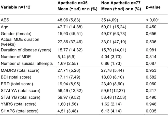
Variable n=112 Apathetic n=35 Mean (± sd) or n (%) Non Apathetic n=77 Mean (± sd) or n (%) p-value
AES 48.06 (5,83) 35 (4,09) < 0,001
Age 47,71 (14,88) 50,01 (15,24) 0,450
Gender (female) 15,93 (45,51) 49,07 (63,73) 0,656
Actual MDE duration
(weeks) 27,86 (37,46) 33,01 (47,19) 0,536
Duration of disease (years) 15,77 (14,32) 15,70 (14,01) 0,981
Number of MDE 5,14 (5,9) 4,04 (3,73) 0,314
Number of suicidal attempts 1,69 (2,55) 0,86 (1,73) 0,087
MADRS (total score) 27,71 (5,26) 27,78 (5,44) 0,953
BDI (total score) 17,11 (7,49) 18,00 (8,10) 0,582
ERD (total score) 19,94 (8,95) 23,40 (8,60) 0,060
STAI YA (total score) 56,49 (12,32) 59,61(12,27) 0,217
STAI YB (total score) 59,97 (9,52) 58,48 (12,53) 0,490
YMRS (total score) 1,60 (1,56) 1,62 (2,14) 0,948
List of figures
Figure 1 – Flow chart
Patients included n = 124
Patients analysed for clinical data
n = 112 Patients excluded n = 12 - 3 without consent - 5 without clinical evaluation - 2 MADRS < 15 - 1 epilepsy - 1 dementia Patients excluded n = 19 - 1 fontal anomaly - 18 dental artifacts Patients analysed for perfusion data
n = 93 Apathetic n = 35 Apathetic n = 30 Non apathetic n = 77 Non apathetic n = 63
Figure 2 – Segmentation of the dorsal striatum with AAL (blue), segmentation of the dorsal striatum with Freesurfer (red)
Figure 4 – Significant hyperperfusion for apathetic depressed patients. Green : left putamen, blue : left caudate, red : bilateral accumbens nucleus
Figure 5 – Significant hyperperfusion for apathetic depressed patients. Blue : left middle frontal gyrus, red : bilateral superior frontal gyrus
CONAN Camille - Apathie dans la dépression : une étude en arterial spin labeling
44 feuilles, 5 figures, 3 tableaux, 30 cm.- Thèse : Médecine ; Rennes 1; 2017 ; N°
Abstract :Introduction - Apathy is defined as a lack of goal-directed behavior. Although it is observed in about 30 % of depressed patients, neurovascular mechanisms underpinning apathy, especially in those patients, remain little-known.
The main objective of this study was to compare the cerebral perfusion of apathetic depressed patients with non-apathetic depressed patients by arterial spin labeling (ASL). ASL is a quantitative and non-invasive perfusion magnetic resonance imaging (MRI) technique that uses blood as an endogenous tracer to quantify tissue blood flow. The secondary objective vas to study the clinical profile of apathetic depressed patients. Methods - This study was conducted from a cohort of depressed patients meeting the DSM IV TR criteria, between November 2014 and June 2016 in Rennes, France (www.clinicaltrial.gov ; NCT02286024). 112 depressed patients were included, of whom 35 were apathetic (AES ≥ 42), 77 non-apathetic (AES < 42). Everyone got a clinical evaluation with scale screenings especially for apathy (AES), anxiety (STAI) and anhedonia (SHAPS) as well as a cerebral MRI for 93 of them. MRI data included a 3D T1-weighted anatomical sequence (3D-T1w) and a pseudo-continuous ASL sequence. The 3D-T1w was segmented into brain tissues while the ASL series were motion corrected and co-registered onto the 3D-T1w before quantification to CBF maps. Both anatomical and perfusion data were normalized to the MNI-space. An analysis was conducted on regions of interest (ROI) to compare the CBF between apathetic and non-apathetic groups. ROIs were defined with Freesurfer atlas.
Results - Apathetic perfused more than non-apathetic in the left putamen (p = 0.03), the left caudate nuleus (p = 0,014), in the left (p = 0,035) and the right (p = 0,023) accumbens nucleus, in the left middle frontal cortex (p = 0,022), and in the left (p = 0,040) and right superior frontal cortex (p = 0,041). From a clinical point of view, apathetic depressed patients were less anhedonic than non-apathetic ones (p = 0,035).
Conclusions - We have shown that perfusional profiles of apathetic depressed patients and non-apathetic ones differ. This study suggests the existence of abnormalities affecting key regions involved in meso-cortico-limbic dopaminergic loop of reward system : the dorsal striatum, the ventral striatum and the frontal gyrus. Each region seems underlying some key functions represented by respectively behavioral, emotional and cognitive dimensions of apathy.
Apathy seems to be a useful biomarker to characterize different phenotypes of depression, with abnormalities of the motivational network for apathetic patients instead of abnormalities of the emotional network for more anhedonic patients in order to better adjust the treatment of depression.
Rubrique de classement : PSYCHIATRIE
Mots-clés :
ASL, dépression, apathie, motivation, récompense
anhédonie, perfusion, striatum
Mots-clés anglais MeSH :
ASL, depression, apathy, motivation, anhedonia,
liking-wanting, reward, perfusion, striatum
JURY :
Président : Monsieur le Professeur Dominique DRAPIER
Assesseurs : Monsieur le Docteur Jean-Marie BATAIL, directeur de thèse Monsieur le Professeur Jean-Christophe FERRE Monsieur le Docteur Gabriel ROBERT