HAL Id: dumas-01611806
https://dumas.ccsd.cnrs.fr/dumas-01611806
Submitted on 6 Oct 2017
HAL is a multi-disciplinary open access
archive for the deposit and dissemination of sci-entific research documents, whether they are pub-lished or not. The documents may come from teaching and research institutions in France or abroad, or from public or private research centers.
L’archive ouverte pluridisciplinaire HAL, est destinée au dépôt et à la diffusion de documents scientifiques de niveau recherche, publiés ou non, émanant des établissements d’enseignement et de recherche français ou étrangers, des laboratoires publics ou privés.
Évaluer la dose optimale de glucocorticoïdes pour traiter
la néphrite lupique : une collaboration entre Bordeaux
et Bilbao
Cécile Saint-Pastou Terrier
To cite this version:
Cécile Saint-Pastou Terrier. Évaluer la dose optimale de glucocorticoïdes pour traiter la néphrite lupique : une collaboration entre Bordeaux et Bilbao. Médecine humaine et pathologie. 2017. �dumas-01611806�
COLLÈGE SCIENCES DE LA SANTÉ
Année 2017
N°3044
Thèse pour l’obtention du
DIPLOME d’ETAT de DOCTEUR EN MEDECINE
Spécialité : MÉDECINE INTERNE
Présentée et soutenue publiquement
Le 15 Mai 2017
Par Cécile Saint-Pastou Terrier
Née le 2 décembre 1986, à Grenoble (38).
Directrice de thèse :
Madame le Professeur Estibaliz LAZARO,
Rapporteur de thèse :
Monsieur le Docteur Pierre DUFFAU,
Membres du Jury :
Monsieur le Professeur Patrick BLANCO,
Président
Monsieur le Professeur Lionel COUZI,
Juge
Monsieur le Professeur Christophe RICHEZ,
Juge
Monsieur le Professeur Guillermo RUIZ-IRASTORZA,
Juge
Madame le Docteur Amaia UGARTE NUÑEZ ,
Invitée
Evaluer la dose optimale de glucocorticoïdes pour
traiter la néphrite lupique :
Une collaboration entre Bordeaux et Bilbao.
Assess the optimal dose of glucocorticoids to treat lupus nephritis: collaboration between Bordeaux and Bilbao.3
“Guérir parfois, soulager souvent, écouter toujours.” Louis Pasteur
4
Remerciements
AU PRESIDENT DU JURY
Monsieur le Professeur Patrick Blanco,
Professeur des Universités, Praticien Hospitalier,
Chef de service d’immunologie et d’immunogénétique,
Groupe Hospitalier Pellegrin, Centre Hospitalier Universitaire de Bordeaux.
Coordinateur du groupe de Recherche « Mécanismes immunitaires de l’auto-immunité » au centre de recherches Immunology from Concept and Experiments to Translation
(ImmunoConcept) CNRS UMR 5164, Université de Bordeaux.
Vous me faites l’honneur de présider ce jury de thèse. Veuillez trouver ici l’expression de mon profond respect et soyez assuré de ma sincère reconnaissance pour juger ce travail.
A MA DIRECTRICE DE THESE
Madame le Professeur Estibaliz LAZARO,
Professeur des Universités, Praticien Hospitalier,
Service de médecine interne et maladies infectieuses,
Hôpital Haut Lévèque – Groupe Hospitalier Sud, Centre Hospitalier Universitaire de Bordeaux. Chercheur centre de recherches ImmunoConcept CNRS UMR 5164, Université de Bordeaux.
Chère Esti, merci de m’avoir si bien guidée et suivie tout au long de cet épanouissant projet. Je te suis très reconnaissante de m’avoir proposé ce travail et de m’avoir fait confiance. J’espère être à la hauteur de tes attentes. Travailler avec toi est un plaisir et un honneur, qui, j’espère, à l’avenir se reproduira. Merci pour ton écoute, ton soutien, ta disponibilité et ton enthousiasme. Ton exigence et ta rigueur m’ont incitée à approfondir ma réflexion. Apprendre à tes côtés m’a permis de découvrir et de mesurer ton savoir, ton sens clinique et tes qualités humaines. Je suis admirative de la médecine que tu enseignes et que tu pratiques au quotidien ainsi que de la volonté que tu as de mener de si beaux projets à terme. Que cette thèse soit l’expression de ma plus grande estime à ton égard et puisse apporter une pierre à l’édifice de la collaboration entre Bordeaux et Bilbao.
A MON RAPPORTEUR
A Monsieur le Docteur Pierre DUFFAU
Maître de conférences des universités, Praticien Hospitalier,
Service de médecine interne et maladies infectieuses
Hôpital Saint-André, Centre Hospitalier Universitaire de Bordeaux,
Chercheur centre de recherches ImmunoConcept CNRS UMR 5164, Université de Bordeaux
Je vous remercie de l’honneur que vous me faites en prenant le temps de lire et de juger ce travail. Veuillez recevoir ici, l’expression de ma sincère reconnaissance et tout mon respect.
5
AUX MEMBRES DU JURY
A Monsieur le Professeur Lionel COUZI,
Professeur des Universités, Praticien Hospitalier,
Service de Néphrologie – Transplantation – Dialyse - Aphérèses
Groupe Hospitalier Pellegrin, Centre Hospitalier Universitaire de Bordeaux.
Chercheur centre de recherches ImmunoConcept CNRS UMR 5164, Université de Bordeaux.
Vous me faites l’honneur d’avoir accepté de faire partie de ce jury et de juger ce travail. Je vous remercie pour votre accueil chaleureux en néphrologie et votre affabilité. Je vous suis très reconnaissante de votre aide et de vos conseils apportés tout au long de ce travail, si précieux à l’élaboration de cette thèse. Votre expertise et votre disponibilité ont été indispensables à sa réalisation. Veuillez trouver ici l'expression de ma gratitude et de ma reconnaissance.
Al Profesor Guillermo RUIZ-IRASTORZA
Profesor Titular en la U.D. Facultad de Medicina y Odontología en Cruces Dpto, Universidad del País Vasco, Euskal Herriko Unibertsitatea Leio, España.
Doctor en Medicina,
Jefe de sección de Enfermedades Autoinmunes, Hospital Universitario Cruces, Barakaldo, País Vasco, España.
Coordinador del Grupo de investigación “Enfermedades Autoinmunes, Inflamatorias e Infecciosas” al Instituto de Investigación Sanitaria de la OSI Ezkerraldea Enkarterri Cruces (Biocruces), Barakaldo, País Vasco, España.
Gracias por el entusiasmo con el que usted ha aceptado a pesar de la distancia, ser parte de este jurado, para mí es un gran honor de que usted juzgue mi tesis. Gracias por permitirme llevar a cabo este gran proyecto entre nuestras dos ciudades.
Que esta tesis pueda aportar un grano de arena al edificio de la cooperación entre Burdeos y Bilbao. Le quiero expresar mi máximo reconocimiento y mi respeto el más sincero.
A Monsieur le Professeur Christophe RICHEZ
Professeur des Universités, Praticien Hospitalier,
Service de médecine interne et maladies infectieuses
Hôpital Saint-André, Centre Hospitalier Universitaire de Bordeaux.
Chercheur centre de recherches ImmunoConcept CNRS UMR 5164, Université de Bordeaux.
Vous me faites l’honneur d’avoir accepté de lire et de juger cette thèse. Veuillez croire en l’expression de mon profond respect et le témoignage de ma gratitude.
A la Doctora Amaia UGARTE NUÑEZ
Doctor en Medicina,Enfermedades Autoinmunes, Hospital Universitario Cruces, Barakaldo, País Vasco, España.
Investigador en el grupo “Enfermedades Autoinmunes, Inflamatorias e Infecciosas” al Instituto de Investigación Sanitaria de la OSI Ezkerraldea Enkarterri Cruces (Biocruces), Barakaldo, País Vasco, España.
Me hace el honor de leer esta tesis. Le quiero expresar mi agradecimiento por su importante aporte y participación en el desarrollo de esta tesis. Muchas gracias por venir hasta Burdeos para asistir a este momento que cuenta tanto para mí.
6
A MES MAITRES D’INTERNAT
Je remercie également l'ensemble des médecins qui m'ont transmis leur savoir, leur savoir-faire et leur savoir-être au cours de mon internat, tout au long duquel vous m’avez accompagnée et qui m’ont confortée dans mes choix d’exercer une si belle spécialité et de pratiquer une médecine humaniste.
A Messieurs les Professeurs Charles Cazanave, Xavier Combes, Joël Constans, Didier Neau, Jean-Luc
Pellegrin, Julien Seneschal, Alain Taieb et Jean-François Viallard.
A Mesdames les Docteurs Isabelle Degasne, Fatima De Oliveira, Anne Gerber, Carine Greib,
Marie-Christine Jaffar-Bandjee, Marie Lagrange, Brigitte Milpied, Sophie Skopinski, Gaëtane Wirth,
Messieurs les Docteurs Olivier Belmonte, Frederic Dauchy, Dominique Ferrandiz, Hamid Laksir,
Frederic Renou, Frederic Revel et Jean-Luc Yvin.
Au Docteur Marie-Pierre Moiton, tu m’as accompagnée avec bienveillance lors de mes premiers pas d’interne. Merci pour ta gentillesse, ta patience, ton écoute et ton envie de partager tes connaissances. Merci d’avoir cru en moi et de m’avoir poussée dans cette belle voie. Travailler avec toi était un plaisir et j’espère que l’occasion se renouvellera à l’avenir.
Au Docteur Loïc Raffray, merci d’avoir toujours su répondre à mes interrogations, mes doutes, d’avoir été de bons conseils et à l’écoute durant mon internat. Merci pour tout ce que tu fais pour notre formation sur notre belle île, pour ton investissement sans faille, pour faire vivre notre spécialité au beau milieu de l’Océan Indien. Ton savoir, ton parcours, tes qualités humaines forcent l’admiration. J’espère un jour avoir l’honneur de travailler à tes côtés.
A Monsieur le Professeur Philippe Gasque, pour m’accompagner dans cette enrichissante année recherche.
A mes assistants et chefs de clinique qui m’ont tant appris et m’ont poussée à me perfectionner : à
Lucile Boursault, Kelly Bagny, Alexandre Maillet, Elena Morello, Sarah Payet, Lucile Sorin, Nathalie Sultan Bichat, Nicolas Traversier, Camille Valette et Marc-Olivier Vareil.
A MES COLLEGUES INTERNES,
A mes belles rencontres réunionnaises et bordelaises : Alix D., Jeanne P., Jonathan C. Léa F., Thomas
G., Audrey Maillot, Aurélie M., Rodolphe M., Pierre L. et sans oublier la dream team des
cardiologues Florianne B., Rémi C. Mon internat a été parsemé de fous rires, de complicité, de réflexions et discussions enrichissantes grâce à vous. J’espère pouvoir échanger encore avec vous. Merci pour votre amitié.
A Fanny B. : A mon binôme de choc, pour ta douceur et ta gentillesse, pour ton écoute, pour toutes les fois où j’ai pu compter sur toi, pour ton amitié.
A Coraline G. : Coco, pour ta gentillesse, pour toujours être là quand il le faut, pour notre amitié. J’admire ta force de caractère et ta volonté qui me pousse à aller de l’avant.
A Christelle P. : Pour les bon repas partagés, pour les fous rires, pour ton écoute pendant mes moments de doutes, pour notre amitié. Pour ton soutien et tes encouragements dans cette dernière ligne droite.
A Laetitia S. : Probablement ma plus belle rencontre bordelaise, j’ai trouvé en toi bien plus qu’une simple consœur avec qui discuter et débattre mais une amie sincère. Merci pour tes conseils, ton soutien inconditionnel, ton écoute et tes encouragements.
7
AUX EQUIPES PARAMEDICALES,
A l’ensemble des formidables équipes des services de médecine qui m’ont toujours si bien accueillie, qui font un travail conséquent et formidable, et sans qui notre quotidien serait bien plus difficile, plus morose et la prise en charge de nos patients impossible. Une pensée particulière aux secrétaires pour ces courriers toujours trop longs, aux aides soignantes pour les malencontreuses tasses de café oubliées et aux infirmiers pour toutes les prescriptions même les plus curieuses, pour avoir su rester soudés dans les bons comme les mauvais moments et pour m’avoir tant appris sur mon métier.
A MA FAMILLE,
A mes parents,
Pour votre amour, pour vos sacrifices et votre indéfectible soutien tout au long du chemin parcouru. Tous deux avez toujours su faire face aux tracas de l’existence tout en gardant à esprit la nécessité de faire partager à chacun de vos enfants ces qualités de volonté, de détermination, de travail, de motivation, de l’importance de profiter de l’instant présent et de ce que la vie nous offre. En complément de toute l’affection que vous m’avez apportée, ces valeurs m’ont menée jusqu’ici. Vous avez remarquablement gâté et encouragé mes ambitions, et vous n’avez pas hésité à m’aider et à me soutenir coûte que coûte pour me rapprocher de mes rêves. Devenue mère je mesure la chance d’avoir pu m’épanouir auprès de vous. J’espère que vous serez fiers de ce que je suis devenue grâce à vous. Je vous dédie tendrement cette thèse pour la belle vie que vous m’avez offerte.
A mon frère,
Julien, merci d’avoir tracé le chemin devant moi et pour l’intérêt que tu as toujours porté à ce que je réalisais. Merci de m’avoir hébergée sans hésiter pendant mes semaines parisiennes et d’avoir eu le courage de relire ces nombreuses pages. Sans oublier ma belle-sœur, Anne-Laure, mon espiègle nièce Eloïse et mon tout petit et beau neveu Augustin qui te ressemble tant. Ton épanouissement personnel et professionnel m’a remplie de fierté. Nos déceptions d’enfants, nos rires, nos bêtises, nos disputes sont loin déjà mais j’en garde un doux souvenir. J’espère que nos enfants partageront autant d’aventures et d’éclats de rire.
A ma tante Ginette et mon oncle René,
Depuis toute petite, vous avez une place imprenable dans ma vie. Merci à vous d’avoir toujours été à mes côtés, de m’avoir soutenue et pour votre bienveillance. Vous êtes aujourd’hui dans mes pensées.
A mes beaux-parents, ma belle-sœur Anne et à toute ma belle-famille.
Vous m’avez accueillie, soutenue, merci pour vos petites attentions.
A mon mari,
Guilhem, pour ce bonheur partagé si rare. Pour tout ce que tu sais et que je ne te dis peut être pas
assez. Merci pour ta patience à toute épreuve, ton écoute, ton indéfectible soutien, pour ton réconfort dans ces moments de doutes, sans toi ces longues années d’études et cette thèse n’auraient pu être menées jusqu’à leur terme. Je suis bien consciente des sacrifices consentis pour réaliser mes projets professionnels. Les épreuves de la vie nous ont rendus plus forts et ta présence à mes côtés m’est indispensable. Ton amour me permet de m’épanouir chaque jour un peu plus. Et pour ne rien gâcher tu es un papa formidable. Pour toute cette richesse que tu m’apportes et pour m’avoir permis de devenir cette personne profondément heureuse que je suis aujourd’hui. Merci pour tout.
8
A ma fille Mahée,
Ma plus grande fierté, ma force.
Je suis bien consciente que nous avons dû faire des sacrifices et que depuis ton plus jeune âge tu as su les accepter. En supportant mes absences tu as contribué à mener à bien ce travail. Tu es sans conteste mon rayon de soleil. Sache que tu es une des plus belles choses qui me soit arrivée dans la vie et que je n’aurai de cesse de te construire un monde plus beau. Tu m’émerveilles un peu plus chaque jour. Je suis si fière de toi. N’oublie jamais que je t’aime.
A notre enfant, qui sera bientôt parmi nous. Je suis impatiente de ce bonheur à quatre.
A MES AMIS,
Edwige G.,
Un de mes plus grands bonheurs c'est notre amitié partagée et sincère et qui se dessine avec un grand « A ». Le temps passe, la terre s'use, l'amitié des âmes, jamais. Merci pour être toujours là dans les moments qui comptent pour moi, pour avoir pu compter sur toi tant de fois, d’être à l’écoute et de savoir trouver les mots justes. J’espère que la vie nous offrira encore des moments formidables.
Anaëlle M.,
A mon amie d’enfance, varçoise de surcroît, il restera toujours des supers souvenirs de notre adolescence. Peu importe la distance, comme l’a dit ton Papa un jour, même si l’on ne se voit pas pendant longtemps, rien ne change. « Le temps confirme l'amitié. »
A Coralie F., Nicolas G., Timothée L., Gaël R., Emmanuelle T. et Léa V. pour ces années médecine.
Même si nous avons chacun suivi notre chemin, je sais que vous êtes tous promis à un bel avenir.
A Laurie B., Lucie C, Philine G., Fabiola J., Julie P., Marion P. pour nos plus belles années, pour les
fous rires et les larmes, même si nos vies se sont un peu éloignées, je sais que vous avez toutes trouvé votre bonheur.
A Eliane & Serge P.,
Vous m’avez vu grandir depuis toute petite. Merci pour votre bienveillance, vos encouragements, votre gentillesse et votre amitié. Je suis très touchée que nous nous retrouvions pour ce moment si particulier dans cette si belle ville qui compte tant à vos yeux.
A ceux présents en ce jour si singulier, venus parfois de loin pour m’encourager, merci pour votre présence et votre soutien sans faille.
9
Glossary
- ACR: American College of Rheumatology
- AMM: autorisation de mise sur le marché (French marketing authorization for a new drug)
- aPL antiphospholipid
- BC: Bordeaux cohort
- BMD: bone mass density
- C-GR: cytosolic glucocorticoid receptor
- CC: Cruces-protocol cohort
- CCL2-chemokine ligand 2
- CCL7: Chemokine (C-C motif) ligand (MCP3).
- CCL8: Chemokine (C-C motif) ligand 8
- COX : Cyclo-oxygenase
- CR: complete remission
- CVEs: cardiovascular events
- CXCL8: CXC-chemokine ligand 8
- CYC: cyclophosphamide
- DC: dendritic cell
- ERA-EDTA: European renal association -European dialysis and transplant association
- ESRD: End-stage renal disease
- EULAR: European League Against Rheumatism
- GCs: Glucocorticoids
- GM-CSF: Granulocyte-macrophage colony-stimulating factor
- GRE: Glucocorticoid Response Element
- GRs: Glucocorticoid receptors
- HAS : Haute autorité de santé
- HC: Historic cohort
- HCQ: Hydroxychloroquine
- IFN: interferon
- IL-1RA: Interleukin-1 Receptor Antagonist
- IL: Interleukin
- IV: intravenous
- KDIGO: Kidney Disease: Improving Global Outcomes
- LN: lupus nephritis
- MCP1: Monocyte chemoattractant protein 1
- MCP2: monocyte chemoattractant protein 2
- MCP3: monocyte-chemotactic protein 3
- mGCR: membrane-bound glucocorticoid receptor
- MHC: Major histocompatibility complex
- MMF: mycophenolate mofetil
- MP: Methylprednisolone
- NF-kB: nuclear factor-kinase B
- NIH: National Institutes of Health
- NK cell: Natural killer cell
- OP: osteoporosis
- PDC: plasmacytoid dendritic cells
- PNDS: Protocol national de diagnostic et de soins
- SLE: Systemic lupus erythematosus
- SNPs: Single nucleotide polymorphisms
- sVCAM-1: soluble vascular cellular adhesion molecule-1
10
- TLR: Toll like receptor
- TNF: Tumor necrosis factor
- vs: versus
11
Table of contents
Table of contents
Introduction ... 13
I. The optimal dose of Glucocorticoids to treat lupus nephritis... 14
1. The role of steroids in managing systemic lupus erythematosus ... 14
1.1 Mechanisms of action of GCs: anti-inflammatory and immunosuppressive actions ... 14
1.2 Glucocorticoid Receptors (GRs) ... 15
1.3 Mechanisms of action of GCs: the genomic and non-genomic pathways ... 15
2. Glucocorticoids are recommended in the treatment of lupus nephritis ... 19
2.1 Induction therapy ... 19
2.2 Maintenance therapy ... 24
3. Adverse effects of steroids ... 26
3.1 Glucocorticoids & Bone health ... 26
3.2 Glucocorticoids & Cardiovascular Events ... 26
3.3 Glucocorticoids & Infections ... 27
3.4 Glucocorticoids & Morbidity Mortality in SLE ... 27
4. Treat lupus with lower doses or without Glucocorticoids ... 28
4.1 Reduce the doses of GCs ... 28
4.2 The new wave: Treat without oral GCs ... 29
II. Suggested Study: A collaboration between Bordeaux & Bilbao ... 30
1. OBJECTIVE ... 30 2. METHODS ... 30 2.1 Cruces-protocol protocol... 30 2.2 Bordeaux protocol ... 30 2.3 Definition of remission ... 30 3. OUTCOMES ... 31 4. RESULTS ... 31 5. CONCLUSION ... 31 6. SUPPLEMENTARY DATA ... 31
6.1 Details of the treatments received in BC ... 31
III. Manuscript ... 35
Conclusion ... 58
12
Table numbers
Table 1 - Recommendations for Focal proliferative Lupus Nephritis Class III & Diffuse proliferative Lupus nephritis class IV. Page 21 - 22
Table 2 - Recommendations for Lupus Nephritis Class IV or IV/V with cellular crescents. Page 23
Table 3 - Recommendations for Membranous Lupus Nephritis Class V. Page 23
Table 4 - Recommendations for maintenance therapy. Page 25
Table 5 - Immunosuppressant Induction therapy received in BC. Page 32
Table 6 - Glucocorticoids Induction Therapy received in BC. Page 32
Table 7 - Glucocorticoids maintenance therapy received in BC. Page 34
Figures
Figure 1: Mechanisms of action of Glucocorticoids: anti-inflammatory and immunosuppressive actions adapted from EM Sternberg et al (21). Page 15
Figure 2: Mechanisms of action of GCs: Genomic and Non-Genomic pathways adapted from Rhen T et al (18). Page 17
Figure 3: The production of type I IFN by pDCs as a common mechanism of pathogenesis of SLE adapted from Ganguly et al (47). Page 18
13
Introduction
Systemic lupus erythematosus (SLE) is a chronic systemic autoimmune disease with a wide range of clinical manifestations due to immunological alterations. General fatigue, skin rash, fever, and arthritis are common symptoms in SLE, but visceral organ involvement such as heart, lung, nervous system and kidney can result in severe disease status. Among these organ complications, lupus nephritis most strikingly influences the course of SLE. Renal involvement may be the first manifestation of SLE, but most commonly, it occurs within a year of diagnosis and almost always within 5 years, even if it can occur at any time throughout the course of the disease (1). It can affect over half of all patients with SLE depending in particular on ethnic origins (2,3). LN occurred in 38.3% of SLE patients, frequently as the initial presentation, in a recent large multi-ethnic inception cohort (4). A study from northwest England yielded incidence and prevalence of 0.40 per 100,000 population per year and 4.4 per 100,000 population, respectively (5). Ten to thirty percent of patients with lupus nephritis (LN) class III and above (according to the classification of glomerulonephritis in SLE revisited (6)) progress to end-stage renal disease (ESRD) resulting in kidney transplantation or haemodialysis (7–9). Renal damage is a severe SLE manifestation, carries excessive mortality and morbidity and is considered as a predictor of mortality in SLE patients, irrespectively of ethnicity (4,9–11).
In the first few years of illness, the major causes of mortality are severe infections due to immunosuppression or deaths from complications of active disease such as LN, while causes of late death include damage from chronic SLE (ESRD), treatment complications, and cardiovascular disease that appeared to be associated with long term steroid usage (8,12).
The therapeutic management of SLE is still a great debate and there is not a common approach for treating SLE. For decades glucocorticoids (GCs) have been the first and the most frequently therapy choice used as immune suppressive agents in SLE via oral route or with pulse therapy in order to abrogate acute deterioration of renal function in lupus nephritis patients. GCs still remain the most important therapy in SLE in daily routine (13,14).
The response to GCs differs widely from one patient to another and long-term therapy is usually associated with side effects. Controversy remains over the optimal use of GCs in LN in terms of their impact on disease activity, clinical outcomes and associated toxicity, partly due to the lack of enough evidence based medicine and randomised trial evidence. Still now, clinicians and physicians are unable to predict the exact and ideal dose and term of therapy for patients suffering from various symptoms and degree of disease activity of SLE. Even if GCs have undeniable benefits, the current trend is to use the lowest dose to control the lupus disease and taper it at soon as possible (13,15).
14
I.
The optimal dose of Glucocorticoids to treat lupus nephritis
Firstly, we will focus on the corticosensitivity of SLE and the role of GCs in managing the disease. Then we will have a look at the present recommendations for the treatment of LN. Next, we will consider the side effects of GCs therapy. Finally we will explore recent data showing positive feelings towards treating LN with a steroid-sparing regimen.
1. The role of steroids in managing systemic lupus erythematosus
1.1 Mechanisms of action of GCs: anti-inflammatory and immunosuppressive actions
GCs (which includes Cortisone, Dexamethasone, Prednisolone, Prednisone, Methylprednisolone) are essential hormones for homeostasis and have anti-inflammatory and immunosuppressive effects, involving GCs-responsive cascades, transcription factors, and cytokines (13).
Synthetic GCs diffuse rapidly into tissues where they are metabolised, particularly in the liver by 11β-hydroxysteroid dehydrogenase enzymes to active 11-β-hydroxyl compounds (16). The GCs hormones are highly lipophilic molecules that easily pass through cellular membranes, reaching the cytosol and interacting with intracellular structures (13,17,18).
1.1.1 Glucocorticoids effects on immune-cell trafficking
These steroid hormones exert their anti-inflammatory and immunosuppressive effects by reducing the expression of cytokines and inhibiting the expression of many cell-adhesion molecules that are involved in cell trafficking. Glucocorticoids also inhibit the production and secretion of chemokines, such as CC-chemokine ligand 2 (CCL2; also known as MCP1) and CXC-chemokine ligand 8 (CXCL8; also known as IL-8) by eosinophils. Finally, GCs downregulate T-cell expression of CCL8 (also known as MCP2) and CCL7 (also known as MCP3), which are chemotactic for many cell types including monocytes, DCs, and natural killer (NK) cells. Thereby they inhibit T cell activation, leukocyte traffic and access to inflammation site by interfering with leukocyte, fibroblast and endothelial cell functions (17,19–27).
1.1.2 Glucocorticoids effects on immune-cell functions
GCs have strong anti-inflammatory and immunosuppressive effects on both acquired and innate immune functions. Almost all primary and secondary immune cells are targets for their effects. In general, GCs suppress maturation, differentiation and proliferation of all immune cells, including DCs and macrophages. Gcs also reduce the capacity of DCs to promote allostimulatory responses and efficient activation of naive T cells, possibly owing to downregulation of expression of MHC class II (Major histocompatibility complex) and co-stimulatory molecules. In addition to inhibiting B and T cell responses and effector functions of monocytes and neutrophils, GCs reduce the overall number of circulating T-cells and diminish the number of circulating monocytes and macrophages by decreasing their myelopoiesis and their release from the bone marrow. Besides, they reduce the expression of Fc receptors. GCs act on immune cells both directly and indirectly to suppress the induction of pro-inflammatory responses by reducing the production of pro-inflammatory cytokines such as Interleukin (IL)-2, IL-6, IL-1β, Tumor necrosis factor(TNF)-α and block prostaglandin synthesis at an early step in the pathway, prior to arachidonic acid synthesis via the inhibition of COX-2 (cyclo-oxygenase 2) activity, while promoting the production of anti-inflammatory cytokines, such as IL-10, by macrophages and DCs. In addition GCs also promote apoptosis of macrophages, DCs and T cells, leading to inhibition of immune response and affect granulocyte function by increasing the number of circulating neutrophils and reducing the amount of eosinophils and basophils (17,19,21,22).
15
Figure 1: Mechanisms of action of Glucocorticoids: anti-inflammatory and immunosuppressive actions adapted from EM Sternberg et al (21).
1.2 Glucocorticoid Receptors (GRs)
The response to GCs depends on the expression of two glucocorticoid receptors (GRs), GRα and GRβ. The biological mechanisms, the anti-inflammatory and immunosuppressive effects of GCs are regulated through activation of these GRs. The GRα is the predominant isoform which binds steroids and regulate transactivation of target genes whereas the GRβ, a C-terminal modified form of the GRα, is not able to bind GCs. These effects are mediated by two different mechanisms of action: cytosolic glucocorticoid receptor (cGCR)-mediated classical genomic and rapid non-genomic effects, membrane-bound glucocorticoid receptor (mGCR)-mediated non-genomic effects and non-specific non-genomic effects (19).
Despite the strong biological action of GCs, the efficiency of GCs fluctuates among SLE patients. The efficiency of GCs not only relays on the doses of GCs but strongly depends on the density and affinity of the glucocorticoid receptors (GRs). Due to the high number of GR isoforms, sensitivity to GCs and therefore their efficiency vary among individuals and even within different tissues from the same individual (13,16). Many studies also showed that the GRα expression correlated with disease activity and the GR gene SNPs (Single nucleotide polymorphisms) (13,28–30).
1.3 Mechanisms of action of GCs: the genomic and non-genomic pathways
The anti-inflammatory and immunosuppressive actions of GCs are exerted by two different mechanisms: the genomic and non-genomic pathways.
1.3.1 Genomic pathway
In the genomic pathway, GCs modulate the expression of proteins via their interaction with the cGR, which is a member of the steroid hormone receptor family, a super-family of ligand-inducible transcription factors.
After reaching the cytosol, GCs are available to bind with high affinity to the cytosolic GR that becomes activated. Upon contact with GCs, GRs undergo a conformational change that triggers their translocation to the nucleus where the GCs-GR complex binds to specific DNA binding sites, the
16
Glucocorticoid Response Element (GRE). GR can therefore alter target genes, which can modify the target cells and organs (13,17,31,32).
The interaction between the GCs-GR complex and GRE determines modulation of gene expression either positively, called transactivation or negatively, called transrepression. Transrepression induced by the GCs-GR complex with the negative GRE binding sites inhibits many pro-inflammatory molecules and it became one of the most important explanations for the immunosuppressive and anti-inflammatory effects of GCs. Activated GR can bind to key transcription factors, like nuclear factor-kinase B (NF-kB) and repress the activity of many important pro-inflammatory genes and of a growing list of immune-regulating transcription factors (13,16,17,33–37).
In contrast, transactivation is responsible for transcription of enzymes linked to gluconeogenesis and other adverse reactions. Therefore, transactivation appears as the main mechanism involved in most adverse effects of GCs (16,17,19). Even if the processes by which the GCs activate then modulate the protein expression take less than an hour, the genomic pathway is a very slow process. In fact the overall immunosuppressive and anti-inflammatory effects via the genomic pathway manifest late and will take hours or days before they are efficient (16,17,38,39).
1.3.2 Non-genomic pathway
On the other side, there is a faster mechanism, known as the non-genomic pathway, which provides part of the rapid immunosuppressive and anti-inflammatory effects. It is well known that high and very high systemic methylprednisolone doses may quickly improve many processes such as systemic autoimmune disease flares (17,40,41).
The non-genomic pathway offers three mechanisms: First, GCs can interact with the cGR in a GR-dependent but non genomic pathway (transcriptional-inGR-dependent mechanism) where the GR complex, after binding to GCs, is able to interact with intracellular proteins, leading to a rapid inhibition of inflammatory mediators such as arachidonic acid. Secondly, GCs may produce their effects by interacting with biological membranes, particularly cellular and mitochondrial membranes and therefore, GCs can reduce ATP that is essential for immune cells to maintain their functions. Thirdly, GCs may interact with a membrane-bound GR (17,40,41).
The cooperation between these transcriptional factors and the monomeric GRs can coordinate the transcriptional activation of genes involved in inflammatory diseases. There are several transcriptional factors, which interact with GRs and contribute to the transrepression such as c-Jun, c-Fos, activator protein 1, NF-kB. At cellular level, the primary anti-inflammatory mechanism of GCs is thought to be NF-kB inhibition. A wide range of anti-inflammatory cytokines are enhanced by GCs, such as Interleukin-1 Receptor Antagonist (IL-1RA), IL-10, secretory leukocyte inhibitory protein and neutral endopeptidase. Pro-inflammatory cytokines (such as IL-1, IL-2, IL-3, IL-6, IL-8, IL-11, IL-12, TNF-alpha, INF-gamma, and GM-CSF) can suppress or inhibit GRs function and may develop the glucocorticoid resistance (13,42,43).
17
Figure 2: Mechanisms of action of GCs: Genomic and Non-Genomic pathways adapted from Rhen T et al (18).
1.3.3 Dose related GCs effect
The capacity to recruit both genomic and non-genomic pathways is different for the different steroids compounds. Besides, genomic effects are related with the degree of cytosolic receptor saturation even though there is no such exact linear relationship between them. At doses up to 30 mg/day of prednisone or equivalent, the cGR is saturated up to 50%. GR saturation reaches 100% above that dose. In other words, between 30 and 100 mg/day of prednisone-equivalent, the genomic way is fully operating. In contrast, the non-genomic pathway is operating, at clinically relevant levels, at doses >100mg/ day of prednisone-equivalent (17,44,45).
1.3.4 Corticoids and IFN signature
SLE is an autoimmune disease characterised by chronic stimulation of the innate immune system by endogenous nucleic acids. The hallmark of the SLE disease is an increased IFN (interferon) signature in the blood which is an increased expression of type I IFN-regulated genes (such as IFN α) and it is tightly associated with levels of autoantibodies and disease activity. Self-nucleic acid recognition by
18
Toll like receptor (TLR) 7 and TLR9 on B cells and PDC (plasmacytoid dendritic cells) is believed to be the key in the pathogenesis of SLE promoting immune complexes (IC) and the production of type I IFN. PDCs migrate then from the blood into inflamed tissues including the kidney. I IFN promotes T cell activation, autoantibody production by B cells and the release of neutrophil extracellular traps (NETs) that consist of immune complexes that are preferentially endocytosed by pDCs via Fc receptors (46–48).
The pathogenic role of type I IFNs was provided by Ronnblom and colleagues when they demonstrated that elevated serum IFN-α levels could be driven by immune complexes.In their study, IFN was produced when immunoglobulin from patients with SLE was combined with plasmid DNA or apoptotic cells and added to peripheral blood mononuclear cells (PBmCs) (48–50). Blanco et al. showed that SLE patients serum had the capacity to induce the maturation of monocytes into DCs in an IFN-α-dependent manner. DC differentiation into antigen presenting cells was correlated with disease activity and depended on the actions IFN-α (51). Richez et al. showed another pathway for IFN-α production during the exacerbation of disease in SLE, in a context of bacterial infection: in a mouse model DCs and human monocytes produce abundant IFN-α following TLR4 engagement, which binds bacterial lipopolysaccharide (LPS), if the cells have been pre-treated either with IFN-β, or with supernatant from DCs activated by RNA-containing immune complexes from lupus patients (52). A recent study suggests that the IFN pathway was not affected in patients treated by oral GCs and only intravenous pulse therapy can normalise the IFN signature. This was correlated with a reduction in PDCs. The inhibition of the IFN-signature by pulse therapy is transient, returning to pre-pulse levels 8 days later (42).
Figure 3: The production of type I IFN by pDCs as a common mechanism of pathogenesis of SLE adapted from Ganguly et al (47).
19
2. Glucocorticoids are recommended in the treatment of lupus nephritis
The treatment of lupus nephritis has changed significantly over the past decade. We will try to summarise and discuss the currently available standard of care GCs treatments for severe LN, focusing on class III (focal proliferative), class IV (diffuse proliferative) and class V (membranous) lupus glomerulopathy.
The objective of treatment for active lupus nephritis is to preserve nephrons by reversing the acute inflammatory process and achieving a state of disease quiescence. The concept of two phases of therapy: first induction then maintenance is widely accepted (3). So the management of patients with active lupus nephritis comprises two parts: in patients with active disease, potent immunosuppressive medications including high-dose of Gcs are administered with the objective of inducing rapid disease quiescence, referred to as the induction phase. A long-term maintenance therapy follows, in which immunosuppressive medications are used at reduced doses to maintain a stable state and prevent further renal flares. Thanks to the selection of treatment and their doses, physicians try to minimise treatment-related adverse effects and should, therefore, include a long-term perspective and take into account a fine balance between efficacy and risk considerations (2). The current guidelines are European (EULAR - ERA EDTA - European League Against Rheumatism, European renal association - European dialysis and transplant association), American (ACR- American College of Rheumatology) and internationally based with the addition of the KDIGO (Kidney Disease Improving Global Outcomes) guideline that is considered to be international. Over the past decade, several randomised controlled trials have been conducted for Class III and IV LN, both in the induction and in the maintenance phase with many clinical trials using GCs as standard-of-care therapy in association with immunosuppressive agents (53,54). All guidelines were published within the same period and based on the same body of evidence, and their main statements are congruent and uniform. However, notable differences between them persist (54). The guidelines offer information on different aspects of the management of lupus nephritis including induction and maintenance treatment of the different histological classes but little is specified concerning doses of GCs, either for pulses, or for oral GCs and even less to guide the rate of tapering, the total duration or the minimal effective dose of initial and ongoing GCs therapy (54,55).
2.1 Induction therapy
Current standard of care induction therapy for active, severe proliferative or membranous lupus nephropathy is dual immunosuppression usually an immunosuppressive agent in combination with oral GCs with or without three pulses of intravenous high-dose methylprednisolone (MP) at start of induction treatment (2,54). The use of pulse MP at induction is not always recommended and is reserved by some of the guidelines for more severe cases (54). GCs still remain the standard of care in this case and has been recommended by many scientific societies (56–58).
2.1.1 GCs induction therapy for Class III-IV Lupus nephritis
Generally, induction treatment for class III–IV lupus nephritis should combined immunosuppression with high-dose corticosteroids and an immunosuppressive agent usually either mycophenolate mofetil (MMF) or cyclophosphamide (CYC).
Therefore proliferative lupus nephritis should initially be treated with a pulse of intravenous GCs: usually methylprednisolone for 3 consecutive days, followed by a high-dose GCs oral regimen. Although, in general, the use of both oral and intravenous GCs has been proven effective, evidence is scarce concerning dose and duration and recommendations are mainly based on expert opinions. In the guidelines, the dose of initial MP pulses varies from 500 to 1000 mg/day. The experts recommend one daily MP pulse for up to 3 consecutive days, but the Europeans allow more pulses if required. Concerning the initial dose of oral GCs, dose varies from 0.5 to 1.0 mg/kg/day. However,
20
there is a variability of opinion on the use of pulse steroid therapy and its optimal dose in patients with less aggressive disease (2,3,54,56–58).
2.1.2 GCs induction therapy for LN Class V
Initial immunosuppression for patients with severe manifestations, especially those who present a nephrotic range proteinuria (>3 g/24 h) should be oral GCs, combined with an immunosuppressive therapy either mycophenolate mofetil or cyclophosphamide, or oral GCs combined with a calcineurin inhibitor (2,56–58). There is also no consensus neither on the daily doses of GCs nor on its duration, although there is agreement that GCs should be included in the regimen (54,56–58).
2.1.3 GCs tapering after induction therapy
There is a lack of clarity over the duration of this treatment, and it is also unclear when to start reducing the dose and what dose should be reached. Furthermore, advice for tapering of GCs is usually fairly general, even if all the scientific societies agree that the dose should be gradually reduced as soon as the clinical conditions allow it in order to reach an effective minimal dose for controlling the disease (2,3,54,56–58).
21
2.1.4 Summary of the current recommendations for the use of Glucocorticoids in Lupus Nephritis in Induction Therapy - ACR: American College of Rheumatology
- EULAR: European League Against Rheumatism
- ERA-EDTA: European renal association - European dialysis and transplant association
- KDIGO: Kidney Disease: Improving Global Outcomes
- PNDS –HAS: Protocol national de diagnostic et de Soins, Haute autorité de santé ; French National Diagnostic and Care Protocol.
Table 1 - Recommendations for Focal proliferative Lupus Nephritis Class III & Diffuse proliferative Lupus nephritis class IV
Scientific Society IV GCs Oral GCs Tapering Level of recommendations
ACR
2012
Guidelines for Treatment and Management of Lupus Nephritis (58)
Daily Pulses IV GCs: - IV Methylprednisolone - 500–1000 mg/day - For 3 doses
IV GCs pulses should be relayed by - Daily oral GCs
- 0.5-1 mg/kg/day
- Oral GCs should be followed by a taper to the minimal amount
necessary to control disease.
- Insufficient data to recommend a specific steroid taper.
Level C
EULAR/ERA-EDTA
2012
Recommendations for the management of adult and paediatric lupus nephritis(59)
Daily Pulses IV GCs:
- IV Methylprednisolone (MP) - three consecutive pulses of IV MP - 500–750 mg
Further course of three intravenous MP pulses can be considered in patients failing to improve within the first 3 months.
IV GCs pulses should be relayed by - Oral prednisone
- 0.5 mg/kg/day - for 4 weeks
- Tapering to ≤10 mg/day by 4–6 months
Higher doses of oral prednisone (0.7–1 mg/kg/day) may be used in severe renal or extra-renal lupus, or when intravenous MP treatment is not feasible.
Oral GCs should be reduced to ≤10 mg/day by 4–6 months
Level C
EULAR
2013
Recommendations on the management of medium to high-dose glucocorticoid therapy
in rheumatic diseases (57)
No specific recommendation on IV GCs
Recommendations only for medium to high-dose (ie, >7.5 mg but ≤100 mg) prednisone equivalent daily.
Select the appropriate starting dose to achieve therapeutic response, taking into account the risk of undertreatment
Keep the requirement for continuing GCs treatment under constant review, and titrate the dose against therapeutic response, risk of undertreatment and development of adverse events
Percentage of the task force members that strongly to fully recommended this proposition: 92%
22
Table 1 - Recommendations for Focal proliferative Lupus Nephritis Class III & Diffuse proliferative Lupus nephritis class IV
Scientific Society IV GCs Oral GCs Tapering Level of recommendations
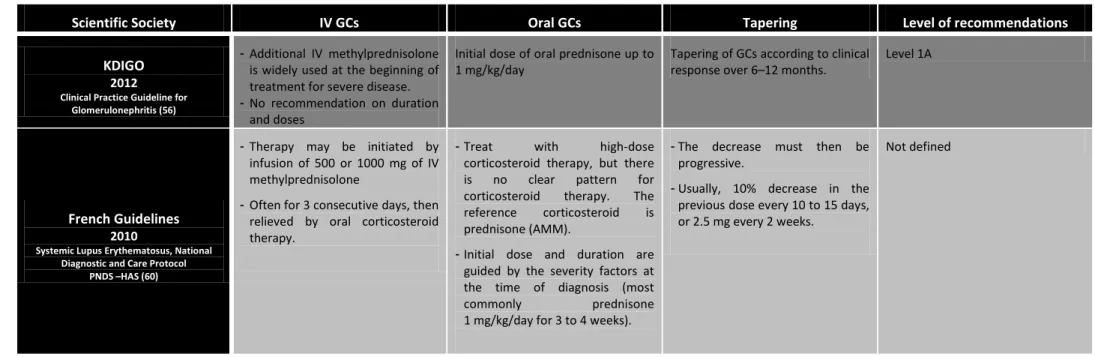
KDIGO
2012
Clinical Practice Guideline for Glomerulonephritis (56)
- Additional IV methylprednisolone is widely used at the beginning of treatment for severe disease. - No recommendation on duration
and doses
Initial dose of oral prednisone up to 1 mg/kg/day
Tapering of GCs according to clinical response over 6–12 months.
Level 1A
French Guidelines
2010
Systemic Lupus Erythematosus, National Diagnostic and Care Protocol
PNDS –HAS (60)
- Therapy may be initiated by infusion of 500 or 1000 mg of IV methylprednisolone
- Often for 3 consecutive days, then relieved by oral corticosteroid therapy.
- Treat with high-dose corticosteroid therapy, but there is no clear pattern for corticosteroid therapy. The reference corticosteroid is prednisone (AMM).
- Initial dose and duration are guided by the severity factors at the time of diagnosis (most commonly prednisone 1 mg/kg/day for 3 to 4 weeks).
- The decrease must then be progressive.
- Usually, 10% decrease in the previous dose every 10 to 15 days, or 2.5 mg every 2 weeks.
23
Table 2 - Recommendations for Lupus Nephritis Class IV or IV/V with cellular crescents
Scientific Society IV GCs Oral GCs Tapering Level of recommendations
ACR
2012
Guidelines for Treatment and Management of Lupus Nephritis (58) Pulses IV GCs: - High doses - no recommended duration Iv pulses relayed by - daily oral GCs - higher range dose - 1 mg/kg/day
No recommendation Level C
Table 3 - Recommendations for Membranous Lupus Nephritis Class V
Scientific Society IV GCs Oral GCs Tapering Level of recommendations
ACR
2012
Guidelines for Treatment and Management of Lupus Nephritis (58)
No recommendation In case of nephrotic range proteinuria only: - prednisone - 0.5 mg/kg/day - for 6 months No recommendation Level A EULAR/ERA-EDTA 2012
Recommendations for the management of adult and paediatric lupus nephritis(59)
No recommendation Oral prednisone (0.5 mg/kg/day) No recommendation Agreement level among experts : 9/10
KDIGO
2012
Clinical Practice Guideline for Glomerulonephritis (56)
No recommendation In case of persistent nephrotic proteinuria, treat with corticosteroids.
No recommendation Level 2D
French Guidelines
2010
Systemic Lupus Erythematosus, National Diagnostic and Care Protocol
PNDS –HAS (60)
No recommendation In case of persistent nephrotic proteinuria, treat with prednisone, 0,5 to 1 mg/kg/day during 1 month and then taper.
Taper after 1 month of oral GCs therapy.
24
2.2 Maintenance therapy
2.2.1 Glucocorticoids and maintenance therapy
Once remission has been induced, maintenance phase therapy should focus on the long-term management of chronic, more or less indolent, disease. GCs remain a major component of treatment in the maintenance phase of lupus nephritis therapy: the current recommendations are based on clinical studies that do not exclude the use of steroids in maintenance therapy (3).
In the maintenance phase of treatment, either mycophenolate or azathioprine is recommended by all guidelines, supported by low-dose oral GCs, but without any consensus concerning the optimal dose, if tapering is needed or the optimal duration (2,56–58). The EULAR committee recommends judicious use of these agents, taking into consideration the potential harms associated with these drugs (59,61). Finally, with respect to the duration of the treatment, guidelines differ: at least 3 years (EULAR/ERA-EDTA) or at least 1 year (KDIGO) after (complete) remission. Due to the length of completed studies, there is no advice on the optimal duration of therapy beyond 3 years (54,56–58).
25
2.2.2 Summary of the current recommendations for the use of Glucocorticoids in Lupus Nephritis in Maintenance Therapy
Table 4 - Recommendations for maintenance therapy
Scientific Society IV GCs Oral GCs Tapering Level of recommendations
ACR
2012
Guidelines for Treatment and Management of Lupus Nephritis (58)
No recommendation Low dose oral GCs No recommendation Not defined
EULAR/ERA-EDTA
2012
Recommendations for the management of adult and paediatric lupus nephritis(59)
No recommendation Low dose prednisone (5 to 7.5 mg/day)
- There is no data to guide duration of treatment beyond 3 years - Continuing treatment for longer
time periods should be individualised with an effort first to withdraw glucocorticoids before immunosuppressive agents. Level C EULAR 2007
Recommendations on the management of systemic glucocorticoid therapy in rheumatic
diseases (61)
No recommendation No recommendation - For prolonged treatment, the GCs dosage should be kept to a minimum
- A GCs taper should be attempted in case of remission or low disease activity
Percentage of the task force members that strongly to fully recommended this proposition: 86%
KDIGO
2012
Clinical Practice Guideline for Glomerulonephritis (56)
No recommendation - Low-dose oral corticosteroids - ≤ 10 mg/day prednisone
equivalent
Maintenance therapy be continued for at least 1 year before consideration is given to tapering the immunosuppression
Oral GCs: Level 1B Tapering: Level 2D
French Guidelines
2010
Systemic Lupus Erythematosus, National Diagnostic and Care Protocol
PNDS –HAS (60)
No recommendation No recommendation - Low doses of GCs (5-10mg/day of prednisone) may be maintained over long term - Maintenance corticosteroids
(5 mg/day or 0.10 to 0.20 mg/kg/day) are often maintained for several years, depending on the initial severity or any previous relapses. - The optimal duration of
maintenance treatment is not defined. The current trend is the extension of maintenance treatment for a period of 5 years.
26
3. Adverse effects of steroids
GCs remains the corner stone treatment and most of SLE patients have received GCs; in some cohorts up to 80% of patients continue indefinitely this treatment as “maintenance” therapy, at low doses of less than 7.5 mg/day. This long-term treatment results in various side effects and contributes to worsen quality of life even years after its withdrawal. Although the risk of side effects is dependent on time and dose and the cumulative dose of GCs is clearly related to adverse effects, intermittent or low dose therapies have also been associated with damages (13,62,63).
In fact GCs use is associated with several significant side effects like osteoporosis, metabolic syndrome, cardiovascular disease, infections, osteonecrosis, myopathy and cataracts, among others (17,62,64,65).
3.1 Glucocorticoids & Bone health 3.1.1 Glucocorticoids & Osteoporosis
GCs therapy results in an early and rapid bone loss and increased fracture risk after beginning of GCs treat. GCs acting through bone GR increase life span of osteoclasts and induce osteocyte apoptosis and decrease bone remodeling. There is an early deleterious effect of GCs that persists throughout the duration of GCs therapy on bone with a transient increase in bone resorption followed by a decrease in bone formation. Osteogenesis is also suppressed leading to bone collapse. Indirect effects of GCs also occur as a result of hypogonadism, decreased intestinal calcium absorption and reduce renal calcium reabsorption, increased intestinal and renal calcium loss and vitamin D insufficiency because of accelerated catabolism of 25(OH)D and 1,25(OH)2D due to GCs (62).
Fracture risk is dose dependent but it increases even with low dose (2.5–7.5 mg of prednisolone or the equivalent) or intermittent therapy (62,66,67). Carli and al showed on univariate analysis that cumulative doses of GCs were correlated with osteoporosis (OP) (p<0.03) (67). Fractures in glucocorticoid induced OP occurs independently of bone mass density (BMD) and can be found in up to 30–50% of patients (62,66). A Spanish study has determined the prevalence of undiagnosed vertebral fractures in women chronically using GCs therapy for auto immune disease : Morphometric vertebral fractures were detected in 13.7 % of SLE patients, 4-times more frequently than self-reported by patients (3.2 %) (68).
3.1.2 Glucocorticoids & Osteonecrosis
Up to 40% of patients can present non traumatic osteonecrosis after long-term GCs therapy. The risk increases with higher doses and prolonged treatment but it may also occur with short-term exposure to high doses (62,64,66). In a recent systematic review of the literature including fifty-seven studies (23,561 patients) it has been reported that osteonecrosis incidence was 6.7% with corticosteroid treatment of >2 g (prednisone-equivalent) and that SLE patients had positive correlations between dose and osteonecrosis incidence. Each 10 mg/day increase was associated with a 3.6% increase in osteonecrosis rate, and >20 mg/day resulted in a higher osteonecrosis incidence. Mean osteonecrosis incidence was 33% in twenty studies evaluating the effects of pulsed corticosteroids based on data from the systematic review (69). The effects of GCs on bone density loss are thought to be rapid with medication initiation, then slower after 6 months of treatment. After withdrawal of GCs therapy, fracture risk returns partially back to baseline levels. Unless the patient had substantial loss in bone mass due to high cumulative doses of GC the risk remains higher (62,66).
3.2 Glucocorticoids & Cardiovascular Events
Patients with SLE are at excess risk of cardiovascular events (CVEs). Reports have suggested that this higher risk is multifactorial, with contributions from traditional risk factors for CVEs, SLE disease
27
activity, SLE-related immunologic factors (recent levels of circulating anti-double-stranded DNA, positive antiphospholipid antibody (aPL), elevated markers of endothelial activation (von Willebrand factor (vWf), soluble vascular cellular adhesion molecule-1 (sVCAM-1) and fibrinogen), and SLE-related medications such as GCs (70–72). GCs major numerous cardiovascular risk factors like obesity, insulin resistance, glucose intolerance, dyslipidaemia and hypertension. GCs promote atherogenesis and affect vascular remodeling and have direct effects on the heart, kidneys and blood vessels, influence vascular function. The risk is related to the development of an exaggerated
metabolic syndrome (17,62).
The glucocorticoids are a primary factor that influences cardiovascular risk. There is a directly proportional relationship between the cumulative GCs dose and the increase in cardiovascular risk with SLE patients(73). Magder and Petri showed that doses of prednisone more than 10 mg per day were associated with a two-fold increased risk and more than 20 mg with a five-fold increased risk. This remained true after adjusting for disease activity (70). The cumulative dose of GCs promote hypertriglyceridemia and insulin resistance and are associated with a higher cholesterol plasma level, higher blood pressure and weight change in lupus patients (74). Petri and al. showed that increases in prednisone dose in SLE patient led to definable changes in risk factors for coronary artery disease, a change in prednisone dose of 10 mg and the duration even at this level was associated with a change in cholesterol (75). Wei et al. found a 2.56-fold increase in risk of cardiovascular events with a prednisone dose of 7.5 mg or more. In the Hopkins Lupus Cohort, an increase in prednisone dose of 10 mg caused an increase in cholesterol of 7.5 mg/dL, weight change of 5.5 lbs and change in blood pressure of 1.1 mmHg (76,77). An ongoing excess cardiovascular risk is sustained after cessation of exogenous GCs administration. Current use or even a past use of GCs increased significantly the risk for heart failure. This might be explained by the mineralo-corticoid effects of GCs which increases
sodium and fluid retention and promotes remodeling through fibrosis of the atria and ventricles (62).
3.3 Glucocorticoids & Infections
GCs exert many immunosuppressive effects that induce cellular immunodeficiency and consequently
increase host susceptibility to a wide variety of infectious pathogens: bacterial infections are most
frequent, followed by viral, then fungal and parasitic infections and also predisposed to opportunistic
infections. Infection is well recognised to be a cause of early and late deaths in lupus (62,78–81).
The risk is associated with daily dose and duration of therapy; some studies highlighted the
importance of the cumulative dose. Numerous studies have demonstrated that corticosteroids,
already with prednisone doses over 7.5-10 mg/day and especially in doses higher than prednisone
20 mg daily (or its equivalent), and also when administered intravenously, significantly increased susceptibility to infection (3,5,9,36–38). A recent cohort study confirmed that damage due to steroids increased over the five years of follow-up while damage independent of steroids therapy remained constant throughout the period of observation (82). Also, concerning infection, the risk decreases after withdrawal of GCs. Limiting steroid administration for less than 21 days also
decreases infectious complications (62,78,79,83–90).
3.4 Glucocorticoids & Morbidity Mortality in SLE
Several longitudinal studies performed in SLE patients have revealed that GCs were the main cause of damage and sequelaes, as determined by the Systemic Lupus International Collaborating Clinics (SLICC)/American College of Rheumatology (91).
Thus the mean SLICC/ACR damage index (SDI) rose from 0.33 at baseline to 1.9 after 15 years of follow-up in an inception cohort, and damage was considered as GCs-related in 16% and 49% of the cases at baseline and last follow-up, respectively (92). Ruiz-Arruza et al. showed that mean doses of
28
prednisone >7.5 mg/day increased the risk of accruing damage after 5 years of follow-up, whilst doses <7.5 mg/day and methylprednisolone pulses did not (93).
Finally, damage accrual was found to be associated with higher risk of mortality, every 1-point increase in SDI being associated with a 1.32 times more risk to die during follow-up (81).
4. Treat lupus with lower doses or without Glucocorticoids
Due to the considerable side effects of GCs, both patients and physicians are concerned with the use
of GCs and there is an urgent need to find new protocol to avoid a prolonged cortisonic impregnation and to reduce the doses of GCs. Research team have been focusing on defining new treatments which minimise therapy related toxicity and reduce in particular the burden of steroid use.
The paradigm of immunosuppressive treatment for lupus nephritis has evolved over the past few decades from corticosteroids alone to corticosteroids combined with immunosuppressants and now to immunosuppressants alone or with less GCs.
4.1 Reduce the doses of GCs
Is reducing or stopping corticosteroids an achievable and reasonable goal? In order to limit GCs toxicity, lower oral doses have been successfully used in lupus nephritis trials in association with other immunosuppressants, such as azathioprine, mycophenolate mofetil or cyclophosphamide, widely used as steroid sparing agents (94).
First indications are given by Zahr et al. who reported on the largest prospective cohort study of predictors of prednisone tapering in SLE patients. This Hopkins Lupus Cohort gathered 866 patients from 1987 through 2009. The analysis was based on patients whose previous prescribed dose of prednisone was 5 mg/day. The authors examined the proportion of times the patient's dose was reduced to below 5 mg/day. Among those patients who tapered oral GCs they analysed the proportion whose prednisone dose remained below 5 mg/day for at least one year.
The analyses showed that Caucasians, younger patients, patients with a higher level of education, lower disease activity, or absence of urine protein were more likely to have a prednisone taper. However, successful tapering, so patients who were treated during at least one year with a dose of prednisone under 5mg/day was not dependent on age, ethnicity, or education but it was more frequent in those with lower disease activity. Successful tapering was achieved more often after the year 2000 (95).
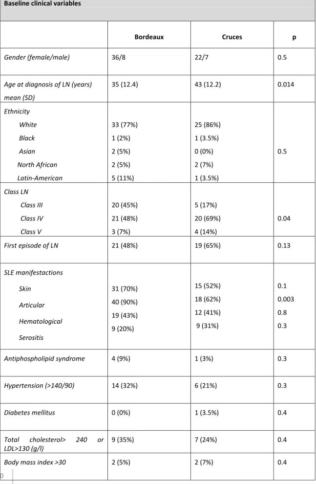
Pr. Ruiz-Irastorza and his team from Bilbao proposed a specific new therapeutic scheme in order to spare oral GCs, the Cruces-protocol cohort (CC) (96). The patients from the CC received medium doses of prednisone (Class II up to 15mg/day, Class III-IV up to 20-30mg/day, Class V up to 15mg/day), combined with methyl-prednisolone pulses (3 consecutive days bolus from 250mg to 500mg and one methylprednisolone 125 or 250 mg pulse was given just before each dose of cyclophosphamide), hydroxychloroquine (HCQ) and immunosuppressive drugs, usually cyclophosphamide. The oral dose of GCs was quickly tapered every 2 weeks until 10 mg/day (class III and IV) or 7.5 mg/day (class II and V), being then reduced by 2.5 mg/day every 4 weeks to a maintenance dose of 2.5–5mg/day. These patients were compared to the historic cohort (HC) who received high-dose prednisone (up to 1mg/kg/day) and followed a NIH (National Institutes of Health) protocol-based scheme (cyclophosphamide for class III and IV or azathioprine or mycophenolate mofetil for others states).
Among the forty-five patients included in the analysis, the CC patients were less exposed to oral GCs and tapering to 5mg/d was quicker. The mean (SD) cumulative doses of prednisone at six months were 1.7 (0.5) g vs. 4.5 (2.1) g (p < 0.001), in the CC and HC, respectively, which resulted in average doses of 9 mg/d and 25 mg/d, respectively. Furthermore the mean (SD) weeks to achieve the
29
reduction of prednisone to 5 mg/day were 16 (±9) weeks in the CC vs. 130 (±146) weeks in the HC (p<0.001). This CC protocol is at least as effective in achieving remission of LN as regimes containing high-dose prednisone. But this protocol causes less toxicity: Global toxicity attributable to GCs was seen in 1/15 patients (7%) in the CC vs. 20/30 patients (67%) in the HC (p<0.0001) (96).
The duration of corticosteroid administration during maintenance therapy in LN remains controversial and non evidence-based. A small pilot study by Galbraith et al. was achieved in order to assess the feasibility of doing a larger trial on whether maintenance corticosteroids are needed in LN. This study included 15 patients with proliferative (membranous) LN induced with cyclophosphamide or MMF who achieved at least a partial response and had been tapered down to 20 mg of prednisone. Patients were randomised to prednisone continuation with low-dose maintenance target (5-7.5 mg/day) (n=8) or to withdrawal (n=7). Patients were followed for a median of 11.9 months, and the end point was renal or major non-renal relapse. Relapse occurred in only one patient from the withdrawal group (14%; one renal flare), but in 50% of the low-dose prednisone patients (three renal flares and one major non renal flare) (97).
4.2 The new wave: Treat without oral GCs
Avoiding oral steroids in the treatment of LN is now the aim. Rituximab might also be effective without long-term corticosteroids. A London research team led by Pr. Liz Lightstone from the Imperial College has piloted a steroid-avoiding protocol (RITUXILUP) for the treatment of biopsy-proven active International Society of Nephrology/Renal Pathology Society (ISN/RPS) class III, IV, or class V LN. In this open-label trial, 50 patients with LN who were not taking long-term oral steroids, were treated with two doses of rituximab (1 g infusion every two weeks) together with intravenous methylprednisolone 500 mg and mycophenolate mofetil at a starting dose of 1 g daily, titrated to achieve a maximum dose of 3 g daily but without any oral GCs. At 52 weeks, the complete response and partial response rates were 52% and 34%, respectively. Patients with class III, IV or V lupus nephritis responded similarly. Adverse events were infrequent, with hospital admission and infection occurring in 18% and 10% of the patients, respectively (98).
Following this study, this regimen is currently tested in a phase III open label randomised multicentre controlled trial against conventional treatment protocol in the ongoing RITUXILUP (Trial of Rituximab and Mycophenolate Mofetil Without Oral Steroids for Lupus Nephritis) that aims to demonstrate whether the addition of Rituximab to MMF therapy is useful in treating a new flare of lupus nephritis and whether it has a long lasting steroid-sparing, beneficial effect with equal efficacy and greater safety than a conventional regimen of MMF and oral prednisolone. Estimated study completion is expected for April 2019 (94,99).
CORTICOLUP is a French ongoing study which objective is to study the possibility of corticosteroids interruption in patients with quiescent SLE treated since at least one year with 5 mg of prednisone per day. The primary outcomes are the occurrence of mild or moderate flares of SLE. The estimated Study Completion is expected for April 2018 (100).