UNIVERSITE DE GENEVE
FACULTE
DE
MEDICINE
Section de médecine clinique
Départment des Neurosciences
Cliniques et Dermatologie
Service d’Oto-Rhino-Laryngologie et
Chirurgie Cervico-faciale
Thése préparée sous la direction du
Docteur Jean-Silvain LACROIX, Chargé de Cours
ETUDE IN VIVO ET IN VITRO DES EFFETS DE
L’HYPOCHLORITE DE SOUDE DANS LE TRAITEMENT DES
RHINOSINUSITES CHRONIQUES
Thése
présentée à la Faculté de Médecine de l’Université de Genève
pour obtenir le grade de Docteur en médecine
par
Tanveer RAZA
DHAKA, BANGLADESH
Thèse N 10442 Genève 2005RESUME
Le traitement médical de la rhinosinusite chronique (RSC) pose un problème de santé publique en raison, entre autres, de la fréquence des récidives infectieuses nécessitant une antibiothérapie et du prix des corticostéroïdes topiques. L’ hypochlorite de sodium (NaOCl) est un agent bactéricide efficace contre toutes les souches bactériennes potentiellement pathogènes. Le but de cette étude a été de déterminer la cytotoxicité du NaOCl sur des cellules épithéliales respiratoires primaires cultivées in vitro. Nous avons déterminé la concentration (0.05 %) et la durée (5 min 2 x par jour) d’exposition au NaOCl n’ayant pas d’effet cytotoxique tout en conservant ses propriétés bactéricides. Nous avons évalué, chez des patients symptomatiques, l’effet thérapeutique de lavages des fosses nasales avec une solution de NaOCl à 0.05 % 2 fois par jour pendant 3 mois. L’intensité subjective des symptômes et les paramètres cliniques endoscopiques ont été évalués par des échelles analogiques visuelles. Nous avons mesuré la résistance respiratoire nasale par rhinomanométrie et la production nasale de monoxyde d’azote (NO) avant et après le traitement.
Le lavage des fosses nasales 2 fois par jour avec une solution de NaOCl 0.05 % semble améliorer de façon significative la plupart des symptômes et des paramètres inflammatoires endoscopiques de la RSC.
INTRODUCTION
Rhino-sinusite chronique :
La rhino-sinusite chronique (RSC) est un des problèmes les plus fréquents de santé au niveau mondial avec des conséquences socio-économiques majeures. L’étiologie de la RSC est probablement multifactorielle. Le rôle et le type des bactéries qui pourraient être impliquées dans la physiopathologie de cette maladie sont l’objet de nombreux travaux de recherche. A l’heure actuelle, le traitement médical classique de la RSC inclut très souvent une antibiothérapie. Bien que le choix de l’antibiotique devrait toujours être accompagné d’un examen bactériologique et d’un antibiogramme, dans la plupart des cas le type d’antibiotique est choisi de façon empirique (1, 2). Un nombre croissant d’antibiotiques ont progressivement perdu de leur efficacité contre les bactéries les plus fréquemment rencontrées dans la RSC. L’émergence de souches bactériennes multirésistantes aux antibiotiques est probablement d’origine multiple et inclut probablement l’usage abusif de ce type de médicaments antimicrobiens. L’introduction de nouveaux antibiotiques a diminué au cours des dernières années et les nouveaux agents développés ont malheureusement été associés au développement rapide de nouvelles souches résistantes (2, 5). C’est ainsi que de nouvelles alternatives thérapeutiques concernant le traitement des rhino-sinusites chroniques associées à la présence de bactéries sont devenues un sujet en plein développement.
Le traitement classique de la RSC comprend des lavages des fosses nasales, l’application de corticoïdes topiques, la prise prolongée d’antibiotiques à large spectre et parfois des antihistaminiques (3, 4). En cas d’échec du traitement traditionnel médical et en présence d’anomalies anatomiques révélées par le scanner du massif facial, on peut proposer une chirurgie endoscopique rhino-sinusienne (5). Selon la littérature disponible, la chirurgie
endoscopique rhino-sinusienne est un geste dont les résultats à long terme sont plutôt satisfaisants (1). Cependant, il existe un pourcentage significatif de patients qui n’ont pas été améliorés par le traitement chirurgical. Dans les cas de surinfection chronique, on peut proposer une antibiothérapie intraveineuse pendant plus de 6 semaines (8), des lavages sélectifs des sinus avec des solutions antibiotiques ou des corticostéroïdes (6,9).
La rhino-sinusite chronique a un impact financier important. Quatre vingt cinq % des patients souffrant de RSC sont en âge de travailler (entre 18 et 65 ans) et les coûts indirects secondaires dus par exemple à l’absentéisme ou au présentéisme, c’est à dire une baisse de la productivité lors de l’activité professionnelle, semblent affecter de façon importante l’aspect économique de cette maladie (1). Les coûts directs incluent le bilan radiologique, l’hospitalisation et le traitement médicamenteux. Aux Etats-Unis, les dépenses annuelles en médicaments pour le traitement de la RSC avant et après chirurgie ont été évalués à 1.220 $ et 629 $ par patient et par an, respectivement (10). Le traitement chirurgical coûte en moyenne 6.490 $ par patient. A la vue de ces chiffres importants, une alternative à ces prises en charge thérapeutiques pourrait se révéler intéressante.
L’hypochloride de soude :
L’hypochloride de soude (NaOCl) est un agent chimique bien connu utilisé essentiellement pour ses propriétés de décoloration et blanchiment du linge ainsi que son action désinfectante (11-19). Ces propriétés bactéricides ont été observées contre de multiples organismes, en particulier le staphylocoque doré (11-16), le pseudomonas aeruginosa (18), le candida albicans (18) et le vibrio vunificus (19). En fait, le NaOCl semble être efficace contre toutes les bactéries connues pour infecter les plaies des patients brûlés (14). L’hypochloride de soude a été utilisé pour le lavage des plaies par Bugnan pendant la 2ème guerre mondiale. Sur
les brûlures, les lavages étaient effectués avec une solution d’hypochloride de soude à 0,2%. Elles étaient ensuite recouvertes par un pansement à base de soie contenant du NaOCl à 0,05%. L’effet antiseptique du NaOCl a déjà été observé par Koch en 1880 et Dakin l’a utilisé pour le traitement des plaies pendant la 1ère guerre mondiale (14, 18, 20). De nos jours, le NaOCl est l’agent le plus utilisé pour les traitements antiseptiques stomatologiques et parodontiques (11, 17, 20-22).
Le mécanisme exact des propriétés bactéricides du NaOCl n’a jamais été déterminé. En présence d’eau, le NaOCl se dissocie en Na+ et OCl-/HOCl (acide hypochlorique). La chlorine active est un puissant agent oxydant. Des observations expérimentales semblent suggérer que la chlorine exerce son effet antimicrobien par une oxydation irréversible des groupes SH présents dans certaines enzymes et en induisant un blocage des fonctions métaboliques essentielles des cellules bactériennes. La chlorine peut également se combiner avec des composants cytoplasmiques pour former des complexes N-chloro qui sont également toxiques pour les micro-organismes (14, 20, 22).
Le choix d’une concentration appropriée de NaOCl dépend d’un bon équilibre entre les propriétés antibactériennes et des éventuelles toxicités tissulaires. Traditionnellement, le choix de la concentration varie entre 0,5% jusqu’à 5,25% avec parfois des concentrations pouvant aller jusqu’à 10% (17). Cependant, les effets bactéricides du NaOCl ont déjà été observés à des concentrations aussi basses que 0,025% lorsqu’il est utilisé sur des pansements occlusifs (23). Dans le cas des irrigations des lésions parodontiques ou stomatologiques, les propriétés antimicrobiennes d’une solution à 0,5% de NaOCl semblent être significativement moins efficaces que les concentrations à 5,25% (22). En augmentant le temps d’exposition à une solution de NaOCl on diminue également la survie des micro-organismes. Si le temps d’exposition est augmenté, une solution à 0,5% de NaOCl semble avoir le même effet
bactéricide qu’une solution à 5,25% (21). Dans les traitements à long terme, il semble qu’une solution à 0,5% ne soit pas appropriée. En effet, après 2 semaines de traitement continu sur des brûlures recouvertes avec de la gaze imbibé de NaOCl, une toxicité marquée a été observée au niveau des cellules de l’épiderme. Les cellules basales de l’épiderme qui ont été isolées dans les régions traitées semblaient avoir une croissance fortement perturbée. Cependant, une solution à 0,1% de NaOCl semble avoir une faible toxicité pour les traitements à long terme.
Avant d’utiliser une solution de NaOCl chez nos patients souffrant de RSC, nous avons d’abord évalué la toxicité de différentes concentrations de NaOCl et de temps d’exposition sur des cultures primaires de cellules épithéliales respiratoires cultivées in vitro.
Effets d’une solution de NaOCl sur la F-actine et les jonctions serrées intercellulaires :
La F-actine est un constituant du cytosquelette qui est impliquée dans la stabilisation de la forme des cellules et joue également un rôle important dans la distribution des protéines membranaires. D’autre part, la F-actine est également impliquée dans les transports transmembranaires et dans la régulation du volume cellulaire (24, 25). La F-actine est également directement liée à de nombreux composants des jonctions serrées intercellulaires. Il semblerait donc qu’une altération de l’expression de la F-actine pourrait avoir des effets importants sur la structure des jonctions serrées intercellulaires (26).
Ezrine :
La polarisation des cellules epithéliales en culture dépend de l’adhésion intercellulaire, de l’organisation du cytosquelette et de la répartition de certaines protéines dans les compartiments baso-latéraux et apicaux. Les protéines spécialisées du pôle apical des
membranes cellulaires dépendent essentiellement de certaines protéines polarisées comme l’ezrine, la radixine et la moesine (ERM) qui sont issues d’un lage pool cytoplasmique. En mettant en évidence l’expression de l’ezrine, on peut suivre assez facilement la différentiation et la polarisation des cellules épithéliales respiratoires cultivées in vitro (27).
Résistance électrique transépithéliale :
Les jonction serrées, qui sont classiquement localisées sur les bordures apico-latérales des cellules épithéliales, régularisent de façon sélective le passage de l’eau, des ions et de certaines molécules neutres ainsi que des cellules inflammatoires à travers les voies de passage paracellulaires. Ces jonctions serrées définissent les limites de certains compartiments liquidiens, qui sont vitaux pour l’homéostase cellulaire et qui contribuent également au maintien de la polarité cellulaire. Une des façons les plus efficaces d’évaluer le bon fonctionnement de ces jonctions serrées est de mesurer sur des cultures primaires de cellules épithéliales in vitro la présence d’une résistance électrique transépithéliale. La résistance électrique transépithéliale générée à travers un épithélium est déterminée par les transports ioniques et l’effet sélectif et restrictif des jonctions serrées (28).
Evaluation clinique :
La RSC et la polypose nasale peuvent être définies comme une inflammation des muqueuses nasales et des sinus paranasaux caractérisée par 2 ou 3 des symptômes suivant pendant plus de 3 mois : obstruction ou congestion des voies aériennes nasales ; écoulement antérieur et/ou postérieur ; douleurs ou pression faciales ; diminution ou perte des performances olfactives. Les signes endoscopiques associés au diagnostic clinique de RSC sont la présence de polypes au méat moyen, un écoulement mucopurulent au niveau du méat moyen ou un œdème
radiologiques sont : un épaississement mucopériosté diffus essentiellement localisé au niveau du méat moyen ou des sinus paranasaux (1).
L’évaluation subjective de l’intensité des symptômes associés à la RSC peut être mesurée en utilisant une échelle analogique visuelle (EAV). Concernant l’obstruction par exemple, l’absence de ce symptôme correspondrait à la valeur 0 sur l’EAV et une obstruction complète pourrait correspondre à la valeur de 10. Les EAV permettent une quantification reproductible des symptômes spécifiques de la RSC (7).
Monoxyde d’azote :
La production de monoxyde d’azote (NO) par les voies respiratoires semble être un indicateur assez spécifique de la présence de cellules inflammatoires et d’un dysfonctionnement du transport mucociliaire. La mesure de la production de NO par les voies respiratoires supérieures a souvent été utilisée comme paramètre permettant d’évaluer l’évolution de la maladie après un traitement chirurgical (1). La production de NO au niveau des fosses nasales est une des plus hautes de l’ensemble du corps humain (29). La production de NO est en général mesurée par la technique de chémiluminescence (30).
Rhinomanométrie :
La rhinomanométrie permet de mesurer de façon objective la résistance respiratoire nasale. Cette mesure objective permet également une bonne évaluation du traitement chirurgical et/ou médical de la rhino-sinusite chronique (31).
L’objectif principal de cette étude a été d’évaluer l’efficacité des lavages des fosses nasales avec une solution d’NaOCl chez les patients souffrant de RSC associée à des infections loco-régionales récidivantes.
Les effets cytotoxiques potentiels de différentes concentrations de NaOCl ou de temps d’exposition ont été évalués sur des cultures primaires de cellules épithéliales respiratoires in vitro.
SUMMARY
Treatment of chronic rhinosinusitis (CRS) is arduous as a result of increasing resistance to antibiotics and expenses of topical corticosteroids among other factors. Finding an alternative therapy is important. Sodium hypochlorite (NaOCl) is an established disinfecting agent with no known bacterial resistance. No literature has been found on NaOCl use for CRS and its effect in all tissues may not be the same. Selection of NaOCl is based on anti-microbial affectivity and cytotoxicity. The aim of this study was to determine the appropriate concentration and application method of NaOCl and evaluate its clinical efficiency in CRS. Two concentrations of NaOCl were prepared by diluting 0.55% NaOCl (Amuchina Med) with normal saline (0.9% NaCl). In vitro tests for cilia and epithelial cell viability were done on reconstituted epithelial cells by primary epithelial cells cultured in vitro. Cells were exposed for 5 and 15 minutes twice daily for 5 consecutive days to one of the following conditions, 1) untreated and 2) 0.5% NaOCl and 0.05% NaOCl. Transepithelial resistance (TER) was measured. In order to visualize cell integrity and test for cilia, immunostaining was done for ezrin and F-actin network and observed with confocal microscopy. After the determination of optimal NaOCl concentration in vitro, in vivo tests were performed. Patients with CRS without any known risk factor for increased infection were selected. Each patient applied NaOCl as nasal lavage twice daily on both nostrils for 3 months. Symptom intensity and endoscopic findings were recorded with visual analogue scale (VAS) and nasal airway resistance (NAR) and nasal Nitric Oxide (NO) level were measured at baseline and after 3 months. Fourteen patients completed the study. F-actin network loss and decreased expression of ezrin was significant in cells exposed to 0.5% than 0.05% NaOCl and more obvious when exposed for longer periods (15 min. and 5 min.) daily. Statistically significant improvement in nasal obstruction (p=0.001), posterior nasal discharge (p=0.018), smell (p=0.007) and
headache (p=0.009) was demonstrated. There was no significant improvement in anterior nasal discharge (p=0.129). Significant improvement was recorded in nasal endoscopic findings of oedema (p=0.001), erythema (p=0.001), purulent discharge (p=0.002) and nasal crusts (p=0.001). There was no significant improvement in nasal NO production. A significant improvement occurred in NAR (p=0.05) as measured by rhinomanometry. We conclude that 0.05% NaOCl is suitable for use in nasal epithelium as a lavage in humans. We have demonstrated that if applied twice daily for 3 months, 0.05% NaOCl lavage may be a good alternative to systemic antibiotics in the treatment of CRS with recurrent infection.
ACKNOWLEDGEMENT
The work included in this thesis was carried out in the Rhinology-Olfactology unit of the Service of Otorhinolaryngology of the University Hospital of Geneva in the period 2004 through 2005. Thanks are due to a number of respected colleagues and friends for their active participation and supports
¾ Dr. Jean-Silvain Lacroix, my supervisor, for his guidance, enthusiasm, competence, devotion, support and friendship that contributed to the initiation of research in this area that ultimately lead to the realization to this thesis. I am grateful to him for sharing and teaching me from his extensive knowledge in Otorhinolaryngology especially Rhinology and Nasal Endoscopic Surgery. I am also grateful to his wife Titane for inviting me and my wife and giving us the taste of Swiss warmth and hospitality.
¾ Dr. Laurence Zulianello for her support and guidance in helping me to carry out and interpret the in vitro part of the study a new area of research for me. I am also grateful to Isabelle Borges for helping me perform the in vitro experiments.
¾ The “Commission des affaires humanitaires” of the Geneva University Hospital for providing me with a scholarship.
¾ Dr. Alma Ricchetti, Dr. Basil Landis and Dr. Selim Bouayed for being good friends and sharing their knowledge and resources and for their support.
¾ Marianne Hugentobler for helping me with Nitric Oxide and Rhinomanometry measurements, for showing me how the machines work and how to take measurements.
¾ Mme Yolande Bosshard for the immense help that she had provided in my research and beyond and thus making our stay in Geneva easier.
¾ I am grateful to Giuseppe Rizzo for his technical support, Christine Geneyne for her gracious assistance, and to Paulo, Fabian and all my other friends in the Rhinology- Olfactology unit for their immense support and help.
¾ Thanks to my friends and colleagues in the ORL service for a wonderful time.
Last but not least
¾ I want to express my appreciation to my parents, Dr. Md. Rakibur Raza and Mrs. Rasheda Raza. My father through his expertise in Otorhinolaryngology has always provided me the enthusiasm to strive harder. His generosity, persistence, personal integrity and respect for the individual remains to be a guidance for me. My mother with her love and care provided the fundamental feeling of stability and warmth.
¾
I want to express thanks to my wife, Nadia for helping me through the thesis and checking the literature of a subject that is foreign to her.Dedicated to my parents
Dr. Md. Rakibur Raza
&
INTRODUCTION
Chronic rhinosinusitis
Chronic rhinosinusitis (CRS) is one of the most common health care problems with major economic implications. The etiology of this disease is multifactorial. The role and type of bacteria in the pathogenesis of CRS is still debatable. Yet the conventional medical therapy of CRS includes antibacterial treatment. Although the choice of antibiotics should be culture directed, in most cases, empiric selection of antibiotic is still preferred [1, 2]. A growing number of antibiotics are being gradually ineffective against the common isolates of CRS. The probable causes of the resistance of micro-organisms to antibiotics are considered to be multiple, including inappropriate use, spread of antimicrobial resistant bacteria across different countries due to increased travel, and increased use in agriculture, fisheries and animal husbandry. The introduction of new antibacterial drugs has declined and any new drug that has been introduced is challenged with rapidly developing resistance [2-5]. Thus novel and alternative therapy for bacteria in CRS is becoming significantly more important.
CRS is usually treated with nasal irrigation or lavage, topical steroids, a long course of broad-spectrum antibiotics and anti histamines as indicated [5, 6]. Sinus diseases resistant to traditional medical therapy are usually managed by endoscopic sinus surgery (ESS) [7]. ESS is a reasonably successful treatment [1]. Nevertheless a significant percentage of patients are not improved after ESS. A number of alternative treatment modality has been reported for management of resistant CRS, including home i.v. antibiotics for 6 weeks [8], selective sinus irrigation with topical antibiotics and steroids [9] and nebulized aerosol therapy [6].
CRS can have a major financial impact in terms of both “direct” and “indirect” costs. With 85% of CRS patients of working age (between 18-65 years old), indirect costs such as “abstenteeism” (missed work days) and “presenteeism” (decreased productivity at work) significantly add to the economic burden of the disease [1]. Direct costs include radiography, hospitalization and medication. The annual expenditure of the pharmacologic management of CRS before surgery and after surgery has been reported to be US$ 1,220 and US$ 629 per patient per year, respectively, in the United States [Figure 1] [10]. Surgical costs averaged US$ 6,490 per patient. Thus, where cost constraints are important careful considerations of alternatives are crucial.
Figure 1: Annual expenditure of the pharmacologic management of chronic rhinosinusitis in the United States [10]
0 200 400 600 800 1000 1200 1400 Pre-Surgery Post-Surgey US Dollars Antibiotics Nasal sprays OTC Sodium hypochlorite
Sodium hypochlorite (NaOCl) is a well known bleaching and disinfecting agent that has been found to be effective against several organisms [11-19]. NaOCl has been found to be effective against Staphylococcus aureus (SA) [11-16] and other organisms including Pseudomonas aeruginosa [18], Candida albicans [18] and Vibrio vulnificus along with other 18 vibrio
species [19]. In fact it has been found to be effective in killing virtually all of the micro-organisms known to infect burn patients [14].
NaOCl was used as an irrigant in treating wounds by Bunyan, who during World War II, used it as a topical antiseptic in burn therapy by initially irrigating with 0.2% NaOCl and then covering with silk occlusive envelopes containing 0.05% NaOCl. However the antiseptic property had been observed by Koch as early as 1880s and Dakin had used various NaOCl solutions for treating wounds during World War I [14, 18, 20]. Nowadays NaOCl is the most commonly used endodontic irrigant because of its physico-chemical and antibacterial properties [11, 17, 20-22].
The exact mechanism of microbial killing of NaOCl has never been determined. It dissociates in water to Na+ and OCl-/HOCl (hypochloric acid). The active chlorine is a strong oxidizing agent. Substantial evidence suggests that chlorine exerts its antibacterial effect by the irreversible oxidation of -SH groups of essential enzymes, disrupting the metabolic functions of the bacterial cell. Chlorine may also combine with cytoplasmic components to form N-chloro compounds, toxic complexes that destroy the micro-organism. However, the first contact oxidation reactions of chlorine with bacteria may lead to the rapid killing of bacterial cells even prior to the formation of N-chloro compounds in the cytoplasm [14, 20, 22].
Selection of appropriate concentration of NaOCl is based on a balance between antimicrobial affectivity and tissue toxicity. The choice of concentration traditionally ranges from 0.5% to 5.25% and sometimes as high as up to 10% [17]. Nevertheless bactericidal effect of NaOCl has been observed in concentration as low as 0.025% when used as a fluid dressing [23]. As an endodontic irrigant, the antimicrobial affectivity of 0.5% NaOCl was significantly less than 5.25% NaOCl [22]. At 0.5% concentration, NaOCl efficiently decontaminated split thickness
cadaveric skin contaminated with methicillin resistant strains of SA. Even though 0.1% NaOCl could significantly decrease the amount of bacteria, organisms could still be isolated from the medium at 10 min [18].
Increasing the time of exposure to NaOCl decreases cell viability. If the irrigation time is increased, 0.5% NaOCl has nearly the same bactericidal effect as 5.25% NaOCl when being used as an endodontic irrigant [21]. It has also been demonstrated [14] that 0.5% NaOCl was inappropriate for long term maintenance. After 2 weeks of continuous treatment of burn wound with NaOCl gauze soaks, a marked toxicity was noticed in the epidermal cells of the treated areas. The basal cell isolates from treated areas were inhibited in their growth and tissue culture. Conversely 0.1% NaOCl was found to be of low toxicity for long term maintenance.
Thus before applying NaOCl in vivo we first evaluated the response of reconstituted nasal epithelium from primary nasal epithelial cells cultured in vitro to different concentration of NaOCl and exposure time.
F-actin network
F-actin based cytoskeleton stabilizes cell shape and plays important roles in the distribution of membrane proteins, in the regulation of transmembrane transport pathways and possibly in sensing cell volume changes. F-actin is directly linked to many components of the tight junction (TJ) complex [24, 25]. Thus alteration in the expression of F-actin may have profound effect on TJ structure and barrier function of epithelial cells and inversely TJ modulation has effect n F-actin [26].
Ezrin
Establishment of epithelial cell polarity is initially directed by cell adhesion molecules followed by organization of the cytoskeleton and sorting of proteins to basolateral and apical compartments. A central role in organizing and regulating specialized apical membrane proteins is played by polarized Ezrin, Radixin and Moesin (ERM) that is derived from a large dormant cytoplasmic pool. Dormant ERM proteins present in a folded conformation masking cortical-membrane and F-actin binding site. When polarized, ERM maintain an activated conformation linking cortical membrane proteins and cytoskeletal actin. Differentiation of the airway epithelium is highly regulated to generate ciliated and secretory goblet cells [27].
Thus tracking the expression of ezrin can help to indicate the polarity and differentiation of ciliated nasal epithelial cells.
Transepithelial electrical resistance
Tight junctions, characteristically located at the apico lateral borders of epithelial cells, selectively regulate the passage of water, ions, neutral molecules, and inflammatory cells through the paracellular pathway (the fluid-filled spaces between cells). These junctions define the limits of distinct fluid compartments, vital to homeostasis, and contribute to the maintenance of cell polarity. The most effective way to assess the functioning of TJ’s is to measure the capacity of an intact epithelium to generate a transepithelial electrical resistance (TER). The TER generated across an epithelium is determined by the ion transport properties of its component cells and the selective and restrictive barrier afforded by the TJ [28].
Clinical evaluation
The European position paper on Rhinosinusitis and Nasal Polyps [1] has defined CRS (including nasal polyps) as inflammation of the nose and the paranasal sinuses (PNS) characterized by two or more symptoms [Table 1] lasting at least for 12 weeks and either endoscopic signs and/or Computed tomographic changes.
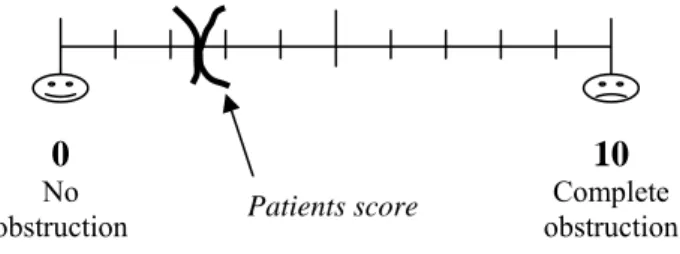
The strength of the subjective symptoms can be estimated using a visual analogue scale (VAS). The VAS is a self-reporting device that measures the magnitude of the state in a line that is horizontally placed with two poles anchored at both ends from 0 (none) to 10 (severe or maximum) [Figure 2]. Participants or the examiner place a mark somewhere along the line that best indicates the magnitude of the state according to the perception of the patient.
VAS offers a reproducible and quantifiable evaluation of the patient’s rhinosinusitis-specific symptoms, which may be a better means of evaluating symptom intensity than simply asking whether the patient feels better, the same or worse [7].
0 No obstruction 10 Complete obstruction Patients score
Figure 2 : Visual analogue scale recording for subjective sensation of nasal obstruction
Nitric oxide
Nitric oxide (NO) found in the upper and lower respiratory tract is a sensitive indicator of the presence of inflammation and ciliary dysfunction and can be used as an outcome measure after therapy [1]. The NO levels produced in the upper airway are among the highest measured in the human body [29]. The chemiluminescence technique for detecting NO in the exhaled nasal breath is currently the most widely accepted method of measurement [30].
Rhinomanometry
Rhinomanometry (Rhm) has been applied successfully for a long time and have gained wide acceptance in the objective assessment of nasal patency by measuring nasal airway resistance (NAR). NAR is measured by assessing nasal flow at a constant pressure. Rhm can assist in making the correct diagnosis, monitor treatment effects and outcome measurements, evaluate new therapeutic strategies and avoid inappropriate surgery [31].
Table 1: Clinical definition of Chronic Rhinosinusitis/nasal polyps [1] Chronic Rhinosinusitis (including nasal polyps) is defined as:
• Inflammation of the nose and the paranasal sinuses characterized by two or more symptoms for more than 3 months:
- Blockage/congestion;
- Discharge: anterior/post nasal drip; - Facial pain/pressure;
- Reduction or loss of smell; and either
• Endoscopic signs: - polyps;
- mucopurulent discharge from middle meatus;
- oedema/mucosal obstruction primarily in middle meatus, and/or
• CT changes:
AIM OF THESIS
The main objective of this study was to evaluate whether NaOCl might be an efficient and cheap method of nasal lavage as an alternate to systemic antibiotics in ameliorating clinical symptoms of chronic rhinosinusitis without significantly diminishing epidermal cell viabilities and causing much discomfort to the patient.
MATERIAL AND METHOD
SODIUM HYPOCHLORITE
Preparation of NaOCl solution
As a source of NaOCl solution, Amuchina Med (ACRAF SpA, Genova-Italia) [Figure 3] containing 0.5% NaOCl was used. This product is available in all pharmacies.
Figure 3 : Commercially available Amuchina ® bottles
The 0.55% NaOCl was diluted with normal saline (0.9% Sodium chloride) to get the desired concentration of 0.05% NaOCl using the following formula,
(0.05 X 100)/ 0.55 = 9.1ml of 0.5% Amuchina in each 100 ml of solution
The 0.05% NaOCl was put into a Sinus Rinse bottle (NeilMed Products, USA) [Figure 4 (A)]. Each bottle can hold up to 240 ml of the solution. For 240 ml of 0.05% NaOCl Solution:
Each patient was demonstrated how to prepare the solution and the method of administration at home. They were given a 20ml syringe to fill up with Amuchina Med (0.55% NaOCl) and add to the sinus rinse bottle. The bottle was then filled up with normal saline (0.9% Sodium Chloride) up to the 240 mark. After gently shaking the bottle they were advised to apply the rinse.
Application
The patients were advised to rinse each of their nasal cavities two to three times, twice daily for three months with the prepared 0.05% NaOCl solution. They will prepare the solution every morning before application and use it twice daily (morning and afternoon) [Figure 4 (B)]. They were advised to discard the remaining solution at the end of the day.
Figure 4 (A) : Sinus Rinse bottle (NeilMed Products, USA)
IN VITRO
Cilia and Epithelial cell viability
The three factors that may influence the toxicity of any individual agent within a in vitro primary cell culture assay are (1) cell number, (2) duration of exposure and (3) nature of the antiseptic diluents [32]. Different dilutions of NaOCl (0.5% and 0.05%) with change in duration of exposure (5min and 15min) were tested in vitro on nasal mucosal cells. All of the treatment was done twice daily for five consecutive days. The viability and regeneration of the treated cells and cilia were recorded.
Airway epithelia
Biopsies of nasal mucosa were obtained from non-involved male and female patients according to the guidelines of the Ethical Committee for Clinical Studies of the Geneva University Hospital (Number 04-096). Informed consent was obtained from all subjects.
Culture of primary epithelial cells from biopsies was according to the protocol described by Karp et al. [33]. In summary, epithelial cells are dispersed from the biopsies and plated at a density of 5 X 10⁵ cells/cm2 onto 0.6 cm2 collagen-coated filters (Millipore, Molsheim, France). Cells are cultured in Dulbecco's modified Eagle's medium (DMEM) -nutrient mixture F-12 (F-12) (invitrogen, Basel, Switzerland), supplemented with 2% Ultroser G (Biosepra, Ciphergen Biosystems, Cergy-Saint-Christophe, France), 100 U/ml penicillin and 100 mg/ml streptomycin. After one day, filters are taken at the air-liquid interface for the next 2-3 weeks.
Measurement of the epithelial barrier
Transepithelial resistance (TER) of the reconstituted epithelia was assessed using a Millicel ERS Volt-ohm meter (World Precision Instruments, New Haven, CT).
Experimental treatments
The apical surface of reconstituted epithelia, featuring equivalent TER were exposed for 5 and 15 min to one of the following conditions twice daily for five consecutive days: 1) untreated, 2) NaOCl solution at a concentration of 0.05% and 0.5%. The experiments were stopped by washing of the epithelia in DMEM-F12 and metabolically active cells were evaluated using a 3-(4,5-dimethylthiazol-2-yl)-2,5-diphenyltetrazolium bromide (MTT) (thiazolyl blue) assay (Sigma).
Immunostaining
The reconstituted epithelia were fixed in 4% paraformaldehyde, permeabilized in 0.1% saponin and incubated for 1 hour to overnight with the rabbit polyclonal ezrin antibodies from Upstake (New York), diluted in (1:800) phosphate-buffer saline-1% BSA. After washing, the tissues were incubated again for 30 min with an appropriate secondary antibody in presence of Texas-red phalloidin from Molecular Probes (Leiden, The Netherlands), in order to label the F-actin network and delineate the cells. Filters were cut off from the culture inserts, mounted in Vectorshield-DAPI (VECTOR) between glass cover slips, and observed with an LSM 510 confocal microscope (Zeiss).
IN VIVO
Patient selection criteria
Adult patients with a history of CRS resistant to classical treatment including saline lavages, topical corticosteroids and short antibiotics were eligible for participating in the study. CRS has been defined according to The European position paper on Rhinosinusitis and Nasal Polyps [Table 1] [1]. They were recruited from the outpatient clinic of the Rhinology-Olfactology unit of the Service of Otorhinolaryngology of the University Hospital of Geneva.
Patients were excluded from the study in case of the presence of any disease or conditions associated with a higher risk of developing infection, except nasal anatomical abnormalities and rhinosinusitis.
In addition to 0.05% NaOCl nasal lavages, each patient were given topically administered nasal steroid, to be applied twice daily along with NaOCl. They were followed up every month for three months starting from the baseline visit. At the end of three months the study was concluded.
This experimental protocol (05-035) was accepted by the institution ethical committee and patients gave their informed consent.
Symptom evaluation
Patients recorded nasal (nasal obstruction, anterior and posterior nasal discharge, olfactory function and headache) symptoms on a VAS during the baseline visit and after three months of therapy.
Endoscopic examination
Nasal endoscopy was carried out with a rigid endoscope 2·7 mm O0(Karl Stortz, Essen,
Germany) by the same physician. The endoscopic images of the nose were evaluated by VAS
twice, at the baseline visit and after three months of therapy. Nasal mucosal oedema,
erythema, purulent discharge, crust and nasal polyps were evaluated. Nasal polyps can be classified in three clinical stages [34]; Stage 1: Polyps confined to the middle meatus, Stage II: Polyps extending beyond middle meatus and Stage III: Polyps totally occluding the entire nasal cavity.
Nitric oxide measurements
The level of NO was measured by chemiluminescence’s analyzer (Ecomedics, Drunten, Switzerland). The analyzer was calibrated daily and ambient NO was recorded before each maneuver. The subjects sat with a Teflon tube inserted inside the nostril ensuring a tight seal and then asked to take in a deep breath and maintain a full inspiratory position (valsalva maneuver). During the measurement subjects were relaxed with their mouths closed without straining. NO value to be recorded Nitric Ox id e pp b time
The breath was held for a minimum of 30s. Closure of the soft palate allowed analysis of the local NO concentration with free flow of ambient air or NO from one nostril to the other and subsequent direction into the analyzer. The value of the last plateau part of the trace of nasal NO was recorded [Figure 5]. Three measurements were taken and the mean recorded as the level of NO in the nose. The method of NO measurement is based on the 1997 European Respiratory Society Guidelines [35, 36]. NO measurements were taken twice, during baseline visit and after three months.
Rhinomanometry measurements
NAR (Pa/cm3/s) was measured by active anterior Rhm (Rhinometer 200; ATMOS, Germany) at the baseline and after three months. Intranasal pressure and flow signals were measured simultaneously, digitized and analyzed to calculate airflow in cm3 per second for each nostril after 10 normal breathings. Total NAR (tNAR) was then calculated with the following formula [37]:
STATISTICAL ANALYSIS
All statistical analysis was performed using SPSS (SPSS Inc. Chicago, Illinois) statistical package (version 10.0) for windows. Data are given as Mean ± Standard error of mean (SEM).
Student t-test was performed to compare the changes of TER of reconstituted nasal epithelial calls. A value of p<0.05 was considered to be statistically significant.
A Wilcoxon signed rank test of paired differences was performed for all pair-wise combinations of VAS scores for subjective symptom and nasal endoscopic findings. The mean paired difference (pre treatment minus post treatment) and p value of the Wilcoxon signed rank test were reported. P< 0.05 was considered to be statistically significant.
For comparison of pre- and post- treatment NO and NAR measurements, Wilcoxon signed rank test was used. P<0.05 was considered to be statistically significant.
RESULTS
Fourteen patients (6 females and 8 males; mean age 41 years, age range 18 - 59 years) completed the study. Except for one, all patients were Caucasians. Eight of the patients were admitted to the hospital during the last three years, all of who underwent surgical procedures of the nose. Three of the patients were employed in the health sector and performed administrative work. None of the patients had any factors that might be associated with increased risk for developing persistent infection. All the patients had monthly follow ups for three months after the initiation of treatment.
IN VITRO
F-actin network
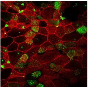
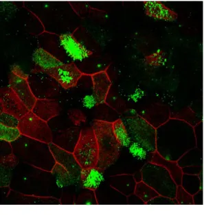
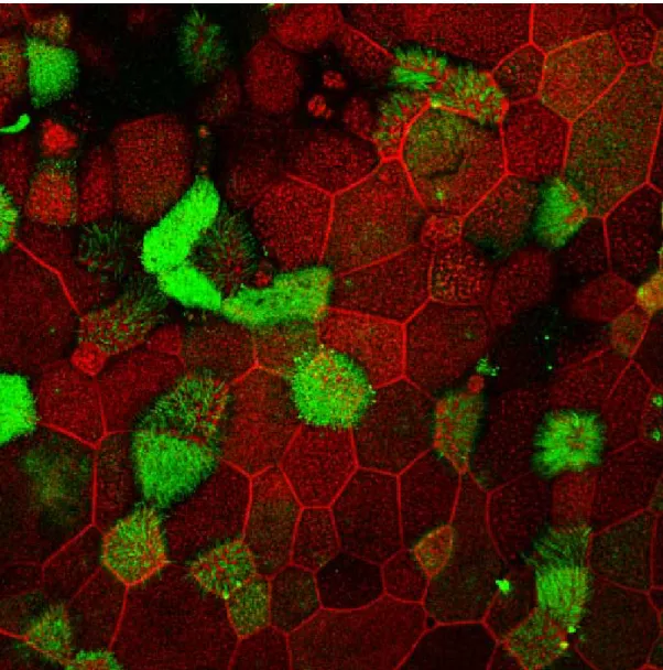
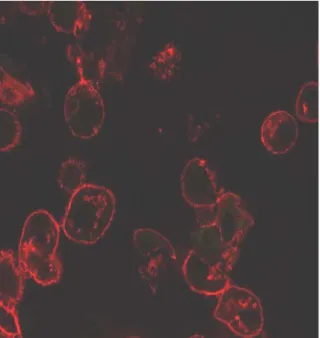
F-actin network labeling forms a distinctive ring at the periphery of cells. The gradual depletion of this ring (red) in cells is comparatively more obvious when treated twice daily for four consecutive days for 15 minutes [Figure 6] than those for 5 minutes [Figure 7]. Depletion in cells treated for 5 minutes is less but more comparable with the cells at the beginning of treatment [Figure 8]. A significant loss of F-actin network and loss of cells is noticeable in cells exposed to the more concentrated NaOCl (0.5% NaOCl) after only 5 minutes.
Day 1 Day 2
Day 3 Day 4
Figure 6: Confocal microscopic images of reconstituted nasal epithelial cells showing expression of ezrin (green) and F-actin network (red) delineating the cell outline after treatment with 0.05% Sodium Hypochlorite solution (prepared with PBS) exposed for 15 minutes, twice daily for four consecutive days
Day 1 Day 2
Day 3
Day 4
Figure 7: Confocal microscopic images of reconstituted nasal epithelial cells showing expression of ezrin (green) and F-actin network (red) delineating the cell outline after treatment with 0.05% Sodium Hypochlorite solution (prepared with PBS) exposed for 5 minutes, twice daily for four consecutive days.
Figure 8: Confocal microscopic images showing expression of ezrin (green) and F-actin network (red) delineating the cell outline in reconstituted nasal epithelial cells at the beginning of treatment.
Expression of ezrin in the apical domain of reconstituted epithelia.
After exposure to undiluted (0.5% NaOCl) and one tenth dilution (0.05% NaOCl) of Amuchina for 5 minutes, the epithelia cells were observed under confocal microscope for ezrin expression. No ezrin was detected in the epithelia exposed to 0.5% NaOCl [Figure 9a] even as early as 5 minutes. On the other hand expression of ezrin was observed in the cells exposed to diluted Amuchina (0.05% NaOCl) [Figure 9b].
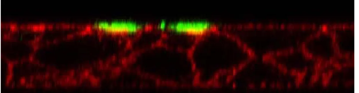
In Figure 10b, Z stack images of confocal microscopy demonstrate polarized apical expression of ezrin in epithelia cells after 4 days of treatment, twice daily for 5 minutes with 0.05% NaOCl which is comparable to the control [Figure 10a]. However, in case of epithelia cells treated for 15 minutes [Figure 10c], the thick apical band expression of ezrin is absent. There is redistribution of Ezrin indicating loss of polarity of cells. Absence of apical ezrin may be due to loss of ciliated cells.
Figure 6 and Figure 7 compare expression of ezrin in cells treated with 0.05% NaOCl, two times daily for four consecutive days, 15 minutes and 5 minutes respectively. Ezrin expression reduction was relatively more pronounced in the epithelia cells treated for 15 minutes. It is mentionable that no ezrin was expressed at all in cells exposed to 0.5% NaOCl for 5 minutes one time only [Figure 9a].
Changes in transepithelial resistance
TER values of cells treated with 0.05% NaOCl exposed for 5 minutes and 15 minutes respectively were compared. In Figure 11 the changes of TER values with time are expressed.
a) 0.5% Sodium hypochlorite
b) 0.05% Sodium hypochlorite
Figure 9: Confocal microscopic images of reconstituted nasal epithelial cells showing expression of ezrin (green) and F-actin network (red) delineating the cell outline after exposure to 0.5% and 0.05% Sodium Hypochlorite solution (prepared with PBS).
a) Control
b) 5 minute
c) 15 minutes
Figure 10: Confocal microscopic Z stack images of reconstituted nasal epithelial cells showing expression of ezrin (green) and F-actin network (red) delineating the cell outline at the end of treatment for 4 days of a) control (PBS) and after exposure for twice daily to 0.05% Sodium Hypochlorite solution for b) 5 minutes and c)15 minutes.
0 500 1000 1500 2000 2500 3000 TER Ω . cm2 Control 5 minutes 15 minutes M A Day 1 M A Day 2 M A Day 3 M A Day 4 M A Day 5 Treatment (days)
Figure 11: Transepithelial resistance changes of reconstituted epithelia at the end of treatment for 4 days of a) control (PBS) and after exposure for twice daily (“M” morning and “A” afternoon) for five days to 0.05% Sodium Hypochlorite solution for b) 5 minutes and c) 15 minutes. Mean ± SEM
Values are given as Mean ± SEM. TER was measured twice daily for five days. A Student t test was performed to compare the changes in TER of control cells and cells treated for 5 minutes and 15 minutes with 0.05% NaOCl with TER value of cells at day Zero at the beginning of treatment. There was a statistically significant (p<0.001) drop in TER of control cells and treated cells (5 and 15 minutes) at the end of day 5 in comparison to TER of cells at day Zero. Sodium hypochlorite was diluted with (phosphate buffered saline) PBS. To validate whether the drop in TER was due to NaOCl or PBS treatments, we performed the experiment
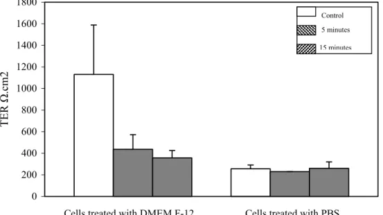
in DMEM-F12 containing 25mM of N-2-hydroxyethylpiperazine-N -2-ethanesulfonic acid (HEPES) pH 7.4 that will stabilize the pH at the apical surface of the epithelia, during treatment [Figure 12]. At the end of 5 days the TER value in control cells treated with DMEM-F12 was 1131 ± 458 Ω/cm2, a 57% decrease from the TER values at the beginning of the experiment. The TER values of cells treated with PBS (255 ± 36 Ω/cm2) had a 90%
decrease from initial TER values.
0 200 400 600 800 1000 1200 1400 1600 1800 Treatment (days) TER Ω .cm2 Control 5 minutes 15 minutes
Cells treated with PBS Cells treated with DMEM F-12
Figure 12: Comparison of transepithelial resistance of reconstituted nasal epithelial cells at the end of treatment for two times per day (“M” morning and “A” afternoon) for five days with 0.05% NaOCl (control, 5 and 15 minutes), and with PBS and DMEM-F12 separately. Mean ± SEM
MTT assay
Treatment with NaOCl induced a reduction in TER, which, however, was not paralleled by a decrease in the viability of epithelial cells, as evaluated by the MTT assay. This drop increased permeability and presumably is correlated to the disorganization of TJ belts that surround epithelial cells. Opening of the paracellular route induces a loss of polarity with redistribution of apical marker to basolateral domains. These preliminary data suggest that disruption of the tight junction might lead to a disorganization of F-actin as well as the redistribution of ZO-1 and other TJ's associated molecules. However, further experiments will be required in order to validate the possible changes in the F-actin cytoskeleton as well as the localization of TJ proteins (occludin and claudins) and ZO-1 during the 5 day treatment.
IN VIVO
Nasal Subjective Symptom Score and Nasal endoscopic findings
The degree of variability of the pre and post treatment data is depicted as a box-and-whiskers plot in Figure 13 and Figure 14. A thick vertical line indicates the median value, a horizontal shaded box represents the interquartile range between the 25th and 75th percentile of the data. Mean of the VAS values (pre and post treatment) is given in Table 2. Table 3 illustrates the mean and confidence interval of the individual difference of VAS at the end of 3 months of treatment with NaOCl. Statistically significant improvement was found in all nasal endoscopic findings (p<0.05). In case of subjective symptoms, statistically significant improvement was found for all symptoms, except only for anterior (Wilcoxon sign rank test, p=0.106) nasal discharge.
A: Nasal obstruction Post Pre S ubj e c ti v e S y m p to m S c or e 12 10 8 6 4 2 0 -2
B: Anterior nasal discharge
Post Pre S u b je c ti ve sym p to m S c o re 12 10 8 6 4 2 0 -2 *** NS
C: Posterior Nasal Discharge
Post Pre S u b je c ti v e S y m p to m S c o re 12 10 8 6 4 2 0 -2 D: Smell Post Pre S u b je c ti v e S y m p to m S c o re 12 10 8 6 4 2 0 -2 * ** E: Headache Post Pre S u b je c ti v e S y m p to m S c o re 12 10 8 6 4 2 0 -2 **
Figure 13. Comparison of subjective symptoms recorded by visual analogue scale, pre and
post treatment. The Box-and-whiskers plot represents the median and the 25th and 75th
A: Oedema Post Pre Su b je c tiv e S y m p to m Sc o re 12 10 8 6 4 2 0 -2 B: Erythema Post Pre Su b je c ti v e Sym p to m S c o re 12 10 8 6 4 2 0 -2 *** ***
C: Purulent Nasal Discharge
Post Pre Su b je c ti v e Sy m p to m Sc o re 12 10 8 6 4 2 0 -2 D: Crust Post Pre S u b je c ti v e S y m p to m s c o re 12 10 8 6 4 2 0 -2 ** ***
Figure 14. Comparison of nasal endoscopic findings recorded by visual analogue scale, pre
and post treatment. The Box-and-whiskers plot represents the median and the 25th and 75th
Table 2 : Mean of the visual analogue scale values of subjective symptoms and nasal endoscopic findings (n=14).
Mean (±SE) Minimum Maximum
Subjective Symptom Score Nasal Obstruction
Pre-treatment 5.78 (±0.72) 2 10
Post-treatment 2.00 (±0.57) 0 7
Anterior Nasal Discharge
Pre-treatment 3.92 (±0.99) 0 10
Post-treatment 2.43 (±0.61) 0 6
Posterior Nasal Discharge
Pre-treatment 4.75 (±0.79) 0 10 Post-treatment 2.00 (±0.56) 0 6 Smell disturbance Pre-treatment 5.39 (±1.07) 0 10 Post-treatment 1.54 (±0.92) 0 10 Headache Pre-treatment 4.29 (±1.14) 0 10 Post-treatment 0.93 (±0.46) 0 5
Nasal Endoscopic Findings Oedema Pre-treatment 6.00 (±0.74) 0 9.5 Post-treatment 1.29 (±0.41) 0 5 Erythema Pre-treatment 6.93 (±0.71) 0 10 Post-treatment 1.04 (±0.30) 0 3 Purulent discharge Pre-treatment 7.75 (±0.94) 0 10 Post-treatment 1.00 (±0.55) 0 7 Crusts Pre-treatment 7.64 (±0.83) 0 10 Post-treatment 0.89 (±0.44) 0 6
Table 3: Mean ± Standard error of mean (SEM) difference in the improvement of visual analogue scale scores of subjective symptoms and nasal endoscopic findings after treatment
Mean of 95% Confidence P value* change (±SEM) Interval
Subjective symptom score
Nasal Obstruction 3.79 (±0.61) 2.48 to 5.09 P = .001 Nasal Discharge Anterior 1.50 (±1.03) -0.72 to 3.72 P = .129 Posterior 2.75 (±0.96) 0.68 to 4.82 P = .018 Smell disturbance 3.86 (±0.96) 1.80 to 5.92 P = .007 Headache 3.36 (±0.94) 1.32 to 5.39 P = .009
Nasal Endoscopic Findings
Oedema 4.71 (±0.70) 3.20 to 6.23 P = .001
Erythema 5.89 (±0.91) 3.93 to 7.85 P = .001
Purulent discharge 6.75 (±0.96) 4.69 to 8.81 P = .002
Crusts 6.75 (±0.81) 5.01 to 8.49 P = .001
Four of our patients had nasal polyps. All of them showed mild improvement in the nasal polyp stage [34]. Two patients had Stage 1 nasal polyposis, which after treatment showed remission to Stage 0. The other two had Stage 2 and 3 which three months after treatment became Stage 1 and 2 respectively [Table 4].
Table 4: Changes in nasal polyp stage [34] and smell disturbances after treatment with Sodium Hypochlorite (n=4)
Pre treatment Post treatment
Polyp Smell Nasal Polyp Smell Nasal
Stage disturbance obstruction stage score obstruction
1 6 4 0 0 0
1 0 9 0 0 7
2 5 5 1 0 4
Nitric Oxide measurement
Pre- and post- treatment NO was measured in 10 patients only due to unavailability of equipment. The pre- and post- treatment NO, Mean ± SEM was 366.75 ± 97.94 ppb and 282 ± 73.26 respectively. The difference was not statistically significant. (p=0.06; Wilcoxon signed rank test). Figure 15 illustrates in error bars the Mean ± 95% CI of pre- and post- treatment NO values.
(p=0.06; W ilcoxon signed rank test).
Post Pre N it ric o x id e ( p a rt s p e r b illio n ) 700 600 500 400 300 200 100 0
Figure 15: Mean ± 95% Confidence Interval of pre- and post- treatment values of Nitric Oxide.
Nasal Airway Resistance measurements
Mean tNAR decreased from 0.45 Pa/cm3/s before therapy to 0.35 Pa/cm3/s after three months of 0.05% of NaOCl, corresponding to a 33% decrease. There was a statistically significant decrease in NAR (p=0.05; wilcoxon signed rank test). Figure 16 illustrates the Mean ± 95% CI of pre- and post- treatment tNAR values.
(*p<0.05; W ilcoxon signed rank test)
Post Pre Na s a l a ir w a y r e si sta n ce ( P a /cm 3 /s) .8 .7 .6 .5 .4 .3 .2 .1 0.0 *
Figure 16: Total Mean ± 95% Confidence Interval of pre- and post- treatment values of nasal airway resistance
Discussion
The main finding of this study is that NaOCl nasal lavages seem to be a safe, cheap and efficient alternative to systemic antibiotics in the medical treatment of CRS. Previous studies have demonstrated the efficacy of treating infection with NaOCl. However no study was found in the literature on the use of NaOCl in treating CRS. Additionally the cellular effects of NaOCl may not necessarily be comparable in all tissues [38]. Hence there was a need to do in vitro test for an appropriate concentration of NaOCl, duration of exposure with nasal epithelium that would not be harmful yet bacteriocidal, before in vivo application.
The depletion of F-actin network in our study indicated that cytotoxic effect of NaOCl depends on both concentration and duration of exposure. Chang et al [38] demonstrated a dose-dependent inhibition of mitochondrial activity by NaOCl and suggested that the cytotoxic effect might be via DNA or RNA synthesis. However the cellular effects of NaOCl may not necessarily be comparable in all tissue.
Expression of Ezrin agreed with alteration in F-actin network. Five minutes after exposure to 0.5% NaOCl, reconstituted epithelia had a reduced number of ciliated cells and the altered ciliated cells failed to express ezrin. Ezrin expression in cells exposed to 0.05% NaOCl was also far more reduced compared to cells exposed for 5 minutes. Ezrin expressed in the apical membrane of epithelial cells lining the airways is an indicator of polarity and cell differentiation. Thus the in vitro response by F-actin network and ezrin expression of reconstituted nasal epithelium have demonstrated that 0.05% NaOCl induced less epithelial
cell cytotoxicity than 0.5% NaOCl. We also demonstrated that 5 minutes exposure twice daily for consecutive days to 0.05% NaOCl was less harmful than 15 minutes exposure.
The alteration of F-actin network and ezrin expression are frequently associated with changes in the appearance and function of TJ’s. We therefore examined the effect on TER. There was a statistically significant drop (p<0.001) in TER at the end of 5 days in control cells and cells treated with 0.05% NaOCl for 5 minutes and 15 minutes daily in comparison to TER values at day zero. The reduction of TER in cells not treated with NaOCl might indicate that the changes have been due to the diluent (PBS) rather than NaOCl. But when dilutions were made in DMEM-F12, the reduction in TER was not so drastic in control cells, at the end of five day treatment. The discrepancy between ezrin expression at the cell surface and F-actin network may be due to a change in the cell polarity, possibly due to the opening of TJ of the reconstituted nasal epithelia. We thus propose to investigate whether, upon treatment, TJ are modulated and if the paracellular route is then made accessible to bacteria or inert particles. Furthermore, it will be interesting to investigate whether bacteria infected nasal reconstituted epithelial cells are still responsive to NaOCl and then, to evaluate the response in term of TER and bacterial killing.
Application of NaOCl in soaked gauze or dressing would have increased the exposure time and thus increase the bactericidal effect [21]. However this may raise the toxicity of NaOCl causing noticeable epithelial cell damage [14]. In addition a soaked gauze or a dressing can cause obstruction in the nasal airway resulting in difficulty of respiration.
We have demonstrated that increasing the time of exposure to 0.05% NaOCl may decrease cell viability. Thus when applied for consecutive days, it is more suitable if the contact period
is not prolonged. Such application can be made by lavage or irrigation where the solution washes the surface area.
Thus to see the clinical efficacy of NaOCl, we administered 0.05% NaOCl twice daily continuously for three months on both nostrils with Sinus Rinse bottle (NeilMed Products, USA). These bottles were easy to use and provided a smooth flow of lavage in the nasal cavity. A lavage has certain advantages over the administration of topical or i.v. antibiotics. Any watery solution removes secretions and crusts from the nose and irrigating the nasal cavity in itself would therefore produce therapeutic effect [5]. The action of the lavage may be effected through mechanical cleansing of the sinus surface, altering the composition of the mucus or reducing local concentrations of bacterial or fungal organisms, their toxins or pro-inflammatory substances released during pro-inflammatory responses to these [6].
The two main sources of NaOCl are pharmaceutical supplies or commercially available household bleaches. NaOCl from commercial sources have been used for many years for clinical purpose without significant problems [17]. Thus the solutions may be available at a very affordable price. Often the price of syringes and needles are greater than the NaOCl it contains [14, 41].
In our study as an alternative therapy for persistent CRS we have demonstrated significant improvements in symptoms with the administration of 0.05% NaOCl. Except for anterior nasal discharge (AND), a statistically significant improvement was recorded for nasal obstruction, posterior nasal discharge, smell and headache. Nasal endoscopic findings demonstrated significant reduction in oedema, erythema, purulent discharge and nasal crusts.
improvement in AND may be explained by increased flow due to decreased crust and widening. Our post treatment CRS symptom scores were similar to sinus symptom scores recorded in 100 normal individuals by Walker and White [42] indicating the efficacy of treatment [Table 5].
Objective Rhm recordings have shown that NAR was significantly reduced at the end of 3 months of treatment. Active anterior Rhm is a reliable method of assessing the functional status of the nasal cavities in different situations [43].
NO levels did not show statistically significant changes between pre- and post- treatment values even though symptomatic and endoscopic findings documented improvements. In fact in our study the post treatment NO level was higher. This lack of improvement in NO level correlates with a study carried out by Cervin et al. [44], who demonstrated no significant improvement in NO level despite of significant improvement in symptoms in patients who received long term low dose erythromycin therapy for CRS after 3 months. After 12 months of therapy there was a trend toward an increase in nasal NO. This can be explained as a result of the gradual restoration of normal epithelium function due to appearance of ciliated cells, reduction of inflammation and reduction in nasal secretion. It will be interesting to follow up our patients for a longer period to monitor if there is an increase or decrease in NO levels.
Table 5. Mean (minimum and maximum) scores from normal subjects [42] and patients three months after treatment with 0.05% Sodium Hypochlorite.
Symptoms Normal subjects [56] Patients post-treatment (n=100) (n=14)
Nasal obstruction 2.3 (0 to 7) 2.0 (0 to 7)
Nasal Discharge 2.0 (0 to 7) 2.2* (0 to 6)
Disturbance of smell 1.7 (0 to10) 1.5 (0 to 10)
Headache 1.2 (0 to 8) 0.9 (0 to 5)
CONCLUSION
Our in vitro test demonstrated that 0.05% NaOCl may be used on nasal epithelium and found it to be effective in treating persistent CRS as an alternative to antibiotic therapy. With no known side effects and risk of developing tolerance as well as due to its cheap cost and method off application, nasal lavage with NaOCl is advantageous over other alternative therapies and should be recommended for the treatment of CRS.
REFERENCES
1. European Academy of Allergology and Clinical Immunology. European position paper on rhinosinusitis and nasal polyps. Rhinol Suppl., 2005;(18):1-87.
2. Kingdom TT, Swain RE Jr.The microbiology and antimicrobial resistance patterns in chronic rhinosinusitis. Am J Otolaryngol. 2004 Sep-Oct;25(5):323-8
3. Tomasz A. Multiple-antibiotic-resistant pathogenic bacteria. A report on the Rockefeller University Workshop. N Engl J Med. 1994 Apr 28;330(17):1247-51.
4. Swartz MN. Use of antimicrobial agents and drug resistance. N Engl J Med. 1997 Aug 14;337(7):491-2.
5. Bachmann G, Hommel G, Michel O. Effect of irrigation of the nose with isotonic salt solution on adult patients with chronic paranasal sinus disease. Eur Arch Otorhinolaryngol. 2000 Dec;257(10):537-41
6. Desrosiers MY, Salas-Prato M. Treatment of chronic rhinosinusitis refractory to other treatments with topical antibiotic therapy delivered by means of a large-particle nebulizer: results of a controlled trial. Otolaryngol Head Neck Surg. 2001 Sep;125(3):265-9
7. Giger R, Landis BN, Zheng C, Malis DD, Ricchetti A, Kurt AM, Morel DR, Lacroix JS. Objective and subjective evaluation of endoscopic nasal surgery outcomes. Am J Rhinol. 2003 Nov-Dec;17(6):327-33.
8. Anand V, Levine H, Friedman M, Krespi Y, Panje W, Schettino R, Stankiewicz J, Tichenor W, Kacker A, Horn C. Intravenous antibiotics for refractory rhinosinusitis in nonsurgical patients: preliminary findings of a prospective study. Am J Rhinol. 2003 Nov-Dec;17(6):363-8.
9. Lavigne F, Tulic MK, Gagnon J, Hamid Q. Selective irrigation of the sinuses in the management of chronic rhinosinusitis refractory to medical therapy: a promising start. J Otolaryngol. 2004 Feb;33(1):10-6.
10. Gliklich RE, Metson R. Economic implications of chronic sinusitis. Otolaryngol Head Neck Surg. 1998 Mar;118(3 Pt 1):344-9
11. Vianna ME, Gomes BP, Berber VB, Zaia AA, Ferraz CC, de Souza-Filho FJ. In vitro evaluation of the antimicrobial activity of chlorhexidine and sodium hypochlorite. Oral Surg Oral Med Oral Pathol Oral Radiol Endod. 2004 Jan;97(1):79-84.
12. Perin FM, Franca SC, Silva-Sousa YT, Alfredo E, Saquy PC, Estrela C, Sousa-Neto MD. Evaluation of the antimicrobial effect of Er:YAG laser irradiation versus 1%
sodium hypochlorite irrigation for root canal disinfection. Aust Endod J. 2004 Apr;30(1):20-2.
13. Mehl A, Folwaczny M, Haffner C, Hickel R. Bactericidal effects of 2.94 microns Er:YAG-laser radiation in dental root canals. J Endod. 1999 Jul;25(7):490-3.
14. Cotter JL, Fader RC, Lilley C, Herndon DN. Chemical parameters, antimicrobial activities, and tissue toxicity of 0.1 and 0.5% sodium hypochlorite solutions. Antimicrob Agents Chemother. 1985 Jul;28(1):118-22.
15. Sassone LM, Fidel R, Fidel S, Vieira M, Hirata R Jr. The influence of organic load on the antimicrobial activity of different concentrations of NaOCl and chlorhexidine in vitro. Int Endod J. 2003 Dec;36(12):848-52.
16. Estrela C, Ribeiro RG, Estrela CR, Pecora JD, Sousa-Neto MD. Antimicrobial effect of 2% sodium hypochlorite and 2% chlorhexidine tested by different methods. Braz Dent J. 2003;14(1):58-62. Epub 2003 Jul 31.
17. Frais S, Ng YL, Gulabivala K. Some factors affecting the concentration of available chlorine in commercial sources of sodium hypochlorite. Int Endod J. 2001 Apr;34(3):206-15.
18. Fader RC, Maurer A, Stein MD, Abston S, Herndon DN. Sodium hypochlorite decontamination of split-thickness cadaveric skin infected with bacteria and yeast with subsequent isolation and growth of basal cells to confluency in tissue culture. Antimicrob Agents Chemother. 1983 Aug;24(2):181-5.
19. Milner SM, Heggers JP. The use of a modified Dakin's solution (sodium hypochlorite) in the treatment of Vibrio vulnificus infection. Wilderness Environ Med. 1999 Spring;10(1):10-2.
20. Zehnder M, Kosicki D, Luder H, Sener B, Waltimo T. Tissue-dissolving capacity and antibacterial effect of buffered and unbuffered hypochlorite solutions. Oral Surg Oral Med Oral Pathol Oral Radiol Endod. 2002 Dec;94(6):756-62.
21. Gernhardt CR, Eppendorf K, Kozlowski A, Brandt M. Toxicity of concentrated sodium hypochlorite used as an endodontic irrigant. Int Endod J. 2004 Apr;37(4):272-80.
22. Ayhan H, Sultan N, Cirak M, Ruhi MZ, Bodur H. Antimicrobial effects of various endodontic irrigants on selected microorganisms. Int Endod J. 1999 Mar;32(2):99-102. 23. Heggers JP, Sazy JA, Stenberg BD, Strock LL, McCauley RL, Herndon DN, Robson MC. Bactericidal and wound-healing properties of sodium hypochlorite solutions: the 1991 Lindberg Award. J Burn Care Rehabil. 1991 Sep-Oct;12(5):420-4
24. Papakonstanti EA, Vardaki EA, Stournaras C. Actin cytoskeleton: a signaling sensor in cell volume regulation. Cell Physiol Biochem. 2000;10(5-6):257-64.
25. Cantiello HF. Role of actin filament organization in cell volume and ion channel regulation. J Exp Zool. 1997 Dec 1;279(5):425-35.
26. Turner JR.'Putting the squeeze' on the tight junction: understanding cytoskeletal regulation. Semin Cell Dev Biol. 2000 Aug;11(4):301-8.
27. Huang T, You Y, Spoor MS, Richer EJ, Kudva VV, Paige RC, Seiler MP, Liebler JM, Zabner J, Plopper CG, Brody SL. Foxj1 is required for apical localization of ezrin in airway epithelial cells. J Cell Sci. 2003 Dec 15;116(Pt 24):4935-45.
28. Godfrey RW. Human airway epithelial tight junctions. Microsc Res Tech. 1997 Sep 1;38(5):488-99.
29. Lundberg JO, Farkas-Szallasi T, Weitzberg E, Rinder J, Lidholm J, Anggaard A, Hokfelt T, Lundberg JM, Alving K. High nitric oxide production in human paranasal sinuses. Nat Med. 1995 Apr;1(4):370-3.
30. Oh CK, Miman MC, Duncavage JA. Current value of nasal nitric oxide measurement in rhinology. Curr Opin Otolaryngol Head Neck Surg. 2004 Feb;12(1):30-3.
31. Hirschberg A. Rhinomanometry: an update. ORL J Otorhinolaryngol Relat Spec. 2002 Jul-Aug;64(4):263-7.
32. Tatnall FM, Leigh IM, Gibson JR. Assay of antiseptic agents in cell culture: conditions affecting cytotoxicity. J Hosp Infect. 1991 Apr;17(4):287-96.
33. Karp PH, Moninger TO, Weber SP, Nesselhauf TS, Launspach JL, Zabner J, Welsh An in vitro model of differentiated human airway epithelia. Methods for establishing primary cultures. Methods Mol Biol. 2002;188:115-37.
34. Lacroix JS, Zheng CG, Goytom SH, Landis B, Szalay-Quinodoz I, Malis DD.Histological comparison of nasal polyposis in black African, Chinese and Caucasian patients Rhinology. 2002 Sep;40(3):118-21.
35. Kharitonov S, Alving K, Barnes PJ. Exhaled and nasal nitric oxide measurements: recommendations. The European Respiratory Society Task Force. Eur Respir J. 1997 Jul;10(7):1683-93
36. Wodehouse T, Kharitonov SA, Mackay IS, Barnes PJ, Wilson R, Cole PJ. Nasal nitric oxide measurements for the screening of primary ciliary dyskinesia. Eur Respir J. 2003 Jan;21(1):43-7
37. Giger R, Pasche P, Cheseaux C, Cantini L, Rossetti A, Landis BN, Lacroix JS. Comparison of once- versus twice-daily use of beclomethasone dipropionate aqueous nasal spray in the treatment of allergic and non-allergic chronic rhinosinusitis. Eur Arch Otorhinolaryngol. 2003 Mar;260(3):135-40. Epub 2002 Nov 6.
38. Chang YC, Huang FM, Tai KW, Chou MY.The effect of sodium hypochlorite and chlorhexidine on cultured human periodontal ligament cells. Oral Surg Oral Med Oral Pathol Oral Radiol Endod. 2001 Oct;92(4):446-50.
39. Vastag M, Neuhofer W, Nagel W, Beck FX. Ammonium affects tight junctions and the cytoskeleton in MDCK cells. Pflugers Arch. 2005 Jan;449(4):384-91. Epub 2004 Sep 8.
40. Jessup W, Shirazi MF, Dean RT. Inhibition of some spontaneous secretory processes in macrophages and fibroblasts by ammonium chloride. Biochem Pharmacol. 1983 Sep 15;32(18):2703-10..
41. Clarkson RM, Moule AJ. Sodium hypochlorite and its use as an endodontic irrigant. Aust Dent J. 1998 Aug;43(4):250-6.
42. Walker FD, White PS. Sinus symptom scores: what is the range in healthy individuals? Clin Otolaryngol Allied Sci. 2000 Dec;25(6):482-4.
43. Weinke T, Schiller R, Fehrenbach FJ, Pohle HD. Association between Staphylococcus aureus nasopharyngeal colonization and septicemia in patients infected with the human immunodeficiency virus. Eur J Clin Microbiol Infect Dis. 1992 Nov;11(11):985-9.
44. Cervin A, Kalm O, Sandkull P, Lindberg S. One-year low-dose erythromycin treatment of persistent chronic sinusitis after sinus surgery: clinical outcome and effects on mucociliary parameters and nasal nitric oxide. Otolaryngol Head Neck Surg. 2002 May;126(5):481-9.
INDEX
Index i
Abbreviations ii
List of Tables iii
List of Figures iv Résumé 3 Introduction (French) 4 Summary 11 Acknowledgement 13 Introduction 16 Aim of thesis 24
Material and method 25
Statistical analysis 32
Results 33
Discussion 51
Conclusion 56
![Figure 1: Annual expenditure of the pharmacologic management of chronic rhinosinusitis in the United States [10]](https://thumb-eu.123doks.com/thumbv2/123doknet/2205388.12955/16.892.129.731.515.767/figure-annual-expenditure-pharmacologic-management-chronic-rhinosinusitis-united.webp)