HAL Id: dumas-01787599
https://dumas.ccsd.cnrs.fr/dumas-01787599
Submitted on 7 May 2018HAL is a multi-disciplinary open access archive for the deposit and dissemination of sci-entific research documents, whether they are pub-lished or not. The documents may come from teaching and research institutions in France or abroad, or from public or private research centers.
L’archive ouverte pluridisciplinaire HAL, est destinée au dépôt et à la diffusion de documents scientifiques de niveau recherche, publiés ou non, émanant des établissements d’enseignement et de recherche français ou étrangers, des laboratoires publics ou privés.
A mild form of oculocutaneous albinism type 1:
phenotypic analysis of compound heterozygous patients
with the R402Q variant of the TYR gene
Solène Monfermé
To cite this version:
Solène Monfermé. A mild form of oculocutaneous albinism type 1: phenotypic analysis of compound heterozygous patients with the R402Q variant of the TYR gene. Human health and pathology. 2017. �dumas-01787599�
Année 2017
Thèse N° 3074
U.F.R. des Sciences Médicales
THÈSE
Pour l’obtention du
DIPLOME D’ETAT DE DOCTEUR EN MEDECINE
Discipline : Ophtalmologie
Présentée et soutenue publiquement le 07 juillet 2017 par
Solène MONFERMÉ
née le 5 aout 1988 à Neuilly-sur-Seine
A MILD FORM OF OCULOCUTANEOUS ALBINISM TYPE 1:
PHENOTYPIC ANALYSIS OF COMPOUND
HETEROZYGOUS PATIENTS WITH THE R402Q VARIANT
OF THE TYR GENE
Directeur de thèse :
Monsieur le Docteur Clément PAYA
Rapporteur de thèse :
Monsieur le Professeur Arnaud SAUER
Membres du jury
Présidente :
Madame le Professeur Marie-Noëlle DELYFER
Juges :
Monsieur le Professeur Benoit ARVEILER
Monsieur le Professeur Jean-François KOROBELNIK
Madame le Docteur Eulalie LASSEAUX
REMERCIEMENTS
A notre respectée coordinatrice de DES et présidente du jury,
Madame le Professeur Marie-Noëlle Delyfer,
Je vous suis extrêmement reconnaissante pour votre investissement dans la formation des internes en tant que coordinatrice du DES d’ophtalmologie, veillant sur chacun d’entre nous, disponible et à l’écoute dès notre première année d’internat et aux étapes clef de notre cursus. Votre professionnalisme, votre rigueur, mais aussi, vos qualités humaines et votre
bienveillance sont pour moi autant d’exemples que je n’aurai de cesse de tenter d’imiter. Merci de me faire l’honneur de juger ce travail et de présider ce jury. Soyez assurée de ma reconnaissance et de ma sincère admiration.
A mon Maître et directeur de Thèse,
Monsieur le Docteur Clément Paya,
Tu as conforté mon goût pour l’ophtalmo-pédiatrie, à l’écoute dès mon premier semestre au CHU, me fournissant déjà d’excellents conseils pour l’internat en général et pour une orientation en pédiatrie d’autre part. Je te dois cette excellente expérience au sein du service du Dr Caputo à la Fondation Rothschild, que tu m’avais recommandé. Merci de m’avoir suggéré ce travail sur un thème qui m’a passionné et beaucoup enrichie au contact de l’équipe de génétique du CHU de Bordeaux. Merci pour tes conseils toujours très justes pendant la rédaction de ce travail, pour ta rigueur scientifique et ton goût de la recherche que tu m’auras j’espère un peu transmis, pour ta patience et ta grande disponibilité, pour tes encouragements et ta confiance. Merci enfin et surtout pour le modèle de médecin que tu es, consciencieux, passionné, patient et à l’écoute avec tes patients de tous âges. J’espère parvenir à m’en inspirer pour ma pratique future.
A mon rapporteur,
Monsieur le Professeur Arnaud Sauer,
Merci de m’avoir fait l’honneur d’accepter de juger ce travail et d’y apporter votre regard critique d’expert en ophtalmo pédiatrie. Merci pour le temps que vous m’avez accordé et pour vos conseils. Recevez toute ma reconnaissance et ma gratitude.
Aux membres du jury
Monsieur le Professeur Benoit Arveiler,
Merci d’avoir accepté de me confier ce sujet que je sais être le fruit d’années de travail et de recherche au sein du laboratoire de génétique du CHU de Bordeaux et qui m’a réellement passionné. Comment aurait il pu en être autrement au contact des membres de votre équipe, qui m’ont accueillie, ont répondu à toutes mes questions, pardonnant mes connaissances si limitées en génétique et m’ont présentée leur travaux et projets, tous avec une passion contagieuse ne pouvant qu’éveiller mon intérêt. Merci de m’avoir confié ce travail et de m’avoir accordé votre confiance. Merci pour vos conseils indispensables, vos relectures attentives et votre disponibilité. J’espère avoir été à la hauteur de votre confiance. J’espère aussi enfin, pouvoir encore à l’avenir collaborer avec votre service, entendant bien enrichir d’ophtalmologie-génétique ma pratique de l’ophtalmo-pédiatrie et de l’ophtalmologie en générale.
Monsieur le Professeur Jean-François Korobelnik,
Merci de me faire l’honneur de juger le travail de thèse venant clôturer 5 ans de formation d’interne. Merci pour votre exigence au sein du service d’ophtalmologie du CHU de bordeaux qui en fait un centre dynamique propice à la formation des internes. Soyez assuré de mon plus profond respect.
Madame le Docteur Eulalie Lasseaux,
Les gens passionnés sont toujours passionnants, et il est certains que te rencontrer aura modifié profondément ma perception d’une science, très obscure pour moi il y a encore 1 an et que j’essai désormais d’apprivoiser un peu tant bien que mal : la génétique ! Un domaine vaste et plein de promesse d’avenir dans lequel je suis bien heureuse que ma thèse m’ait plongée. Merci d’avoir accepté de me confier un sujet sur lequel tu as tant travaillé et de m’avoir donner les clefs pour l’appréhender. Merci pour ta grande patience, ta disponibilité, ta bonne humeur et ton exemplaire motivation.
Aux praticiens de toute la France qui ont acceptés et ont pris le temps de me transmettre les dossiers de leurs patients
Merci pour la confiance que vous avez témoignée dans ce travail en acceptant d’y participer et merci pour les efforts que vous avez consacrez à cette aide qui nous était indispensable. Merci :
Au Pr Hamel de Montpellier
Au Pr Dollfus et à Madame Pelletier de Strasbourg Au Dr Defoort de Lille Au Dr Vincent-Delorme de Lille Au Dr Duncombe-Poulet de Caen Au Dr Zanlonghi de Nantes Au Pr Bonneau d’Anger Au Dr Thauvin de Dijon Au Dr Pallot de Dijon Au Dr Derrieux de Caen
Au Dr Demurger et au Dr Quelin de Rennes Au Dr Gambarelli de Marseille
Au Dr Morice-Picard Fanny : Merci pour ton aide indispensable dans le domaine de la dermatologie. Merci pour tes explications, tes conseils, pour le temps que tu m’as accordé et pour toutes ces belles photographies que tu as accepté de me confier pour illustrer les
SOMMAIRE GÉNÉRAL
I. ARTICLE ... 7
RESUMÉ ... 8
ABSTRACT ... 9
INTRODUCTION ... 10
PATIENTS AND METHODS ... 12
RESULTS ... 17
DISCUSSION ... 27
REFERENCES ... 35
II. ANNEXE 1 : Rappels sur l’albinisme oculocutané ... 41
III. ANNEXE 2 : Rappels sur l’hypoplasie fovéolaire et étiologies ... 57
IV. ANNEXE 3 : Fiche de renseignements cliniques pour les demandes de génotypage adressées au laboratoire de génétique moléculaire du CHU de Bordeaux ... 68
SOMMAIRE DES FIGURES ET TABLEAUX
Figure 1: A), Illustration of the normal foveal structural features detectable, using optical coherence tomography. B), Foveal hypoplasia severity scale according to structural features detectable using optical coherence tomography ... 16Table 1: Genotypes of all patients of the laboratory with two mutations identified for the TYR gene ... 18
Figure 2: Representative sample of the hair, iris and retinal pigmentation phenotypes in oculocutaneous type 1 patients, compounds heterozygous with one classical TYR mutation and the R402Q variant ... 23
Table 2: Demographics and phenotypic characteristics of patients oculocutaneous albinism du to TYR mutations and so consider as having oculocutaneous albinism type 1 (OCA1). Comparison between compound heterozygous patients with the R402Q variant and another pathogenic variant of the TYR gene (R402Q-OCA1), and patients with two pathogenic variants of the TYR gene, other tan R402Q (classical-OCA1) ... 24
Figure 3: Representative sample of foveal morphologies in oculocutaneous type 1 patients, compounds heterozygous with one classical TYR mutation and the R402Q variant (R201Q-OCA1 patients) ... 26
ABRÉVIATIONS
AROA : autosomal recessive ocular albinism ELM : external limiting membrane
FHONDA syndrome : Foveal Hypoplasia, Optique Nerve Décussation defect and Anterieur segment dysgenesis
GCL : ganglion cell layer
HPS : Hermansky-Pudlak syndrome (HPS1 – HPS10 = type 1 – 10) INL : inner nuclear layer
IPL : inner plexiform layer IZ : interdigitation zone
MRI: magnetic resonance imaging
NGS : next-generation sequencing
OA1 : ocular albinism type 1
OCA : oculocutaneous albinism (OCA1 – OCA7 = type 1 – 7) OCT : optical coherence tomography
ONL =:outer nuclear layer OPL : outer plexiform layer
PIS and POS : photoreceptor inner segments and photoreceptor outer segments separated by IS/OS line
RNFL : retinal nerve fiber layer RPE : retinal pigment epithelium.
SD-OCT : spectral domain-optical coherence tomography VEP: visual evoked potentials
I. ARTICLE
A MILD FORM OF OCULOCUTANEOUS ALBINISM TYPE 1:
PHENOTYPIC ANALYSIS OF COMPOUND HETEROZYGOUS PATIENTS
WITH THE R402Q VARIANT OF THE TYR GENE
*****************************
UNE FORME MODÉRÉE D’ALBINISME OCULO-CUTANÉE DE TYPE 1:
ANALYSE PHÉNOTYPIQUE DES HÉTÉROZYGOTES COMPOSITES
PORTEURS DU VARIANT R402Q DU GÈNE TYR
AUTEURS:
Monfermé Solène (Service d’ophtalmologie, CHU de Bordeaux) Dr Paya C. (Centre d’ophtalmologie du Palais Gallien, Bordeaux) Pr Korobelnik J-F. (Service d’ophtalmologie, CHU de Bordeaux)
Pr Taieb A. (service de dermatologie et dermatologie pédiatrique du CHU de Bordeaux) Dr Morice-Picard F. (Service de génétique médicale, Unité de dermato-pédiatrie du CHU de Bordeaux)
Pr Dollfus H. et à Madame Pelletier V. (Centre des affections rares en génétique ophtalmologique, CHU de Strasbourg)
Pr Hamel C. (Service d’ophtalmologie, Equipe maladies sensorielles génétiques, CHU de Montpellier)
Dr Defoort (Service d’exploration de la vision et neuro-ophtalmologie, CHRU de Lille) Dr Vincent-Delorme C. (Clinique de génétique Guy Fontaine, Pole de biologie pathologie génétique, CHRU de Lille)
Dr Duncombe-Poulet C. de Caen (Cabinet d’ophtalmologie, rue du Château d’eau, Caen) Dr Zanlonghi X. de Nantes (Clinique ophtalmologique Sourdille, Nantes)
Pr Bonneau d’Anger (Service de génétique, CHU d’Anger)
Dr Thauvin C. de Dijon (Centre de génétique, Hopital des enfants, CHU de Dijon) Dr Pallot C. de Dijon (Service d’ophtalmologie, CHU de Dijon)
Dr Derrieux L. de Caen (Cabinet d’ophtalmologie, Rue Bailey, Caen)
Dr Demurger F. et Dr Quelin C. de Rennes (Service de génétique médicale, CHU de Rennes) Dr Gambarelli N. de Marseille (Centre d’ophtalmologie Monticelli-Paradis, Marseille) Dr Lasseaux E. (Laboratoire de génétique Moléculaire, CHU de Bordeaux)
RÉSUMÉ
OBJECTIFS: L’albinisme oculo-cutané (OCA) est un groupe hétérogène d’affections
génétiques caractérisées par une hypopigmentation congénitale de la peau, des cheveux et des yeux. L’OCA type 1 est du à des mutations du gène TYR. R402Q est un variant thermosensible du gène TYR dont un rôle est suspecté dans des formes modérées d’OCA1. L’objectif de l’étude était de définir le phénotype associé à ce variant dans une large série.
METHODE: Une étude rétrospective a inclus tous les patients hétérozygotes composites
présentant le variant R402Q et un autre variant pathogène du gène TYR (groupe R402Q-OCA1, n=122) comparés à un groupe contrôle de patients présentant deux variants pathogènes autres que R402Q (groupe classical-OCA1, n=119). Les données cliniques ont été recueillies sur dossiers.
RESULTAS: Les patients R402Q-OCA1 présentaient le plus souvent des cheveux blancs ou
blanc-jaunes à la naissance (65,31%), blonds plus tard (62,96%), un phototype clair mais avec possibles naevi ou tendance à pigmenter (71,43%), des yeux bleus (82,73%). Leurs peau, cheveux et iris étaient significativement plus pigmentés que dans le groupe classical-OCA1 avec des teintes allant jusqu’au brun. Tous les patients du groupe R402Q-OCA1 présentaient des atteintes ophtalmologiques de l’albinisme. Les prévalences de nystagmus (84,75%), photophobie (78,13%) et d’hypopigmentation rétinienne (92,59%) dans ce groupe étaient cependant plus faibles que dans le groupe classical-OCA1. La sévérité des scores de transillumination irienne et d’hypoplasie fovéolaire était plus faible dans le groupe R402Q-OCA1. Enfin, l’acuité visuelle y était plus élevée avec une moyenne de 0,38±0,21 LogMAR (environ 20/50 Snellen) et au moins 20/40 Snellen chez 50% des patients.
CONCLUSION: Le variant R402Q est responsable de formes d’intensité variable et souvent
modérées d’albinisme possiblement sous-diagnostiquées. Un bilan et suivi ophtalmologique précoce permet d’optimiser le diagnostic et le pronostic visuel.
ABSTRACT
PURPOSE: Oculocutaneous albinism (OCA) is a heterogeneous group of genetic
abnormalities that typically presents with congenital hypopigmentation and affects skin, hair and eyes. OCA type 1 is due to TYR mutations. R402Q is a thermosensible variant of the TYR gene that has been reported to be responsible for mild forms of OCA1. The aim of our study was to define the phenotype associated to this variant.
METHODS: A retrospective study included all compound heterozygous patients harboring
the R402Q variant with one other pathogenic variant of the TYR gene (R402Q-OCA1 group, n=122) compared with a control group of patients with two pathogenic variants other than R402Q (classical-OCA1 group, n=119). Clinical data were collected from medical records.
RESULTS: R402Q-OCA1 patients more often presented with white or yellow-white hair at
birth (65.31%), blond hair later (62.96%), a light phototype but with possible pigmented neavi or a tendency to tan (71.43%), and blue eyes (82.73%). Their skin, hair and iris were significantly more pigmented than in the classical-OCA1 group with possible ginger or brown hair, green or brown eye. All patients from the R402Q-OCA1 group presented with ocular features of albinism. However the prevalences of nystagmus (84.75%), photophobia (78.13%) and retinal hypopigmentation (92.59%) in this group were significantly lower than in the classical-OCA1 group. The severity scores of iris transillumination and foveal hypoplasia were also lower in the OCA1 group. Finally, visual acuity was higher in the R402Q-OCA1 group with a mean visual acuity of 0.38±0.21 LogMAR (about 20/50 Snellen) and at least 20/40 Snellen in 50% patients.
CONCLUSION: The R402Q variant leads to variable but generally mild forms of albinism
whose less typical presentation may lead to underdiagnosis. Early ophthalmologic examination and follow-up are useful to optimize the diagnosis and the ophthalmologic prognosis.
INTRODUCTION
Albinism is a heterogeneous group of genetic abnormalities that presents with congenital hypopigmentation and more often affects both skin, hair and eyes (oculocutaneous albinism (OCA)), but can involve exclusively or predominantly the eyes (ocular albinism (OA)). Its estimated worldwide prevalence is 1/17 000 (1). The definition of albinism includes optic disorders in varying proportions: decreased visual acuity, refractive errors, strabismus, photophobia, nystagmus, hypopigmentation of the iris and iris transillumination, hypopigmentation of the retina, foveal hypoplasia, anomalies of the optic nerve head and an excess of optic nerve fibers decussation at the chiasm (2–6).
The common physiopathology consists in an absent or reduced melanin synthesis (7–10). Mutations in six genes have been reported to be responsible for different types of OCA with an autosomal recessive mode: the tyrosinase-encoding gene (TYR) responsible for OCA type1 (MIM# 203100), the OCA2 gene (P gene) in OCA type 2 (MIM# 203200), the tyrosinase-related protein-1 gene (TYRP1) in OCA type 3 (MIM# 203290), SLC45A2 in OCA type 4 (MIM# 606202), SLC24A5 in OCA type 6 (MIM# 113750) and C10ORF11 (MIM# 615179) in OCA type 7. Of note the locus for OCA type 5 (MIM# 615312) has been mapped to
chromosomal region 4q24 but the gene has not been identified yet (11).Some mutations are
responsible for syndromes associating a clinical phenotype of OCA with additional health consequences. These are Hermansky-Pudlak Syndrome (HPS1, AP3B1 (HPS2), HPS3, HPS4, HPS5, HPS6, DTNBP1 (HPS7), BLOC1S3 (HPS8), PLDN (HPS9) and AP3D1 (HPS10)) and Chediak-Higashi Syndrome (LYST). Mutations in one gene, GPR143, are responsible for X-linked ocular albinism, OA1 (MIM#300500) (3,12–16).
Among these different forms of albinism, OCA type 1 (OCA1) due to TYR mutations is the second most common form (after OCA2) worldwide with a prevalence estimated at 1/40 000 (17). This prevalence fluctuates widely in the different populations and is higher in Caucasians among whom OCA1 is the most common subtype found and accounts for more than 50% of all cases (18). OCA1 presents with usual features found in all types of albinism. It however exhibit variations in its clinical presentation, from the most severe form associated to no melanin synthesis in any tissue and sometime referred as OCA1A, to a phenotype associated with minimal amounts of melanin synthesis in the hair, skin, and eyes and sometime referred as OCA1B (19).
Since 1978 an autosomal recessive form of ocular albinism (AROA) has been described (20). Screening patients with AROA, mutations were found in the TYR gene, OCA2 or TYRP1
(21,22). In their series, Hutton et al found 56% of patients with TYR gene mutations among thirty-six patients with a clinical diagnosis of AROA. Most of them were compound heterozygous with one severe OCA1 mutant alleles and the R402Q variant of the TYR gene. The R402Q variant is a common variant of the TYR gene (located in 11q14.3) that has been described in the general population and particularly in the Caucasian populations (23). The age of the derived non-synonymous R402Q allele was estimated to be around 20 400 or 29 400 years and this allelic variant is mainly present in Europe and North Africa (24). Its global prevalence is about 17.7% and is higher among Caucasians. Indeed, its allele frequency is about 26.48% among European population whereas it is lower among Hispanics (9.16%), Africans (4.63%) and Asians (6.08% to 0.01%) (25).
R402Q is widely regarded as a neutral polymorphism rather than as a pathogenic variant and
do not lead to albinism in a homozygous statesince the prevalence of R402Q homozygous is
about 7.01% in the European population without albinism (Allele frequency in the European population (except Finnish)2 = 0.26482 = 7.01%) (25). However the role of this variant in mild forms of OCA1 has been suspected (26–28) since 1991.
The R402Q is a thermosensitive variant which renders the tyrosinase enzyme thermolabile, with only 25% of normal catalytic activity at body temperature of 37°C but approaching a normal activity at a lower temperature (23,29). This poor enzymatic activity is explained by a nearly absolute and irreversible endoplasmic reticulum retention at the restrictive temperature of 37°C while a 31-32 °C temperature permits endoplasmic endothelium exit of at least a fraction of the newly synthesized tyrosinase and subsequent transport to late endosomes or melanosomes (30,31).
There is currently no study in the literature analyzing the phenotype of OCA1 patients who are compound heterozygous with one TYR mutation and the R402Q variant in a large cohort. The aim of our study was to define the phenotype and estimate the ophthalmological prognosis associated to this specific genotype in a large series of patients.
SUBJECTS AND METHODS
Subjects
The molecular genetics laboratory of Bordeaux University hospital has a fifteen years long experience in albinism. It receives samples of patients with a clinical diagnosis of albinism
from all over France and occasionally from other,mainly European, countries.
The genotypes of all the patients whose genotyping has been performed by the laboratory are listed in a register.
The population of our study included all the patients of this register fulfilling the following criteria for the cases and controls groups. Were eligible as cases, patients with a clinical
diagnosis of albinism and who were compound heterozygous for the R402Q variant of the
TYR gene and one other pathogenic variant. These patients are referred as “R402Q-OCA1”.
Were eligible as controls in the present study, patients with a clinical diagnosis of albinism and who had two pathogenic variants of the TYR gene. These patients are referred as
“classical-OCA1”. Patients for whom no clinical data could be obtained were excluded from
the phonotypic study.
Genetic analysis and their use for genetic research were performed under conditions established by the French law. Informed consent was obtained from all participants or their parents in the case of minors.
Molecular genetic analysis
DNA was extracted from peripheral blood lymphocytes.
Patients’ genotypes were established by Sanger sequencing (TYR, OCA2, TYRP1, SLC45A2,
GPR143, HPS 1) (before 2013) or by Next-generation Sequencing (NGS) using a panel of
genes involved in syndromic and non-syndromic OCA (TYR, OCA2, TYRP1, SLC45A2,
SLC24A5, C10ORF11, GPR143, HPS 1 to 10, LYST, SLC38A8) (after 2013). The SLC38A8 gene is involved in FHONDA syndrome (Foveal Hypoplasia, Optique Nerve Decussation defect and Anterieur segment dysgenesis) sharing similarities with albinism concerning the ophthalmologic features. It was so included in the systematic sequencing panel.
NGS was performed using the IonTorrent technology with an AmpliSeq panel (Thermo Scientific – Life Technologies) that covered all exons of the targeted genes including 25bp of flanking intronic sequences.
The NGS coverage of our panel was 97.43% of the targeted genes. The average base coverage
depth was 300. Missed bases were covered by Sanger sequencing. Sequences were run on a
Personal Genome Machine (Thermo Scientific – Life Technologies, Saint Aubin, France). Experimental conditions are available from the authors upon request. Bioinformatic analyses were performed using software Cartagenia Bench (Agilent) and Alamut (Interactive Biosoftwares) for visualization, annotation, and prioritization of the variants.
The parents’ genotypes, when available, confirmed that the two TYR variants observed were in trans.
Phenotypic data
Demographic and phenotypic data included age at diagnosis or at the time of prescription of the genetic test, sex, ethnicity, hair color at birth and evolution, skin color and ability to tan, presence of nevi (indicating if pigmented or not), medical history of skin cancer, best-corrected distance and near visual acuity, refraction, presence of strabismus, presence or absence of clinically detectable nystagmus, photophobia, iris transillumination and its intensity, retinal hypopigmentation and its intensity, presence or absence of foveal hypoplasia and its severity.
Clinical data were collected on a standardized questionnaire filled by practitioners at the time of genetic test prescription and/or through consultation reports. Ophthalmological data, in particular the grade for Iris transillumination, for retinal hypopigmentation and for foveal hypoplasia, were often missing. Clinical geneticists and ophthalmologists were solicited by mail or phone to provide missing data, more recent ophthalmological descriptions and when available, portrait photographs, retinophotography and optical coherence tomography (OCT) images.
The phototype description was inspired by Fitzpatrick classification scale (32) and relied on the skin color, ability to tan, presence of pigmented or unpigmented nevi but it did not take into account the hair and eye color which were analyzed independently.
The visual acuity was tested using age-appropriate methods chosen by clinicians. The visual acuity measurements were converted into logarithm of the minimum angle of resolution values (LogMAR) for comparison. The study focused on the acuity of the better eye since binocular acuity, even if more relevant, was often missing.
The refraction data used in the study corresponded to autorefractor measurements or to optic correction prescriptions. The cycloplegic refraction was preferentially selected when available.
Were considered as significant refractive errors: a myopia ≤-0.75D, a hypermetropia ≥1D and an astigmatism ≥1D.
The iris transillumination was analyzed from clinical descriptions or photographs when available. Summer et al. suggested in 1988 a grading scale (33), with grade 1 representing a marked amount of iris pigment and a minimal punctuated transillumination, grade 2 representing a moderate amount of iris pigment and a diffuse punctuated iris transillumination, grade 3 representing a minimal amount of iris pigment and almost complete transillumination and grade 4 representing no iris pigment and full iris transillumination. Because of the difficulties to distinguish grades 3 and 4 thanks to the clinical descriptions (and because photographs were rarely available) we used a simplified scale combining grades 3 and 4, thus providing a scale ranging from 1 to 3.
The retinal hypopigmentation was analyzed according to the clinical description or the retinal photographs when available. It was classified according to a simplified version of the grading scale of Käsmann-Kellner et al. (34) restricted to the distinction between grade 1 representing peripheral hypopigmentation (restricted to outside vascular arcs), and grade 2 representing both peripheral and central hypopigmentation (outside and inside vascular arcs).
The foveal hypoplasia analysis relied on optical coherence tomography (OCT) slides. A majority of those slides was obtained by Spectral Domain-OCT (SD-OCT), but some were obtained by Time Domain-OCT which has a slightly lower resolution. Our grading scale is
described in Figure 1. It was inspired byThomas et al grading scale which relies on SD-OCT
(35), but differs from it because it does not take outer nuclear layer (ONL) widening into account. Indeed, ONL widening was considered too difficult to appreciate on many OCT slides available, in part because of the nystagmus and sometime because Time Domain-OCT had been used instead of SD-OCT, leading to a lower picture resolution.
Statistical analysis
The prevalence, expressed in percentage, of each phenotypic feature was calculated among R402Q-OCA1 patients and among classical-OCA1 patients. The comparison of these prevalences between the two groups was performed with a Chi-2 test.
For visual acuity, expressed in LogMAR for statistical analyses, the mean and standard deviation were calculated and the comparison between the two groups was performed with a Student’s t-test.
Grading scale scores were considered as ordinal data. Therefore comparisons between the two groups concerning these data were performed with a Mann-Whitney U test.
The correlation between visual acuity and the severity of foveal hypoplasia was evaluated thanks to the Pearson correlation test.
Values of P < 0.05 were considered significant.
For the analysis of each clinical feature, patients with a missing data for this feature were excluded.
For the analysis of hair color at last description, patients younger than 12 months were excluded since the chances to observe any modification from hair color at birth was considered too low. Hair color at birth was evaluated separately. Patients under 12 months were also excluded from the analysis of skin color since the ability to tan and the presence of nevi had few chances to be noticed among young babies. There inclusion could have introduced a bias in the comparison of the two groups due to an uneven distribution of these young babies.
For the estimation of visual acuity, the analysis focused on patients aged five years or older for different reasons. The first reason was the lack of compliance encountered with pre-school children. The second reason was the use of specific tests for this population, which complicates the comparison with adults and older children. The third reason was that visual acuity has been reported to improve during childhood. Several studies agree on the fact that most of this improvement occurs before the age of five (36,37).
A patient was assigned a diagnosis of nystagmus if a nystagmus had been noticed at any time of his/her medical history, even if it had disappeared at the latest description.
Among R402Q-OCA1 patients, the mean visual acuity was compared between patients with
missense variants and patients with supposedly more deleterious variants (nonsense, splicing,
frameshift variants, partial deletions of the TYR gene), in trans of R402Q (Chi-2 test).
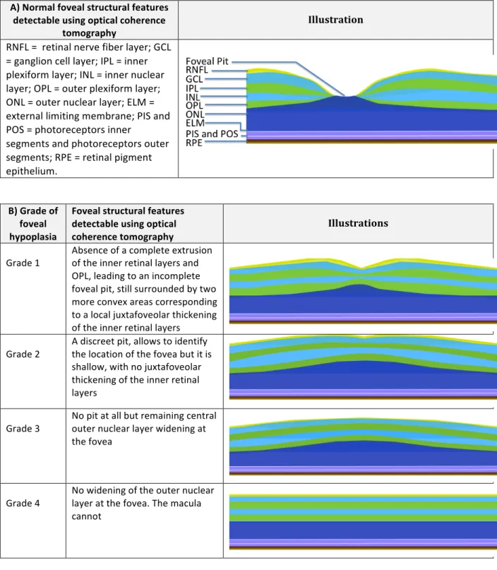
A) Normal foveal structural features detectable using optical coherence tomography Illustration RNFL = retinal nerve fiber layer; GCL = ganglion cell layer; IPL = inner plexiform layer; INL = inner nuclear layer; OPL = outer plexiform layer; ONL = outer nuclear layer; ELM = external limiting membrane; PIS and POS = photoreceptors inner segments and photoreceptors outer segments; RPE = retinal pigment epithelium. B) Grade of foveal hypoplasia Foveal structural features detectable using optical coherence tomography Illustrations
Grade 1 Absence of a complete extrusion of the inner retinal layers and OPL, leading to an incomplete foveal pit, still surrounded by two more convex areas corresponding to a local juxtafoveolar thickening of the inner retinal layers Grade 2 A discreet pit, allows to identify the location of the fovea but it is shallow, with no juxtafoveolar thickening of the inner retinal layers
Grade 3 No pit at all but remaining central outer nuclear layer widening at the fovea
Grade 4 No widening of the outer nuclear layer at the fovea. The macula cannot
Figure 1: A), Illustration of the normal foveal structural features detectable, using optical coherence tomography. B), Foveal hypoplasia severity scale according to structural features detectable using optical coherence tomography.
RESULTS
Genetic studyIn our series of patients, 268 patients were diagnosed with OCA1. 122 (45.5%) were compound heterozygous and harbored one pathogenic variant of TYR and the R402 polymorphic variant of the TYR gene. They constituted the R402Q-OCA1 group. 146 patients harbored two pathogenic variants of the TYR gene, other than R402Q. Due to the absence of any clinical report available for 27 of these 146 patients, only 119 could be included in the phenotypic analysis. These 119 patients constituted the classical-OCA1 group.
For all these 268 patients, an extensive analysis of the 18 known albinism genes (and
SLC38A8) had been performed. None of them was found to have two pathogenic variants (or
one for GPR143) in any of the other 17 genes or in SLC38A8. This excluded that they had
another form of albinism than OCA1.
Table 1 presents the mutations found among R402Q-OCA1 patients and among classical- OCA1 patients. c.1118C>A and c.1A>G were the most frequent mutations among R402Q-OCA1 patients (allele frequency respectively 23.8% and 6.6%). Those same mutations were first (9.9%) and third (5.5%) most frequent mutations among the classical-OCA1 patients whereas c.1037-7T>A was the second most frequent mutation (8.6%). All the mutations present in the classical-OCA1 population with an allele frequency higher than 2.5% were also present in the R402Q population except for c.832C>T of which half resulted from genotyping of people living in Sri-Lanka from blood samples sent in France for analysis (Which could constitute a recruitment bias).
Table 1: Genotypes of all patients of the laboratory with two mutations identified for the TYR gene
Mutations associated to the R402Q variant among R402Q-OCA1 patients (122 patients, 122 alleles)
Kind of mutation TYR mutation sequence name Protein Name Number of allele
Missense (n=88, 72.1%) c.62C>T p.Pro21Leu 2 c.71G>A p.Cys24Ty 1 c.107G>A p.Cys36Tyr 2 c.140G>A p.Gly47Asp 5 c.230G>A p.Arg77Gln 3 c.242C>T p.Pro81Leu 4 c.244T>C p.Ser82Pro 1 c.290G>T p.Gly97Val 1 c.307T>C p.Cys103Arg 1 c.415A>C p.Thr139Pro 1 c.595G>T p.Asp199Tyr 2 c.613C>A p.Pro205Thr 1 c.616G>A p.Ala206Thr 1 c.635G>A p.Arg212Lys 1 c.649C>T p.Arg217Trp 2 c.650G>A p.Arg217Gln 1 c.715C>T p.Arg239Trp 2 c.816G>C p.Trp272Cys 1 c.823G>T p.Val275Phe 5 c.1007C>A p.Ala336Asp 1 c.1017C>G p.Ser339Arg 1 c.1036G>A p.Gly346Arg 1 c.1058G>A p.Gly353Glu 1 c.1064C>T p.Ala355Val 1 c.1099C>T p.His367Tyr 1 c.1111A>G p.Asn371Asp 1 c.1118C>A p.Thr373Lys 29 c.1138T>C p.Ser380Pro 1 c.1146C>A p.Asn382Lys 4 c.1217C>T p.Pro406Leu 1 c.1250C>A p.Pro417His 1 c.1306G>T p.Gly436Cys 1 c.1315T>G p.Phe439Val 1 c.1336G>A p.Gly446Ser 3 c.1342G>A p.Asp448Asn 1 c.1454G>A p.Gly485Gln 1 c.1469C>A p.Ala490Asp 1 Nonsense (n=11, 9%) c.239G>A p.Trp80* 1 c.346C>T p.Arg116* 1 c.533G>A p.Trp178* 1 c. 655G>T p.Glu 219* 1 c.732_733delTG p.Cys244* 2 c.1036G>T p.Gly346* 1 c.1204C>T p.Arg402* 1 c.1392_1393insT p.Lys465* 3 Frameshift (n=8, 6.6%) c.216delA p.Val74Trpfs*46 1 c.841delG p.Glu281Serfs*38 1 c.1386_1387insAA p.Tyr463Asnfs*23 3 c.1467dup p.Ala490Cysfs*20 3 Splice site mutation (n=13, 10.7%) c.1A>G p.? 8 c.1037-1G>A p.? 1 c.1037-2A>G p.? 1 c.1037-7T>A p.? 2 c.1184+1G>A P.? 1 Deletion of exons (n=2, 1.6%) Deletion exon 2 Deletion exon 3 P.? P.? 1 1
Mutations fond among classical 0CA1 patients (146 Patients, 292 alleles): Part 1
Kind of mutation TYR mutation sequence name Protein name Number of allele
Missense (n=167 57,2%) c.56 A>G p. His 19 Arg 1 c.61C>T p.Pro21Ser 5 c.71G>C p.Cys24Ser 1 c.98 A>C p. Lys 33 Thr 3 c.107G>A p.Cys36Tyr 1 c.124G>A p.Asp42Asn 1 c.136T>C p.Cys46Arg 1 c.140G>A p.Gly47Asp 11 c.164G>A p.Cys55Tyr 1 c.229C>T p.Arg77Trp 5 c.230G>A p.Arg77Gln 4 c.241C>T p.Pro81Ser 1 c.242C>T p.Pro81Leu 4 c.272G>A p.Cys91Tyr 1 c.325G>A p.Gly109Arg 1 c.451A>T p.Ile151Phe 1 c.547G>T p.Val183Leu 1 c.613C>A p.Pro205Thr 1 c.616G>A p.Ala206Thr 5 c.617C>T p.Ala206Val 4 c.635G>A p.Arg212Lys 2 c.649C>T p.Arg217Trp 5 c.650G>A p.Arg217Gln 3 c.679G>A p.Gly227Arg 1 c.710A>C p.Asp237Ala 1 c.715C>T p.Arg239Trp 2 c.755T>G p.Met252Arg 2 c.816G>C p.Trp272Cys 2 c.823G>T p.Val275Phe 8 c.866 G>C p.Cys289Ser 1 c.895C>T p.Arg299Cys 1 c.896 G>A p.Arg 299 His 3 c.982G>C p.Glu328Gln 2 c.996 G>A p.Met332Ile 1 c.1012T>G p.Phe338Val 2 c.1036G>A p.Gly346Arg 1 c.1037 G>A p.Gly346Glu 1 c.1058 G>A p.Gly353Glu 1 c.1064C>T p.Ala355Val 3 c.1111A>T p.Asn371Tyr 6 c.1118C>A p.Thr373Lys 29 c.1146C>A p.Asn382Lys 6 c.1147G>A p.Asp383Asn 3 c.1171_1172delinsTT p.Ala391Leu 2 c.1200G>T p.Trp400Cys 1 c.1217 C>T p.Pro406Leu 6 c.1255G>A p.Gly419Arg 1 c.1264C>T p.Arg422Trp 1 c.1265G>A p.Arg422Gln 2 c.1336G>A p.Gly446Ser 8 c.1342G>A p.Asp448Asn 5 c.1432C>T p.Leu478Phe 1 c.1469C>A p.Ala490Asp 1 Nonsense (n=37, 12.7%) c.255T>A p.Tyr85* 1 c.273C>A p.Cys91* 1 c.346C>T p.Arg116* 4 c.488C>G p.Ser163* 1 c.571G>T p.Gly191* 2 c.732_733delGT p.Cys244* 6 c.741C>A p.Cys247* 2 c.753C>A p.Tyr251* 1 c.815G>A p.Trp272* 2 c.832C>T p. Arg 278* 8 c.1204C>T p.Arg402* 4 c.1392dup p.Lys465* 5
Mutations fond among classical 0CA1 patients (146 Patients, 292 alleles): Part 2
Kind of
mutation TYR mutation sequence name Protein name Number of allele
Frameshift (n=26, 8.9%) c.286 dupA p.Met96Asnfs*73 1 c.326del p.Gly109AspfS*11 1 c.334delT p.Cys112Alafs*8 1 c.338_339del p.Thr113Argfs*55 1 c.505_507delGAC p.Asp169del 1 c.525_527dup p.Phe176dup 1 c.567_571delTGGGG p.Gly190Ilefs*2 1 c.573delA p.Ser192Leufs*34 4 c.649delC p.Arg217Glyfx*9 1 c.773_774delCA p.Thr258Lysfs*3 1 c.841delG p.Glu281Serfs*38 1 c.911_914del p.His304Profs*14 1 c.1164delT p.His389Thrfs*96 1 c.1177delG p.Val393fs*92 1 c.1386_1387insAA p.Tyr463Asnfs*23 2 c.1467dup p.Ala490Cysfs*20 6 c.1472dupT p.Leu492Alafs*18 1 Splice site mutation (n=57, 19.5%) c.-111_-109dup p.? 1 c.1037-1G>A p.? 1 c.1037-7T>A p.? 25 c.1184+1G>A p.? 2 c.1185-1 G>T p.? 6 c.1366+1 G>A p.? 1 c.1A>G p.? 16 c.2T>C p.? 3 c.820-1_820delinsTG p.? 1 c.820-3delC p.? 1 Deletion of exon or of the gene (n=5, 1.7%) Deletion exon 1 p.? 1 Deletion exon 2 p.? 1 Deletion 3-4-5 p.? 1 Deletion TYR p.? 1 Deletion 11q14.3 p.? 1
Phenotypic study
R402Q-OCA1 patients were aged between <1 month and 60 years and classical-OCA1 patients between <1 month and 81 years at the latest description.
Data concerning ethnicity were available for 105 R402Q-OCA1 patients, 95.41 % of whom were Caucasian. They were available for 109 classical-OCA1 patients, 77.14 % of whom were Caucasian.
Figure 1 illustrates the wide spectrum of possible phenotypic presentations in R402Q-OCA1 patients. It can be noted that the level of depigmentation, with regard to the skin, to the hair and to the eyes, is highly variable, ranging from a visible hypopigmentation to an apparently “normal” pigmentation.
Table 2 presents the comparative description of demographic and phenotypic characteristics of R402Q-OCA1 and classical-OCA1 patients.
Skin, hair and eyes colors were found to differ significantly between the two groups with a statistically more pigmented phenotype in the R402Q-OCA1 group. Nystagmus and photophobia were found to be significantly less frequent in the R402Q-OCA1 group. The prevalence of refractive error was very high with only 2 patients with no reported refractive error in the R402Q-OCA1 group and none in the classical-OCA1 group. It did not differ between the two groups for hypermetropia and astigmatism which both had a high prevalence rate of more than 70%. Myopia was rarer in both group but more frequent in the classical-OCA1 group. The prevalence of iris transillumination was more than 90% in both groups and did not significantly differ between the two groups whereas the severity score was lower in the R402Q-OCA1 group.
The visual acuity of the best eye was significantly higher in the R402Q-OCA1 group. It was 0.9 to 0.1 LogMAR (20/160 to 20/25 Snellen) with a mean of 0.38±0.21 LogMAR (slightly higher than 20/50 Snellen) in the R402Q-OCA1 group whereas it was 1.3 to 0.15 LogMAR (20/400 to about 20/27-28 Snellen) with a mean of 0.76±0,24 LogMAR (about 20/115 Snellen) in the classical-OCA1 group.
Foveal hypoplasia was reported in 100% of classical-OCA1 patients and 92.59 % of R402Q-OCA1 patients for whom this data was available. Its severity score was significantly lower in the R402Q-OCA1 group.
Macular OCT slides were available for only 19 classical-OCA1 patients and 53 R402Q-OCA1 patients. Figure 2 illustrates the various morphological aspects of the macula revealed by macular OCT imaging in R402Q-OCA1 patients. Nearly all patients with an available
OCT had a foveal hypoplasia at least grade 1, with the exception of one patient (with genotype c.1336G>A + c.1205G>A) from the R402Q-OCA1 group.
A statistically significant correlation between visual acuity and the grade of foveal hypoplasia was found among R402Q-OCA1 patients. Hence visual acuity appears to decrease (increase of LogMAR measure) with rising grade of foveal hypoplasia (r= 0,37 IC95%[0,11-0,59], p= 0,007).
Comparing the two studied groups, foveal hypoplasia was significantly more severe in classical-OCA1 patients (p< 0,001).
5 patients had a medical history of skin cancer in the classical-OCA1 group whereas there was none in the R402Q-OCA1 group.
Finally, assuming that the type of mutation in trans with the R402Q variant may impact of the phenotype expressivity and penetrance we tried to compare 2 groups of R402Q-OCA1 patients which separate patients with missense variants and patients with truncating, supposedly more deleterious, variants (nonsense, splicing, frameshift variants, partial deletions of the TYR gene), in trans of R402Q. Patients with a missense mutation were found to have a higher mean visual acuity (respectively 0.36±0.20 LogMAR which means about 20/45 Snellen versus 0.44±0.20 LogMAR which means about 20/55 Snellen). However, this difference was not statistically significant (P = 0.14).
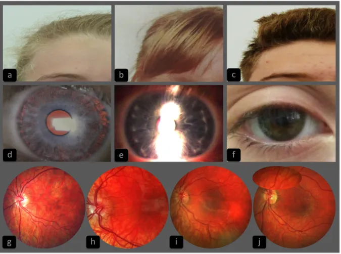
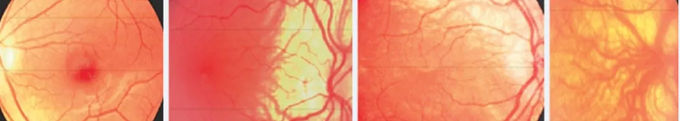
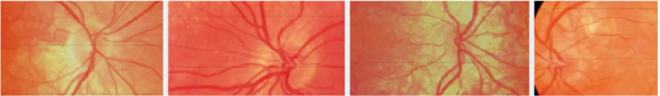
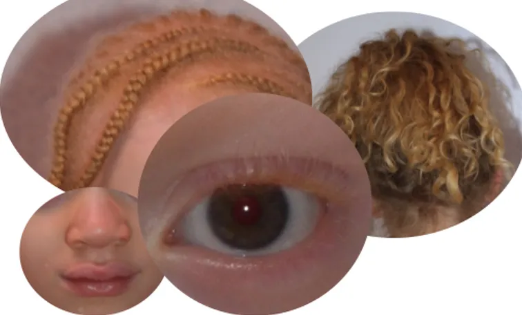
Figure 2: Representative sample of the hair, iris and retinal pigmentation phenotypes in oculocutaneous type 1 patients, compounds heterozygous with one classical TYR
mutation and the R402Q variant.
The first line presents the variety of hair colour in this population with different shades of blond (a) (patient c.649C>T + c.1205G>A (R402Q)), ginger (b) (patient c.1386_1387insAA + c.1205G>A ) and brown (c) (patient c.649C>T + c.1205G>A). The second line presents the variety of iris color from blue (d) (patient c.1306G>T + c.1205G>A) and (e) (patient c.823>C + c.1205G>A) to green or brown (f) (patient c.649C>T + c.1205G>A) and iris transillumination severity ranging from total or almost total transillumination (grade 3 in our classification) (d) to discreet punctuate transillumunation (grade 1) (e) and to no transillumination at all (grade 0) (f). The third line presents the variable severity of retinal hypopigmentation which may extend to the whole retina including the macular zone (g) (patient c.649C>T + c.1205G>A) or sparing partially the macular region (h) (patient c.655G>T + c.1205G>A) (combined into grade 2 in our classification), which also may be restricted to outside vascular arcs (grade 1) (I) (patient c.823G>T + c.1205G>A) and (j) (patient c.649C>T + c.1205G>A) or may be absent.
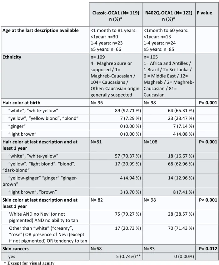
Table 2: Demographic and phenotypic characteristics of patients with oculocutaneous albinism due to TYR mutations (OCA1). Comparison between compound heterozygous patients with the R402Q variant and another pathogenic variant of the TYR gene
(R402Q-OCA1), and patients with two pathogenic variants of the TYR gene, other than R402Q (classical-OCA1). Classic-OCA1 (N= 119) n (%)* R402Q-OCA1 (N= 122) n (%)* P value Age at the last description available <1 month to 81 years: <1year: n=30 1-4 years: n=23 ≥5 years: n=66 <1month to 60 years: <1year: n=13 1-4 years: n=24 ≥5 years: n=85 Ethnicity n= 109 4= Maghreb sure or supposed / 1= Maghreb-Caucasian / 104= Caucasians / Other: Caucasian origin generally suspected n= 105 1= Africa and Antilles / 1 Brazil / 2= Sri-Lanka / 6 = Middle East / 12= Maghreb / 2= Maghreb-Caucasian / 81= Caucasian Hair color at birth N= 96 N= 98 P= 0.001 “white”, ”white-yellow” 89 (92.71 %) 64 (65.31 %) “yellow”, “yellow blond”, “blond” 7 (7.29 %) 23 (23.47 %) “ginger” 0 (0.00 %) 7 (7.14 %) “light brown” 0 (0.00 %) 4 (4.08 %) Hair color at last description and at least 1 year N=81 N=108 P< 0.001 “white”, ”white-yellow” 57 (70.37 %) 18 (16.67 %) “yellow”, “light blond”, “blond”, ”dark-blond” 17 (20.99 %) 68 (62.96 %) “yellow-ginger” “ginger” “ginger-brown” 4 (4.94 %) 14 (12.96 %) “light brown”, ”brown” 3 (3.70 %) 8 (7.41 %) Skin color at last description and at least 1 year N= 82 N= 98 P< 0.001 White AND no Nevi (or not pigmented) AND no ability to tan 75 (79.27 %) 28 (28.57 %) Other than “white” (“creamy”, “rose”) OR presence of Nevi (except if not pigmented) OR tendency to tan 17 (20.73 %) 70 (71.43 %) Skin cancers N=68 N=83 P= 0.012 yes 5 (0.74%)** 0 (0.00%)
* Except for visual acuity
** 1 patients with basal cell carcinoma (c.1A>G + c.1118C>A, ), 1 with basal cell carcinoma and melanoma (c.140G>A + c.1037-7T>A), 1 with melanoma (c.1118C>A + c.1467dup), 1 with squamous cell carcinoma (c.140G>T + c.325G>A) and 1 with unspecified skin cancer (c.1118C>A + c.1264C>T).
(Table 2 continuing) Classic-OCA1 (N= 119) n (%) * R402Q-OCA1 (N= 122) n (%)* P value Iris color N=99 N= 109 P= 0.006 “grey”,”blue-grey”,”blue”,”bleu-green” 94 (94.95 %) 91 (82.73%) “green”, green-brown”,”brown” 5 (5.05 %) 19 (17.27 %) Presence of Iris transillumination N= 84 N= 109 P= 0.053 yes 79 (94,05 %) 93 (85.32 %) Severity score of iris transillumination if present N= 30 N= 44 P< 0.001 Stage 1 0 (0.00%) 15 (34.09%) Stage 2 16 (38.10 %) 19 (43.18%) Stage 3 29 (96.67%) 10 (22.73%) Photophobia N=91 N=96 P< 0.001 yes 88 (96.70 %) 75 (78,13 %) Nystagmus N= 104 N= 118 P= 0.011 yes 99 (95.19 %) 100 (84.75 %) Strabismus N= 75 N= 108 P= 0.058 yes 31 (40,79 %) 60 (55,56 %)
Refractive error (patient at least 1 year) N=61 (60 for astigm.) N=94 (93 for astigm.)
None 0 (0.00%) 2 (2.13%) P= 0.252 Myopia (≤ -0,75D) 16 (26.23%) 12 (12.77%) P= 0.033 Hyperopia (≥ 1D) 44 (72.13%) 74 (78.72%) P= 0.436 Astigmatism (≥ 1D) 46 (76.67%) 72 (77.42%) P= 0.686 Visual acuity (LogMAR) (patient ≥5years) N= 44 N= 74 P< 0.001 Mean ±standard deviation 0.76 ±0,24 0.38 ±0,21 Median [min-max] 0.70 [1.3-0.15] 0.35 [0.9-0.1] % with VA≥20/40 Snellen (LogMAR=0,3) 4.55 % 50.00 % Presence of retinal hypopigmentation N= 80 N= 108 P= 0.013 yes 80 (100.00 %) 100 (92.59 %) Severity score of retinal hypopigmentation if present N= 36 N= 67 P=0.013 Stage 1 2 (5.56 %) 17 (2.37 %) Stage 2 34 (94.44 %) 50 (74.63 %) Presence of foveal hypoplasia N= 50 N= 109 P= 0.335 Yes (reported with or without OCT) 50 (100.00 %) 107 (98.17 %) Severity score of foveal hypoplasia if OCT available N= 19 N= 53 P< 0.001 Stage 0 0 (0.00 %) 1 (1.89 %) Stage 1 1 (5.26 %) 7 (13.21 %) Stage 2 1 (5.26 %) 10 (18.87 %) Stage 3 2 (10.53 %) 21 (39.62 %) Stage 4 15 (78.95 %) 14 (26.41 %) Mean score ±standard deviation 3.63 ±0.19 2.75 ±0.14
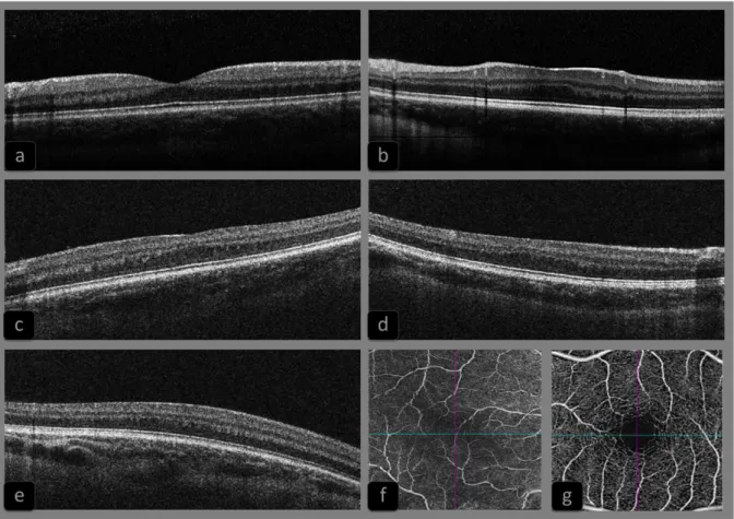
Figure 3: Representative sample of foveal morphologies in oculocutaneous type 1 patients, compounds heterozygous with one classical TYR mutation and the R402Q variant (R201Q-OCA1 patients).
Slides a) to e) corresponds to spectral domain optical coherence tomography (OCT) imaging of R402Q-OCA1patients’fovea, illustrating our classification for foveal hypoplasia. Grade 0 (a) (patient c.1336G>A + c.1205G>A (R402Q)), grade 1 (b) (patient c.1469C>A + c.1205G>A), 2 (c) (patient c.649C>T + c.1205G>A), grade 3 (d) (patient c.239G>A + c.1205G>A), grade 4 (e) (patient c.655G>T + c.1205G>A). Picture (f) is an Angio-OCT imaging of the fovea of a R402Q-OCA1 patient compared with a normal fovea (g). It confirms the clinically suspected vascular modifications of the macular region in albino patients, mainly characterized by the absence of a normal foveal avascular zone.
DISCUSSION
This study is shedding light on the specific phenotype observed in the R402Q-OCA1 group of patients, which represents a significant part of OCA1 patients (45.5% in our study).
General phenotypic characteristics
The R402Q-OCA1 phenotype significantly differs from the classical-OCA1 phenotype. Clinical signs in preschool-aged children are less evocative of oculocutaneous albinism in the R402Q-OCA1 children than among the classical-OCA1 patients. Skin and hair color, are often fair at birth, but not as systematically as in classical-OCA1, and generally tend to darken over the years. Iris transillumination when present is less severe and notably seldom total (22.73% vs 96.67% grade3, p<0,001), contrary to classical-OCA1 patients, and nystagmus is slightly less frequent (84,75 % vs 95,19 %, p=0,011). As a consequence, the diagnosis may not be obvious at birth particularly in a Caucasian population in which blue eyes, fair skin, and blond hair are frequent in babies. Neither is it later in life, based upon skin and hair pigmentation criteria. Indeed, most of the R402Q-OCA1 patients present pigmented nevi and/or have a tendency to tan, which is rarer among classical-OCA1 patients. Moreover, few of them have the classical albinoid white-platinum blond hair. Instead, they more often present with a wide panel of hair colors ranging from yellow blond to ginger and brown. This may be the raison why some patients in the R402Q-OCA1 group where diagnosed in adolescence or adulthood by an ophthalmologist whereas the diagnosis of albinism had never been evoked before. It is difficult to estimate the mean age at the diagnosis and the proportion of patients with a late diagnosis at adolescence or adulthood. But it is a frequent observation of practitioners that can be illustrated with the case of one of our patients (c.216delA + c.1205G>A (R402Q)), who had benefit of an ophthalmological follow-up for congenital esotropia and for whom the diagnosis of albinism was finally evocated at the age of twenty. This patient had fair skin, ginger-brown hair and blue-green eyes. His complete ophthalmological examination found in addition to the known strabismus: a discreet nystagmus compatible with the diagnosis of congenital esotropia, a bilateral iris transillumination, an hypopigmented retina and a grade 2 foveal hypoplasia in both eyes. His visual acuity was 20/30 in one eye and 20/40 in the other eye.
An interesting result of this study is the lower frequency of skin cancer in the R402Q-OCA1 group (n= 5 (0,74%)) as compared with the classical-OCA1 group (n= 0). It could be
be an important prognosis data. Nevertheless, the limit of our study, concerning this conclusion, is the low number of elderly patients in the study population, particularly in the R402Q-OCA1 group whose eldest patient was 60. Thus, it is important to remember that previous studies have demonstrated that an increased risk of photoinduced skin cancers is related to albinism and that a photoprotection adapted to the phototype is particularly recommended for the albino patients (39–41).
Ophthalmologic features
Focusing on the ophthalmological symptoms and impairments, which also appear to be milder in R402Q-OCA1 patient, it is particularly interesting to notice the difference in the level of visual acuity, which is a key factor of the prognosis of this disease. R402Q patients, five years or older, had a mean visual acuity of 0,38 LogMAR (about 20/45 on Snellen chart), significantly higher than classical-OCA1 patients (0,76 LogMAR, P< 0,001). Half of them had a visual acuity of their better eye of 20/40 or more, which is the acuity threshold for
driving (Rees 2015) whereas only 4,55 % reached this threshold in the classical-OCA1 group.
All R402Q-OCA1 patients five years or older had a visual acuity higher than 20/200 which is, since 2006, the definition of blindness according to the International Statistical Classification of Diseases (Dandonna 2006), whereas 3 patients in the classical-OCA1 group had a visual acuity below this threshold and nine more just at this level of 20/200.
Photophobia and nystagmus were also less frequent in R402Q-OCA1 patients than among classical-OCA1 patients. Moreover, among the R402Q-OCA1 patients reported to have a nystagmus, some were described to have a “subtle” nystagmus or “micronystagmus” which frequently decreased with age. One patient had a history of nystagmus in his childhood, which disappeared when he was an adolescent.
In the same way, iris transillumination was less severe in R402Q-OCA1 patients, with a tendency to decrease with age as reported by some practitioners.
The frequency of refractive errors was high in both R402Q-OCA1 and classical-OCA1 patients, with astigmatism being the most frequent refractive error and hyperopia being more frequent than myopia. These results are consistent with those of previous studies, which all agree on the high frequency of astigmatism in albinism but report a predominance of myopia sometime or of hyperopia (42–44).
Concerning strabismus, we have no explanation for its higher frequency among R402Q-OCA1 patients but suspect a possible bias leading to an underestimation of strabismus prevalence in classical-OCA1 patients. Indeed, the presence of a marked nystagmus makes
the evaluation of strabismus difficult. The higher rate and higher intensity of nystagmus in the classical-OCA1 group could explain this difference.
The foveal hypoplasia can be objectified by OCT since the early 2000s (45,46). In our study the presence of a foveal hypoplasia confirmed by OCT imaging appeared to be very sensitive for the diagnosis of the OCA1. Indeed, only one patient, out of the total of 72 patients with an available OCT, had no foveal hypoplasia. He was belonging to the R402Q-OCA1 group (c.1336G>A + c.1205G>A (R402Q)). This highlights the interest of OCT for the exploration of suspected albinism and notably for the exploration of congenital nystagmus as reported by previous articles (47–49). In particular, Lee et al demonstrated the benefit of handheld OCT in this indication when examining young children (48). Moreover, SD-OCT has a prognostic interest since the severity of foveal hypoplasia is correlated with visual acuity as demonstrated in the present study as well as in some previous ones (35,49–52).
Optical coherence tomography angiography is a more recent imaging tool that confirms the vascular modification accompanying foveal hypoplasia, a feature that was less precisely observed on fundoscopy or retinophotography. Foveal hypoplasia is indeed accompanied by a reduced or absent foveal avascular zone (53).
The mechanism of retinal and foveal anomalies in albinism is a topical issue that is not yet fully clarified. Neither is normal foveal development. But many studies addressed these questions (54).
Concerning normal foveal development, Yodelis and Hendrickson since 1986 demonstrated from histological analyses that morphological maturation of the human fovea is marked by three general developmental events: a peripheral migration of the inner retinal layers to form the foveal depression starting during the gestation and not reaching completion until about 15 months postpartum, a central migration of cones condensing them into the macular area so that the rod-free zone decreases in size from gestation to adulthood and resulting in an about six-fold increase in foveolar cell packing density, and a concomitant elongation of each cones particularly marked in the fovea, leading to a thicker ONL at the fovea (55,56). OCT analyses in premature babies and children were consistent with this foveal development (57–59). In 1978, Fulton and al, thanks to an histological analysis of the retina of an albino child, confirmed the clinically presumed absence of a foveal pit and found no rod-free zone and no foveal cone packing (60). In 2006, Kelly and Weiss findings from multifocal electroretinographs were consistent with a homogeneous density of cone photoreceptors across the central retina in albinism (61). In vivo the study with adaptive optics scanning light ophthalmoscopy in albino patients revealed lower peak cone densities (62). Finally, OCT was
widely used to explore foveal anomalies in albino patients (35,47–52,62,63), revealing some quite specific signs such as a decreased foveal pit and a thinner ONL. It showed a broad range of foveal morphologies which have similarities with the successive aspects of the fovea throughout the normal foveal development, supporting the idea of an uncompleted development (64). In 2015, Lee et al studied the evolution of the foveal morphology with OCT in albino children from birth to the age of 6 and showed continuing regression of the inner retinal layers and a continuing elongation of the photoreceptor layers (63). Those findings suggest a possible residual plasticity of these patients’ retina after birth since it is still developing and its morphology is not fixed yet. They are so encouraging for a treatment at the earliest stages of the condition, which could maybe improve retinal development and optimize vision as experimented in animals model (65).
The mechanisms and signaling pathways implicated in the normal foveal development and in guiding the retinal axons to their specific regions of the midbrain, have not yet been fully elucidated. Albinism has proved to be an interesting model to explore those issues and revealed the role of tyrosinase and of the menanin synthesis in the visual system development
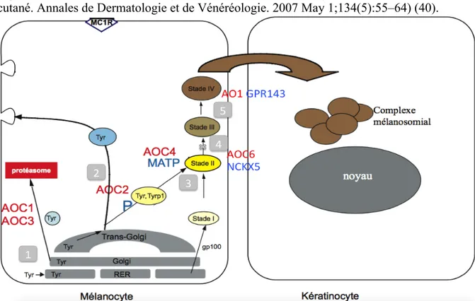
(65–67). Dihydroxyphenalanine (L-Dopa) is an intermediate in the synthesis of melanin,
generated by tyrosinase in pigmented retinal pigment epithelium (RPE). L-Dopa has been hypothesized to be involved in the development of the retina (68) and subsequently in the retinal and foveal abnormalities observed in OCA (69). L-DOPA and dopanmine signaling have also been hypothesized to be involved in the excess of optic nerve fiber decussation at the chiasm (70).
The chiasm anomalies represent a characteristic feature of albinism that can be explored by Magnetic Resonance Imaging (MRI) (71,72) or by Visual evoked potentials (VEP) recording (33,73,74) but for which we had only little data in our study. Indeed, VEP results were available for only 49 patients (31 R402Q-OCA1 patients and 18 classical-OCA1 patients) and only 14 (28.57%) (8 R402Q-OCA1 patients and 6 classical-OCA1 patients) revealed a characteristic crossed asymmetry whereas numerous results where reported as interpretable or showing non-specific abnormalities. The low number of available VEP results is partly explained by the fact that a VEP test is often performed only once at the time of the initial work-up but is not repeated during the follow-up. Besides VEP testing is less systematically performed with the emergence of OCT. But more importantly, VEP recording is difficult in
patients with albinism: specific parameters are needed (flash VEP and pattern onset/offset
stimulation) (74) and results may be disturbed by a poor visual acuity and by the presence of
Interrogations regarding the penetrance
Further investigations are needed to deepen the knowledge about R402Q penetrance. In 2009, Oetting et al raised the issue of the absence of an albinoid phenotype in some parents of patients who had the same genotype as their child (75). Variable expressivity and incomplete
penetrance are possible explanations.
Fourteen parents of patients included in our study in the R402Q-OCA1 group, had the same genotype as their child and were so compound heterozygous with the R402Q variant and one other pathogenic variant of the TYR gene in trans. As in Oetting and al’s study, none of them was reported to have albinism. Nevertheless, we had no complete clinical records for those
parents and tried to get some more information about them. Only two patients’ mothers
accepted to come to Bordeaux for clinical examination at the time of the survey and no clinical signs of albinism were found in these two women, except a discreet iris transillumination in one. Both reported difficulties to tan and a tendency to burn in the sun. One father of whom a photograph was available had dark-blond hair and fair skin while he reported his brother to have brown hair and to tan more easily than himself. Such a sibling should have been interesting to examine. Finally, we received one clinical description of a patient’s mother whose diagnosis of albinism was confirmed both clinically and genetically after her daughters were diagnosed. Indeed, she presented an R402Q-OCA1 genotype (c.1386_1387insAA + c.1205G>A (R402Q)), and clinically presented with a light phototype,
yellow-blond hair, blue transilluminable iris, nystagmus, photophobia, a divergent strabismus, a 20/40 visual acuity, a retinal hypopigmentation grade 2, a foveal hypoplasia grade 3 and a
crossed asymmetry on the VEP recording. Concerning this woman, the diagnosis of albinism
had never been suspected or evoked before. The hypothesis of similar cases of undiagnosed parents should not be excluded in the absence of a complete clinical examination.
As previously explained, the symptoms of albinism in R402Q-OCA1, are variable and rather mild, in such a way that R402Q albinism is probably under-diagnosed, especially in the Caucasian population. One patient in our cohort (c.1336G>A + c.1205G>A (R402Q)), in spite of being really an index case clinically suspected of albinism and then genetically confirmed, presented at the age of ten years a very mild form of albinism. This Caucasian boy presented with a phototype 2 skin, dark-blond hair, blue eyes with no iris transillumination, a subtle nystagmus, a grade 1 retinal hypopigmentation, a 20/25 visual acuity in both eyes and no foveal hypoplasia as confirmed by OCT. He was suspected of albinism at the age of 2 month because of the association of nystagnus, light skin and white-platinum hair, blue transilluminable iris and hypopigmentation of the retina, and the diagnosis was genetically
confirmed. Most of the evocating characteristics decreased with aging. Obviously this boy could have never been diagnosed without the expert advice of a specialist of ophthalmologic genetic troubles. This patient illustrates the possibility of very mild forms of R402Q-OCA1, at a frontier between normal and pathologic.
We may so make the hypotheses of an incomplete penetrance and of a variable expressivity with a continuum from the more severe forms of the disease to the absence of significant signs and symptoms.
Limits of the study
We acknowledge that there are limitations associated with our study. In spite of a total population of 241 patients, very few data were available concerning some studied clinical features such as the severity of iris transillumination and that of foveal hypoplasia. This high level of missing data is explained by the retrospective design of our study and the difficulty to collect data from multiple centers and multiple practitioners.
The collection of data from multiple practitioners is also responsible for the heterogeneity of those data, which is another important limitation of our study. It is to limit this heterogeneity that some clinical scales were simplified and that the grading of foveal hypoplasia was restricted to patient for whom macular OCT slides were available.
It is to mention that the few number of marcular OCT slides available is probably multifactorial. First, this exam may have been performed once and the result notified on the consultation report without saving the images. Then the OCT which is nowadays a wide spread imaging tool was not some years ago and neither was it consider as a reference for the exploration and diagnosis of albinism. Moreover, its realization may be impossible in babies and young children in the lack of specific handheld devices. Even in an adult, the recording of OCT slides may be compromised by the presence of a marked nystagmus.
Finally, concerning the choice of the studied data, some criticisms may be leveled. Binocular acuity would have been a more pertinent method to evaluate visual acuity, especially in a population of patients with nystagmus, and whose visual binocular acuity is as a consequence often significantly better than the monocular acuity of the better eye. Nevertheless, the binocular visual acuity was often missing in consultation reports and the monocular acuity of the better eye was considered as the best “alternative”.
As mention previously, it would have been interesting to study VEP but we renounced because of the lack of data available.