Aspects of postural alignment and postural control relevant for the evaluation and the treatment of Idiopathic Scoli osis patients.
Par Karl F. Zabjek
Programme Sciences Biomédicales Faculté de Médecine, Université de Montréal
Thèse présentée à la Faculté des études supérieures en vue de l’obtention du grade de Ph.D. en sciences biomédicales Novembre 2003 © Karl F. Zabjek, 2003 I ‘-9 3 2CV
U
050
Direction des bibliothèques
AVIS
L’auteur a autorisé l’Université de Montréal à reproduire et diffuser, en totalité ou en partie, par quelque moyen que ce soit et sur quelque support que ce soit, et exclusivement à des fins non lucratives d’enseignement et de recherche, des copies de ce mémoire ou de cette thèse.
L’auteur et les coauteurs le cas échéant conservent la propriété du droit d’auteur et des droits moraux qui protègent ce document. Ni la thèse ou le mémoire, ni des extraits substantiels de ce document, ne doivent être imprimés ou autrement reproduits sans l’autorisation de l’auteur.
Afin de se conformer à la Loi canadienne sur la protection des
renseignements personnels, quelques formulaires secondaires, coordonnées ou signatures intégrées au texte ont pu être enlevés de ce document. Bien que cela ait pu affecter la pagination, il n’y a aucun contenu manquant.
NOTICE
The author of this thesis or dissertation has granted a nonexclusive license allowing Université de Montréal to reproduce and publish the document, in part or in whole, and in any format, solely for noncommercial educational and research purposes.
The author and co-authors if applicable retain copyright ownership and moral rights in this document. Neither the whole thesis or dissertation, nor substantial extracts from it, may be printed or otherwise reproduced without the authors permission.
In compliance with the Canadian Privacy Act some supporting forms, contact information or signatures may have been removed from the document. While this may affect the document page count, it does not represent any loss of content from the document.
Cette thèse intitulée:
Aspects of postural alignment and postural control relevant for the evaluation and the treatment of Idiopathic Scoliosis patients.
présentée par: Karl F. Zabjek
a été évaluée par un jury composé des personnes suivantes:
Dr. Guy Grimard
président-rapporteur
Dr. Charles Hilaire Rivard, M.D.
directeur de recherche Dr. Francois Prince, Ph.D. codirecteur Dr. Denis Gravel membre du jury Dre. Julie Côté examinateur externe Dr Jean Dansereau représentant du doyen de la FES
L’objectif général de cette thèse était d’identifier les paramètres liés à l’alignement postural et au contrôle postural qui aideront à l’évaluation et au traitement de la scoliose idiopathique (SI). Pour cela, deux types d’évaluation posturale ont été élaborés. La première approche correspond à un modèle postural d’alignement et implique l’évaluation de la position et de l’orientation des segments du corps dans l’espace. La seconde, est une évaluation du contrôle postural en mettant en application une technique d’estimation du centre de masse (CdeM) des patients atteints de la SI.
Une évaluation de la posture debout sur des sujets atteints de la SI et de sujets contrôles a indiqué que les positions angulaires et linéaires moyennes du bassin, du tronc et des épaules pourraient être représentées à l’aide d’une période d’acquisition d’une durée aussi courte qu’une seconde. Dans un groupe de patients atteints d’une SI, des caractéristiques uniques pour chaque type de SI ont été trouvées. Les caractéristiques posturales les plus évidentes servant à décrire la posture des patients atteints de scoliose thoracolombaire gauche (ThLG), thoracique droite (ThD) et thoracique droite lombaire gauche (ThDLG) sont respectivement les suivantes l’inclinaison du bassin, le décalage latéral des épaules par rapport à la base du support, la rotation relative et le décalage latéral des épaules par rapport au bassin, la rotation relative de la ceinture scapulaire par rapport au bassin et la rotation et l’inclinaison des épaules par rapport à la ceinture scapulaire.
Avec l’utilisation d’un corset souple, posé de manière spécifique pour chaque type de courbure rachidienne, il y avait une diminution significative de l’amplitude de cette dernière pour chaque type de scoliose. De plus, il y avait un changement postural spécifique pour chaque type de courbure. Pour les patients avec une courbure ThD, ces changements comportent la rotation opposée des épaules par
rapport à la ceinture scapulaire, accompagnée par un mouvement couplé d’une inclinaison des épaules et du décalage latéral de Ti par rapport à Si. Pour les patients ThLG, il y avait un changement de l’inclinaison relative des épaules, de la rotation des épaules et de la ceinture scapulaire par rapport au bassin. Les patients avec une courbure ThDLG ont eu un changement dc rotation du bassin, une rotation et l’inclinaison des épaules par rapport au bassin, une rotation des épaules par rapport à la ceinture scapulaire et finalement, un décalage latéral de Ti par rapport à Si.
L’utilisation d’une nouvelle technique pour estimer la position et le déplacement du CdeM chez les patients atteints d’une SI, a permis d’identifier des différences significatives en comparaison avec le CdeM estimé par un modèle anthropométrique. L’utilisation de la technique basée sur les plates-formes de force a souligné des différences importantes au niveau du contrôle postural pour les patients atteints d’une SI. La racine carrée de la valeur moyenne du carré entre le centre de pression et le centre de masse (CdeP-CdeM) était plus grande chez les SI que chez les contrôles. Les deux groupes ont démontré une tendance à se comporter comme un pendule inversé, cependant, le mouvement angulaire relatif, reflète une stratégie posturale unique aux patients atteints d’une SI.
Mots-clés: Scoliose Idiopathique, Posture, Équilibre, Biomécanique, Développement, Adolescence
C
Abstract
The general objective of this thesis was to identify postural alignment and postural control parameters that will assist in the assessment, and treatment of Idiopathic Scoliosis (IS). This specifically involved applying two approaches for the postural assessment of IS. The first is a postural alignment model, and implicates assessing the position and orientation of the body segments in space. The second, is the evaluation of postural control through implementing a novel technique to estimate the Centre of mass (COM) in IS patients and control subjects.
An initial evaluation of the quiet standing posture of IS and control subjects revealed that the mean angular and linear position of the pelvis, trunk and shoulders over 120 second penod could be represented by that obtained during a short trial acquisition period of 1 second. The postural alignment evaluation of 15 patients revealed unique postural characteristics measured in reference to the base of support and relatively between body segments specific to each type of IS curvature. The postural parameters that were important for charactenzing right thoracic (RTh), left thoracolumbar (LThL), and right thoracic left lumbar (RThLL) curvatures included the tilt of the pelvis and lateral shift of the shoulders in reference to the base of support, relative rotation, tilt and lateral shift of the shoulders in reference to the pelvis, relative rotation of the shoulder blades in reference to the pelvis, and rotation and tilt of the shoulders in reference to the shoulder blades.
With the use of a non-ngid brace specific for each type of spinal curvature, there was a significant decrease in the amplitude of spinal curvature accompanied by a change in the postural parameters. For the RTh patients these changes involved the opposite rotation of the shoulder girdie in reference to the shoulder blades accompanïed by a coupled movement of shoulder tilt and lateral shift of Ti in reference to Si. For the LThL patients there was a change in the relative tilt of the
shoulders, rotation of the shoulders and shoulder blades in reference to the pelvis. The RThLL patients had a change in the rotation of the pelvis, tilt of the shoulders, rotation and tilt of the shoulders in relation to the pelvis, rotation of the shoulders in reference to the scapula and lateral shift of T 1 in reference to S 1.
The application of a technique to estimate the position and dispiacement of the COM from a Force Plate, ïdentified signifïcant differences when compared with the COM estimated from an anthropometnc model. Using this Force Plate based technique to evaluate the postural control of IS subjects, a greater foot mean square of the COP-COM signal was found in comparison to control subjects. Both the IS and control subjects demonstrated a tendency to behave as an inverted pendulum, however, the significantly greater relative angular movement reftects a postural control strategy that is unique to IS patients.
Keywords: Idiopathic Scoliosis, Posture, Balance, Biomechanics, Development, Adolescence
Table of contents
RÉSUIVIÉ
ABSTRACT iii
TABLE 0F CONTENTS V
LIST 0F TABLES viii
LIST 0F FIGURES x
LIST 0f SYMEOLS AND ABBREVIATIONS xiv
DEDICATIONS xvii
ACKNOWLEDGMENTS xviii
FORWARD xix
INTRODUCTION 1
CHAPTER I: Idiopathic Scoliosis
1.1 Natural history 3
1.2 Clinical evaluation 5
CHAPTER II: Posture
2.1 Prïnciples of postural evaluations 11
2.2 Postural alignment 12
2.3 Postural control 16
2.4 Postural control growth and development 19
2.5 Postural control and Idiopathic Scoliosis 20
OBJECTIVES 23
CHAPTER III: Manuscript #1
Evaluation of segmental postural characteristics during quiet standing in control and Idiopathic Scoliosis patients 24
CHAPTER IV: Manuscript #2
Postural alignment charactenstics ofldiopathic Scoliosis patients 51
CHAPTER V: Manuscript #3
The change in posture induced by a non-rigid brace fitted on Idiopathic Scoliosis patients: A comparison of different curve
types $0
CHAPTER VI: Manuscript #4
Estimation of the Centre of Mass for the study of postural control in Idiopathic Scoliosis patients: Companson of an anthropometric and
force-plate based model 120
CHAPTER VII: Manuscript #5
Postural control in adolescent Idiopathic Scoliosis and control
CHAPTER VIII: General Discussion .170 8.1 Angular and linear dispiacement ofthepelvis and shoulder girdie
dunng quiet standing in control and Idiopathic Scoliosis patients 171 8.2 Postural alignment charactenstics specific to the type of spinal
curve 172
8.3 Postural alignment changes induced by a non-rigid brace 173 2.4 Estimation of the centre of mass and evaluation of postural control 175
8.5 Limitations of the present research 180
8.6 Future studies 182
CHAPTER IX: Conclusion 183
REFERENCES 186
APPENDIX 1 Ethics Committee Approval xx
List of tables
Chapter III, manuscript #1
Table 1 11e mean angular and linear position of global and relative body segment parameters for the 15 and control subjects (Avg = Average; -95 %, + 95 % =
Confidence Interval) 45
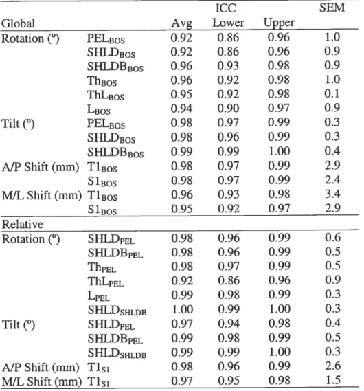
Table 2 11e Intra-class conelation coefficient (1CC) and Standard Error of Measurement (SEM) for the mean angular and linear position of 4 repeat trials for the IS and control subjects together (Avg = Average; Lower = Lower Bound; Upper
UpperBound) 46
Chapter IV, manuscript #2
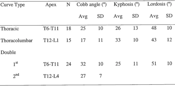
Table 1 Radiological characteristics of untreated IS patients (Avg = average, SD
Standard Deviation) 72
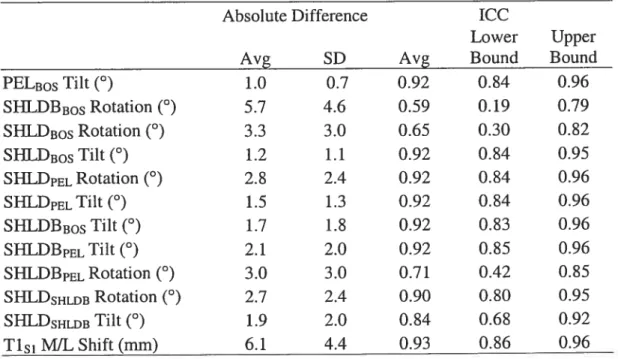
Table 2 Average absolute difference and Intra-Class Correlation Coefficient for postural measures calculated with the stereovideographic system and the sequential digitisation system. (Avg = Average; SD = Standard Deviation 1CC = Intra Class Correlation, Lower Bound = -95 % Confidence Interval, Upper Bound = + 95 %
Confidence Interval) 73
Chapter V, manuscript #3
Table 1 Initial radiological charactenstics 108
Table 2 Initial and BMCMP conditions 109
Chapter VI, manuscript #4
Table 1 COM radius, and COM proportions as proposed by Jensen (1989) and
Table 2 The mean absolute difference (MABD), RMS amplitude difference (RMSD), and Range of Dïfference (ROD) between COMgIBOS, andCOManthBOS....142
Chapter VII, manuscrïpt #5
Table 1 Angular measurements in the sagittal plane. (Avg = Average, SD =
Standard Deviation) 163
Table 2 Angular measurements in the frontal plane (Avg = Average, SD =
Standard Deviation) 164
Table 3 The correlation between the COMacc and COP-COM, for control and Idiopathic Scoliosis patients.(Avg = Average, SD = Standard Deviation) 165
List of figures
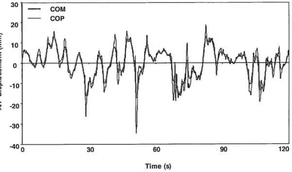
Chapter IIFigure 1 The COP and the COM AJP dispiacement (mm) dunng quiet standing of a typical subject dunng a period of 120 seconds. The thick une represents the COM,
and the thin une the COP 17
Chapter III, manuscript #1
Figure 1 Postural geometry of an idiopathic scoliosis patient depicting the base of support, lower extremilies, pelvis, shoulders and spinous processes represented in the Transverse (Apical), Frontal (Postenor-Anterior), and Sagittal planes (Right
Lateral) 47
Figure 2 The transverse view (apical) of the Iandmarks which define the shoulder blade (SHLDB = inferior angle of each scapula), the shoulder (SHLD acromions),
and the une of reference of the base of support, and the relative angle between the two segments (øi= SNLDB0S, 02= SHLDBB0s, 03 SHLDSHLDB (øi -02=03) 48
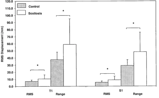
Figure 3 RMS and range of displacement for TiBos and S1cs in the A/P
direction for the IS and the control subjects (* = p<O.O5) 49
Figure 4 The angular dispiacement over a period of 120 seconds for a control subject. The angles measured are rotation of the pelvis (PELBOS) (solid line) and shoulder blades (SHLDBB0S) (Solid-Dotted une) in reference to the base of support,
and relative rotation of the shoulder blades in reference to the pelvis (SHLDBpL)
Chapter IV, manuscrïpt #2
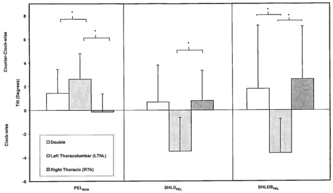
Figure 1 Angular parameters in the frontal plane for the riglit thoracic (RTh), lcft thoracolumbar (LThL), and nght thoracic left lumbar (RThLL) patients (*p<O.OS).
Reference to base of support (BOS): PELB0S = Pelvis. Reference to pelvis (PEL):
SHLDpBL= Shoulders; SNLDBpEL= Shoulder blade 74
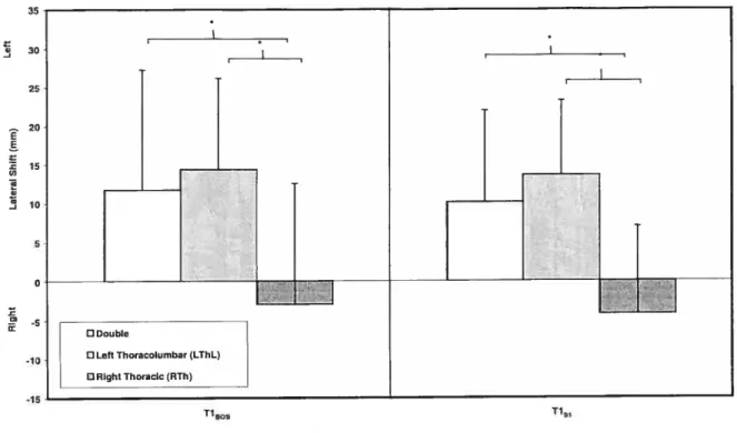
Figure 2 Linear parameters in the frontal plane for the right thoracic (RTh), left thoracolumbar (LThL), and right thoracic left lumbar patients (RThLL) (*p<O.O5)
Reference to base of support (BOS): TiBos = 1st Thoracic spinous process.
Reference to 1stsacral prominence (Si): T15i = 1st Thoracic spinous process 75
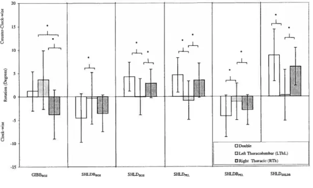
Figure3 Angular parameters in the Transverse plane for the right thoracic (RTh), left thoracolumbar (LThL), and right thoracic left lumbar (RThLL) patients (*p<O.OS).
Reference to base of support (BOS): GIBBB0s = Gibbosity; SHLDBBOS = Shoulder
blade; SFILD805 = Shoulder. Reference to the pelvis (PEL): SHLDPEL = Shoulder;
SHLDBp= Shoulder blade Reference to shoulder blade: SHLDSHLDB =
Shoulder 76
Figure 4 A typical postural representation of a left thoracolumbar patient 77
Figure 5 A typical postural representation of a right thoracic patient 7$
Figure 6 A typical posture of a right thoracic left lumbar patient 79
Chapter V, manuscript #3
Figure 1 Corrective movement principal demonstrated on a nght thoracic
patient 110
Figure 2 The corrective movement principal favorised by the SpineCor
Figure 3 The amplitude of spinal curvature as measured with the angle of Cobb for the initial and bi-ace maintained corrective movement principle (BMCMP)
conditions 112
Figure 4 The amplitude of shoulder tilt in reference to the base of support (SBLD305) for the initial and brace maintained corrective movement principle
(BMCMP) conditions 113
Figure 5 The amplitude of shoulder rotation in reference to the pelvis (SHLDPEL) for the initial and brace maintained corrective movement principle (BMCIvIP)
conditions 114
figure 6 The amplitude of shoulder rotation in reference to the shoulder blade
(SHLDsHLDB) for the initial and brace maintained corrective movement pnnciple
(BMCMP) conditions 115
Figure 7 Apical and Posterior-Anterior view of a typical right thoracic patient, without the BMCMP and with the BMCMP. Note that due to the brace the thorax
couÏd not be defined in the nght graph 116
Figure 8 Apical and Postenor-Anterior view of a typical Ieft thoracolumbar
patient, with and without the BMCMP 117
figure 9 Apical and Postenor-Anterior view of a typical left lumbar patient,
without the BMCMP and with the BMCMP 11$
Figure 10 Apical and Postenor-Antenor view of a typical Double curve patient, without and with BMCMP. Note that due to the brace the thorax could flot be
Chapter VI, manuscript #4
Figure 1 Mean MIL position of the COMgisi for Single, Double and Control
subjects (*p<O.O5) 143
Figure 2 The COMgi, and COManth in the AJP direction for a typical 15
patient 144
Chapter VII, manuscript #5
Figure 1 The ankle, hïp and neck angle in the A/P direction with the bias removed for a typical 15 patient. The solid thick dotted une represents the ankle, the thin une the hip, and the thin dotted une is the neck 166
Figure 2 The COP and COM in the A/P direction for a typical 15 patient. The solid thick line is the COM and the light thin une is the COP 167
Figure 3 RMS amplitude (mm) of COP, COM and COP-COM dispiacement in the AJP direction. Solid bars define the 15 group, light bars represent the control group. A significant difference (p<O.O5) is identified as an asterisk 16$
Figure 4 RMS amplitude (mm) of COP, COM and COP-COM dispiacement in the MiL direction. SoÏid bars define the 15 group, light bars represent the control group. A significant difference (p<O.O5) is identified as an asterisk 169
List of symbols and abbreviations
ANOVA Analysis of Variance
AIP Antenor-Posterior
ASIS Anterior Superior Iliac Spine
AVG Average
BMCMP Brace Maintained Corrective Movement Principle
BOS Base of Support
B Brace
CI Confidence Interval
cm Centimeter
C1VIP Corrective Movement Pnnciple
Cobb Angle of Cobb
COM Centre of Mass
COMacc Centre of Mass Acceleration
COMgi Centre of Mass estimated from a force plate
COMaflth Centre of Mass estimated from an anthropometric model
COP Centre of Pressure
COPr Centre of Pressure of the Right Foot
COP1 Centre of Pressure of the Left Foot COP-COM Centre of Mass— Centre of Pressure
CT Computerized Tomography
D Double
FZr Vertical ground reaction force of the nght foot Fz1 Vertical ground reaction force of the left foot
G Group
GIBB Gibbosity (Rib Hump)
Hz Hertz
Ç
ILIO IÏïac IS Idiopathic Scoliosis kg Kilogram L Lumbar m Meters itt IVIassMRI Magnetic Resonance Imaging
MIL Medial-Lateral
mm Millimetre
P/A Posterior-Anterior
PEL Pelvis
PSIS Posterior Superior Iliac Spine
r Conelation coefficient
REL Relative
$D Standard Deviation
SHLD Shoulders
SHLDBOS Shoulders in reference to the base of support
SHLDp Shoulders in reference to the pelvis
SHLDSHLDB Shoulders in reference to the shoulder blade SHLDBBOS Shoulder Blade in reference to the base of support SHLDBPEI. Shoulder Blades in reference to the pelvis
Si Sacral spinous process
S1BOS 1stSacral spinous process in reference to the base of support
Th Thoracic
ThL Thoracolumbar
T Tïme
Ti 1StThoracic spinous process
TiBos 15tThoracic spinous process in reference to the base of support
T151 1StThoracic spinous process in reference to the 1st Sacral spinous process
Degree
2-D Two Dimensional
3-D Three Dimensional
RMS Root Mean Square
R1VISD Root Mean Square amplitude Difference
ROD Range of Difference
s Seconds
RThLL Right thoracic left lumbar
RTh Right Thoracic LTh Left thoracic LThL Leftthoracolumbar RThL Right thoracolumbar LL Leftlumbar RL Right lumbar x Position
Dedications
Acknowledgments
I greatly appreciate the opportunity provided by Dr. Charles-Hilaire Rivard and Dr. Francois Prince to pursue my doctoral studies in a unique multi-dïsciplinary clinical setting at the Research Centre of Sainte-Justine Hospital. The clinical guidance of Dr. Rivard and expert knowledge of Dr Francois Prince in biomechanics and postural control has assisted me in developing an in depth understanding of Idiopathic Scoliosis.
I am also thankful for the time and insight that Dr. Michel Leroux, Dr. Christine Coillard and Johanne Badeaux have shared with me through the course of my studies. The expertise of Dr. Leroux in biomechanics, and the clinical expenence of Dr. Coillard and J. Badeaux has assisted me in appreciating the value of clinical observation in the development of pertinent biomechanical techniques for the evaluation and treatment of Idiopathic Scoliosis.
I would also like to thank the personel of the Research Centre of Sainte Justine Hospital and Souad Rhalmi for their support through the duration of my studies.
Forward
The advent of new technologies in human movement science has provided the unique opportunity to improve the comprehension of disorders to the musculo skeletal system. The challenge faced by rehabilitation experts, clinicians and researchers is to apply appropriate techniques that wilI improve the diagnosis, prognosis and treatment of these pathologies.
As a kinesiologist I have had the unique opportunity to develop an in depth understanding of the principles involved in the kinematic and kinetic analysis of human movement. The possibilïty to apply this knowledge within a multi disciplinary setting studying Idiopathic Scoliosis motivated me to pursue my doctoral research at Sainte-Justine Hospital.
Introduction
Idiopathic scoliosis (IS) is recognized as a spinal pathology of unknown aetiology that affects approximately 1.2% of the population (Rogala et al., 1978; Stirling et al., 1996; Willner & Udén 1982). The greatest risk of progression of this pathology is found in young immature patients (Lonstein & Carlson 1984) dunng periods of growth and development (Littie et al., 2000; Risser 1958) and is most notably marked by a complex rotation and deviation of the spinal column and thoracic cage (Stokes et al., 1987; Stokes et al., 1989). The presence of additional postural abnormalities (Manganiello 1987; Millis & Hall, 1979; Masso & Gorton, 2000), dysfunction to the proprioceptive (Cook et al., 1986; Keesen et al., 1992), vestibular (Jensen & Wilson 1979) vestibulo-spïnal systems (Sahlstrand 1979), and postural equilibrium (Sahistrand 1978; 1979; Lidstrom et al., 198$) have also been implicated in the characterization and aetiology of this pathology.
The quiet standing position lias served to evaluate many aspects of 15, which includes postural alignment (De la Huerta et al., 199$; Denton et al., 1992; Le Blanc et al., 1996), postural sway (Lidstrom et al., 1988; Sahlstrand et aï., 197$), and spinal deviation (Cobb 1948; Stokes et al., 1988). Although the amplitude of spinal deformation measured radiographically has served as the principal tool to diagnose and prescribe non-surgical treatment for IS patients, it provides little insight into the postural changes that accompany treatment, nor the mechanisms responsible for the correction or stabilisation of the spinal curvature. Recent development of postural evaluation techniques to evaluate postural control and postural aÏignment have demonstrated potential to improve the evaluation of IS patients. However, little emphasis lias been placed on identifying the clinical utility of these models through characterizing the posture of 15 patients and how this posture changes under treatment.
This thesis revïews the current techniques involved in the clinical evaluation and non-operative treatment of IS, as well as the pertinent concepts reÏated to the evaluation of postural alignment and postural control of IS patients. The general scope and objectives of this thesis are outlined followed by manuscnpts that address each objective indivïdually. The principal result of each manuscnpt is then discussed in relation to pertinent literature as well as the strengths and limitations of the methodology utilised, and future research is outlined.
Chapter I: Idiopathic Scoliosis.
1.1 Natural history
Idiopathic scobosis is recognized as a spinal pathology of unknown aetïology that evolves duiing periods of growth and development (Littie et al., 2000; Risser 1958). The progression of this disease is marked by a complex rotation and deviation of the spinal column and thorax (Stokes et al., 1987; Stokes et al., 1989). Although, subtie postural asymmetries and deviations of the spinal column have been found to have a prevalence of 14-38% (Nissinen et al., 1989; Vercauteren et al., 1982) and 2.7 to 14% (Brooks et al., 1975, Kane 1977; Stirling et al., 1996), 15 is not diagnosed until a lateral deviation of the spine exceeds 10°, and is accompanied by axial rotation (Kane, 1977). According to this definition, the prevalence of 15 is 1-2% of the population (Rogala et al., 197$; Stirling et al., 1996; Willner & Udén 1982). The ratio of female to male is 1:1 for curves of 5-6°, however this ratio changes as the amplitude of curvature increases to 20° with a ratio of 5.4:1 (Rogala et al., 1978).
The risk of progression of spinal curvatures has been associated with the severity of the initial (Bunneli 1986; Lonsetin & Carlson 1984) type of curve (Bunnell 1986; Lonsetin & Carlson 1984), age (Bunnefl 1986), menarche (Bunneli 1986; Lonstein & Canson 1984; Peterson et al., 1995), skeletal matunty (Bunneli 1986; Lonstein & Carlson 1984; Peterson et al., 1995) and gender of the patient (Bunnell 1986; Peterson et al., 1995). The risk of progression was found to be greater when these factors were considered together (Lonstein & Carlson 1984). It was found that with a measure of the amplitude of spinal curvature between 20-29° referred to as Cobb angle (Cobb, 1948), and an indice of maturity of 0, lreferred to as Risser sign (Risser, 1958), or an age of 12 years, the risk of progression is 68% and 61% respectively. Whereas a more mature patient with a Risser sign of 2, 3 or 4, or
an age of 15 years, the risk of progression decreased considerably and was estimated to be 1.6% and 4% rcspectively.
Although the greatest degree of curve progression may occur dunng periods of rapid growth, the spinal curve has also been found to progress after skeletal maturity. The progression 15 years after skeletal maturity was reported by Ascani et al., (1986) to be as high as 20° for curves Ïarger than 40°, and 100 for curves Iess than
20°. In a retrospective study of patients evaluated between 1932 and 1948, Weinstein et al., (1981) reported at 20 — 30 years follow-up, an average progression of spinal
curvature of greater than 5° for 44% of the 1$ patients. The largest amplitude of progression has been found in patients with right thoracic curvatures between 50 and 80° who progressed an average 11°, and the lumbar component of Double curves between 50 and740
who progressed 7°.
IS patients have been reported to expenence back-pain (17-73 %) (Joncas et al, 1996; Mayo et al., 1994; Ramirez 1997; Weinstein et al., 1981; Weinstein et al., 2003), shortness of breath or dyspnoea (22- 29%) (Ascani et al., 1986; Weinstein et al., 1981), psychological disorders (19%) (Ascani et al., 1986) and have visible postural deformities (Weinstein et aI., 1981). The postural deformities were found to be present in IS patients for 72% of the thoracic curves between 20° and 156° (average 92.4°), 71% of the Double curves between 30° and 109° (upper curve average of 67.3°), 25° and 103° (lower curve average of 61.2°), 58% of the thoracolumbar curves between 45° and 145° (average 72°) and 46% of the lumbar curves between 16° and 780 (average 36.4°). A survival analysis of 15 patients
evaluated between 1927-1937 revealed an increased cumulative number of observed deaths between 40 and 80 years (Pehrsson et al., 1992). A positive correlation between time of onset and death also suggests that the earlier IS appears, the greater risk of severe dyspnea (Branthwaite 1986) and mortality (Branthwaite, 1986; Pehrsson et al., 1992).
Clinical evaluation.
The evaluation of IS patients involves obtaining appropnate information that wïll assist the clinician in assessing the severity of spinal deformity, its risk of progression and determining the optimal treatment approach. The subjective observation of the patient focuses on identifying structural postural abnormalities that may include limb length discrepancy, scapular asymmetry and lateral trunk shift of the C7 or Ti spinous process in reference to the sacral base. For the most part these observations have been subjective, with the exception of trunk shift that is quantified by using a plumb une and ruler, and trunk deformation estimated in the forward bending position with a scoliometer or similar device.
The radiological evaluation of the patient serves as the principal tool to identify a number of parameters that will assist the clinician in making clinical decisions. A frontal plane and lateral plane radiograph of the trunk and pelvis is essential to estimate the necessary spinal parameters. The severity of spinal curvature may be measured using the technique described by Cobb and often referred to as the angle of Cobb (Cobb, 194$). This angle is the angle between two-end vertebras that define the limits of a spinal curve (Stokes 1994). There are two-end vertebrae, one supenor and one inferior. The supenor vertebra is defined as the vertebra with a supenor surface that is maximaÏÏy angled towards the concave aspect of the curve. The inferior vertebra is defined as the vertebra with an inferior surface that is maximally angled towards the concave aspect of the curve (Stokes 1994). The inter-rater correlation coefficient of the Cobb angle detection has been reported to be 0.9$, reflecting a strong reliability (Goldberg et al., 1988). The average between evaluator measurement error has ranged from 2° to 5° (Dutton et al., 1989; Carman et al., 1990; Goldberg et al., 1988). However, Carman et aÏ., (1990) suggested that the 95 % confidence interval for between evaluator agreement was 8° for frontal plane measures, and 7° for sagittal plane measurements. Using tolerance limits, it was
suggested that the clinically detectable change was 1O, and 110 for the frontal and
sagittal planes respectively (Carman et al., 1990).
Additional radiological measurements include the Risser sign, and the degree of vertebral rotation. The Risser sign was originally identified on an Anterior-Postenor radiograph and is an index of maturity rated on a scale of 0-5. This index refers to the amount of ossification of the iliac epiphyses that is closeiy synchronized with the development of the vertebral growth plates (Risser 1958). This process of ossification may take 2-3 years, with completion averaging around 14 years for girls, and 16 years for boys. The estimation of the amplitude of vertebral rotation has been performed using a variety of techniques (Ho et al., 1993; Perdriolle & Vidal 1985). The most common is the technique developed by Perdirolle & Vidai (1985) that utilises the Torsion meter. When placed on a Postenor-Anterior radiograph this meter utilises the outer edges of the vertebra, and the longitudinal axis of the pedicles to measure vertebral rotation (Perdnolle & VidaI 1985).
Classification of the scoliosis curves is performed through the identification of the location and the side of the apical vertebra. A thoracic curve is defined by an apex situated between T2 and T11-T12 disc, a thoracolumbar curve has an apex situated between T12 and Li, or T12-L1 disc, and a lumbar curve is between L1-L2 disc and
li-L5 disc.
1.3 Treatment.
The treatment of IS has focused on altenng the natural history of spinal deformity progression. Historically, axial traction andlor lateral forces were applied to the patient, achieved through a range of table/tree pulley devices that date back to Hippocrates (Kumar 1996). The basic principles of these techniques may be found in modem day bracing techniques where active and passive axial traction andlor the
The first modem day rigid brace was developed for post-surgïcal neuromuscular scoliosis in 1945 (Blount et al., 1958). Although the material that is used bas evolved, the basic components of this brace are stili in use today. This includes a pelvic girdle, one anterior aluminium and two posterior stainless steel columns that mn the length of the tmnk, a neck ring with throat mould and two posterior occipital pads. A refinement of the Milwaukee Brace was made by Hall et al., (1975). For this modified brace, the exoskeleton was removed and replaced by a polypropylene sheli, with polyethylene foam lining. With this concept, the principle of axial traction was replaced by the notion that the three-point pressure pnnciple is effective in reducing the spinal curvature. Additional rigid bracing techniques made of thermo-plastics that rely on similar principles include the Wilmington brace (Bunneli et al., 1980) and Charlston brace (Price et al., 1990). In general these braces are prescribed for curves of 30°-40° if growth still remains, and for curves 200 - 29° if there is proven
progression and there is stiil substantial growth potential. With the exception of the Charlston brace which is wom at night, the braces are wom for 22-24 hours per day, from the brace fitting until the patient reaches skeletal maturity, growth has terminated and 2-1 $ months post—menarche for females.
Other non-bracing techniques included surface (Axelgaard et al., 1983) or indwelling electncaÏ muscle stimulation (Bobechko et al., 1979). The implantable electrodes were placed above and below, and laterally to the apex of the spinal curvature in the longissimus muscle (Bobechko et al., 1979). The muscle stimulation was performed at night with 10.5 second stimulation cycles (1.5 second stimulation, 9 second relaxation). The surface stimulation technique utilised surface electrodes placed on the convex side of the curvature at the level of the apical vertebra, 5 cm electrode disks were utilised to stimulate the muscle over a 6 second penod with 6 seconds relaxation (Axelgaard & Brown 1983).
The effectiveness of therapeutic approaches in changing the natural history of IS has been the focus of both prospective and retrospective studies. The principal
Cobb angle bas served as the principal measurement for evaluating treatment outcome. Nachemson & Peterson (1995), followed 129 patients by observation only, 111 braced patients and 46 patients treated with surface electrical stimulation within the context of a multi-centre study during a 4-year period. The participants of the study were girls diagnosed with IS, with a skcletal age between 10 and 15, with a Cobb angle bctween 25-35° for a single curve between T8 and Li. A failure in the treatment method was defined as an increase in the spinal curvature of greater than 6°. A survival analysis performed by Nachemson & Peterson (1995) on these patients found a successful treatment rate of 74% for patients treated with a brace, 34% for the observation patients and 33% for the patients treated with surface electrical stimulation. Although this was a prospective study, the main limitation is that each centre treated patients with one approach, so there was no randomization between groups.
A companson between bracing techniques, observation and electncal surface stimulation was made in a Meta-Analysis of treatment efficacy by Rowe et al., (1997). This study reported a weighted mean proportion of success of 0.60, 0.62, and 0.93 for bracing 8, 16, and 24 hours a day respectively, and 0.49 for observation, and 0.39 for lateral electrical surface stimulation. However, this meta-analysis included the prospective study by Nachemson & Peterson (1995), serving as the only source of data for the observation group, and was based on a number of studïes that are retrospective in nature. Retrospective studies have the advantage of surveying a large number of patients, over an extended time period. Thcy have compared control untreated to treated patients (Miller et al., 1984), compared bracing techniques (Montgomery & Willner 1989), and long-term follow-up of patients (Carr et al., 1980; Lonstein & Winter 1994; Noonan et al., 1996). These studies have reported an overali average stabilization of the curve of 1-4° for the Milwaukee brace (Carr et al., 1980; Lonstein & Winter 1994; Noonan et al., 1996); with an aggravation of the curve during follow-up (Noonan et al., 1996). Emans et al., (1986) reported on 295 patients treated with the Boston brace, with 49% of the patients stabilized (no change
of greater than 5°) and 39% had a correction of 5-1 5°. Thc overali failure rate of both bracing techniques lias been reported to range from 11% (Emans et al., 1986), to 22%
for the Milwaukee brace (Lonstein & Winter 1994). These studies have also highlighted some important prognostic factors related to successful outcome with brace treatment. Olafsson et al., (1995) identified that a 7° correction of the curve in patients with an initial in brace correction of greater than 50%. Poorer initial colTection was associated with a poor post-treatment outcome. However, interpretation of the resuits of retrospective studies must be made wïth caution. Limitations exist in controlling the technique in which the brace was appfied, the selection criteria used for including the patients in the studies, as well as the available information and how it was recordcd.
Apart from assessing a change in the amplitude of spinal curvature using the Cobb angle, vertebral rotation is the second most common radiological measurement utilised to assess the effect of treatment with a bracing system. Using a 3D reconstruction technique of the spine, Labelle et al., (1996) noted that there vas no change in vertebral rotation of IS patients evaluated prior to and 1 month after being fitted with the Boston brace. There was also no change found in vertebral rotation between the initiation of treatment with a ngid brace and during follow-up (Emans et al., 1986; Olafsson et al., 1995; Willers et al., 1993). However, a decrease in the amplitude of the Cobb angle as well as vertebral rotation in brace has been associated with a positive outcome (Olafsson et al., 1995). A negative outcome lias been found for 93% of the patients who had an increase in brace Cobb angle and for 63% who had no change in Cobb angle or vertebral rotation (Olafsson et al., 1995).
The presence of a rib hump has been positively conelated to the amplitude of spinal deformation (Stokes et al., 1987; Stokes et al., 1988), however very few studies have evaluated if treatment with a brace will change this aspect of 15. An immediate effect on rib hump when weanng the Boston brace was flot found (Labelle et al., 1996). However, a decrease in nb hump was found out of brace in patients followed
up for an average penod of 3.8 years (Theologis et al., 1993), and a decrease in trunk decompensation 4 years after the initiation of treatment (Korovessis et al., 2000).
Although the utilization of a ngid brace lias become an accepted means of treating IS, there are some shortcomings and negative effects of this treatment approach. When a rigid brace is applied it lias been found to constrain respiratory functions, such as decreasing vital capacity, functional residual capacity and total lung capacity (Kennedy et al., 1989), and also changing respiration dynamics with an ïncrease in upper rib cage movement, and a decrease in abdominal wall and lower nb cage movement (Kennedy et al., 1989). Although, during long — term follow-up IS
patients treated with a brace have been found to have similar to normal back pain and function during daily activities (Danïelsson & Nachemson 2003). The treatment with a ngid brace lias been found to affect the psychological health of the patient as seen through a lower self-esteem and greater depression (Freidel et al., 2002).
Recently a non-rigid brace was developed at Sainte-Justine hospital to address the limitations imposed by ngid bracing systems. This brace consists of a pelvic base and bolero that serves as an anchor for elastic bands that are fitted on the patient (Coillard et al., 1999; Coillard et al., 2003). The brace was designed to treat patients with a spinal curvature greater than 30° who are stili growing, and for patients with curvatures under 30° who are flot mature, have documented progression, and significant growth potential remaïning. A survival analysis of the first patients treated consecutively with the non-ngid brace SpineCor found a probability of success of 0.92 and 0.88 for a combined treatment and follow-up period of 4 years for spinal curves less than 30° and greater than 30° respectively (Coillard et al., 2003). Evaluation with a scoliometer, has identified a significant decrease in thoracic and thoracolumbar rib humps for patients fitted with the non-ngid brace (Griffet et al., 2000). However, no studïes have been performed to understand what the mechanisms and principles of curve correction are, and what are the postural changes that occur with the application of the brace.
Chapter II: Posture.
2.1 Principles of postural evaluations.
The evaluation of posture plays a fundamental role in assisting researchers to understand the function and dysfunction of the neuro-musculo-skeletal system, and assists clinicians in obtaining pertinent information to diagnose and treat neuro muscular related pathologies. Posture is referred to as the general position of the body and its segments in relation to each other and to a vertical axis that is defined by gravity (Winter 1995). The basic elements that define an individuals posture include skeletal morphology and the active/passive properties of muscles. The control of the body segments for the purpose of stability and orientation is defined as postural control (Shumway-Cook & Woollacott 2000). This process involves relating somatosensory information to the CNS so that appropriate reflexes, postura] synergies or planned movement strategies may be performed.
The comrnon objectives of postural evaluations are to: 1) comprehend the normal neuromuscular controÏ of posture inclusive of changes that occur with growth, maturation and degeneration with ageing; 2) identify changes that occur with specific pathologies, and define these changes as either adaptations or contributors to the pathogenesis of the pathology; 3) to provide the clinician with a non-invasive evaluative tool to assess a pathology and follow changes associated with treatment and pathology progression. However, the emphasis placed on evaluating specifîc aspects of posture and postural control has varied greatly between pathological populations and between the healthy young and elderly. The predominant structural aspect of IS has solicited considerable research into quantifying the position, orientation and deformation of skeletal structures. This has been pursued through medical imaging techniques using 2-D, or 3D radiography, computed tomography (CT) or magnetic resonance imaging (MRI). Non-invasive approaches focus on
evaluating specific aspects of the underlying skeletal structures through visible surface anatomical landmarks. These techniques include surface imaging techniques such as Moiré Topography, raster-stereophotography, video-stereography, torso scans and landmark digitïsation using an electrogoniometer, electromagnetic fields and ultras ound.
The mechanisms responsible for the maintenance of postural control have been evaluatcd in a quiet standing position, aÏtered sensory conditions and expected and unexpected perturbations applied to the body. These approaches are based on a non-invasive assessment of the external forces on the body measured with a Force Platform, the kinematics of body movement, measured with a 2-D or 3-D opto electronic system or the measurement of muscle activation obtained through electromyography.
2.2 Postural alignment.
There have bcen a number of techniques developed to quantify the postural alignment characteristics of IS patients. The objective of these techniques has been two fold; firstly to identify patients with IS so that they may be refelTed for doser foflow-up and secondly to provide sufficiently accurate information to assess the amplitude of spinal curvature and changes that may occur during natural evolution and treatment.
The relationship of back surface rotation, vertebral rotation and vertebral lateral deviation was investigated by Stokes et al., (1988). A strong positive relationship was found between back surface rotation with vertebral deviation (r=O.79), and a moderate relationship between back surface rotation with vertebral rotation (r=O.70). The overail ratio between back surface rotation and vertebral rotation was found to be 0.55 (Stokes et al., 1988), and with surface rotation and Cobb angle it was found to be 0.49 (Stokes et al., 198$).
The amplitude of the relationship of the back surface and spinal deviation has varied according to the type of instrument used, the region of spinal curvature, and the nature of the parameter calculated. An overail moderate to strong positive conelation has been found for rib hump asymrnetry (Pearsall et al., 1992; Duval —
Beaupère 1992; Stokes et al., 1987; Stokes et al., 1988; Weisz et al., 198$) and surface angle which is the angle between two spinous processes defining the limits of a spinal curvature (Dawson et al., 1993, Thometz et al., 2000). A significant but weak correlation has been found for additional parameters of spinal deformity (Inami et al., 1999).
However, regression equations have been developed which report a relatively strong relationship between the digitized spinous processes, torso scanning and Cobb angle. In an evaluation of the surface digitization of spinous processes by Letts et al., (199$), correlation with the Cobb angle was 0.90, with a standard error of 6.6°. In an evaluation of the surface scanner, Jaremko et al., (2001; 2002b,c) predicted the Cobb angle using rib hump, left-right differences in torso width and age with a correlation of 0.91, and standard error of 6.1°. Jaremko et al., (2002a) also applied a neural network model to estimate the amplitude of spinal deformity with a correlation of 0.93 and standard error of 6°.
Recently, the quantification of postural alignment of the pelvis, shoulders, and thorax bas been proposed as potentially useful for the evaluation of IS (Dao et al., 1997; Le Blanc et al., 1996; De la Huerta et al., 199$; Nguyen et al., 1998; Zabjek et al., 1999). The basis of this approach is to calculate angular measurements of rotation (transverse plane), tilt (frontal plane), version (sagittal plane) and linear measures of anterior- posterior (AIP) and medial-lateral (MIL) shift for each body segment. The measurements are made in reference to the base of support as well as relative to the pelvis and shoulder blades. The two techniques that have been applied include a video bascd acquisition (Beaudoin et al., 1998; De la Huerta et al., 1998; Nguyen et al., 199$; Zabjek et al., 1999), and a landmark digitisation technique using magnetic
fields (Dao et al., 1997; Le Blanc et al., 1996) and ultrasound (Zabjek et al., 1999). An evaluation of inter-session reliability between 2 repeat visits with the video-based system (2 week interval) by De la Huerta et al., (199$) found an Intra Class ColTelation (1CC) for IS patients and control subjects to range from 0.83 to 0.88, for rotationltilt of the pelvis and shoulders. The inter-session differences were 1.2-1.5° for rotation, and 0.3° - 1.1° for tilt, and 6.7 to 8.4 mm for A/P and M/L shift of the
pelvis and shoulders (De la Huerta et al., 1998). The intra-observer reliability using a sequential digitisation technique also found angular differences for rotation and tilt between 0.1 to 1.8° (Dao et al., 1997).
These evaluation techniques were originally applied to compare the postural alignment between 15 and control subjects (Le Blanc et al., 1996; De la Huerta et al., 1998). In an evaluation of patients with a Cobb angle of less than 20° by De la Huerta et al., (1998), a greater clock-wise rotation and left lateral shift of the pelvis in reference to the base of support, and a greater absolute rotation of the shoulders was found when compared to an age matched control group. In patients with a Cobb angle that ranged from 24 -47°, a greater shoulder tilt, rotation of the shoulders in reference to the shoulder blades, and shoulder blades in reference to the pelvis and laboratory reference system was found to be greater than the control subjects (Le Blanc et al., 1996). Although these studies do highlight postural alignment parameters other than rib liump as being different in IS patients than control subjects, they did flot address the question if the differences are specific to the type of spinal curvature. Nguyen et al., (1998) compared left thoracolumbar IS patients with an average Cobb angle of 25°, with a group of age matched control subjects. The left thoracolumbar patients were found to have a counter-clockwise tilt of the pelvis and a left lateral shift of Ti and T1 in reference to Si that was greater than the control subjects. A further evaluation of a combined group of left thoracolumbar, left lumbar, and right thoracic left lumbar patients found acute radiological and postural changes when a shoe lift was applied (Zabjek et al., 2001). The noted radiological changes included a decrease in frontal plane sacral tilt and Cobb angle (7°),
C
accompanied by postural alignment changes that included a decrease in pelvic tilt and relative version (sagittal plane rotation) of the iliac bones, and relative tilt of the shoulders in reference to the pelvis (Zabjek et al., 2001).These studies underline a potential benefit of applying these non-invasive evaluation techniques to assist in characterizing the postural alignment characteristics of IS patients, predictïng the amplitude of spinal deformation, as well as evaluating the changes in posture that may accompany treatment. However, these techniques are based on obtaining a sample of a patients posture in a fraction of a second such as from a photograph (Denton et aI., 1992; Stokes et al., 1987), extended scanning periods of 5 to 15 seconds (Dawson et al., 1993; Jaremko 2001), a 1 to 2 minute sequential landmark digitisation period (Leblanc et al., 1996; Zabjek et al., 1999), or a short 1 second duration sample (De la Huerta et al., 1998; Zabjek et al., 1999; 200 1). The source of error attributed to the evaluation techniques bas been natural sway of the body (Dao et al., 1997; Goldberg et al., 2001; Zabjek et al., 1999). A companson of the A/P and MIL positions of body segments of a rigid mannequin with control subjects using a video-based system vs. a sequential digitisation technique found differences of 2 to 10 mm respectively (Zabjek et al., 1999). However, there has been no study performed to evaluate the segmental displacement of these body segments dunng quiet standing such that a sample duration may be justified, and a better understanding of the techniques limitations with regards to postural sway be obtained. Also, the estimation of the position and orientation of the individual body segments bas only been applied to a limited sampf e of the 15 population, with no emphasis placed on the type of spinal curvature, nor the changes that may occur with the most common form of treatment which is bracing.
2.3 Postural control.
The evaluation of postural control in the quiet standing position has principally focused on evaluating the ldnematics of the centre of pressure (COP) and the centre of mass (COM). The COP is the point location of the three components of the vertical ground reaction force vector, and in a motor control context represents the neural control output of the anlde and hip muscles (Winter, 1990, Winter et al., 1998). The COM is a balance point on a segment that is equal to the total segment mass, and a mathematical weighted average of the COM of each body segment in space provides the total body COM position (Winter 1990).
The movements of the COM and COP has been noted to be closely related to each other with the implication of the COP being the control variable and the COM the controlled variable (Winter et al., 1998). The inverted pendulum model is defined as the basis of this relationship, where there is a strong correlation between the horizontal acceleration of the COM and the COP-COM signal (Winter et al., 1998). Figure 1 presents the movement of the COP and the COM of a subject dunng quiet standing. The COP oscillates to either side of the COM to maintain it within the base of support.
The strategies employed to control the movement of the COP in the A/P and the M/L directions have been found to implicate different muscle groups. In the AJP
direction the ankle plantar and dorsi-flexors are responsible for the movement of the COP while in the MIL direction the hip abductors and adductors are responsible for the movement of the COP through a load unload response of each limb (Winter et al., 1993, Winter et al., 1996).
o
E E w w E w o u) Dfigure 1: The COP and the COM A/P dïsplacement (mm) dunng quiet standing of a
typical subject dunng a period of 120 seconds. The thick une represents the COM, and the thin une the COP.
Obtaining an accurate estimation of the position and the displacement of the COM has been the focus of numerous studies. These studies have directly measured the position of the COM and moment of inertia in cadavers (Dempster & Gaughran 1967) and on live subjects, through a vanety of techniques that includes stereophotogramnietry (Zatsiorsky & $eluyanov 1983; 1985), cross-sectionaÏ modelling of body segments (Jensen 1978; Jensen 1986; Jensen 1989), and medical imaging techniques (Pearsali et aI., 1996). 0f these techniques the cadaver studies werc principally performed on aduits and the remainder on aduit subjects with the exception of the cross-sectional elliptical approach employed by Jensen (1989), who studied chïldren and adolescents. The research by Jensen (1989) revealed that dunng growth and development there is a decrease in the COM proportion of the head and an increase in the COM proportion of the arms and legs. The positions of the COM
relative to the proximal joint centres also shifted proximally in the upper legs forearms and arms (Jensen 1989).
Techniques have been proposed to estimate the COM and overcome thc difficulties of anatomical landmark detection, and the estimation of anthropometnc variables (segment COM and moment of inertia). These techniques involve estimating the position of the COM based on measurements obtained from a force plate. The techniques generally include 1) a filtering technique of the COP (Benda et al., 1994), 2) Newtonian mechanics based equations (Morasso et aÏ., 1999), 3) filtenng technique combined with a mathematical relationship between the COP and COM (Caron et al., 1997), 4) double integration of the horizontal ground reaction forces (King & Zatsiorsky 1997; Zatsiorsky &, King 1998). These techniques have been developed and initially applied to estimate the dispiacement of the COM in aduit subjects in a quiet standing position with eyes open (Benda et al., 1994: Caron et al., 1997; King & Zatsiorsky 1997), voluntary oscillations (Caron et aÏ., 1997; King & Zatsiorsky 1997) and one legged stance (Zatsiorsky & King 1998; Shimba 1984). Recently in a simulation study, Lenzi et al., (2003) compared the technique of Caron et aL, (1997), Zatsiorsky & King (1998), and Shimba (1984), with a link segment model based on anthropometnc parameters obtained from Wïnter (1990). Through changing the body segment parameters and evaluating each model in simulated quiet standing, ankle sway, hip sway and the sit to stand task the sensitivity of each technique was tested. The technique by Zatsiorsky & King (1998) was found to be unaffected by changes in body segment parameters, unlike the link segment model which was most sensitive to changes in body segment parameters across conditions (Lenzi et al., 2003). This independence to anthropometric parameters provides a possibility of overcoming the limited source of anthropometric data available for IS patients and healthy adolescents.
2.4 Postural control, growth and development.
The time and frequency characteristics of the COP during quiet standing have been evaluated during growth and development from early childhood through adolescence up until adulthood. These studies have suggested that there is a general age related improvement in the characteristics of the COP. An initial study that evaluated subjects from 2 to 93 years old (Hayes et al., 1984), identified chiidren between the ages of 2 to 5 years old to have the largest amplitude of A/P and MIL dispiacement. The foot mean square (RMS) dispiacement in A/P and MIL dispiacement over a 20 second period was found to decrease between the ages of 2 and 15 years old, with a significant but small negative correlation of age and RMS amplitude to be respectively -0.48 and -0.44 in the AJP and MIL directions (Riach & Hayes 1987). Additional analysis that included height and weight, added littie to decrease the un-explained variance in the regression. However, this may be related to the strong inter-conelation between age, height and weight that was greater than 0.90 (Riach & Hayes 1987). Sakaguchi et al., (1994) evaluated the COP total path length, COP area and the ratio of AJP to M/L sway in a young population aged 4-18 years old with a control group of aduits 20-28 years old. Ah three measurements demonstrated a decrease in amplitude with an increase in age, and were significantly larger for children under 12 years for COP path length and COP area, and under 9 years for the A/P and MiL ratio.
The velocity of the COP bas been evaluated by Riach & Starkes (1994), in a population of 81 children aged 4 to 13 years old. The age of 7-8 years old was found to be the period where there was the most significant decrease in COP velocity in the AIP and MIL directions. Chuidren between the ages of 4 and 7 were found to have comparable veÏocities that were larger in amplitude and group variability than chiidren between the ages of 8-13 years old. Wolf et al., (1998), also noted that children between the ages of 5-6 years old had the largest postural sway, and 15-18 year olds the smallest. This was reflected through a decrease of 33% path length,
27% radial displacement, 25% M/L range of dispiacement, 54% area per second and 61% short term diffusion. The most significant change was found between the 5-6 year olds and the 7-8 year olds for the COP area per second (29%), and short-term diffusion rate (41 %).
The development of mature postural control strategies has been investigated under altered sensory conditions and in the presence of an extemal perturbation (Forssberg & Nashner 1982; Foudriat et al., 1993; Haas et al., 1986, 1989; Woollacott et al., 1987). In the presence of a base of support perturbation, muscle activation pattems similar to those of aduits was detected in children as young as 3 years old (Woollacott et al., 1987). However, the latency of response was found to be significantly greater and more variable, up until the ages of 7 — 12 when there are
greater similarities with adults (Woollacott et al., 1987; Forssberg & Nasliner 1982). The early dependence on the visual and vestibular systems to provide information for postural control of infants was found to decrease through the ages of 3 —6 (Foudriat et al., 1993, Shumway-Cook & Woollacott, 1985) with more emphasis placed upon somatosensory information. This transition was reflected through a decreased latency in neck muscle activation in the presence of a base of support perturbation in chiidren 2-3 years old, and 4-6 years old (Woollacott et al., 1987).
2.5 Postural control and Idiopathic Scoliosis.
The characteristics of the COP in IS patients have been investigated by a number of authors, during quiet standing with the eyes open (Adler et al., 1986; Byl & Gray 1993; Chen et al., 1998; Gauchard et al., 2001; Gregoric et al., 1981; Lidstrom et al., 1988; Nault et al., 2002; Sahlstrand et al., 1978; Sahlstrand & Lidstrom 1980), and under altered sensory conditions (Byl et al., 1993). IS were found to have a greater sway area than controls, (Chen et al., 1998; Nault et al., 2002; Sahistrand et al., 1978), sway radius (Chen et al., 1998), and sway amplitude (Chen et al., 1998) defined by the sum of the A/P and M/L COP position over a 30 s time
period (Chen et al., 1998). However, this is in contrast to the greater amplitude in
AIP RMS, and sway area found in control subjects by Lindstrom et al., (1988), and no difference between control or IS for a dispersion factor (Adier et la., 1986; Byl & Gray 1993), or sagittal sway area, mean radius or path length (Gregonc et al., 1981). Ail of these studies had similar amplitudes in spinal curvature, however, the type of curvature was flot specified, which may account for the observed differences. Gauchard et al., (2001) divided a group of IS into double, thoracic, thoracolumbar and lumbar groups. In this study, there was a significant difference found between groups for the sway path area, where the Double curves had the smallest area, and the lumbar curves had the largest sway area. $ahlstrand & Lidstrom (1980) evaluated progressive and non-progressive curves and did not find any difference between the groups.
Altering the sensory information from the vestibular or the visual systems has been found to affect the postural sway of 15 patients more than control subjects. When the 15 patients were evaluated in a quiet standing position with their eyes closed, Sahlstrand et al., (197$) found that there was a greater sway area and sway amplitude in both A/P and MIL compared to the control subjects. Stimulation of the vestibular labyrinth on the convex side of the curvature also tended to increase postural sway in 15 children when compared to control (Sahistrand et aL 1979). The effect of altering the visual condition was flot found to affect the 15 patients by Byl & Gray (1993). Only when additional tasks were added to eyes closed conditions such as standing on one foot, both feet in tandem, or an unstable surface with the head turning, there was a difference between the 15 and control subjects identified. The addition of tasks to the eyes closed condition was also found to increase the postural sway of 15 patients (Chen et al., 1998). Conditions were added by Chen et al., (199$) that included a maximal trunk flexion and extension, addition of a 2 kg mass close to the trunk and with the arms flexed forward. In contrast to the above findings, Lidstrom et al., (1988), and Adler et al., (1986), found that the eyes closed condition did not increase the postural sway greater than the control subjects.
The propnoceptive accuracy during a finger-pointing task was found to highlight a significant inaccuracy for the right arm in progressive IS patients, and non-progressive subjects with spinal asymmetry during a finger-pointing task (Keesen et al., 1992). The upper and lower extremities also had asymmetries in the threshold to detect joint motion, and a larger threshold in IS than control subjects (Barrack et al., 1984; Cook et al., 1986). These studies also identified that IS subjects found it more difficuit to reproduce joint angles than control subjects (Banack et al.,
Objectives
The general objective of this thesis is to identify postural alignment and postural control parameters that will assist in the assessment, and treatment of TS. This specifically involves applying two approaches to the assessment of IS. The first is a postural alignment model, and implicates assessing the position and orientation of the body segments in space. The second, is the evaluation of postural control through implementing a novel technique to estimate the COM in IS patients and control subjects. The specific objectives that will be addressed in this thesis include:
1) Compare the linear and angular position and dispiacement of the pelvis, trunk and shoulders of IS patients and control subjects during quiet standing, and measure the influence of varying sample duration.
2) Compare the postural alignment characteristics of IS patients with different types of spinal curvature, and compare two techniques used to quantify these postural alignment parameters.
3) Quantify the change in postural alignment of IS patients with different curve types when fitted with a non-rigid brace.
4) Estimate the position of the COM in 1$ patients and control subjects using a force plate technique and anthropometnc model and compare the difference between both models.
5) Evaluate the postural control of IS and adolescent control subjects through companng the COP-COM RMS difference.
Chapter III: Manuscript #1
Evaluation of segmental postural characteristics during quiet standing in control and Idiopathic Scoliosis patients.
The quiet standing position has served as a basis to evaluate aspects related to spinal deformity and back surface asymmetry from childhood through to adolescence in healthy and individuals with IS. To perform these evaluations a variety of techniques have been utilised which includes medical imagery, surface topography, or torso scans, landmark digitisation and stereovideography. The premises of these techniques is to obtain a representation of the bodies skeletal configuration through one image or photograph, an extended scanning time of 5 to 15 seconds, or consecutive landmark digitisation. Since quiet standing is not static, body sway is a possible limitation to obtaining an accurate image of a patients postural alignment. The focus of this study is to evaluate the position and dispiacement dunng quiet standing of the pelvis, thorax and shoulders, and Ïnvestigate the effect of changing the sample duration within the same 120-second trial.
Title: Evaluatiori of segmental postural characteristics during quiet standing in control and Idiopathic Scoliosïs patients.
Subrnitted To: Clinical Biomechanics Type of Article: Original
1,3 Authors: Karl F. Zabjek, M.Sc
3 Michel A Leroux, Ph.D. 3 Chnstine Coillard, MD 1,3 Charles-H Rivard, MD 1,2,3 François Prince, Ph.D.
1- Faculté de médecine, Université de Montréal, C.P. 612$, Succursale Centre-ville Montréal, Québec, Canada H3C 317 2- Département de kinésiologie, Université de Montréal, C.P. 6128,
Succursale Centre-ville Montréal (Québec), Canada H3C 317
3- Centre de Recherche, Centre de réadaptation Marie Enfant, Hôpital Sainte Justine, 5200 Bélanger Est, Montréal (Québec), Canada, HiT 1C9
Address of Correspondence: Dr. François Prince
Département de kinésiologie Université de Montréal
C.P. 612$, Succursale Centre-ville Montréal (Québec), Canada H3C 317 Phone: +1 514-374-1710, ext. 8604. Fax: +1 514-723-7116
funding:
Hospital For Sick-Children Foundation, Toronto, On, Canada Research Centre, Sainte-Justine Hospital, Montreal, Qc, Canada
Acknowledgments: The Sick Children Foundation, NSERC, FRSQ and Sainte Justine Hospital are acknowledged for their financial contribution in scholarship
Abstract
Background: Idiopathic scoliosis is characterized by a deviation of the spine, that progresses during periods of rapid growth and developrnent. The complex nature of this pathology poses a challenge to the clinician to non-invasively evaluate and discriminate IS patients from non-pathological children. The primary objective of this study is to evaluate the linear and angular position and amplitude of dispiacement of the pelvis, thorax and shouldcrs of Idiopathic Scoliosis (15) patients and control subjects during quiet standing. The secondary objective is to evaluate the effect of data collection duration on the estimation of these postural measures.
Methods: Eighteen healthy adolescent female, (age: 11±2 years) and 22 subjects with Idiopathic Scoliosis (age: 12±2 years, Cobb angle: 210±50) were recruited to
participate in this study. The quiet standing posture of the subjects was evaluated with the assistance of infra-red emitting diodes placed on the body and tracked by an Optotrak 3020 position sensor over a period of 120 seconds (20 Hz), with 4 repeat trials. Angular measures of rotation, tilt and linear measures of Antenor-Postenor (A/P) and Medial-lateral (M/L) shift were calculated for the pelvis, thorax and shoulders in reference to the base of support, and for the thorax and shoulders in reference to the pelvis. The mean position, root mean square (RMS) amplitude, range of each parameter over the duration of the 120 second (s) trials, and die mean of 4 trials for each of these parameters was calculated for both groups individually. An Intra Class correlation was also calculated for both groups together. The effect of sample duration, ls, vs. 15s, vs. 30s, vs. 60s, vs. 90s was compared to 120s. A minimum sample time was chosen and evaluated for stability over the 120s period, and between trial reliability evaluated.
Resutts: There was a strong 1CC that ranged from 0.81 to 0.99 for the mean position of die linear and angular parameters. A comparison of the RMS amplitude of displacement revealed a significantly larger AÏP displacement of Ti and Si spinous
processes in reference to the base of support (p<O.02). There was no difference for the RMS or range of angular dispiacement of tilt and rotation for the pelvis, shoulders and thorax between groups. The mean value of the angular parameters revealed a significant difference between shoulder blade rotation in reference to the base of support and to the pelvis. There was no difference between groups for the remainder of the parameters with the control subjects having an asymmetry of 2-3° for rotation and tilt, which was similar to the scoliosis group as a whole, who had a slightly greater range of asymmetry between 2-5°. There was no difference between the sample durations of is, 15s, 30s, 60s 90 s or 120s to estimate the mean position of the body segments, however during these time periods the RMS increased significantly. A sample duration of 1 second with 4 repeat trials had good to excellent trial reliabiïity with an 1CC ranging from 0.84 to 0.92.
Conclusion: The IS and control subjects demonstrated similar angular and linear postural parameter dispiacement characteristics with the exception of AIP dispiacement. A rotation of the shoulder blades in reference to the base of support and the pelvis were the most evïdent postural deviations in the 15 population, but considerable variability across subjects does exist. A representative sample of the mean position and orientation of the body segments is possible with repeated trials and sample durations as short as 1 second.