1
Title: Tissue regeneration in dentistry: can salamanders provide insight?
Authors: Fadi Sader1, Jean-François Denis1 and Stéphane Roy 1,2*
Affiliations: 1Department of Biochemistry and Molecular Medicine, Faculty of Medicine, Université de Montréal, Montreal (Que) CANADA.
2
Department of Stomatology, Faculty of Dentistry, Université de Montréal, Montreal (Que) Canada.
Key words: regeneration, wound healing, Axolotl, periodontal disease, dentistry, tgf-β.
*corresponding author: Stéphane Roy PhD
Department of Stomatology Faculty of Dentistry
Université de Montréal
P.O. box 6128 downtown branch Montreal (Que)
H3C-3J7 Canada
e-mail: [email protected]
2 Abstract
The ability to regenerate damaged tissues would be of tremendous benefit for medicine and dentistry. Unfortunately, humans are unable to regenerate tissues such teeth, fingers or to repair injured spinal cord. With an aging population, health problems are more prominent and dentistry is no exception as loss of bone tissue in the orofacial sphere from periodontal disease is on the rise. Humans can repair oral soft tissues exceptionally well, however hard tissues, like bone and teeth, are devoid of the ability to repair well or at all. Fortunately, Mother Nature has solved nearly every problem that we would like to solve for our own benefit and tissue regeneration is no exception. By studying animals that can regenerate, like Axolotls (Mexican salamander), we hope to find ways to stimulate regeneration in humans. We will discuss the role of the transforming growth factor beta cytokines as they are central to wound healing in humans and regeneration in Axolotls. We will also compare wound healing in humans (skin and oral mucosa) to Axolotl skin wound healing and limb regeneration. Finally, we will address the problem of bone regeneration and present results in salamanders which indicate that in order to regenerate bone you need to recruit non-bone cells. Fundamental research, such as the work being done in animals that can regenerate, offers insight to help understand why some treatments are successful while others fail when it comes to specific tissues such as bones.
3 Introduction
Regenerative medicine has emerged in the last 25 years or so as a new field of medicine that promises (or hopes) to cure most ailments resulting from tissue destruction/degeneration. As one can imagine the list of ailments which would benefit from this is quite long. Most of the images that come to mind when talking regeneration are associated with amputees or spinal cord injuries, however, with an aging population it is becoming quite apparent that being able to stimulate tissue regeneration would be, to say the least, very useful. Most of the age related diseases are due to loss of function resulting from tissue degeneration (Alzheimer’s, osteoporosis, arthritis, Parkinson’s, cardiac insufficiency etc.). Medicine is not the only health related discipline that awaits the new applications promised by regeneration, dentistry is also facing multiple similar challenges. Of course we would all like to be able to regenerate those lost teeth due to our childhood sugar craves, but it is likely that this will have to wait for a few decades at least. At the moment one of the biggest challenges in dentistry is the increased prevalence of periodontal disease (Ebersole et al., 2016, Eke et al., 2015). Many labs are working on finding solutions to the loss of bone and cementum but it remains a challenge to this day (Ripamonti, 2016). Periodontal diseases and tooth loss are not the only aspect of dentistry that would benefit from regeneration; oral cancers often result in surgical removal of large segments of the tongue or jaw. Therefore, improved wound healing or regenerating complex tissue sections would be beneficial. At the moment regenerative medicine is more a subject found in research labs using model organisms than clinical applications.
Wound healing is an essential process that enables living organisms to recover from injuries. This process leads to formation of scar tissues in human (and mammals in general) in most cases (Martin, 1997). Although rapid healing of open wounds is important to reduce the
4 risks of infection it is most often associated with scarring. Scarring is the deposition of abnormally organized collagen fibers in the extra cellular matrix (ECM) that is responsible for many problems from esthetic appearances to the loss of function. Understanding how humans could heal with minimal scarring is a prominent part of the current skin research field. When looking at wound healing in humans, one interesting exception to scarring is noticed in the outstanding healing capacity of the oral mucosa (Denis et al., 2013). Following surgery/injury in the mouth, the healing is operated with very little fibrosis. This lead to the idea that, even in humans, the molecular pathways needed for near perfect healing (often referred to as regeneration) are present, although restricted to a specific area.
For practical and obvious ethical reasons wound healing is studied in model organisms. In some of these models, the wound healing process is far better, yielding perfect regeneration and recovery of function of the affected organ instead of scarring. One of the best examples of a model organism displaying scarless wound healing and regeneration is the Axolotl (Ambystoma
mexicanum) (figure 1a). It is capable of regenerating most of its organs and this, throughout its
life. This animal model can heal wounds without scarring (Levesque et al., 2007, Levesque et al., 2010) and is capable of regenerating an entire limb following amputation (Roy & Lévesque, 2006). This latter process is perfect as all tissue types are correctly replaced and the size of the regenerated limb matches the size of the animal. Recently, we demonstrated that this animal can also heal large mandibular excisional punches through sections of the jaw and regenerate the lost tissues without any scars (Charbonneau et al., 2016).
In these examples, the regeneration process begins with a wound closure phase which is initiated with keratinocyte migration that is analogous to wound healing observed in humans. A major difference between the two is that the cellular migration covering the wound in this
5 regenerating animal is faster then what is observed in humans and other mammals (Han et al., 2005). Inflammation is also less prominent and no scar tissue is formed following the wound healing process (Levesque et al., 2010, Seifert et al., 2012). These differences are not due to the presence of unique molecular pathways used in Axolotls that are absent in humans or vice versa. In fact, it seems that they are linked to the way these pathways are modulated/recruited in response to wounding (Roy & Lévesque, 2006).
Using animal models to study wound healing/regeneration is a necessity. It is presently impossible to reproduce the complexity of the interactions between tissues, ECM, nerves and the immune system response occurring during wound healing in in vitro settings. In addition, one has to take in consideration that not every epidermis of the body heals the same. A good example of this latter point is the oral mucosa in humans which heals to near perfection (Szpaderska et al., 2003). It is important to compare what is found in experimental animal models to what is known about human wound healing. As we understand more how perfect healing is orchestrated in model organisms, we start to observe many similarities with what is known about oral healing in humans (figure 1b). In addition to identify the similarities, differences are also being observed which are important to help our understanding of what may be the best approaches to stimulate perfect healing in humans.
This review will focus on underlining the similarities and differences between Axolotls and humans keeping in mind what differentiates the “perfect” wound healing of Axolotls to what happens in a human skin wound. The emphasis will be on a well characterized signaling pathway, the transforming growth factor beta (tgf-ß) pathway. It is an important signaling pathway as it is linked to cellular proliferation and migration, inflammation and scarring (Branton & Kopp, 1999, Heldin et al., 1997, Klass et al., 2009, Massagué, 1987). It has been
6 described in human skin wound healing and oral wound healing as well as during the regeneration process in Axolotls. How tgf-ß signaling occurs in both models will be addressed along with its importance in the regulation of the ECM, the immune response and implications in perfect healing and regeneration. Finally, bone regeneration which represents one of the major impediments in dentistry will also be discussed as it represents a tissue of exception in Axolotls. The lack of bone regeneration in humans is accentuated in periodontal diseases which can lead to tooth loss or in situations where critical gaps in bones occur and healing/repair becomes impossible. Bone represents an interesting tissue and the Axolotl may offer important insight on why our efforts to stimulate its regeneration have been paved with difficulties and clinical results have not yet reached the desired outcome.
TGF-β
The transforming growth factor beta (tgf-ß) superfamily comprises a large number of structurally related polypeptide growth factors capable of regulating a multitude of cellular processes including cell proliferation, lineage determination, differentiation, motility, adhesion and death. Expressed in complex temporal and tissue-specific patterns, tgf-ß and related factors play a prominent role in development, homeostasis, and repair of virtually all tissues in organisms from fruitfly to human. Together, these factors account for a substantial portion of the intercellular signals governing cell fate (Hoffmann, 1991, Hogan, 1996). Tgf-ß signaling has been shown to be important during development, wound healing, bone fracture healing and in compensatory liver hyperplasia following partial hepatectomy (Braun et al., 1988, Gabbiani, 2003, Massague, 2000, Zentella & Massague, 1992). Different tgf-ß family members have been associated with different aspects of wound healing: for example tgf-ß1 favours more rapid
7 wound closure with the formation of scar tissue in mammals and tgf-ß3 favours a slower and less fibrotic healing process (Branton & Kopp, 1999, Martin, 1997). Tgf-ß2 on the other hand is the least modulated during mammalian wound healing and studies have shown that it is not a major participant in the tgf-ß signaling observed during wound healing (Frank et al., 1996).
The tgf-ß signal is transduced by a pair of transmembrane serine/threonine kinases, known as type I (TβR-I) and type II (TβR-II) receptors (Cheifetz et al., 1987, Massague, 1998). According to the current model of tgf-ß signaling, the binding of tgf-ß to the TβR-II, a constitutively active kinase, leads to the recruitment and phosphorylation of the TβR-I resulting in its activation. The activated TβR-I then propagates the signal inside the cell by phosphorylating Smad2 and/or Smad3. Phosphorylated Smad2 and/or Smad3 are released from the receptor and form a complex with Smad4. This heteromeric Smad complex then translocates to the nucleus and interacts with DNA to regulate target gene expression by recruiting transcriptional co-activators and co-repressors (Cheifetz et al., 1987, Wrana & Attisano, 2000, Roberts, 1998).
Interestingly, in wound healing tgf-ß has some positive effects in stimulating cellular migration, but also has an important role in scar formation (Branton & Kopp, 1999, Leask & Abraham, 2004). To make matters more complicated, recent work by us and others has shown that tgf-ß signaling is also essential for tissue regeneration in different organisms (Ho & Whitman, 2008, Levesque et al., 2007).
TGF-β in oral wound healing
The role of tgf-ß in wound healing has been described in detail in multiple publications (Diegelmann & Evans, 2004, Martin, 1997). Dormant in the matrix, it is activated by an array of
8 enzymes including matrix metalloproteases (MMPs) and fibrin. As previously mentioned, the expression of tgf-ß is linked to scar tissue formation. The closest to scarfree healing we find in adult humans is oral wound healing. Interestingly, oral wound healing quality is preserved, even at an advanced age (Denis et al., 2013). Although tgf-ß1 is associated with the formation of scar tissue, it is present during oral wound healing. However, a second isoform (tgf-ß3) is also expressed. It has been reported that the ratio of tgf-ß3 on tgf-ß1 in oral wound healing is much higher when compared to other types of wound healing (Eslami et al., 2009). Expression of
tgf-ß, following oral injury, is rapidly increased but does not persist compared to what is observed in
other wounds. This could potentiate a different gene expression pattern, leading to better healing. In addition, scar tissue formation is often associated with a strong immune response (Harty et al., 2003, Stramer et al., 2007). In the case of oral wound healing, the inflammation response is low compared to the response observed in cutaneous wounds corroborating the fact that these wounds heal with low amounts of scarring could be partly due to the low immune response (Szpaderska et al., 2003).
TGF-β in regeneration and scarless wound healing
Because of the important role of tgf-ß in wound healing, our lab has been interested for many years in the role this cytokine could play in regeneration. We have shown that tgf-ß is essential for regeneration: if it is inhibited with SB-431542 (tgf-ß inhibitor) regeneration does not occur (Levesque et al., 2007). In Axolotls, tgf-ß is quickly activated following an amputation similar to what has been reported in oral wounds as mentioned above (Levesque et al., 2007). When looking at excisional skin wounds (punch wounds) the same thing is observed as in regenerating limbs (Levesque et al., 2010). Even in the case of a punch wound through the
9 mandibular area, the same thing is observed (Charbonneau et al., 2016). What’s interesting in these cases is that the Axolotl does not form a scar following injuries even if tgf-ß is highly up regulated. In a skin wound tgf-ß is usually associated with scarring which is clearly not the case in this regenerating animal model. In the Axolotl regenerative process, tgf-ß1 is the cytokine driving the cells to replace the lost tissues since expression of tgf-ß3 isoform is hardly detectable and the expression of tgf-ß2 isoform is not regulated during regeneration (Roy’s lab unpublished data). Expression of tgf-ß1 in humans is critical for wound healing to occur, but its sustained expression over multiple days leads to the formation of scar tissue. This is most likely due to the fact that the collagen type I promotor contains a SBE motif (Smad binding element) and is a known target of Smad3. Nevertheless, in oral wound healing, activation of tgf-ß does not induce significant fibrosis and little scar tissue can be observed (Larjava et al., 2011, Szpaderska et al., 2003). We have been working over the last decade at understanding what causes that difference in response to tgf-ß during Axolotl tissue regeneration. Many hypotheses have been put forth to explain this lack of scar tissue. In our regeneration model, we showed that following amputation,
Smad3 and pSmad3 is maintained at a very low level even when tgf-ß is active. Therefore tgf-ß
can signal through pSmad2 (which is not associated with fibrosis). Is there, in salamanders, a response following injury that could maintain pSmad3 to a low level even with a high level of
tgf-ß? There is also the possibility that the differences reside in the amount of tgf-ß stored in the
matrix. The response to tgf-ß, as for many other cytokines, can be modulated depending on the quantity that is present in the ECM. It could be that Axolotls have lower amounts of residing
tgf-ß which may modulate a response that is less pro-fibrotic. It is, at present, difficult to fully
10 differences that could be noticed may be due, for example, to a lower affinity of the antibodies for the Axolotl protein.
TGF-β target in ECM
The ECM plays a key role in wound healing and scarring. The “improper” remodeling of the matrix results in scarring and a lack of flexibility (Degen & Gourdie, 2012). Molecules like hyaluronic acid (HA) and fibronectin provide a favorable environment for migration and remodelling while myofibroblasts, that express alpha-smooth muscle actin (α-SMA) and collagens, are associated with scar formation (Gabbiani, 2003). Collagens produced during wound healing are the main element present in the scar tissue (Degen & Gourdie, 2012). The process of ECM remodelling is an important step in wound healing. The MMPs, known tgf-ß targets, are the major enzyme group involved in the remodelling process (Toriseva & Kahari, 2009). This section will cover the importance and the differences between the ECM components and enzymes responsible for its remodelling.
When tissues are injured, they produce new ECM at the wound site. The major components include collagens and fibronectin (Degen & Gourdie, 2012). The expression of collagen type I and collagen type III has been well studied in different wound healing models (Seifert et al., 2012, Wong et al., 2009). In general, collagen type I is the main ECM component that forms a mesh with good tensile strength but lower flexibility when compared to mesh formed by collagen type III. Scar tissue is mostly composed of collagen type I that has not been properly remodelled or set down. In oral wounds, little scar tissue is observed and this correlates with a higher ratio of collagen type III to type I than what is observed in skin wound healing
11 models (Larson et al., 2010). In the Axolotl skin wound healing process, collagen type III is deposited first and is slowly replaced by collagen type I (Seifert et al., 2012).
Other important components of the ECM include HA and fibronectin. In oral wound healing, HA and fibronectin are abundant creating a permissive environment for cellular migration (Degen & Gourdie, 2012). This correlates with a better healing process than what is observed in other wound healing models. During Axolotl regeneration, the presence of fibronectin is also important (Christensen & Tassava, 2000). The aberrant presence of fibronectin is also detected in animals injected with bleomycin, indicating that scar formation in Axolotl is possible but is not linked to the deposition of collagen type I and they are not hypertrophic (Levesque et al., 2010). These components of the ECM are produced mostly by fibroblasts that are implicated in the formation of scar tissues. Some fibroblasts are capable of producing considerable amounts of tgf-ß following injury and are also responsible of expressing collagens. These specialized fibroblasts are often termed myofibroblasts because they express α-SMA and are capable of contracting the wound (Gabbiani, 2003). In oral wound healing, few myofibroblasts are detected (Shannon et al., 2006). Similarly, during Axolotl regeneration and skin wound healing, no cells expressing α-SMA are observed (Levesque et al., 2007, Levesque et al., 2010). Some α-SMA expressing cells can be observed when animals are depleted of macrophages which leads to a loss of regenerative capacity and scar tissue deposition composed of collagens (Godwin et al., 2013).
In addition to ECM proteins, some enzymes are essential for the wound healing process. The protease family MMP and inhibitor TIMP (tissue inhibitor of MMP) are highly expressed following wounding (Kahari & Saarialho-Kere, 1997). Depletion of some of these MMPs can lead to abnormal wound healing (example in knock-out (KO) mice) (Cho et al., 2016). In oral
12 wound healing high levels of numerous MMPs are observed (McKeown et al., 2007, Stephens et al., 2001). This provides a migration permissive environment that promotes fibroblast and immune cell invasion. In the Axolotl regeneration process and skin wound healing MMPs are also observed (Ashcroft et al., 1999, Denis et al., 2016, Seifert et al., 2012, Yang & Bryant, 1994, Yang et al., 1999). Some of these enzyme are regulated by tgf-ß (namely MMP2 and MMP9) (Denis et al., 2016) and they are essential for this process since inhibition of MMP activity leads to a loss of regenerative capacity (Vinarsky et al., 2005). These observations point out that regulation of collagens and enzymes responsible for their remodelling are co-regulated. Although tgf-ß plays a central role, other signals are required to ensure proper deposition and remodelling of ECM.
Tgf-ß canonical signaling operates via two intracellular proteins, Smad2 and Smad3 as
mentioned above. These two proteins are known to be activated by the tgf-ß receptors but their role in wound healing is clearly different. In normal wound healing, activation of Smad3 in mesenchymal cells is associated with the expression of ECM components such as collagens which eventually leads to the formation of scar tissue (Flanders, 2004). On the other hand, the activation of Smad2 is associated with cellular migration and proliferation of mesenchymal cells (Brown et al., 2007). In order to understand the role of these proteins, different KO mouse models were produced targeting components of the tgf-ß pathway. However, most of these mutants were embryonic lethal (Song et al., 2009). The exception is the Smad3 KO which is viable and fertile (Zhu et al., 1998). These mice also display an interesting wound healing phenotype: they have faster re-epithelialization, less scar tissue formation and a weak immune response following injury (Ashcroft & Roberts, 2000, Ashcroft et al., 1999). In palatal wound healing, they show accelerated wound healing with a lower expression of tgf-ß1 following injury
13 (Jinno et al., 2009). When Smad2 is overexpressed in keratinocytes, gingival wound closure is slower and more scar tissue is formed (Tomikawa et al., 2012). In this case however, cells overexpressing Smad2 are eliminated by apoptosis which could explain the increase in scarring and slower rate of wound closure (Fujita et al., 2012). Since limb regeneration in Axolotls occurs without the formation of scar tissue, we have looked at the activation of these two Smad proteins in these animals. During Axolotl limb regeneration, activation of Smad3 is rapid but weak and short lived compared to the activation of Smad2. Smad2 is activated when mesenchymal cells are migrating and proliferating to give rise to the regenerative structure known as «blastema». We also demonstrated that the activity of Smad3 is not essential for the regeneration process to occur and we showed that overexpression of the protein leads to apoptosis (Denis et al., 2016). In addition, the immune response is weak following amputation which is reminiscent of what is observed in the Smad3 KO mouse (Mescher & Neff, 2006).
Bone regeneration
As discussed in the previous sections, the quality of oral wound healing in humans is very good and is achieved with minimal scarring. Therefore, why should we try to improve oral wound healing? Does anything relating to it need improvement? The biggest issue following an injury of the oro-facial sphere is not soft tissue repair, it is related to the bone repair which has been historically difficult to treat for every types of bone (Griffin et al., 2015). We can use the example of periodontal diseases which has been related with age and inflammation (Ebersole et al., 2016). A study in 2015 confirmed a high prevalence of periodontitis among adults in the USA (Eke et al., 2015). The study showed that 46% of adults had periodontitis with 8.9% suffering from a severe form. When they looked at attachment of the teeth, 19.3% of sites with
14 periodontitis showed signs of attachment loss (Eke et al., 2015). In addition to periodontal disease, which ultimately leads to bone loss if untreated, other conditions can also lead to bone loss. In many situations bone loss is a serious problem for human health, osteoporosis being the most prominent example but other problems also exist (e.g. bone fracture or bone cancer). For example, when a bone fracture or the excision of a tumor requires the removal of a fragment of bone, it will often lead to the creation of a gap that is too large to heal on its own. Such gaps are referred to as critical gaps which means they will not heal (Schmitz & Hollinger, 1986). Some solutions have been developed but they have their limits. The use of bone graft and Emdogain are two examples of attempts to fix critical gaps (Esposito et al., 2009). However, although these latter compounds present some success in the clinic, there are still many instances where the lack of bone regeneration represents a major hurdle preventing the desired clinical outcome (Ghanbari & Vakili‐Ghartavol, 2016).
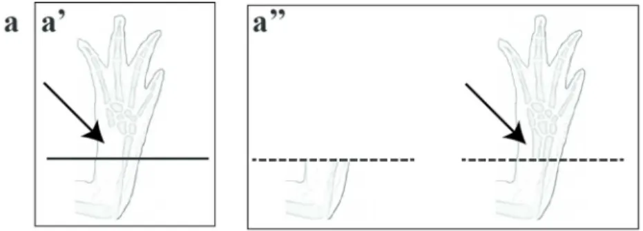
We know Axolotls and other salamanders are champions of regeneration. This remarkable ability to regenerate represents the main reason for studying them in order to understand the fundamentals of epimorphic tissue regeneration. What is particularly interesting in the case of the Axolotl is that even though it can perfectly regenerate a full limb following an amputation (removing all tissue types: soft and hard like bones), if you only remove a critical size section of bone this section will not regenerate (Hutchison et al., 2007). Various examples have been reported showing that a specific lack of bone regeneration exist in these animals when only the bone is amputated. Richard Goss, a pioneer in the field of regeneration, showed a specific lack of regeneration in bones (Goss, 1969). One experiment of particular interest that he performed was a simple extirpation of the ulna in the forelimb of salamanders (Goss, 1956). Following this extirpation the bone did not regenerate. However, when these forelimbs
15 containing only one bone were amputated regeneration occurred normally in the section of forearm that regenerated. The interesting thing is that the missing part of the ulna, proximal to the amputation site, did not regenerate at all, nevertheless, the extirpated part of the bone distal to the amputation site did regenerate normally just as if the bone had been there in the stump (figure 2a) (Goss, 1956). Another experiment of interest in the literature has also shown the lack of bone participation in the regeneration process by using an entirely different approach. Studies demonstrated that X-ray irradiation inhibits limb regeneration in salamanders (Maden & Wallace, 1976). Dunis and Namenwirth devised an intricate experiment where after irradiation with X-ray, they grafted a non-irradiated skin cuff around the circumference of part of the arm (the graft came from a triploid Axolotl to allow tracing of grafted cells). Once the skin graft healed they proceeded to amputate the irradiated limb through the non-irradiated skin graft and the limb was able to regenerate normally except for muscles (Dunis & Namenwirth, 1977). This experiment showed that the skin (epidermis and dermis) had everything essential to promote perfect regeneration (including bones that were triploid in the regenerated section) except for muscles (figure 2b). Recently, the group of Tanaka reproduced these results using transgenic GFP animals which confirmed the work by Dunis and Namenwirth where regenerated bone can be provided from cells derived from the dermis (Kragl et al., 2009).
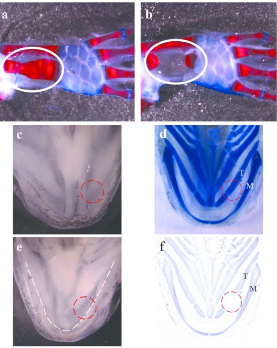
Our lab has published, a few years ago, a paper on Axolotl bone healing. In this paper, the surgical procedures were simple: first a simple fracture was inflicted with scissors on the ulna without removing anything; and second, part of the ulna was removed leaving a critical gap between the remaining parts (4mm)(figure 3). The results went in the same direction as the aforementioned studies in that the bone did not regenerate on its own. The bone was not able to regenerate and the separated parts did not rejoin even 6-7 months post-surgery (figure 3b)
16 (Hutchison et al., 2007). In addition, it showed that healing of a non-stabilised bone fracture in Axolotls healed just as in mammals (Hutchison et al., 2007).
All the above examples were done in salamander limb but what happens in the case of an oral injury. Salamanders have amazing regenerating capacities, and their oral tissues are also capable of regeneration. It was Goss and Stagg in 1958 that first surgically removed part of the jaw in a Newt (Notophthalmus viridescens). Their observations showed that the mandible and teeth could regenerate when the amputation had a rostro-caudal orientation. The second type of injury they did was to remove the mouth floor without touching the mandible. Recently, our group published a paper that also looked at oral tissue regeneration. In this paper, a 4mm punch biopsy was used to make a full excision in the mandibular region. All tissues, including a part of the tongue (which in Axolotls contains cartilage), were removed (Charbonneau et al., 2016). This study presented an extensive time course (up to 180 days following injury) with an in depth histological analysis of the regeneration process. All tissues were reformed but one; the tongue cartilage could not regenerate, even 6 months after the punch was made (Charbonneau et al., 2016) (figure 3c-f). This result is very similar to the critical gap in the forelimb observed by our lab in 2007 (figure 3b)(Hutchison et al., 2007).
All these data combined point to one thing: bones do not regenerate well, if at all, when they are specifically targeted. During the regeneration process, it seems like bones do not participate to the appearance of new tissues. Mesenchymal cells, acting as progenitors, are necessary to reform the new bone as demonstrated with the rescue of regeneration with the triploid skin graft on irradiated limbs (Dunis & Namenwirth, 1977) and the use of transgenic Axolotls by Tanaka’s group which confirmed these evidences in a very elegant way using GFP marked cells (Kragl et al., 2009).
17 Conclusion
Oral wound healing is an example of the capacity of humans to heal with low scarring. This strongly suggests that all the necessary signals for near perfect healing are present in humans. To achieve similar wound healing in other sites, we need a better understanding of how this perfect healing is orchestrated at the cellular and molecular level. The Axolotl is an excellent model to study since it has perfect healing capacities and shares many similarities with oral wound healing. Another important reason to study different organisms is that we can discover fundamental processes governing how, in our case, tissues are repaired or why they failed to regenerate. For example, the difficulty researchers have had in inducing bone regeneration maybe due to a fundamental resistance or complete lack of regenerative potential of bone itself. Obviously, bone can repair fractures and it is constantly renewing itself through the action of osteoblasts and osteoclasts. However, when a fracture reaches a critical size, it becomes very difficult to induce its regeneration. From what has been observed in Axolotls, an animal that can perfectly regenerate an entire limb, it seems that bones do not participate to the regeneration process (at least not on their own). Looking at how Mother Nature has solved tissue regeneration in different organisms could be useful in guiding where we should invest our research efforts. Maybe what needs to be done in order to stimulate bone regeneration is to figure out which cells are responsible for bone regeneration in animals such as Axolotls and try to identify whether similar cells exist in humans in order to recruit them to the task. Research can sometimes seem very fundamental and academic with limited applications, but often this type of work is what
18 yields important discoveries about how biology works which can lead to applications in the clinic.
Acknowledgements
The authors would like to thank the Roy lab members for their comments and for reviewing the manuscript. Work performed in the Roy lab is funded by the Canadian Institute for Health Research (CIHR) and the Fond Québécois de la Recherche Nature et Technologie (FQRNT). F Sader was supported by a scholarship from the Faculty of Graduate Studies at the Université de Montréal and JF Denis is supported by a scholarship from the network of Recherche en Santé Buccale et Osseuse (RSBO).
Author contribution
F Sader participated in the writing of the manuscript and made the figures. JF Denis helped with the writing and S Roy wrote and supervised the work leading to this manuscript.
19 References
Ashcroft GS and Roberts AB (2000). Loss of Smad3 modulates wound healing. Cytokine
Growth Factor Rev 11: 125-31.
Ashcroft GS, Yang X, Glick AB, Weinstein M, Letterio JL, Mizel DE, Anzano M, Greenwell-Wild T, Wahl SM, Deng C and Roberts AB (1999). Mice lacking Smad3 show accelerated wound healing and an impaired local inflammatory response. Nat Cell Biol 1: 260-6. Branton MH and Kopp JB (1999). TGF-beta and fibrosis. Microbes Infect 1: 1349-65.
Braun L, Mead JE, Panzica M, Mikumo R, Bell GI and Fausto N (1988). Transforming growth factor beta mRNA increases during liver regeneration: a possible paracrine mechanism of growth regulation. Proc Natl Acad Sci U S A 85: 1539-43.
Brown KA, Pietenpol JA and Moses HL (2007). A tale of two proteins: differential roles and regulation of Smad2 and Smad3 in TGF-beta signaling. J Cell Biochem 101: 9-33.
Charbonneau AM, Roy S and Tran SD (2016). Oral-Facial Tissue Reconstruction in the Regenerative Axolotl. J Exp Zool B Mol Dev Evol 326: 489-502.
Cheifetz S, Weatherbee JA, Tsang ML-S, Anderson JK, Mole JE, Lucas R and Massagué J (1987). The transforming growth factor-ß system, a complex pattern of cross-reactive ligands and receptors. Cell 48: 409-415.
Cho H, Balaji S, Hone NL, Moles CM, Sheikh AQ, Crombleholme TM, Keswani SG and Narmoneva DA (2016). Diabetic wound healing in a MMP9-/- mouse model. Wound Repair
Regen 24: 829-840.
Christensen RN and Tassava RA (2000). Apical epithelial cap morphology and fibronectin gene expression in regenerating axolotl limbs. Dev Dyn 217: 216-24.
Degen KE and Gourdie RG (2012). Embryonic wound healing: a primer for engineering novel therapies for tissue repair. Birth Defects Res C Embryo Today 96: 258-70.
Denis J, F., Levesque M, Tran S, Camarda A, J. and Roy S (2013). Axolotl as a model to study scarless wound healing in vertebrates: Role of the transforming growth factor beta signaling pathway. Adv. Wound Care 2: 250-260.
Denis JF, Sader F, Gatien S, Villiard E, Philip A and Roy S (2016). Activation of Smad2 but not Smad3 is required for mediating TGF-beta signaling during limb regeneration in axolotls.
Development 143: 3481-3490.
Diegelmann RF and Evans MC (2004). Wound healing: an overview of acute, fibrotic and delayed healing. Front Biosci 9: 283-9.
Dunis DA and Namenwirth M (1977). The role of grafted skin in the regeneration of X-irradiated axolotl limbs. Dev. Biol. 56: 97-109.
Ebersole JL, Graves CL, Gonzalez OA, Dawson D, 3rd, Morford LA, Huja PE, Hartsfield JK, Jr., Huja SS, Pandruvada S and Wallet SM (2016). Aging, inflammation, immunity and periodontal disease. Periodontol 2000 72: 54-75.
Eke PI, Dye BA, Wei L, Slade GD, Thornton-Evans GO, Borgnakke WS, Taylor GW, Page RC, Beck JD and Genco RJ (2015). Update on Prevalence of Periodontitis in Adults in the United States: NHANES 2009 to 2012. J Periodontol 86: 611-22.
Eslami A, Gallant-Behm CL, Hart DA, Wiebe C, Honardoust D, Gardner H, Hakkinen L and Larjava HS (2009). Expression of integrin alphavbeta6 and TGF-beta in scarless vs scar-forming wound healing. The journal of histochemistry and cytochemistry : official journal of the
20 Esposito M, Grusovin MG, Papanikolaou N, Coulthard P and Worthington HV (2009). Enamel matrix derivative (Emdogain(R)) for periodontal tissue regeneration in intrabony defects.
Cochrane Database Syst Rev: CD003875.
Flanders KC (2004). Smad3 as a mediator of the fibrotic response. Int J Exp Pathol 85: 47-64. Frank S, Madlener M and Werner S (1996). Transforming growth factors beta1, beta2, and beta3 and their receptors are differentially regulated during normal and impaired wound healing. J Biol
Chem 271: 10188-93.
Fujita T, Alotaibi M, Kitase Y, Kota Y, Ouhara K, Kurihara H and Shuler CF (2012). Smad2 is involved in the apoptosis of murine gingival junctional epithelium associated with inhibition of Bcl-2. Arch Oral Biol 57: 1567-73.
Gabbiani G (2003). The myofibroblast in wound healing and fibrocontractive diseases. J Pathol 200: 500-3.
Ghanbari H and Vakili‐Ghartavol R (2016). Bone Regeneration: Current Status and Future
Prospects.
Godwin JW, Pinto AR and Rosenthal NA (2013). Macrophages are required for adult salamander limb regeneration. Proc Natl Acad Sci U S A 110: 9415-20.
Goss RJ (1956). The relation of bone to the histogenesis of cartilage in regenerating forelimbs and tails of adult Trituris viridescens. J. Morph. 98: 89-123.
Goss RJ (1969). Principles of Regeneration, Academic Press: New York.
Griffin KS, Davis KM, McKinley TO, Anglen JO, Chu T-MG, Boerckel JD and Kacena MA (2015). Evolution of Bone Grafting: Bone Grafts and Tissue Engineering Strategies for Vascularized Bone Regeneration. Clinical Reviews in Bone and Mineral Metabolism 13: 232-244.
Han M, Yang X, Taylor G, Burdsal CA, Anderson RA and Muneoka K (2005). Limb regeneration in higher vertebrates: developing a roadmap. Anatomical record. Part B, New
anatomist 287: 14-24.
Harty M, Neff AW, King MW and Mescher AL (2003). Regeneration or scarring: an immunologic perspective. Dev Dyn 226: 268-79.
Heldin CH, Miyazono K and ten Dijke P (1997). TGF-beta signalling from cell membrane to nucleus through SMAD proteins. Nature 390: 465-71.
Ho DM and Whitman M (2008). TGF-beta signaling is required for multiple processes during Xenopus tail regeneration. Dev Biol 315: 203-16.
Hoffmann FM (1991). Transforming growth factor-beta-related genes in Drosophila and vertebrate development. Curr Opin Cell Biol 3: 947-52.
Hogan BL (1996). Bone morphogenetic proteins in development. Curr Opin Genet Dev 6: 432-8. Hutchison C, Pilote M and Roy S (2007). The axolotl limb: a model for bone development, regeneration and fracture healing. Bone 40: 45-56.
Jinno K, Takahashi T, Tsuchida K, Tanaka E and Moriyama K (2009). Acceleration of palatal wound healing in Smad3-deficient mice. J Dent Res 88: 757-61.
Kahari VM and Saarialho-Kere U (1997). Matrix metalloproteinases in skin. Experimental
dermatology 6: 199-213.
Klass BR, Grobbelaar AO and Rolfe KJ (2009). Transforming growth factor beta1 signalling, wound healing and repair: a multifunctional cytokine with clinical implications for wound repair, a delicate balance. Postgrad Med J 85: 9-14.
Kragl M, Knapp D, Nacu E, Khattak S, Maden M, Epperlein HH and Tanaka EM (2009). Cells keep a memory of their tissue origin during axolotl limb regeneration. Nature 460: 60-5.
21 Larjava H, Wiebe C, Gallant-Behm C, Hart DA, Heino J and Hakkinen L (2011). Exploring scarless healing of oral soft tissues. J Can Dent Assoc 77: b18.
Larson BJ, Longaker MT and Lorenz HP (2010). Scarless fetal wound healing: a basic science review. Plast Reconstr Surg 126: 1172-80.
Leask A and Abraham DJ (2004). TGF-beta signaling and the fibrotic response. Faseb J 18: 816-27.
Levesque M, Gatien S, Finnson K, Desmeules S, Villiard E, Pilote M, Philip A and Roy S (2007). Transforming growth factor: Beta signaling is essential for limb regeneration in axolotls.
PLoS ONE 2: e1227.
Levesque M, Villiard E and Roy S (2010). Skin wound healing in axolotls: a scarless process. J
Exp Zool B Mol Dev Evol 314B: 684-697.
Maden M and Wallace H (1976). How x-rays inhibit amphibian limb regeneration. J Exp Zool 197: 105-13.
Martin P (1997). Wound healing--aiming for perfect skin regeneration. Science 276: 75-81. Massague J (1998). TGF-beta signal transduction. Annu Rev Biochem 67: 753-91.
Massague J (2000). How cells read TGF-beta signals. Nat Rev Mol Cell Biol 1: 169-78.
Massagué J (1987). The TGF-ß family of growth and differentiation factors. Cell 49: 437-438. McKeown ST, Barnes JJ, Hyland PL, Lundy FT, Fray MJ and Irwin CR (2007). Matrix metalloproteinase-3 differences in oral and skin fibroblasts. J Dent Res 86: 457-62. Mescher AL and Neff AW (2006). Limb Regeneration in Amphibians: Immunological Considerations. TSW Development & Embryology 1: 1-11.
Ripamonti U (2016). Redefining the induction of periodontal tissue regeneration in primates by the osteogenic proteins of the transforming growth factor-beta supergene family. J Periodontal
Res 51: 699-715.
Roberts AB (1998). Molecular and cell biology of TGF-beta. Miner Electrolyte Metab 24: 111-9. Roy S and Lévesque M (2006). Limb Regeneration in Axolotl: Is It Superhealing? TSW
Development & Embryology 6: 12-25.
Schmitz JP and Hollinger JO (1986). The critical size defect as an experimental model for craniomandibulofacial nonunions. Clin Orthop: 299-308.
Seifert AW, Monaghan JR, Voss SR and Maden M (2012). Skin regeneration in adult axolotls: a blueprint for scar-free healing in vertebrates. PLoS ONE 7: e32875.
Shannon DB, McKeown ST, Lundy FT and Irwin CR (2006). Phenotypic differences between oral and skin fibroblasts in wound contraction and growth factor expression. Wound Repair
Regen 14: 172-8.
Song B, Estrada KD and Lyons KM (2009). Smad signaling in skeletal development and regeneration. Cytokine Growth Factor Rev 20: 379-88.
Stephens P, Davies KJ, Occleston N, Pleass RD, Kon C, Daniels J, Khaw PT and Thomas DW (2001). Skin and oral fibroblasts exhibit phenotypic differences in extracellular matrix
reorganization and matrix metalloproteinase activity. Br J Dermatol 144: 229-37.
Stramer BM, Mori R and Martin P (2007). The inflammation-fibrosis link? A Jekyll and Hyde role for blood cells during wound repair. J Invest Dermatol 127: 1009-17.
Szpaderska AM, Zuckerman JD and DiPietro LA (2003). Differential injury responses in oral mucosal and cutaneous wounds. J Dent Res 82: 621-6.
Tomikawa K, Yamamoto T, Shiomi N, Shimoe M, Hongo S, Yamashiro K, Yamaguchi T, Maeda H and Takashiba S (2012). Smad2 decelerates re-epithelialization during gingival wound healing. J Dent Res 91: 764-70.
22 Toriseva M and Kahari VM (2009). Proteinases in cutaneous wound healing. Cell Mol Life Sci 66: 203-24.
Vinarsky V, Atkinson DL, Stevenson TJ, Keating MT and Odelberg SJ (2005). Normal newt limb regeneration requires matrix metalloproteinase function. Dev Biol 279: 86-98.
Wong JW, Gallant-Behm C, Wiebe C, Mak K, Hart DA, Larjava H and Hakkinen L (2009). Wound healing in oral mucosa results in reduced scar formation as compared with skin: evidence from the red Duroc pig model and humans. Wound Repair Regen 17: 717-29.
Wrana JL and Attisano L (2000). The Smad pathway. Cytokine Growth Factor Rev 11: 5-13. Yang EV and Bryant SV (1994). Developmental regulation of a matrix metalloproteinase during regeneration of axolotl appendages. Dev. Biol. 166: 696-703.
Yang EV, Gardiner DM, Carlson MR, Nugas CA and Bryant SV (1999). Expression of Mmp-9 and related matrix metalloproteinase genes during axolotl limb regeneration. Dev Dyn 216: 2-9. Zentella A and Massague J (1992). Transforming growth factor beta induces myoblast
differentiation in the presence of mitogens. Proc Natl Acad Sci U S A 89: 5176-80.
Zhu Y, Richardson JA, Parada LF and Graff JM (1998). Smad3 mutant mice develop metastatic colorectal cancer. Cell 94: 703-14.
23 Legends
Figure 1. Comparison between Axolotl wound healing, limb regeneration and human oral wound healing. (a) Picture of an albino Axolotl. Tail (T); Limb (L); Lower jaw (J) are capable of regeneration following an amputation. (b) Comparison of gene expression and ECM elements between Axolotl tail excisional wound (wound is circled), regenerating limb and human oral injury. Blastema: an accumulation of dedifferentiated cells mostly composed from mesenchymal and muscle cells which migrated under the wound epithelium and are proliferating to give rise to the regenerated tissue.
Figure 2. Schematic representation of bone extirpation and limb irradiation in Axolotl. (a) Ulna extirpation experiment. (a’) Following extirpation no bone regeneration is observed (black arrow points to where the missing ulna should be). (a’’) After amputation the regenerated part is perfect including the ulna (black arrow points to the ulna that is present only in the regenerated portion of the limb). (b) Limb irradiation and skin graft experiment. (b’) X-ray irradiation (red lightning) followed by amputation results in absence of regeneration. (b’’) X-ray irradiation followed by a non-irradiated skin graft (green area). After amputation through the skin graft the limb is perfectly regenerated except for muscles.
Figure 3. Forelimb bone gap and oral punch experiments. (a-b) Alizerin red/alcian blue coloration of forelimb bone fractures. (a) Clear cut union fracture wound 7 months post-injury showing bone healing and a callus formation (white oval). (b) 4mm bone gap 7 months post-injury showing no healing (white oval). (c) Ventral view of Axolotl mandibular region. (d)
24 Victoria blue staining (cartilage) of ventral mandibular region. Mandible (M) and tongue cartilage (T) are visible. (e) Inside view of Axolotl mandibular region (tongue contour is highlighted with dotted line). (f) Cartoon showing 180 days post-injury showing how tongue cartilage was not reformed. Red circle shows punch wound area.
Figure 1: Comparison between Axolotl wound healing, limb regeneration and human oral wound healing. 199x216mm (150 x 150 DPI)
Figure 2:Schematic representation of bone extirpation and limb irradiation in Axolotl. 210x211mm (150 x 150 DPI)
Figure 3. Forelimb bone gap and oral punch experiments. 182x223mm (150 x 150 DPI)