Université de Montréal
Validité et fidélité de la combinaison de l’anamnèse et de
l’examen physique pour le diagnostic des pathologies
communes au genou
par Simon Décary, pht., M.Sc. École de réadaptation Faculté de médecine Thèse présentée à la Faculté de médecine en vue de l’obtention du grade de Philosophiae Doctor (Ph. D.) en Sciences de la réadaptation 31 Août 2017 © Simon Décary, 2017
U
niversité de Montréal Faculté des études supérieures Cette thèse intitulée : Validité et fidélité de la combinaison de l’anamnèse et de l’examen physique pour le diagnostic des pathologies communes au genou présentée par : Simon Décary, pht., M.Sc. A été évaluée par un jury composé des personnes suivantes : M. Nicolas Dumont, pht., Ph. D. Président-rapporteur M. François Desmeules, pht., Ph. D. Directeur de recherche Dr. Pascal-André Vendittoli, MD, M.Sc., FRSC Codirecteur de recherche Mme. Carole Fortin, pht., Ph. D. Membre interne du jury M. Yannick Tousignant-Laflamme, pht., Ph. D. Examinateur externe Natacha Trudeau, Ph. D. Représentante de la doyenne de la FESRésumé
Les douleurs au genou font partie des motifs de consultation les plus fréquents auprès d’un médecin ou d’un professionnel de la santé. Les pathologies communes au genou incluent celles d’origine traumatique telles les déchirures du ligament croisé antérieur ou les déchirures méniscales et celles d’apparition progressive telles les déchirures méniscales dégénératives, l’ostéoarthose du genou ou le syndrome fémoro-patellaire. Les données probantes démontrent qu’un diagnostic initial valide, basé sur une évaluation musculosquelettique bien accomplie par un intervenant ayant une formation adéquate, permet l’initiation rapide d’un traitement ciblé. Cependant, le manque de connaissances dans l’évaluation musculosquelettique et les erreurs diagnostiques fréquentes dans notre système de santé favorisent l’utilisation inappropriée des tests d’imagerie médicale et augmentent les références non pertinentes en chirurgie orthopédique. Les données probantes sont actuellement limitées et incomplètes concernant la validité et la fidélité de l’évaluation musculosquelettique combinant l’anamnèse et l’examen physique pour orienter le diagnostic différentiel des pathologies au genou. L’amélioration des connaissances en lien avec l’évaluation musculosquelettique des adultes souffrant de douleurs au genou est donc nécessaire afin d’améliorer l’efficience de nos soins de santé.
Cette thèse propose deux objectifs : 1- évaluer l’accord diagnostique entre un physiothérapeute utilisant une évaluation musculosquelettique standardisée sans imagerie et des médecins experts pour les différentes pathologies communes au genou; 2- évaluer la validité de la combinaison de l’anamnèse et de l’examen physique afin de développer une série d’outils valides permettant d’orienter le diagnostic différentiel de quatre pathologies communes au genou.
Nous avons évalué deux cent soixante-dix-neuf participants, présentant 359 diagnostics primaires et secondaires incluant : 43 participants ayant une déchirure du ligament croisé antérieur, 80 participants présentant une déchirure méniscale, 129 participants atteints
d’ostéoarthrose du genou, 75 atteints d’un syndrome fémoro-patellaire et 32 participants présentant une autre pathologie au genou.
Le physiothérapeute qui exécutait une évaluation musculosquelettique standardisée a démontré un excellent accord diagnostique avec des médecins experts dont le diagnostic était basé sur une évaluation musculosquelettique combinée aux résultats des tests d’imagerie (k=0,89; IC95%: 0,83-0,94). Sur la base de l’évaluation musculosquelettique réalisée indépendamment par le physiothérapeute, nous avons pu développer une série de combinaisons d’éléments de l’anamnèse et de tests de l’examen physique pour orienter le diagnostic différentiel des pathologies au genou. Nous avons démontré qu’un individu qui consultait pour une douleur au genou dont l’origine est un traumatisme du genou en pivot, ayant ressenti un « pop » au moment du traumatisme et chez qui les tests de Lachman ou du
pivot shift sont positifs, a une probabilité élevée de souffrir d’une déchirure du ligament
croisé antérieur avec un rapport de vraisemblance positif de 38,4 (IC95%: 16,0-92,5). Si un individu décrit un traumatisme au genou avec pivot accompagné d’une douleur localisée au côté médial du genou et qu’il démontre une douleur à la palpation de l’interligne articulaire interne, celui-ci a une probabilité élevée de souffrir d’une déchirure méniscale avec un rapport de vraisemblance positif de 8,9 (IC95%: 6,1-13,1).
Un individu présentant une douleur d’apparition progressive au côté médial du genou présente dans les activités qui nécessitent des pivots et qui démontre à l’examen physique un alignement neutre du membre inférieur ou une flexion passive complète du genou, celui-ci a une probabilité modérée d’être atteint d’une déchirure méniscale dégénérative avec un rapport de vraisemblance positif de 6,4 (IC95%: 4,0-10,4). Si toutefois un individu consultant pour une douleur d’apparition progressive est âgé de plus de 50 ans, qu’il a un indice de masse corporel supérieur à 30 et qu’il démontre à l’examen physique la présence d’un alignement au genou en varus ou en valgus, des crépitements à la palpation du genou ou une limitation de l’amplitude articulaire passive en extension, il a une probabilité élevée d’être atteint d’ostéoarthrose du genou avec un rapport de vraisemblance positif de 13,6 (IC95%:
6,5-28,4). Finalement, un individu qui présente une douleur isolée en antérieur du genou accompagnée de difficultés dans les escaliers, d’une douleur à la palpation des facettes rotuliennes et d’une extension passive complète du genou, celui-ci a une probabilité élevée de souffrir d’un syndrome fémoro-patellaire avec un rapport de vraisemblance positif de 8,7 (IC95%: 5,2-14,6). En résumé, les résultats de cette thèse démontrent qu’il existe un accord diagnostique élevé entre un physiothérapeute qui procède à une évaluation musculosquelettique sans recours à des tests d’imagerie et des médecins experts qui basent leur diagnostic sur l’examen physique et les résultats des tests d’imagerie chez des adultes souffrant de douleurs au genou. Les combinaisons des éléments de l’anamnèse et de l’examen physique développées dans cette thèse permettent d’orienter le diagnostic différentiel de différentes pathologies communes au genou avec une validité considérée modérée à élevée. Ces combinaisons devront être validées dans d’autres contextes cliniques, notamment en première ligne, avant une utilisation clinique répandue.
Mots clés : genou, diagnostic, anamnèse, examen physique, ligament croisé antérieur,
Abstract
Knee complaints are among the most common reasons for consulting a healthcare practitioner. Common knee disorders include those of traumatic onset such as anterior cruciate ligament or meniscal tears, or those of progressive onset such as degenerative meniscal tears, knee osteoarthritis or patellofemoral pain syndrome. An early diagnosis to guide toward an efficient management is advocated to prevent persistence of pain, functional limitations and loss of quality of life in affected individuals. However, evidence currently shows an overreliance on medical imaging tests and inappropriate orthopaedic surgery referrals, thus delaying initiation of treatment. Evidence is currently limited concerning the validity and reliability of combining history elements and physical examination tests to support the differential diagnosis of common knee disorders. To answer this evidence gap, this thesis had two objectives: 1- to assess inter-rater diagnostic agreement between a physiotherapist using only a standardized musculoskeletal examination and expert physicians using a musculoskeletal examination in combination with imaging tests for the diagnosis of common knee disorders; 2- to assess the diagnostic validity of clusters combining history elements and physical examination tests and produce a series of tools to support the differential diagnosis of four common knee disorders.
We prospectively recruited two hundred and seventy-nine participants presenting 359 primary and secondary diagnoses including: 43 participants with an anterior cruciate ligament tear, 80 subjects had a meniscal tear, 129 suffered from knee osteoarthritis, 75 were diagnosed with patellofemoral pain syndrome and 32 presented other knee diagnoses. The physiotherapist achieved high inter-rater agreement with the expert physicians for the diagnosis of common knee disorders (k= 0.89; 95%CI:0.83-0.94).
Multiple clusters combining history elements and physical examination tests were developed to support the differential diagnosis of knee disorders. Our results show that an individual consulting for a knee complaint following trauma during a pivot and describing a “popping
sensation” during the trauma as well as showing a positive Lachman or pivot shift tests has a high probability of having an anterior cruciate ligament tear with a positive likelihood ratio of 38.4 (95%CI: 16.0-92.5). If this individual consulting for a knee complaint following trauma during a pivot experience medially located knee pain, confirmed with medial joint line tenderness at palpation, the subject has a high probability of having a meniscal tear of traumatic origin with a positive likelihood ratio of 8.9 (95%CI: 6.1-13.1).
An individual consulting for a complaint of progressive onset with isolated medially located pain during activities requiring a pivot, who also presents a normal knee alignment or full passive knee flexion, has a moderate probability of having a degenerative meniscal tear with a positive likelihood ratio of 6.4 (95%CI: 4.0-10.4). If this individual complaining of knee pain of progressive onset is older than 50 years old, has a body mass index higher than 30 and at physical examination presents knee crepitus at palpation, a valgus or varus knee misalignment or a restricted passive knee extension, he has a high probability of having symptomatic knee osteoarthritis with a positive likelihood ratio of 13.6 (95%CI: 6.5-28.4). Lastly, an individual consulting for isolated anterior knee pain with difficulty descending stairs, palpable patellar facets tenderness and full passive knee extension has a high probability of having a patellofemoral pain syndrome with a positive likelihood ratio of 8.7 (95%CI: 5.2-14.6).
Overall, the results demonstrated high inter-rater diagnostic concordance between providers and that combining selected history elements and physical examination tests was moderately to highly valid to support the differential diagnosis of common knee disorders. The proposed diagnostic clusters will require external validation in various clinical contexts, including primary care, before widespread implementation in clinical practice.
Key words: knee, diagnosis, history elements, physical examination, anterior cruciate
Tables des matières
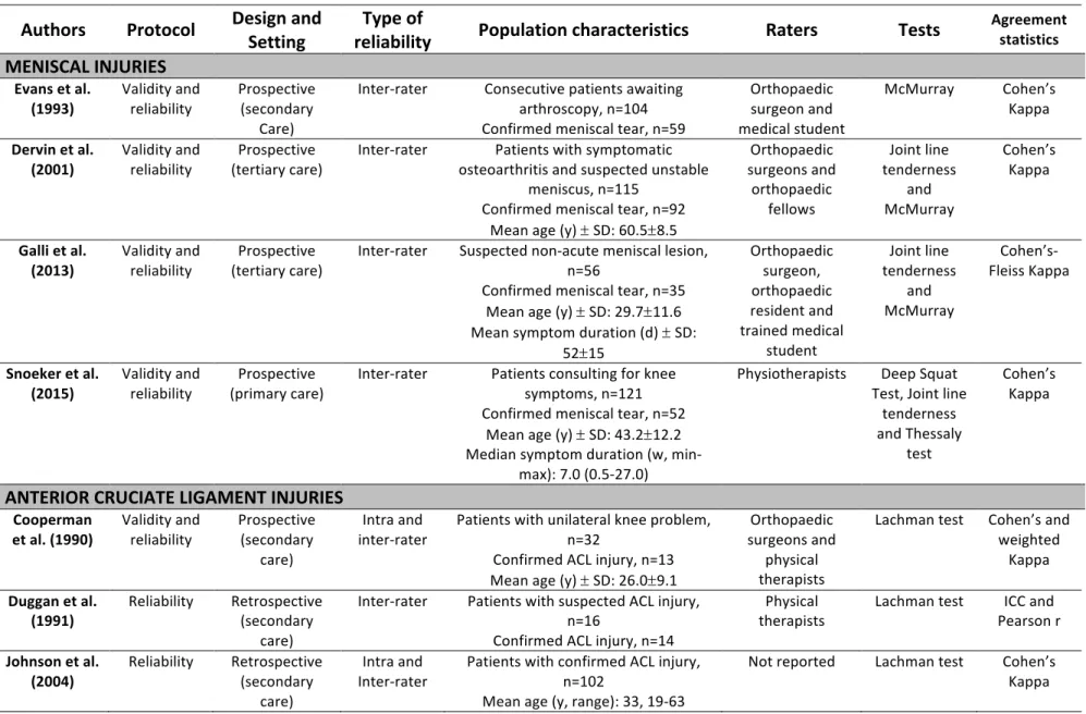
Résumé iii Abstract vi Tables des matières viii Liste des tableaux xi Liste des figures xiv Liste des sigles et abréviations xv Dédicace xvi Remerciements xvii Chapitre 1: Introduction 1 Chapitre 2 : Revue de la littérature 3 2.1 Épidémiologie des pathologies communes au genou 3 2.2 Validité et fidélité de l’évaluation musculosquelettique 4 Article 1: Diagnostic validity of physical examination tests for common knee disorders: an overview of systematic reviews and meta-analysis 5 Article 2: Reliability of physical examination tests for the diagnosis of knee disorders: Evidence from a systematic review 31 2.3 Validité de la combinaison de l’anamnèse et des tests de l’examen physique. 57 2.3.1 Déchirures partielles ou complètes du LCA 57 2.3.2 Déchirures méniscales 57 2.3.3 Ostéoarthrose du genou 59 2.3.4 Syndrome fémoro-patellaire 59 2.4 Validité et fidélité inter-évaluateur du diagnostic émis par un physiothérapeute 59 2.5 Conclusions de la revue de la littérature et limites des études dans le domaine 60 2.6 Objectifs et hypothèses de la thèse de doctorat 62 Chapitre 3 : Méthodologie 64 3.1 Devis de l’étude et milieux de recrutement 64 3.2 Sélection des participants 64 3.3 Collecte des données 65 3.3.1 Portrait clinique des participants et anamnèse 65 3.3.2 Sélection des tests de l’examen physique 66 3.3.3 Procédure pour la collecte des données de l’examen physique 67 3.4 Définition du standard de référence 683.5 Analyses statistiques 69 3.5.1 Évaluation de la fidélité inter-évaluateur 69 3.5.2 Développement des modèles diagnostiques combinant l’anamnèse et l’examen physique 70 3.5.3 Évaluation de la validité des modèles diagnostiques 72 3.6 Taille d’échantillon 73 Chapitre 4 : Résultats 74 Article 3: Diagnostic validity and triage concordance of a physiotherapist compared to physicians’ diagnoses for common knee disorders 75 Article 4: Diagnostic validity of combining history elements and physical examination tests for partial or complete anterior cruciate ligament tears 92 Article 5: Diagnostic validity of combining history elements and physical examination tests for traumatic and degenerative symptomatic meniscal tears 115 Article 6: Initial derivation of diagnostic clusters combining history elements and physical examination tests for symptomatic knee osteoarthritis 140 Article 7: Validity of combining history elements and physical examination tests to diagnose patellofemoral pain 160 Chapitre 5: Discussion générale 180 5.1 Caractéristiques de la cohorte et représentativité 180 5.2 Synthèse des résultats et comparaison avec les données probantes de la littérature 181 5.3 Forces de l’étude 184 5.3.1 Forces liées au mode de recrutement 184 5.3.2 Forces liées au standard de référence 186 5.3.3 Forces liées à la collecte des tests index 187 5.3.4 Forces liées aux méthodes statistiques 187 5.4 Limites de l’étude 188 5.4.1 Biais potentiels et limites liés à la population à l’étude et à la généralisation des résultats 189 5.4.2 Biais potentiels et limites liés au standard de référence 190 5.4.3 Biais potentiels et limites liés aux tests index 192 5.4.4 Biais potentiels et limites liés aux méthodes statistiques 194 5.5 Retombées cliniques 195 5.6 Avenues futures de recherche 196 5.7 Conclusion générale 198 Bibliographie 200
Annexes 218 1. Detailed search strategy with keywords and descriptors for article 1 218 2. Detailed search strategy with keywords and descriptors for article 2 221 3. Liste des variables de l’anamnèse 223 4. Formulaire de l’anamnèse 224 5. Questionnaire Knee Injury and Osteoarthritis Outcome Score (KOOS) 230 6. Questionnaire Kessler-6 234 7. Guide de standardisation de l’examen physique 236 8. Flow chart of patients’ recruitment in article 3 242 9. 2x2 contingency tables to assess the validity and inter-rater reliability of the complete musculoskeletal examination for each knee disorder in article 3 243 10. 3x3 contingency table to assess the inter-rater reliability of the triage of surgical candidates and conservative care in article 3 244 11. Flow chart of patients’ recruitment in article 4, 5, 6 and 7 245 12. 2x2 contingency tables for the diagnostic validity of history elements and physical examination tests when individually performed for partial or complete ACL tears 246 13. 2x2 contingency tables for diagnostic validity of history elements and physical examination tests when individually performed for complete ACL tears only in article 4 247 14. 2x2 contingency tables of diagnostic clusters using recursive partitioning for partial or complete ACL tears in article 4 248 15. 2x2 contingency tables of diagnostic clusters using recursive partitioning for complete ACL tears only in article 4 249 16. 2x2 contingency tables of diagnostic clusters using recursive partitioning for traumatic and degenerative symptomatic meniscal tears in article 5 250 17. Description of other physical examination tests techniques for symptomatic degenerative meniscal tears in article 5 251 18. 2x2 contingency tables of diagnostic clusters using recursive partitioning for symptomatic knee osteoarthritis in article 6 252 19. 2x2 contingency tables of diagnostic clusters using recursive partitioning for patellofemoral pain in article 7 253 20. Certificat d’éthique 254 21. Formulaire de consentement 255
Liste des tableaux
Article 1 Table 1: Characteristics of the included reviews ... 13 Table 2: Assessment of the methodological quality of the included systematic reviews (AMSTAR) ... 15 Table 3: Description of diagnostic properties for selected tests for meniscal injuries based on results from included reviews ... 20 Table 4: Description of diagnostic properties for physical examination tests for Anterior Cruciate Ligament (ACL) injuries based on results from included reviews ... 21 Table 5: Description of diagnostic properties for selected tests for knee fractures based on results from included reviews ... 23 Table 6: Description of diagnostic properties for selected tests for osteoarthritis based on results from included reviews ... 24 Table 7: Description of diagnostic properties of the history taking and physical examination for selected knee disorders based on results from included reviews ... 25 Article 2 Table 8: Characteristics of included reliability studies according to different knee disorders .. 38 Table 9: Methodological quality of included studies using the QAREL checklist ... 43 Table 10: Summary of reliability measures from included studies evaluating meniscal injury tests ... 47 Table 11: Summary of reliability measures from included studies evaluating anterior cruciate ligament injury tests ... 48 Table 12: Summary of reliability measures from included studies evaluating osteoarthritis physical tests ... 49 Table 13: Summary of reliability measures from included studies evaluating common patellofemoral pain tests ... 51 Article 3 Table 14: Characteristics of participants (n=179) ... 84 Table 15: Clinical and imaging diagnoses of participants (n=179) ... 85 Table 16: Concordance between the physiotherapist and physicians’ composite or imaging only diagnoses (n=179) ... 86 Table 17: Diagnostic validity of the musculoskeletal examination performed by the physiotherapist compared to the physicians’ composite diagnosis ... 87 Table 18: Concordance between the physiotherapist and physicians for the triage recommendation following consultation (n=179) ... 88Article 4 Table 19: Description of physical examination tests for ACL tear ... 98 Table 20: Characteristics of participants (n=279) ... 102 Table 21: Description of clinical diagnoses and imaging findings for partial or complete ACL tears participants (n=43) ... 103 Table 22: Clinical variables associated with the diagnosis of partial or complete ACL tears at maximal AUC identified through penalized logistic regression (n=279) ... 105 Table 23: Diagnostic validity and inter-rater reliability of history elements and physical examination tests when individually performed to diagnose or exclude partial or complete ACL tears (n=43) or complete ACL tears only (n=22) ... 107 Table 24: Diagnostic clusters combining history elements and physical examination tests to diagnose partial or complete ACL tears (n=43) or complete ACL tears only (n=22) ... 109 Table 25: Diagnostic cluster combining history elements and physical examination tests to exclude partial or complete ACL tears (n=43) ... 110 Article 5 Table 26: Description of physical examination tests for meniscal tear ... 123 Table 27: Characteristics of participants with a knee complaint (n=279) ... 125 Table 28: Description of clinical diagnoses and imaging findings for SMT participants ... 126 Table 29: Clinical variables associated with the diagnosis of SMT identified through penalized logistic regression in participants with a knee complaint (n=279) ... 127 Table 30: Diagnostic clusters combining history elements and physical examination tests to diagnose or exclude traumatic SMT (n=35) ... 130 Table 31: High specificity diagnostic clusters combining history elements and physical examination tests to diagnose degenerative SMT (n=45) ... 132 Table 32: High sensitivity diagnostic clusters combining history elements and physical examination tests to exclude degenerative SMT (n=45) ... 134 Article 6 Table 33: Characteristics of participants (n=279) ... 149 Table 34: Description of clinical diagnoses and imaging findings for participants with SOA (n=129) ... 150 Table 35: Clinical variables associated with the diagnosis of SOA identified through penalized logistic regression in participants with a knee complaint (n=279) ... 151 Table 36: High specificity diagnostic clusters combining history elements and physical examination tests to rule in SOA ... 153 Table 37: High sensitivity diagnostic clusters combining history elements and physical examination tests to rule out SOA ... 154
Article 7 Table 38: Characteristics of participants (n=279) ... 170 Table 39: Clinical variables associated with the presence or absence of PFP at maximal AUC identified through penalized logistic regression (n=279) ... 171 Table 40: High specificity diagnostic clusters combining history elements and physical examination tests to diagnose PFP ... 173 Table 41: High sensitivity diagnostic clusters combining history elements and physical examination tests to exclude PFP ... 175
Liste des figures
Figure 1: Bibliographic search flowchart. ... 12 Figure 2: Bibliographic search flowchart ... 37 Figure 3: Diagnostic clusters to rule in or rule out a traumatic SMT. ... 131 Figure 4: Diagnostic clusters to rule in degenerative SMT. ... 133 Figure 5: Diagnostic clusters to rule out degenerative SMT. ... 135 Figure 6: Diagnostic clusters to rule in SOA ... 155 Figure 7: Diagnostic clusters to rule out SOA. ... 156 Figure 8: Diagnostic clusters to rule in patellofemoral pain ... 174 Figure 9: Diagnostic clusters to rule out patellofemoral pain. ... 176Liste des sigles et abréviations
Français IRM : Imagerie par résonnance magnétique LCA : Ligament croisé antérieur OA : Ostéoarthrose RV- : Rapport de vraisemblance négatif RV+ : Rapport de vraisemblance positif SFP : Syndrome fémoro-patellaire Se : Sensibilité Sp : Spécificité VPN : Valeur prédictive négative VPP : Valeur prédictive positive Anglais AMSTAR: Assessment of methodological quality of multiple systematic reviews ACL: Anterior cruciate ligament injuries DOR: Diagnostic odds ratio LR: Likelihood ratio MA: Meta-analysis NPV: Negative predictive value OA: Osteoarthritis SOA: Symptomatic knee osteoarthritis PCL: Posterior cruciate ligament injuries PFP: Patellofemoral pain PPV: Positive predictive value PRISMA: Preferred Reporting Items for Systematic Reviews and Meta-Analyses Se: Sensitivity SMT: Symptomatic meniscal tear Sp: Specificity SR: Systematic-reviewDédicace
À Annie et Xavier. Merci de m’offrir la plus belle vie au monde. À ma famille. Merci pour votre support inébranlable.Remerciements
La rédaction d’une thèse doctorale est l’aboutissement d’un long processus au cours duquel le candidat se développe tant sur le plan professionnel que personnel. J’aimerais débuter par remercier ma partenaire de vie, Annie, de me soutenir, de me calmer, mais aussi de m’aider à catalyser mes idées les plus folles. Merci d’être une maman merveilleuse pour notre petit Xavier. Mon fils, merci d’être arrivé dans nos vies, tu nous remplis de bonheur et nous rappelles quotidiennement les valeurs importantes de la vie. J’aimerais aussi remercier mes parents, mon frère et ma sœur pour leur compréhension lorsque je ne suis pas toujours présent. Merci d’avoir respecté mes idées, mes projets et mes folles ambitions pendant toutes ces années.
Mes premiers remerciements professionnels vont à mon directeur de recherche, professeur François Desmeules. Merci d’avoir fait confiance à l’électron libre surexcité que je suis. La liberté que tu m’as offerte m’a permis de développer ma confiance professionnelle pour devenir, je l’espère, un professeur enthousiaste et dévoué pour ses étudiants. Merci pour ton pragmatisme et ton encadrement qui m’ont donné les outils nécessaires pour mener à terme ce projet. J’aimerais ensuite remercier mon codirecteur, Dr Pascal-André Vendittoli de m’avoir fait confiance et de m’avoir ouvert les portes de l’univers des orthopédistes. Merci pour ton regard intéressé sur mes travaux, qui m’oriente et m’amène à me dépasser. Merci à mes mentors, professeure Debbie Feldman et Dre. France Légaré. Merci pour votre temps et votre écoute. Merci d’être des guides pour ma jeune carrière, vous êtes une source d’inspiration et un modèle d’excellence pour la nouvelle génération de chercheurs.
Cette thèse n’aurait aussi tout simplement pas eu lieu sans l’apport inconditionnel des collaborateurs au projet. Merci à Dr Michel Fallaha, Dr Bruno Pelletier, Dr Pierre Frémont et Dr Sylvain Belzile d’avoir si gentiment accepté de partager votre expérience et expertise. Cela a été un honneur d’être votre étudiant et j’espère bientôt pouvoir être votre collègue et poursuivre nos projets et idées pour améliorer la santé des patients. Merci à tous les patients d’avoir accordé leur confiance à un jeune professionnel et d’avoir partagé leurs mille et une
histoires. Un merci spécial à Sylvie pour ton support au quotidien et sans qui l’organisation de ce projet aurait été impossible. Finalement, merci à Véronique, à Philippe et à Roula. Vous êtes les collègues de travail en qui j’ai le plus confiance.
Finalement, j’aimerais remercier tous les organismes qui ont accepté de financer un jeune étudiant chercheur passionné par son travail, mais n’ayant pas encore fait ses preuves. Les Fonds de Recherche en Santé du Québec (FRQS), Les Instituts de Recherche en Santé du Canada (IRSC), la Faculté des Études Supérieures (FESP), l’École de Réadaptation et le Réseau provincial de recherche en adaptation-réadaptation (REPAR). Lorsque vous acceptez de nous financer ou lorsque vous acceptez de donner des prix aux étudiants, vous contribuez à faire grandir notre confiance tant personnelle que professionnelle.
Chapitre 1: Introduction
Près d’un individu sur deux souffrira de douleur au genou au cours de sa vie, ce qui en fait une raison de consultation fréquente en médecine et en réadaptation [1-4]. Les pathologies communes au genou incluent celles d’origine traumatique telles les déchirures du ligament croisé antérieur (LCA) ou les déchirures méniscales [5, 6] ainsi que celles d’apparition progressive telles les déchirures méniscales dégénératives, l’ostéoarthrose du genou (OA) ou le syndrome fémoro-patellaire (SFP) [6]. Un diagnostic initial valide est proposé comme étant une composante essentielle d’une prise en charge efficiente pour ces pathologies [7-23].
Malheureusement, le diagnostic initial est souvent incertain ou erroné chez un nombre important d’individus, ce qui résulte en des trajectoires de soins non optimales [6, 8, 9, 18, 24]. Une première trajectoire non optimale concerne l’utilisation inappropriée des tests en imagerie médicale, principalement l’utilisation de l’imagerie par résonnance magnétique (IRM) [9]. L’utilisation de l’IRM a triplé au cours de la dernière décennie, les genoux représentant 44% de tous les motifs de consultation en IRM pour la catégorie « extrémités » et jusqu’à 50% des examens d’IRM pour les pathologies au genou seraient non justifiés [25]. En plus d’engendrer des coûts importants pour les systèmes de santé, ce test d’imagerie a aussi un fort potentiel de surdiagnostic [9, 25].
Cette incertitude diagnostique mène aussi fréquemment à la référence en spécialité, principalement en chirurgie orthopédique. En effet, on estime qu’entre 55% et 90% des références en chirurgie orthopédique ne seraient pas justifiées puisque ces patients ne nécessiteraient pas de traitements chirurgicaux, mais plutôt une approche conservatrice [26-28]. Dans ce contexte, le chirurgien orthopédique agit principalement à titre de consultant expert pour émettre le diagnostic approprié ce qui amène des délais de prise en charge aux niveaux d’un traitement conservateur médical ou en réadaptation [26, 27, 29]. En effet, en combinant les temps d’attente pour l’obtention d’un examen d’imagerie et subséquemment celui d’un rendez-vous avec un orthopédiste, les délais de prise en charge atteignent souvent
plus de 6 à 12 mois [25]. Cette situation expose les patients à une chronicisation de leur pathologie et une prolongation de leur invalidité.
Ces trajectoires sont souvent causées par une évaluation musculosquelettique initiale incomplète ou erronée en première ligne de soins [25]. Or, il est démontré que dans bien des cas la validité diagnostique de l’évaluation musculosquelettique obtenue par des experts pourrait être équivalente à celle obtenue par les tests d’imagerie pour les pathologies au genou [6]. Afin d’améliorer cet écart dans la capacité diagnostique des cliniciens experts et ceux notamment de première ligne, plusieurs auteurs ciblent le développement d’outils d’aide au diagnostic des pathologies au genou par l’identification de combinaisons valides d’éléments de l’anamnèse et de l’examen physique qui constituent l’évaluation musculosquelettique [6, 9, 25]. Cette thèse explorera donc la validité et la fidélité de
l’évaluation musculosquelettique combinant l’anamnèse et l’examen physique afin de développer une série d’outils permettant d’orienter le diagnostic différentiel des pathologies communes au genou.
Rôle du candidat et structure de la thèse
Cette thèse débutera par une revue de la littérature (Chapitre 2) qui inclut deux revues systématiques sur la validité (Article 1) et la fidélité (Article 2) des tests de l’examen physique pour le diagnostic des pathologies au genou. Le Chapitre 3 présentera le protocole utilisé. Le Chapitre 4 présentera les résultats sous la forme de cinq articles scientifiques (Articles 3 à 7). Finalement, une discussion sera présentée au Chapitre 5.
Chapitre 2 : Revue de la littérature
La revue de la littérature dressera d’abord un portrait des données probantes sur la validité et la fidélité de l’évaluation musculosquelettique des pathologies au genou afin d’identifier les limites de la littérature qui mèneront à l’élaboration des objectifs de la thèse. 2.1 Épidémiologie des pathologies communes au genouLa douleur au genou serait présente chez plus de 20% de la population et représente le symptôme le plus commun pour plusieurs pathologies au genou [3, 25, 30]. Ces pathologies sont généralement classifiées entre celles d’origine traumatique ou celles d’apparition progressive [6]. Parmi les pathologies d’origine traumatique, les deux structures du genou les plus couramment lésées sont le ligament croisé antérieur (LCA) et les ménisques [6]. Le LCA est une structure stabilisatrice du genou qui protège contre les forces excessives en translation antérieure et en rotation interne du tibia [19, 31]. La déchirure du LCA correspond à 4% de toutes les atteintes au genou en première ligne [6]. Elle survient, dans environ 70% des cas, lors d’une activité sportive chez des jeunes adultes [19, 32-36]. Une autre structure couramment lésée lors d’un traumatisme au genou est les ménisques. Ceux-ci sont responsables de la distribution des forces de compression et de pivot, améliorant ainsi la stabilité du genou lors des mouvements [37]. Les déchirures méniscales traumatiques correspondent à approximativement 10% de toutes les pathologies au genou en première ligne et surviennent fréquemment dans un contexte de blessures sportives chez des jeunes adultes de façon concomitante à une déchirure du LCA [6, 38]. Parmi les pathologies d’apparition progressive, les ménisques peuvent aussi subir des lésions considérées d’usure, ou dégénératives, typiquement chez une population adulte plus âgée, et correspondent à environ 30% de toutes les pathologies au genou [6, 11, 37, 39-41]. Outre le fait que les déchirures méniscales dégénératives soient associées à une symptomatologie spécifique, certaines données probantes placent cette pathologie dans un continuum concomitant et progressif vers le développement de l’ostéoarthrose du genou (OA) [13, 42]. L’OA du genou correspond à environ 35% de toutes les pathologies et affecte 12,5% des
adultes âgés de plus de 45 ans [43-45]. Cette prévalence est d’ailleurs en augmentation en lien avec le vieillissement de la population et l’augmentation de la prévalence de l’obésité [43-45]. Contrairement aux autres pathologies du genou, l’OA, bien qu’elle puisse affecter préférentiellement les compartiments fémoro-tibiaux ou fémoro-patellaire, est considérée comme une pathologie qui affecte l’articulation dans son ensemble [46]. Une dernière pathologie d’apparition progressive est le SFP. Cette pathologie correspond à environ 20% de toutes les pathologies au genou et se retrouve principalement chez une population de jeunes adultes [47-50]. Les patients atteints rapportent de la douleur au niveau de la rotule lors d’activités qui augmentent les forces de compression à l’articulation fémoro-patellaire, telles la montée ou la descente d’escaliers, la marche ou la course à pied [47-50]. Des données de la littérature démontrent que plus de 50% des individus atteints développeraient des douleurs chroniques [7, 8, 47, 51-55]. Les pathologies au genou ont un impact significatif sur la qualité de vie des individus puisque la douleur engendre fréquemment des limitations fonctionnelles dans les activités de la vie quotidienne, la vie professionnelle ainsi que dans les activités physiques ou sportives [14-21, 56-59]. Dans ce contexte, un diagnostic initial valide basé sur une évaluation musculosquelettique combinant l’anamnèse et l’examen physique est requis afin de prendre en charge les individus atteints de façon efficiente et ciblée.
2.2 Validité et fidélité de l’évaluation musculosquelettique
L’objectif des revues systématiques est de déterminer les propriétés psychométriques des éléments de l’anamnèse et de l’examen physique pour le diagnostic des pathologies communes au genou. Ces propriétés incluent la validité et la fidélité des éléments constituant l’évaluation musculosquelettique. La validité réfère à la précision d’un élément pour identifier les cas et les patients n’ayant pas une pathologie d’intérêt [60]. La fidélité réfère à la reproductibilité des éléments par un ou plusieurs évaluateurs. Puisque l’étude de la validité et de la fidélité nécessite des méthodes distinctes, ces propriétés sont traitées séparément dans
Article 1: Diagnostic validity of physical examination tests for common knee disorders: an overview of systematic reviews and meta-analysis. Simon Décary 1, 2 Philippe Ouellet 1, 2 Pascal-André Vendittoli 2, 3 Jean-Sébastien Roy 4, 5 François Desmeules 1, 2*
1 School of Rehabilitation, Faculty of Medicine, University of Montreal, Montreal, Quebec,
Canada
2 Orthopaedic Clinical Research Unit, Maisonneuve-Rosemont Hospital Research Center,
University of Montreal Affiliated Research Center, Montreal, Quebec, Canada
3 Department of Surgery, Maisonneuve-Rosemont Hospital, University of Montreal, Montreal,
Quebec, Canada
4 Department of Rehabilitation, Faculty of Medicine, Laval University, Quebec City, Quebec,
Canada
5 Centers for Interdisciplinary Research in Rehabilitation and Social Integration, Quebec City,
Canada
Article publié dans la revue Physical Therapy in Sport. 2016 Aug 5. Doi: 10.1016/j.ptsp.2016.08.002.
En tant que premier auteur, j’ai réalisé le développement du protocole de cette revue systématique, la collecte des données, l’analyse des résultats ainsi que la rédaction du manuscrit. FD, PAV et JSR ont contribué au développement du protocole, à l’analyse des résultats et à la révision du manuscrit. PO a contribué à la collecte de données, à l’analyse, à la rédaction et à la révision du manuscrit.
ABSTRACT
Rationale:
More evidence on diagnostic validity of physical examination tests for knee disorders is needed to lower frequently used and costly imaging tests. Objective: To conduct a systematic review of systematic reviews (SR) and meta-analyses (MA) evaluating the diagnostic validity of physical examination tests for knee disorders. Methods:
A structured literature search was conducted in five databases until January 2016. Methodological quality was assessed using the AMSTAR.
Results:
Seventeen reviews were included with mean AMSTAR score of 5.5±2.3. Based on six SR, only the Lachman test for ACL injuries is diagnostically valid when individually performed (Likelihood ratio (LR+): 10.2, LR-:0.2). Based on two SR, the Ottawa Knee Rule is a valid screening tool for knee fractures (LR-:0.05). Based on one SR, the EULAR criteria had a post-test probability of 99% for the diagnosis of knee osteoarthritis. Based on two SR, a complete physical examination performed by a trained health provider was found to be diagnostically valid for ACL, PCL and meniscal injuries as well as for cartilage lesions. Conclusions: When individually performed, common physical tests are rarely able to rule in or rule out a specific knee disorder, except the Lachman for ACL injuries. There is low-quality evidence concerning the validity of combining history elements and physical tests.
Introduction
Knee disorders and injuries are common reasons for consultation in primary care [4]. The lifetime prevalence of knee pain is 45%, and at least 31% of the affected individuals will consult a health care practitioner [1, 2]. Common knee disorders include traumatic injuries such as meniscal injuries [61], anterior cruciate ligament (ACL) injuries [5], fractures [62] or overuse or degenerative disorders like osteoarthritis [59], patellofemoral pain (PFP) [63] and tendinopathies [64]. Knee disorders often result in disabilities as well as in a decrease in health-related quality of life and may lead to workplace absenteeism [58, 65]. Efficient management of patients suffering from knee disorders is often lacking because the initial diagnosis is either erroneous or incomplete. Too often clinicians rely on medical imaging which in turn increase healthcare costs and may incur unnecessary delays in diagnosis and initiation of care [29]. Moreover, evidence suggests that medical imaging may be less valid than a complete physical examination in a large proportion of cases [6]. Clinicians rely on thorough patient history elements and physical examination tests to make a diagnosis where the patient’s responses and findings are combined to make a valid diagnosis. This process remains the cornerstone for optimal, fast and efficient management of patients with musculoskeletal disorders [6]. However, evidence indicates that the ability to make a valid diagnosis for common knee disorders based on a complete physical examination in primary care remains a challenge [66]. Therefore, it is important to synthesize the evidence of the diagnostic validity of physical examination to better help clinicians in making a valid diagnosis. In recent years, systematic reviews of primary diagnostic studies for all diagnoses and tests for hip [67, 68] and shoulder disorders [69] have been published. Also, many new systematic reviews (SR) and meta-analysis (MA) regarding the diagnosis of meniscal injuries [38, 70-72], ACL injuries [34, 73], knee osteoarthritis [46] and patellofemoral pain (PFP) [74] have been published. However, evidence for knee disorders has not been synthesized in one clinically useful format. Therefore, the objective of this study is to provide updated information to
clinicians working with individuals affected by knee disorders by systematically reviewing all the SR and MA reporting the diagnostic validity of physical examination tests performed individually or in combination with patient history elements for diagnosis of common knee disorders.
Methods
1.Study design
The Preferred Reporting Items for Systematic Reviews and Meta-Analyses (PRISMA) guidelines were used to guide the design of this systematic review [75, 76]. The tool is a list of 27 items, which assesses the adequate transparent reporting of the results of a systematic review or a meta-analysis.
2. Literature search and study identification
A literature search was performed in five bibliographical databases: Pubmed, Medline, CINAHL, Embase, the Cochrane Database of Systematic reviews and Pedro using relevant and MESH based keywords. The keywords were adapted to the various databases, as demonstrated in Appendix 1. Databases were searched from their date of inception to January 2016. References lists of included studies and important textbooks on musculoskeletal diagnosis were also investigated to verify the completeness of the current search [77, 78].
3. Data extraction and quality assessment
Study selection and data extraction
Each article, title and abstract were screened by two independent reviewers to determine eligibility. To be included, articles needed to 1- be a systematic review or a meta-analysis, 2- report on the diagnostic properties of at least one physical test for at least one knee disorder and 3- be written in English or French.
Data extraction of the selected SR/MA included: the study design (i.e: SR or MA), the physical tests evaluated to diagnose a specific knee disorder, the number of studies included in the SR, the number of studies pooled for a MA, and the total number of participants included in the review. The diagnostic properties of the clinical tests under study were extracted and included (when reported): sensitivity (Se), specificity (Sp), positive and negative predictive values (PPV/NPV), positive and negative likelihood ratios (LR+/-) and diagnostic odds ratio (DOR (LR+/LR-)).
Assessment of methodological quality of the included systematic reviews (AMSTAR)
The methodological quality of the included SR and MA was assessed with the AMSTAR tool [79]. The AMSTAR is a validated and highly reliable tool aimed at assessing the quality of systematic reviews [80, 81]. Each of the 11 methodological items are marked as “yes”, “no”, “cannot answer” (when unclear or insufficient information to answer) and “not applicable” [79]. Two raters independently assessed the methodological quality of each included review, then compared ratings and resolved any differences if present. In order to achieve consensus, a structured process was employed, where rechecking of the facts in the text was initially performed, followed by a discussion of the adherence to standards as well as the use of an independent third rater in case of persistent disagreement.
A total score out of 11 can be calculated adding the number of “yes” answers [80, 81]. To objectively synthesize and formulate recommendations, the methodological quality was used to establish the strength of evidence. A review with an AMSTAR score ³ 8/11 was considered of high-quality, between 5 and 7/11 was considered of moderate-quality and < 5 was considered of low-quality. No systematic reviews or meta-analysis were excluded based on methodological quality.
4. Data analyses
The mean methodological score of each review was calculated. Cohen’s kappa was used to calculate pre-consensus inter-rater agreement on individual methodological items of the
AMSTAR tool. Overall results and related 95% confidence intervals for each diagnostic property statistic extracted from the reviews were directly extracted when pooled results were presented in the original papers. If no pooled results for a given diagnostic property statistic were presented, the range of estimates reported in the SR was extracted. For each relevant diagnostic test, the range of point estimates for Se, Sp, LR+/- and DOR (where applicable) across all included SR/MA was reported for a qualitative assessment of the evidence.
Sensitivity (Se) and specificity (Sp) relate respectively to the proportion of true positive and true negative when a test is performed and do not inform on the probability of a patient having a disorder if a test is positive or negative, while PPV/NPV inform on that probability considering the prevalence of the studied sample [71, 82]. Because of these issues, the positive and negative likelihood ratio (calculated using both Se and Sp and representing the odds for a patient of having or not having a disorder) is advocated to guide clinical decision-making [70, 82]. Although no universal agreement exists, a test was considered valid if it reaches a LR+ of 5 or more and a LR- of 0.2 or less in order to formulate recommendations regarding its validity [82]. These values produce a moderate shift in post-test probability, signifying that a disorder is present if the test is positive and absent if the test is negative, and when a test performs up to these values, it is useful to make a valid diagnosis [74, 82].
Results
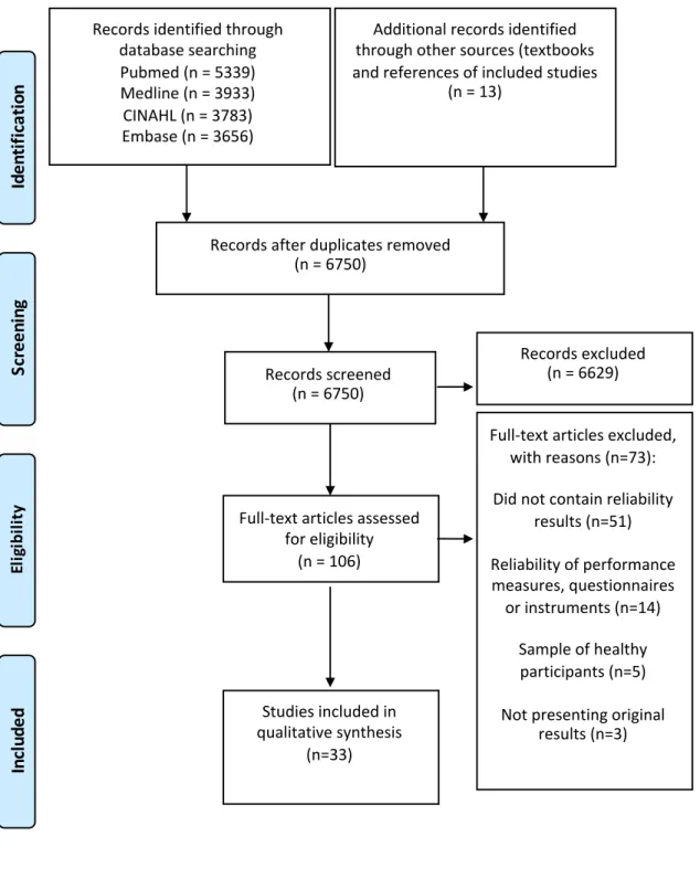
1. Overall description of included reviewsAs shown in Figure 1, 6750 potential studies were initially identified, 6669 articles were excluded and 17 reviews were ultimately included. Table 1 presents the overall characteristics of the included reviews. Eleven reviews included a MA while the six others were SR without a MA. Eight reviews evaluated meniscal injuries, and the most common tests were the McMurray, the Apley’s manoeuver and the joint line tenderness tests (Table 3). Six reviews assessed ACL injuries, and the most common tests evaluated were the Lachman, the anterior
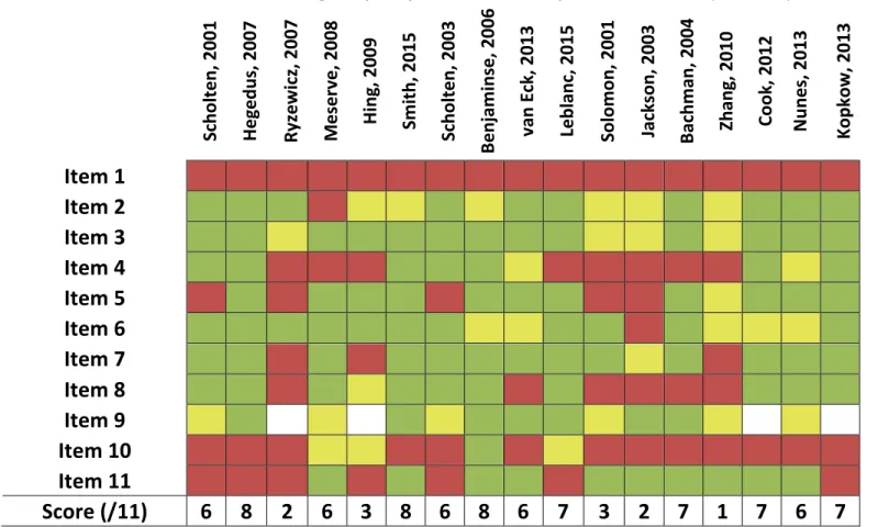
drawer and the pivot shift tests (Table 4). One clinical prediction rule for the screening of knee fractures was evaluated in two reviews (Table 5). One clinical prediction rule and one set of diagnostic criteria to diagnose knee osteoarthritis were the focus of two reviews (Table 6). Combinations of tests for ACL and posterior cruciate ligament (PCL) tears, meniscal injuries and for cartilage defects were appraised in two reviews (Table 7). 2. Methodological quality of included reviews The AMSTAR scores for the assessment of methodological quality of the SR/MA are presented in Table 2. AMSTAR score ranged from 1 to 8 (out of 11) with a mean of 5.5 ± 2.3 indicating a moderate methodological quality. Seven reviews reached a score of 7 or higher. More than ten out of seventeen reviews performed a comprehensive literature search (item 3), provided a list of studies (item 5), provided characteristics of the included studies (item 6) and assessed the scientific quality of the included studies (item 7). Between seven and ten out of seventeen reviews duplicated study selection and data extraction (item 2), used the status of publication as an inclusion criteria (item 4), used the scientific quality of the included studies in the conclusion (item 8), used appropriate methods to combine findings (item 9) and stated potential conflict of interest (item 11). However, only one review assessed the likelihood of publication bias (item 10) while no reviews provided an “a priori” design (item 1). The average inter-rater reliability for individual item was substantial (kappa=0.69). The majority of items reached substantial agreements (k>0.6) (items 1, 4, 5, 6, 7, 8, 9, 11). All other items reached a moderate agreement (k>0.4) (items 2, 3, 10). After discussion between the two raters, consensus was always achieved.
Figure 1: Bibliographic search flowchart Records identified through database searching Pubmed (n = 5339) Medline (n = 3933) CINAHL (n = 3783) Embase (n = 3656) Sc re en in g In cl ud ed El ig ib ili ty Id en tif ic at io n Additional records identified through other sources (textbooks and references of included studies (n = 1) Records after duplicates removed (n = 6750) Records screened (n = 6750) Records excluded (n = 6669) Full-text articles assessed for eligibility (n = 81) ) Full-text articles excluded, with reasons (n=64): Not a systematic review or a meta-analysis (ex: narrative review) (n=33) Systematic review on other subject (ex: reviews on functional questionnaire or prognostic factors) (n=25) Clinical practice guidelines (n=6) Studies included in qualitative synthesis (n=17)
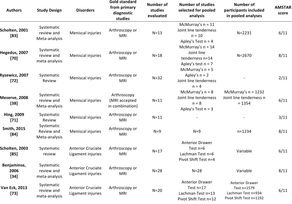
Table 1: Characteristics of the included reviews Authors Study Design Disorders
Gold standard from primary diagnostic studies Number of studies evaluated Number of studies selected for pooled analysis Number of participants included in pooled analyses AMSTAR score Scholten, 2001 [83] Systematic review and Meta-analysis
Meniscal injuries Arthroscopy or MRI N=13
McMurray’s n = 11 Joint line tenderness n = 10 Apley’s Test n = 4 N=2231 6/11 Hegedus, 2007 [70] Systematic review and meta-analysis Meniscal injuries Arthroscopy or MRI N=18 McMurray’s n = 14 Joint line tenderness n=14 Apley’s test n = 7 N=2670 8/11 Ryzewicz, 2007
[72] Systematic Review Meniscal injuries Arthroscopy or MRI N=32
McMurray’s n = 5 Apley’s n = 2 Joint line tenderness n = 4 - 2/11 Meserve, 2008 [38] Systematic review and Meta-analysis Meniscal injuries Arthroscopy (MRI accepted in combination) N=11 McMurray’s n = 8 Joint line tenderness n = 8 Apley’s Test n = 3 McMurray’s n = 1232 Joint line tenderness n = 1354 6/11 Hing, 2009
[71] Systematic Review Meniscal injuries Arthroscopy or MRI N=11 - - 3/11
Smith, 2015 [84] Systematic Review and Meta-analysis Meniscal injuries Arthroscopy or MRI N=9 N=9 n=1234 8/11 Scholten, 2003
[85] Systematic review Anterior Cruciate Ligament injuries Arthroscopy or MRI N=17
Anterior Drawer Test n=6 Lachman Test n=6 Pivot Shift Test n=4 Variable 6/11 Benjaminse, 2006 [34] Systematic review and meta-analysis Anterior Cruciate
Ligament injuries Arthroscopy or MRI N=28 N=28 Variable 8/11
Van Eck, 2013 [73] Systematic review and meta-analysis Anterior Cruciate
Ligament injuries Arthroscopy or MRI N=20
Anterior Drawer Test n=17 Lachman Test n=13 Pivot Shift Test n=12 Anterior Drawer Test n=1579 Lachman Test n=934 Pivot Shift Test n=1192 6/11
Leblanc, 2015 [86] Systematic review and meta-analysis Anterior Cruciate
Ligament injuries Arthroscopy or MRI N=8 Pivot Shift n=4 Lachman n=5 n=1196 7/11
Solomon, 2001 [87] Systematic Review Anterior Cruciate Ligament injuries and Meniscal Injuries Arthroscopy or
MRI N=23 Meniscal n=9 ACL n=15 Variable 3/11
Jackson, 2003 [6] Systematic review and meta-analysis Acute Knee Disorders, Anterior Cruciate Ligament injuries and Meniscal Injuries Arthroscopy/M RI/radiography/ clinical diagnosis
N=35 Meniscal n=4 ACL n=11 Variable 2/11
Bachmann, 2004 [88] Systematic review and meta-analysis Knee fracture with the Ottawa Knee Rule Radiography or follow-up N=11 N=6 N=4249 7/11 Zhang, 2010 [46] Systematic Review, Meta-anaysis and Delphi consensus Knee Osteoarthritis Clinical features
and radiographs N=313 Variable Variable 1/11
Cook, 2012
[74] Systematic review Patellofemoral pain
Arthroscopy or clinical or imaging (accepted by authors) N=9 - - 7/11 Nunes, 2013 [89] Systematic review and meta-analysis Patellofemoral pain Unreported N=5 N=2 N=145 6/11 Kopkow, 2013
[90] Systematic review Posterior Cruciate Ligament injuries Arthroscopy or MRI N=11 - - 7/11 MRI: magnetic resonance imaging
Table 2: Assessment of the methodological quality of the included systematic reviews (AMSTAR) Sc holte n, 2001 He ge dus, 2007 Ry ze w ic z, 2 00 7 Me se rv e, 2 00 8 Hing, 2009 Sm ith, 2015 Sc holte n, 2003 Be nj am inse , 2006 va n Ec k, 2 01 3 Le blanc , 2015 Solom on, 2001 Ja cks on , 2 00 3 Bac hm an, 2004 Zh an g, 2 01 0 Co ok, 2 01 2 Nune s, 2013 Ko pko w , 2 01 3
Item 1
Item 2
Item 3
Item 4
Item 5
Item 6
Item 7
Item 8
Item 9
Item 10
Item 11
Score (/11)
6 8 2 6 3 8 6 8 6 7 3 2 7 1 7 6 7
Item 1: Was an “a priori” design provided?; Item2: Was there duplicate study selection and data extraction?; Item 3: Was a comprehensive literature search performed?; Item 4: Was the status of publication used as an inclusion criterion?; Item 5: Was a list of studies provided?; Item 6: Were the characteristics of the included studies provided?; Item 7: Was the scientific quality of the included studies assessed?; Item 8: Was the scientific quality of the included studies used in conclusion?; Item 9: Were the methods used to combine the findings appropriate?; Item 10: Was the likelihood of publication bias assessed?; Item 11: Was the conflict of interest stated? Green = Yes; Red = No; Yellow = can’t say/unclear; Blank = N/A
3. Summary of findings
Meniscal Injuries
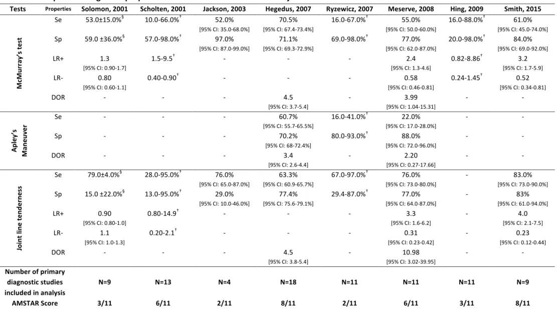
Table 3 presents the diagnostic properties of the physical tests used for the diagnosis of meniscal injuries. Eight SR/MA provided data on the diagnosis of meniscal injuries [6, 38, 70-72, 83, 84, 87] with AMSTAR scores ranging from 2 to 8 out of 11 with a mean of 4.8. Based on the data extracted from the review by Hegedus et al. [70], the highest quality review with the most studies included in the meta-analyses (8/11, n=18), the McMurray’s test demonstrated the highest Se with a score of 70.5% (95% CI: 67.4-73.4%) [70]. The point estimates for Se varied across all included SR/MA (range: 52.0-70.5%). Also based on the data by Hegedus et al., the joint line tenderness demonstrated the highest Sp with 77.4% (95% CI: 75.6-79.1%) [70]. Again, the point estimates for the Sp of this test varied across all included SR/MA (range: 29.0-83.0%). Based on the data extracted from Smith et al. [84], the most recent and highest quality review to provide data for the LR+/- (8/11, n=9), the joint line tenderness also demonstrated both the highest LR+ with 4.0 (95% CI: 2.1-7.5) and the lowest LR- with 0.23 (95% CI: 0.12-0.44). The point estimates of the joint line tenderness LR+/- varied across others included SR/MA for both LR+/- (range LR+: 0.9-4.0; range LR-: 0.23-1.1). The Thessaly’s test diagnostic validity was also presented for the first time by the review of Smith et al. [84], with a reported LR+ of 5.6 (95% CI: 1.5-21.0) and LR- of 0.28 (95% CI: 0.11-0.71). Overall, authors’ recommendations from all included SR/MA were that clinicians should not use these tests individually because of their poor diagnostic validity and advised combining the results of the tests even though no evidence was presented to support this approach [6, 38, 70-72, 83, 84, 87]. Anterior Cruciate Ligament Injuries
Table 4 presents the diagnostic properties of the physical examination tests used for the diagnosis of ACL injuries. Six SR/MA provided data on the diagnosis of ACL injuries [6, 34, 73, 85-87] with AMSTAR scores ranging from 2 to 8 out of 11 with a mean of 5.3. Based on the data extracted from Benjaminse et al. [34], the highest quality meta-analysis with the most studies included in analysis (8/11, n=28) to also provide 95% CI for LR+/-, the Lachman test
demonstrated the highest Se with 85.0% (95% CI: 83.0-87.0%). For Se, the point estimates were relatively similar across all included SR/MA (range: 81.0-89.0%). In terms of LR, the Lachman test also reached the highest LR+ and lowest LR- with 10.2 (95% CI: 4.6-22.7) and 0.20 (95% CI: 0.10-0.30) respectively [34]. The point estimates for the LR+ varied across the included SR/MA (range: 4.5-42.0), but was more consistent for the LR- (range: 0.10-0.22). The Pivot Shift test demonstrated the highest Sp with a score of 98.0% (95% CI: 96.0-99.0%) [34], but important variations in the point estimates across all included SR/MA was observed (range: 81.0-98.0%). Overall, authors’ recommendations from all included SR/MA were that the Lachman test is of high diagnostic value both to rule in or out an ACL injury while a positive Pivot Shift test can be used to rule in an ACL injury [6, 34, 73, 85, 87]. One review concluded that the anterior drawer test may be used to rule in an ACL injury, but not rule out, and could be used instead of the Lachman in situations where the evaluator has less training performing the test [34]. Knee Fractures Table 5 presents the diagnostic properties for the Ottawa Knee Rule, a clinical prediction rule used to exclude a knee fracture and avoid unnecessary radiograph of the knee at the emergency [88]. Two reviews providing data on this clinical prediction rule with AMSTAR score of 2 and 7 out of 11 [6, 88]. Both reviews reported similar diagnostic properties for the rule. Bachmann et al. calculated 98.5% (95% CI: 93.2-100%) for Se, 48.6% (95% CI: 43.4-51.0%) for Sp and 0.05 (95% CI: 0.02-0.23) for LR- [88]. Overall, the authors’ recommendations from both included SR/MA are that this clinical prediction rule, with its low LR-, is considered useful to exclude a fracture if all the rule’s criteria are negative; if this is not the case, the clinician cannot rule out a fracture and should order a knee radiograph (Table 5).
Knee osteoarthritis
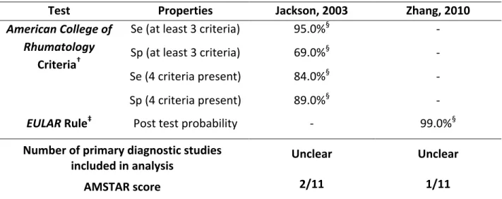
Table 6 presents the diagnostic properties of one set of diagnostic criteria and one clinical prediction rule for the diagnosis of knee OA. Two reviews were found providing data on such tools with AMSTAR scores of 1 and 2 out of 11 [6, 46]. The clinical criteria from the American
College of Rheumatology for the diagnosis of knee OA include: age ≥ 50 years, stiffness ≤ 30
minutes, crepitus, bony tenderness, bony enlargement, and no palpable warmth [6]. If at least three criteria are present, the Se is 95.0% and Sp is 69.0%. If a fourth criterion is present, the Se drops to 84.0%, but the Sp increases to 89.0%. The study by Zhang et al. (EULAR rule) is presented as both a MA with the results of the validation of the rule in two cross-sectional studies [46]. For the clinical prediction rule, they reported a post-test probability of 99% for the diagnosis of OA (Kellgren Lawrence ≥2) if six criteria were present based on an estimated prevalence of 12.5% in adults aged ≥ 45 years [46]. The criteria include three symptoms: knee pain, limited morning stiffness, functional limitations, and three signs: knee crepitus, restricted knee range of motion and bony enlargements (table 6). In both reviews, the authors concluded that a clinical diagnosis of knee OA can be done with the clinical criteria, but knee radiography remains necessary to assess the radiological grading of OA. Patellofemoral pain Two reviews evaluated the diagnostic validity of 25 tests for PFP (AMSTAR score: 7 and 6/11) [74, 89]. Both reviews used the same five studies and one included four more for a total of nine studies [74, 89]. Overall, five tests had a LR+³5: the active instability test (LR+: 249.0), pain during stair climbing (LR+: 11.6), Clarke’s sign (LR+: 7.4), pain during prolonged sitting (LR+: 7.5), and patellar tilt (LR+: 5.4, 95%CI: 1.4-20.8) [74, 89]. They also reported that pain during squatting demonstrated a LR-=0.20 (95%CI: 0.1-0.4) [74, 89]. Combining tests had a mitigated effect on improving the diagnosis of PFP [74, 89]. However, they acknowledged that the primary diagnostic studies included in their SR reported heterogeneous results with an overall high risk of bias [74, 89]. The authors of the included reviews proposed to view PFP as a diagnosis of exclusion [74, 89]. No individual tests can be recommended at this time to diagnose or exclude a PFP.
Posterior cruciate ligament injuries
The SR by Kopkow et al. evaluated the diagnostic validity of 11 tests for PCL injuries (AMSTAR score: 7/11) [90]. In their review including eleven studies, they reported that the posterior
drawer test was the most frequently studied test with a Se ranging from 22% to 100% [90]. They reported that the included primary study in their SR with the lowest risk of bias (i.e: moderate) showed a LR+ of 50.1 (95% CI: 7.1-351.7) and a LR- of 0.11 (95% CI: 0.03-0.40) for this test [90]. The authors also reported that the quadriceps active test appeared to be the most specific test with a Sp ranging from 96% to 100% based on two included studies from their SR [90]. Kopkow et al. concluded that at this time, evidence was insufficient to recommend any physical tests to diagnose or exclude a PCL injury [90].
History taking and physical examination for the diagnosis of common knee disorders
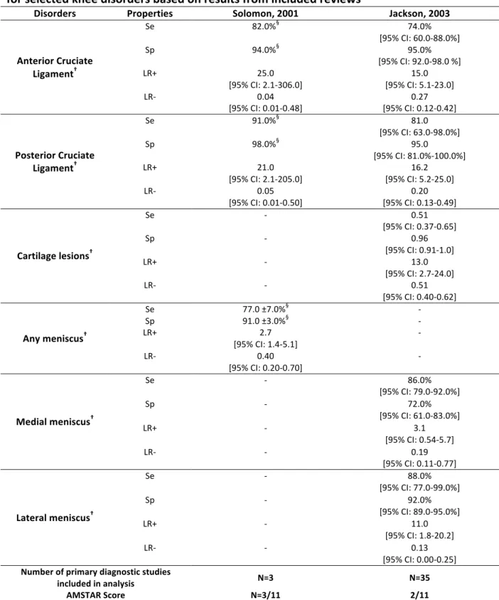
Table 7 presents the diagnostic properties extracted from reviews on the complete physical examination (including a thorough history and physical tests) for the diagnosis of various knee disorders [6]. We found two reviews providing data on such evaluation for four knee disorders with AMSTAR scores of 2 and 3 out of 11 [6, 87]. For ACL injuries, Jackson et al. [6] reported point estimates for LR+ and LR- of 15.0 (95% CI: 5.1-23.0) and 0.27 (95% CI: 0.12-0.42) respectively. For PCL injuries, Jackson et al. [6] also reported point estimates for LR+ and LR- of 16.2 (95% CI: 5.2-25.0) and 0.20 (95% CI: 0.13-0.49) respectively. Therefore, a complete physical examination appears valid to diagnose an ACL injury and diagnose or exclude a PCL injury, although the definition of what constitutes a complete physical examination was not provided. Likewise, for the diagnosis of cartilage lesions, the complete physical examination may be considered valid with LR+ of 13.0 (95%CI: 2.7-24.0) and a LR- of 0.51 (95% CI: 0.40-0.62) [6]. For the diagnosis of meniscal injuries, Solomon et al. reported in their review a LR+: 2.7 (95% CI: 1.4-5.1) and a LR-: 0.40 (95% CI: 0.20-0.70) [87] and concluded that a complete physical examination is not valid to diagnose or exclude a meniscal injury. However, the review by Jackson et al. concluded that a complete physical examination might be valid to identify the presence of lateral meniscal injury or to exclude a medial or lateral meniscus injury [6]. Overall, studies that reviewed the complete physical examination for various knee disorders concluded that a complete physical examination is probably superior to individual tests but it remains unclear how well this approach may perform [6, 87]. Based on this evidence, a complete physical examination may be diagnostically superior to individual tests but further research is needed.
Table 3: Description of diagnostic properties for selected tests for meniscal injuries based on results from included reviews
Tests Properties Solomon, 2001 Scholten, 2001 Jackson, 2003 Hegedus, 2007 Ryzewicz, 2007 Meserve, 2008 Hing, 2009 Smith, 2015
Mc Mu rr ay ’s te st Se 53.0±15.0%§ 10.0-66.0%† 52.0% [95% CI: 35.0-68.0%] 70.5% [95% CI: 67.4-73.4%] 16.0-67.0%† 55.0% [95% CI: 50.0-60.0%] 16.0-88.0%† 61.0% [95% CI: 45.0-74.0%] Sp 59.0 ±36.0%§ 57.0-98.0%† 97.0% [95% CI: 87.0-99.0%] 71.1% [95% CI: 69.3-72.9%] 69.0-98.0%† 77.0% [95% CI: 62.0-87.0%] 20.0-98.0%† 84.0% [95% CI: 69.0-92.0%] LR+ 1.3 [95% CI: 0.90-1.7] 1.5-9.5† - - - 2.4 [95% CI: 1.3-4.6] 0.82-8.86† 3.2 [95% CI: 1.7-5.9] LR- 0.80 [95% CI: 0.60-1.1] 0.40-0.90† - - - 0.58 [95% CI: 0.46-0.81] 0.24-1.45† 0.52 [95% CI: 0.34-0.81] DOR - - - 4.5 [95% CI: 3.7-5.4] - 3.99 [95% CI: 1.04-15.31] - - Ap le y’ s Ma ne uv er Se - - - [95% CI: 55.7-65.5%] 60.7% 16.0-41.0% † 22.0% [95% CI: 17.0-28.0%] - - Sp - - - 70.2% [95% CI: 68-72.4%] 80.0-93.0%† 88.0% [95% CI: 72.0-96.0%] - - DOR - - - 3.4 [95% CI: 2.6-4.4] - 2.20 [95% CI: 0.27-17.66] - - Jo in t l in e te nd er ne ss Se 79.0±4.0% § 28.0-95.0%† 76.0% [95% CI: 65.0-87.0%] 63.3% [95% CI: 60.9-65.7%] 67.0-97.0%† 76.0% [95% CI: 73.0-80.0%] - 83.0% [95% CI: 73.0-90.0%] Sp 15.0 ±22.0%§ 13.0-95.0%† 29.0% [95% CI: 10.0-46.0%] 77.4% [95% CI: 75.6-79.1%] 29.4-87.0%† 77.0% [95% CI: 64.0-87.0%] - 83% [95% CI: 61.0-94.0%] LR+ 0.90 [95% CI: 0.80-1.0] 0.80-14.9† - - - 3.3 [95% CI: 1.6-6.2] - 4.0 [95% CI: 2.1-7.5] LR- 1.1 [95% CI: 1.0-1.3] 0.20-2.1† - - - 0.31 [95% CI: 0.23-0.42] - 0.23 [95% CI: 0.12-0.44] DOR - - - 4.5 [95% CI: 3.8-5.4] - 10.98 [95% CI: 3.02-39.95] - - Number of primary diagnostic studies included in analysis N=9 N=13 N=4 N=18 N=11 N=11 N=11 N=9 AMSTAR Score 3/11 6/11 2/11 8/11 2/11 6/11 3/11 8/11 Se: sensitivity, Sp: specificity, LR: likelihood ratio, DOR: diagnostic odds ratio. § Data presented as mean ± SD (standard deviation), calculated by authors. † Indicates that the authors could not pool the data and did not calculate a mean value with standard deviation. Therefore, we presented the range of values based on the data presented in the article.
Table 4: Description of diagnostic properties for physical examination tests for Anterior Cruciate Ligament (ACL) injuries based on results from included reviews
Tests Properties Solomon, 2001 Jackson, 2003 Scholten, 2003 Benjaminse, 2006 van Eck, 2013¥ Leblanc, 2015
La ch m an T est Se 84.0±15.0%§ 87.0% [95% CI: 76.0-98.0%] 86.0% [95% CI: 76.0-92.0%] 85.0% [95% CI: 83.0-87.0%] 81.0%¥ 89.0% [95% CI: 76.0-98.0%] Sp 100% 93.0% [95% CI: 89.0-96.0%] 91.0% [95% CI: 79.0-96.0 %] 94.0% [95% CI: 92.0-95.0 %] 81.0% ¥ - LR+ 42.0 [95% CI: 2.7-651.0] - 2.0-102.1† 10.2 [95% CI: 4.6-22.7] 4.5¥ - LR- 0.10 [95% CI: 0.00-0.40] - 0.10-0.40† 0.20 [95% CI: 0.10-0.30] 0.22¥ - DOR - - - 70.0 [95% CI: 23.0-206.0] - - Pi vo t S hi ft T es t Se 38.0 ±28.0%§ 61.0% [95% CI: 40.0-82.0%] 18.0-48.0 %† 24.0% [95% CI: 21.0-27.0%] 28.0% ¥ 79% [95% CI: 63.0-91.0%] Sp - 97.0% [95% CI: 93.0-99.0%] 97.0-99.0 %† 98.0% [95% CI: 96.0-99.0%] 81.0% ¥ - LR+ - - 8.2-26.9† 8.5 [95% CI: 4.7-15.5] 5.35¥ - LR- - - 0.50-0.80† 0.90 [95% CI: 0.80-1.0] 0.84¥ - DOR - - - 12.0 [95% CI: 5.0-31.0] - -