Open Archive TOULOUSE Archive Ouverte (OATAO)
OATAO is an open access repository that collects the work of Toulouse researchers and
makes it freely available over the web where possible.
This is an author-deposited version published in :
http://oatao.univ-toulouse.fr/
Eprints ID : 16807
To link to this article : DOI : 10.1016/j.surfcoat.2013.08.039
URL :
http://dx.doi.org/10.1016/j.surfcoat.2013.08.039
To cite this version : Mungkalasiri, Jitti and Bedel, Laurent and
Emieux, Fabrice and Cara, Aurelia Vettese-Di and Freney, Jean and
Maury, Francis and Renaud, François N. R. Antibacterial properties
of TiO2–Cu composite thin films grown by a one step DLICVD
process. (2014) Surface and Coatings Technology, vol. 242. pp.
187-194. ISSN 0257-8972
Any correspondence concerning this service should be sent to the repository
administrator:
[email protected]
Antibacterial properties of TiO
2
–Cu composite thin films grown by a one
step DLICVD process
☆
Jitti Mungkalasiri
a,b, Laurent Bedel
b, Fabrice Emieux
b, Aurelia Vettese-Di Cara
c, Jean Freney
c,d,
Francis Maury
a,⁎
, François N.R. Renaud
caCIRIMAT, 4 allée E. Monso, BP 44362, 31030 Toulouse Cedex 4, France bL2CE/DTM, CEA Grenoble, 17 rue des Martyrs, 38054 Grenoble, France
cMATEIS/I2B, Nosoco.tech, Faculté de Pharmacie Laboratoire de Microbiologie, 8 Av. Rockefeller, 69373 Lyon Cedex 3, France dUniversité Lyon 1, Ecologie Microbienne, 8 Av. Rockefeller, 69373 Lyon, France
a b s t r a c t
Keywords: Nanocomposite coatings Metallic nanoparticles Antibacterial activity Bactericidal surfaces DLICVDThe correlations between microstructural features, chemical compositions and antibacterial properties of coat-ings containing metallic Cu particles embedded in a titanium dioxide matrix have been determined. A Direct Liquid Injection Chemical Vapor Deposition (DLICVD) process was used for the one step growth of TiO2–Cu com-posite coatings on various substrates. Titanium tetra-iso-propoxide (TTIP) and copper bis(2,2,6,6-tetramethyl-3,5-heptationate) (Cu(tmhd)2) were used as titanium and copper molecular sources, respectively. This growth process allows a good control of the quantity of metalorganic precursors injected into the CVD reactor and thus of the coating composition. The deposition occurs at 683 K under low pressure (800 Pa). The influence of the main features of the coatings on their antibacterial properties was investigated in order to produce bacteri-cidal surfaces that are durable, non-toxic and containing a minimum amount of active agent. The antibacterial ac-tivity on Staphylococcus aureus without any photon activation was measured according to the JIS Z 2801:2000 standard method. An antibacterial activity was detected for a low metal content of ca. 1 at.% Cu, and was found to increase with the Cu content. It was maximal for 3.5 at.% Cu, i.e. TiO2–Cu composite coatings exhibit bac-tericidal behavior against S. aureus for this optimal composition (relative activity = 100%). In order to better characterize the microbiological behavior of the coatings more discriminating methods derived from the litera-ture were tested to assess the performances of these CVD coatings in terms of efficiency, release of antibacterial agent and accelerated aging.
1. Introduction
Antibacterial properties of silver and copper have been well known for many centuries. As a result these metals are frequently used as active agents in coatings to produce antibacterial surfaces. Silver has been used to treat wounds, ophthalmia neonatorum, and more recently, in combi-nation with sulfadiazine, burns (for a review see[1]). Silver ions (Ag+)
interact with disulfide or sulfhydryl groups of enzymes, leading to inhibition of metabolic processes[2,3]. Silver also binds bacterial DNA (deoxyribonucleic acid) inhibiting replication and transcription[4,5]. Copper leads to the collapse of some lipopolysaccharide (LPS) patches and alters the permeability and functionality of the outer cell membrane
[6]. Due to their small size (b100 nm), nanoparticles (NPs) present a larger surface area/volume ratio and greater chemical activity than larger particles. Silver nanoparticles exhibit an antibacterial activity at low
concentration[7,8]. It has been demonstrated that their activity is size
[7,9]and shape dependent[10].
Free NPs penetrate inside the bacteria after their attachment to the plasma membrane and they interact with sulfur-containing proteins and the phosphorus containing-DNA. Then, the respiratory chain and cell division are blocked[1]. The nanometric size of silver NPs, i.e. small-er than 10 nm, also produces electronic effects[11,12]. It is still difficult to distinguish the bactericidal activity of NPs from that of metallic ions released by nanoparticles[13–15]. The action mechanism of copper nanoparticles is less understood than silver ones[16,17], due to less studies than for silver element.
Several processes are developing to immobilize NPs on surfaces and the main way is to incorporate them in a thin matrix to form composite coatings. Thereby nanocomposite films have been synthesized by sol– gel deposition[18–20]. In all cases, the active species, i.e. Ag+cations,
are released from NPs immobilized on the support. Furno et al.[21]
dissolved organometallic precursors in supercritical carbon dioxide in order to obtain a homogenous distribution of NPs in a silicone matrix. Egger et al.[22]used an industrial flame spray pyrolysis process to syn-thesize a SiO2–Ag nanocomposite material. Kelly et al.[23]deposited ☆ This article was supposed to be a part of the 19th European Conference on Chemical
Vapor Deposition (EuroCVD19), Varna, Bulgaria, 1st - 6th September 2013 special issue published in volume 230.
⁎ Corresponding author. Tel.: +33 534323401; fax: +33 534323499.
E-mail address:[email protected](F. Maury).
TiN–Ag by pulsed magnetron sputtering. Brook et al.[24]and Sheel D.W. et al.[25]synthesized silver and TiO2–Ag duplex coatings with
antibacterial properties by means of Chemical Vapor Deposition (CVD) while Page et al.[26]deposited TiO2–Ag antimicrobial composite films
on glass. As already mentioned, there is less report on Cu than on Ag as an active antibacterial agent.
The use of a photocatalytic oxide matrix is another route to produce antibacterial coatings but UV activation is generally required. For in-stance Cu/WO3 was added as visible-light-sensitive photocatalyst to
PTFE particulate composite material to overcome this limitation and a synergistic effect was also found on antibacterial performance. In fact the antibacterial activity of this Cu/WO3-added PTFE particulate
com-posite material decreases significantly with the visible light intensity
[27]. The remarkable photo-excited properties of TiO2films have been
described in many reviews where the photocatalytic sterilization process is also discussed [28]. The antimicrobial efficiency of TiO2
coatings by photocatalytic reactions was investigated under UV-A irra-diation but its application is limited under UV light[29]. It was demon-strated that TiO2/cordierite foam irradiated with UV-A light possessed
both a long-term bactericidal action and a high photocatalytic degrada-tion capability making this material a good candidate for air-cleaning filters but, again, UV light is required[30].
In previous studies, we developed a one step Direct Liquid Injection Chemical Vapor Deposition (DLICVD) process using metalorganic sources for the growth of nanostructured TiO2–Ag[8] and TiO2–Cu [31]coatings on various substrates. In both cases, the titanium precursor for the growth of the titanium oxide matrix was titanium tetra-iso-propoxide (TTIP). For the copper containing composite coatings copper bis(2,2,6,6-tetramethyl-3,5-heptationate) (Cu(tmhd)2) was used as
molecular precursor. The metallic Cu particles were incorporated into the TiO2matrix and uniformly distributed over the entire thickness of
the film. For low Cu content, TiO2 presented the anatase structure
while by increasing the Cu content the nucleation and growth of rutile occurred. A preliminary investigation of the behavior of these nanocom-posite TiO2–Cu coatings had shown an antibacterial activity[31].
The aim of this paper is to report a thorough investigation on the in-fluence of the main features of the films including thickness and Cu con-tent as well as reproducibility and aging effect on the antibacterial properties of these TiO2–Cu nanocomposite coatings without any UV
activation. It was found that the JIS Z 2801:2000 standard method did not allow two composite films to be distinguished in spite of different features. Then, more discriminatory microbiological methods derived from the literature were implemented to assess the performances of these coatings.
2. Experimental
2.1. Growth of nanocomposite films
For DLICVD of TiO2–Cu films, TTIP was diluted in xylene with a
con-centration of 1 mol·L−1. The injection parameters were maintained
constant: injection frequency 2 Hz and opening time 2 ms. Cu(tmhd)2
was dissolved in pure xylene at a concentration between 0.01 and 0.05 mol·L−1
depending on the level of Cu desired in the films. The cop-per incorporated into the coatings was also controlled by the injection frequency of the Cu(tmhd)2injector regulated between 0.5 and 6 Hz
while the opening time was fixed at 2 ms. The substrates were silicon wafers, glass and stainless steel 316L. The total pressure was maintained at 800 Pa and the reactor wall and substrate holder were heated at 523 and 683 K, respectively. The film thickness was controlled by varying the deposition time. More details are reported in[31].
2.2. Film characterization
The crystalline structure of the films and the average crystallite size were determined by X-ray diffraction (Seifert 3000TT diffractometer;
Bragg–Brentano configuration; Cu Kα radiation). The morphology and thickness of the films were observed using a scanning electron micro-scope (Leo 1530 FEG-SEM) equipped with an X-ray energy dispersive spectroscopy analyzer (EDS; Tracor analyzer). The size and the distribution of metal NPs were determined by transmission electron mi-croscopy (TEM; JEOL JEM 2010 microscope). The relative composition of the films were analyzed by electron probe microanalysis (EPMA; Cameca SX50), secondary ion mass spectroscopy (SIMS; Cameca IMS 4F6 spectrometer) and X-ray photoelectron spectroscopy (XPS) using a VG ESCALAB MKII spectrophotometer, which operated with a non-monochromatized Mg Kα source (1253.6 eV). Atomic composition of the layers was determined by XPS after Ar+sputtering for 10 min to
clean the atmospheric contamination of the surface.
2.3. Microbiological tests
Antibacterial properties of active films deposited on glass were measured according to the JIS Z 2801:2000 standard [32] using
Staphylococcus aureusstrain (CIP 4.83). The method was slightly modi-fied to accommodate the size of the samples (25 × 25 mm2instead of
50 × 50 mm2
). The test duration was also changed for a specific series of samples in order to investigate kinetic effects. Briefly, a bacterial sus-pension with 3 × 10⁵CFU·mL−1was prepared in 1/500 nutrient broth.
Then, 200 μL (≈6 × 10⁴CFU) was spread on 25 × 25 mm2samples
and covered with a sterile plastic film. The samples were incubated in a humid chamber (RH 90%) from 3 h to 24 h at 37 °C. After incubation the samples were washed in a universal neutralizer solution (Fisher Scientific Bioblock, ref W1801L) to enable bacterial enumeration by means of a plate count. A control (reference) consisting of an uncoated sample was also tested for each analyzed sample. The antibacterial activity was calculated by the following formula:
Antibacterial activity ¼ Log A=Bð Þ ð1Þ where A and B are the numbers of CFUs (Colony Forming Units) on the surface of the reference and coated samples, respectively. For great-er convenience, we calculated also a relative antibactgreat-erial activity according to:
Relative activity ¼ Log A=B½ ð Þ= Log Að Þ% & 100: ð2Þ This relative activity means that (i) when it is equal to 100% (no CFU detected after the test) the surface is bactericidal, i.e. the number of CFUs is reduced from about 6 × 10⁶CFU·mL−1(the amount usually counted on the control) to zero, (ii) in the range 0–100% the surface exhibits an antibacterial behavior and (iii) zero means that the surface is inactive.
The retained bacterial enumeration for each sample was the average value of three identical samples prepared in the same CVD run. The measure could not be replicated on the same sample because to avoid contamination the samples were autoclaved at 120 °C after the microbi-ological test and therefore their characteristics are likely to have evolved. Uncertainty in bacteriology is usually one logarithm that is about 15% for this test (1 log compared to 6–7 logarithms for the num-ber of CFUs on the control). If one of the three measures deviated from this range the average was made on the other two samples. Conse-quently, each experimental data is reported hereafter with a 15% error bar representative of the standard deviation.
Moreover, we adapted the method of Haldar et al.[33]to test for bactericidal action of TiO2–Cu composite coatings under different
con-ditions. Briefly, TiO2–Cu films 60 nm thick were deposited on
75 × 25 mm2glass samples. They were placed vertically at 15 cm in
front of a spray nozzle and sprayed until the slide was uniformly wet (aerosol flow 10 mL·min−1) with a S. aureus suspension (approximately
5 × 10³cells·mL−1). Then, the slides were dried in air for 2 min in a
agar growth medium (thickness 2 mm)[33]. After 24 h of incubation at 37 °C, CFUs were counted, each CFU corresponding to at least one surviv-ing bacterium. Each test included one inactive control (a glass slide with-out TiO2–Cu composite coating). The number of colonies on a sample
must be lower than approximately 120–150 to facilitate bacterial enu-meration. If there is not bacteria on the sample this means it is active, if the number of bacteria is the same as the control it is inactive.
2.4. Diffusion test
In order to determine if active antibacterial agent could release or diffuse from the films to the environment, the active face of coated sam-ple with a composition of 3.5 at.% Cu was placed in contact with the sur-face of an agar plate previously inoculated with an Escherichia coli suspension containing 106CFU·mL−1. After 24 h of incubation the
pos-sible occurrence of an inhibition zone around the sample is sought as a signature of diffusion of the antibacterial agent[34].
To complete this test of release or diffusion in an aqueous medium, three TiO2–Cu films (100 nm thick) deposited on glass substrate were
immersed separately in 20 mL double distilled water respectively at 20 °C and 40 °C for 24 h and at 40 °C for 7 days. The solutions were gently stirred during immersion. Then, traces of copper in the water were analyzed by inductively coupled plasma atomic emission spec-troscopy (ICP-AES). Three analyses have been performed for each sam-ple and the average value was retained. A control solution of 5 ppm Cu was analyzed to check the sensitivity and the calibration of ICP-AES spectrometer and a test with an uncoated glass substrate was carried out as blank test. The detection limit for Cu is typically 5 ppb.
2.5. Aging tests
An evaluation of the aging of such DLICVD TiO2–Cu composite
coat-ings was conducted during 5 months. Two sets of glass samples (75 × 25 mm2
) coated on one side with a very thin film (60 nm thick) which contained 9 at.% Cu were placed in a climatic chamber in dark at 20 °C with a relative humidity (RH) of 40% (condition # 1) and at 60 °C with an RH of 100% (condition # 2). The antibacterial activity was evaluated on 3 series of samples for each aging condition, respec-tively immediately after the deposition and after 2 and 5 months, according the JIS Z 2801 standard with S. aureus.
3. Results
3.1. Influence of Cu content on the antibacterial activity
Although the accuracy is not high the XPS technique was used to de-termine the composition of copper inside the TiO2matrix because of the
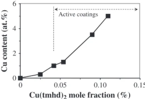
very small thickness of the films. The Cu content increases with the mole fraction of the copper precursor Cu(tmhd)2as shown inFig. 1.
For instance, for a mole fraction of 0.025%, the Cu content in the film is around 0.3 at.% then it increases to ca. 5 at.% Cu for a mole fraction of 0.12%. The Cu incorporation in the coatings can be increased above this value but the goal of this work was to obtain bactericidal surfaces for the lowest levels of the active agent.Fig. 1was partially reproduced from the data of Fig. 7 in[31]because we noticed an error in this figure where the two Y axes were reversed, i.e. the XPS data corresponded to the black triangles instead of the black squares (which did not change the comments of this earlier paper).
The antibacterial activity for different series of samples measured by the JIS method with a contact time of 24 h was detected for a Cu(tmhd)2
mole fraction higher than 0.04% which corresponded to a film composi-tion of ca. 1 at.% Cu (Fig. 2). It was maximal for a Cu(tmhd)2mole
frac-tion near 0.1% which corresponded to ca. 3.5 at.% Cu. Three series of samples were prepared using the same DLICVD conditions for 3 differ-ent deposition times. Consequdiffer-ently, the film thicknesses were equal to 100, 150 and 200 nm. XPS technique was used to analyze the Cu content in these thin films. The relative antibacterial activity increased from 0 at
ca. 1 at.% Cu to its maximum (100%) at ca. 3.5 at.% Cu. No biological
ac-tivity was observed when the Cu content was below the detection limit of XPS (bca. 1 at.%). The results inFig. 2exhibit a good reproducibility whatever the thickness of the film in the range 100–200 nm. Other data confirmed the good reproducibility and an activity threshold close to 1 at.% Cu.
3.2. Influence of the thickness and microstructure of films
Fig. 2 revealed already an effect of the film thickness on the antibacterial activity since the activity seemed to be better for the thickest coatings. We studied the influence of the film thickness for the same mole fraction of Cu(tmhd)2on another series of samples.
The relative activity increased from 45% for a thickness of 35 nm (antibacterial activity) to 100% (bactericidal activity) for a thickness of about 107 nm (Fig. 3). Above this critical value the thickness has no ef-fect on the antibacterial behavior as mentioned above since it is already at its maximum (100%).
Surface SEM micrographs showed that large Cu particles (average size 200–300 nm) partially protruded from the film surface because they are larger than the film thickness. As a result, they were preferen-tially situated near the external surface (Fig. 4). In a previous paper we had also shown by transmission electron microscopy that smaller Cu particles (nanoparticles) were embedded into the oxide matrix to form these composite coatings[31].
0 2 4 6 0 0.05 0.10 0.15
Cu content (at.%)
Cu(tmhd)
2mole fraction (%)
Active coatingsFig. 1.Effect of Cu(tmhd)2mole fraction on the Cu content in TiO2–Cu composite coatings
as determined by XPS analyses. Antibacterial activity was observed for mole fractions higher than 0.025; typically for Cu content ≥1 at.% as indicated by the arrow.
Partially reproduced after permission from Elsevier[31].
0 20 40 60 80 100 0 0.05 0.10 0.15
Cu(tmhd)
2mole fraction (%)
Relative activity (%)
0.3Cu at. % (XPS)
200 nm 100 nm 150 nmThickness
No activity
1 1.4 3.5 9Fig. 2.Influence of Cu(tmhd)2mole fraction on the antibacterial activity determined by
the JIS method for three series of coatings with different thicknesses reported in the inset. The estimated Cu content of the films determined by XPS is also reported on
the upper x-axis, as deduced fromFig. 1. An error bar is estimated at 15% for all samples
(except for samples 100% active since there was no CFU detected) but it is given only for a representative data of each series for clarity.
3.3. Antibacterial activity by different methods 3.3.1. Modified JIS standard as kinetic method
The antibacterial activity of TiO2–Cu composite films was tested by
other microbiological methods to assess their effectiveness through ki-netic information. The JIS method was adapted and the antibacterial ac-tivity was measured for different contact times. Two films of 100 nm thick containing 1.3 at.% Cu and 3.5 at.% Cu respectively were tested by the means of JIS Z 2801 standard[32]. Each of them exhibited a bac-tericidal activity after 24 h of incubation. However by changing the con-tact time they revealed different behaviors. Fig. 5 shows that the bactericidal effect (relative activity = 100%) was observed after 24 h of incubation when the Cu concentration was low (1.3 at.% correspond-ing to 0.054% mole fraction of Cu(tmhd)2) and after only 3 h for samples
containing a larger amount of Cu (3.5 at.% Cu corresponding to 0.074% mole fraction of Cu(tmhd)2).
3.3.2. Competition method (Haldar method)
When the bacteria were deposited directly onto the surface of TiO2–
Cu composite films by means of an aerosol[33], their inhibition was measured by counting the CFUs developed on the agar after 24 h of in-cubation. The results from three experiments showed that no antibacterial effect was noted for a Cu content of 3.5 at.% (no inhibition observed), whereas the same coating was bactericidal according to the JIS method. For the sample containing 9 at.% Cu the relative activity
significantly increases to reach 32%.Table 1summarizes these results. The number of CFUs on the control slide was intentionally reduced to around 150 to allow their development under optimal conditions.
3.3.3. Diffusion test
The absence of diffusion zone around an active TiO2–Cu sample
placed on the agar plate previously inoculated with E. coli clearly re-vealed that no inhibition zone was observed which would result from the diffusion of antibacterial agent from the sample (Fig. 6).
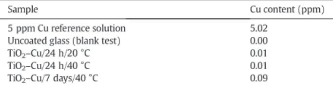
FurthermoreTable 2shows ICP-AES analyses of the water after im-mersion of TiO2–Cu films (100 nm thick; deposited on glass substrate)
for 24 h at 20 °C and 40 °C, and 7 days at 40 °C. The data did not give evidence for Cu release in pure water.
3.3.4. Aging tests
Before starting the aging tests, the relative activity of the selected as-deposited TiO2–Cu samples was 100% as measured by the JIS standard
method (Fig. 2) and it was the highest by the Haldar method (Table 1). The variation of the antibacterial properties determined by the JIS standard method is presented inFig. 7for different aging condi-tions. A decrease of the efficiency was observed with aging. This de-crease was higher for the more severe condition (# 2), i.e. 60 °C/RH
0 20 40 60 80 100 300 100 200
bactericidal
Antibacterial
0r
A
Antibacte
bactericidal
rial
bactericidal
A
Film thickness (nm)
Relative activity (%)
Antibacterial
Fig. 3.Influence of the film thickness on antibacterial activity: the coatings were deposited
using the same Cu(tmhd)2mole fraction. An antibacterial intermediate zone between
in-active and bactericidal surfaces is indicated.
200 nm
c
2 µm
Cu
particles
200 nm
b
a
Fig. 4.Surface (a, b) and cross section (c) SEM micrographs of TiO2–Cu coatings (3.5 at.% Cu) showing Cu metal particles emerging from the surface of the film.
Partially reprinted with permission from[31].
0 20 40 60 80 100 0 6 12 18
3.5
24Contact time (h)
Relative activity (%)
3.5 at. %
1.3 at. % Cu
Fig. 5.Kinetic method (modified JIS Z 2801 standard) showing that for high Cu content
(3.5 at.%) the bactericidal effect is achieved after 3 h of incubation whereas for low Cu con-tents (1.3 at.%) the bactericidal action requires 24 h of incubation revealing a lower activity.
100%, than for condition # 1 (20 °C/RH 40%). For instance the relative activity after 5 months of aging was approximately 50% versus 65%, respectively. This means that the performances of coatings decreased by only 35% after 5 months of aging in condition close to room temperature.
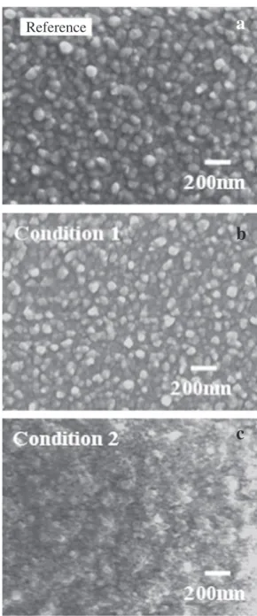
SEM observations were made after 2 and 5 months for both aging conditions and were compared with micrographs before aging (Fig. 8). At 60 °C/RH 100% (condition # 2) the surface morphology of the coating was already damaged after 2 months of aging and it is even more evi-dent after 5 months. The SEM analysis showed that no change has been observed under condition # 1 for 5 months while under more se-vere condition (# 2) the average size of grains on the surface decreased from about 60 to 30 nm and formed agglomerates (Fig. 8c). X-ray dif-fraction pattern under grazing incidence confirmed no structural change for samples aging under condition # 1 while after 5 months under the severe condition # 2 the diffraction peaks of Cu were not de-tected and the intensity of those of anatase was reduced.
4. Discussion
4.1. Influence of copper content and film thickness on the antibacterial activity
In this DLICVD process the Cu content of TiO2–Cu composite films is
controlled by the Cu(tmhd)2mole fraction injected into the reactor
(Fig. 1). The Cu incorporation in the coatings increases with the precur-sor mole fraction and it can reach several atomic percents but the goal was to investigate the biological behavior of the coatings without UV light for the lowest content of active agent. According to the JIS method, an antibacterial effect appears abruptly from a Cu content of ca. 1 at.% (Fig. 2). There is no antibacterial activity when Cu was not detected in the composite coating by XPS and EDS, i.e. when Cu was below the de-tection limit of about 1 at.%. A good reproducibility of the DLICVD pro-cess was found to control the film composition and, thereby, the Cu(tmhd)2mole fraction is a key parameter to control the antibacterial
activity.
Beyond a Cu(tmhd)2mole fraction of 0.04% the antibacterial activity
increases sharply and the films become bactericidal for a mole fraction of ca. 0.07% (Fig. 2). This evolution makes the control of the antibacterial activity between inactive and bactericidal (relative activity 100%) diffi-cult. Nevertheless, the activity can be controlled by the mean of the film thickness for a constant Cu(tmhd)2 mole fraction as shown in Fig. 3but only for the thicknesses below 100 nm. Indeed above this crit-ical thickness, the antibacterial activity is maximal (bactericidal). In a preliminary study[31], we showed that the dispersion of Cu particles was uniform on the surface of TiO2–Cu composite films (SEM analyses)
while a surface enrichment was observed in 300 nm thick layer (SIMS profile analyses). However different Cu particle sizes were observed ranging from 20 to 400 nm and the population density of the smallest particles (20–100 nm) was significantly greater than the largest ones (100–250 nm and N250 nm). The largest particles emerge from the sur-face of the coating (Fig. 4). Due to direct interaction with atmosphere the large Cu particles are probably very active in the microbiological mechanism. One hypothesis is that the largest Cu particles grow slowly and require a minimum film thickness, i.e. duration, to be formed. Solid state diffusion of copper can play a role in the formation of these largest particles since the deposition occurs around 683 K. Above a thickness threshold, probably near 100 nm, the distribution of Cu particles pro-truding from the surface is optimum and the film thickness effect disappears.
4.2. Other methods to determine biological activities
Because the antibacterial activity increases sharply between 1 and 3.5 at.% Cu most of the films have developed antibacterial behavior and they appear bactericidal by the JIS standard method for Cu N 3.5 at.%. Around and above this Cu content the performances of the coatings are difficult to compare. We used a kinetic method based on the JIS 2801 standard method.Fig. 5shows that the bactericidal ac-tivity (100%) was reached faster when the film content was 3.5 at.% Cu compared to 1.3 at.% Cu: respectively, in 3 and 24 h. This modified method could be used in order to distinguish 2 films showing a
Table 1
Relative antibacterial activities determined by the Haldar method of two TiO2–Cu
composite coatings with different Cu contents (the CFU values reported for each sample are the average of 3 measurements).
Cu(tmhd)2mole fraction (%) 0a 0.076 0.15
Cu content (at.%) 0 3.5 9.0
CFU 133 129 27
Relative activity (%) 0 0.6 32
aThis sample used as reference was a pure TiO
2film without co-deposition of copper.
a
b
Fig. 6.Diffusion test showing that (a) no inhibition zone is observed around a 25 × 25 mm2TiO
2–Cu sample whereas (b) there is such inhibition zone (gray area) around a reference
sam-ple consisting of a piece of plastic treated with triclosan as antibacterial agent.
Table 2
ICP-AES analyses of the water after immersion of TiO2–Cu coatings (100 nm thick)
deposited on glass substrate. The immersion conditions were 24 h at 20 °C, 24 h at 40 °C, then 7 days at 40 °C. The data are compared to a 5 ppm Cu solution as reference.
Sample Cu content (ppm)
5 ppm Cu reference solution 5.02
Uncoated glass (blank test) 0.00
TiO2–Cu/24 h/20 °C 0.01
TiO2–Cu/24 h/40 °C 0.01
bactericidal activity after 24 h. Nevertheless this method is both time and sample consuming and therefore difficult to implement.
The Haldar method[33]allows determining the antibacterial power of surfaces. The principle of this test is totally different from that of JIS Z 2801. Indeed, the bacteria are in contact with the surface during 24 h before their numeration like in JIS method, but both the antibacterial ef-fect of the coating surface and the growth of the bacterium in its culture medium take place simultaneously (competitive routes). Preliminary data are reported inTable 1. Two TiO2–Cu composite films that are
bac-tericidal according the JIS method were tested. Their composition is re-spectively 3.5 at.% and 9 at.% of Cu. This Haldar method shows that with the lowest Cu concentration (3.5 at.%) the surface is inactive (relative activity b 1%). The number of CFUs after the test is approximately the same on TiO2–Cu surface as on the reference sample (Table 1). By
con-trast, for 9 at.% Cu the relative antibacterial activity is 32%. This method seems to be less sensitive than the JIS standard method because it is not able to detect an antibacterial activity for a sample that exhibited a bac-tericidal activity by the mean of JIS standard method. However it can be used to distinguish different efficiencies for two surfaces which were found at their maximum of antibacterial activity by the JIS standard method. The Haldar method involves a competition mechanism be-tween the antibacterial effect of the surface and the development of bacteria bring by the culture medium. For the coating which contains the higher Cu content the antibacterial effect of Cu became stronger than the development of bacteria. This test is more differentiating and it can be applied usefully in addition to JIS standard method.
The antibacterial tests used in this paper involve planktonic bacteria and not adherent organisms. This could be a disadvantage when we want to study the prevention of biofouling. But, these are free bacteria that come first in contact with the surface. To prevent biofilm formation, a surface can act at different levels: (1) by preventing the adhesion thanks to surface properties, (2) by killing the bacteria in a relatively short time, before they multiply and synthesize their polysaccharide, and (3) by inhibiting the growth of adhered bacteria. The two microbi-ological methods used in this paper involve free bacteria but are not based on the same principle. First, in the JIS method, the bacteria are in close contact with the surface with very little nutritional factors (growth medium diluted 500 times). For example, active films which contain at least 3.5 at.% Cu kill all the bacteria deposited (≈3 × 10⁵mL−1) within
3 h while the bacterial growth is 2 log on uncoated substrate. One might consider that these coatings avoid bacterial growth by killing them rap-idly. Secondly, in the competition method[33], free bacteria are in con-tact with the surface for a very short time (2 min) and before being covered with pre-cut agar growth medium containing a great amount of nutriments. Therefore bacterial growth is competing with the action of the antibacterial agent and this is why this method is less sensitive than the first one. None of the 2 methods allows the development of
biofilm with adherent bacteria. For testing the capabilities of anti-bacterial coating to remove a preformed biofilm, adherent bacteria should be used.
4.3. Cu release from the coatings
The diffusion test is commonly used to demonstrate the release of an antiseptic compound from a solid support. An inhibition area around the support means that (i) the compound has migrated from the sup-port, (ii) it is soluble in the water of the culture medium and (iii) the bacterial strain is able to be inhibited by the substance. Since no inhibi-tion area around the sample has been observed, it means that no antibacterial element significantly diffuses from active TiO2–Cu
com-posite coatings to the environment (Fig. 6).
However this test is not sensitive enough and this is why we have an-alyzed the Cu traces in pure water that could originate from an im-mersed sample for different times. The ICP-AES data inTable 2did not
0 20 40 60 80 0 2 4 6 100
Duration (month)
Relative activity (%)
20 °C / RH 40 % 60 °C / RH 100 %Fig. 7.Variation of the biological activities under 2 experimental aging conditions:
condi-tion # 1 (20 °C/RH 40%) and condicondi-tion # 2 (60 °C/RH 100%). The decrease of the activity is greater for the most severe conditions.
Reference
a
b
c
Fig. 8.Surface SEM micrographs of TiO2–Cu coatings before (reference sample) and
after aging test for 5 months under condition # 1 (20 °C/RH 40%) and condition # 2 (60 °C/RH 100%).
give evidence for Cu diffusion in water. This is in agreement with the fact that Cu metal is not soluble in water. Only traces of Cu (9 × 10⁻²ppm)
were found after immersion for 7 days at 40 °C under gentle stirring. In these relatively drastic conditions Cu particles can be detached from the coating although immobilized by the oxide matrix and may explain these traces above the detection limit (5 × 10⁻³ppm). These results
in-dicate a good stability of the TiO2–Cu coatings. This is clearly an
advan-tage of using metallic nanoparticles as antibacterial agent embedded in an inorganic matrix compared to other hybrid organic–inorganic sys-tems. Furthermore they can serve as reservoirs of active agent.
4.4. Aging
The loss of activity during aging was the most important under the most severe conditions: 60 °C/RH 100%. As shown inFig. 8, the surface has been altered after aging with probably the formation of a copper oxide. The XRD patterns (not shown) revealed that metallic Cu struc-ture was detected before and after aging under condition # 1 while it disappeared after aging under the more severe condition # 2. This is cer-tainly due to the formation of copper oxide that acts as a barrier layer and therefore, the observed decrease of antibacterial activity may be due to an oxidation of the Cu particles on the surface of the sample. At 20 °C/RH 40%, the conditions were softer and the oxidation was limited compared with 60 °C/RH 100% and without consequence for the activity.
4.5. Comparison of the films containing either Cu or Ag particles
In previous studies, we developed a DLICVD process for the growth of nanostructured TiO2–Ag films[8]related to the one used for TiO2–Cu [31]. In the case of Ag nanocomposite coatings, the antibacterial action was higher than in the case of Cu-based coatings for a same metal con-tent. With Ag element, the relative activity determined by the JIS stan-dard method was 100% for a silver concentration below 1 at.%. The transition between inactivity and bactericidal effect was more abrupt than for copper, so that we did not observe any significant effect of the film thickness: thicknesses as low as 20 nm already showed significant activity. TEM analyses of films containing Ag showed that metal particles were uniformly distributed in the film thickness with an average size below 10 nm and a narrow size distribution. This is significantly different than the microstructure of TiO2–Cu composite coatings prepared by this
DLICVD process. 5. Conclusion
In this study, we developed, by DLICVD, composite coatings contain-ing metallic Cu particles embedded in a TiO2matrix. The materials
exhibited high antimicrobial properties against S. aureus tested by the mean of the JIS Z 2801:2000 standardized method. The low Cu content (3.5 at.%), and the fact that the antibacterial agent is not released in the environment suggests that these engineered surfaces do not exhibit toxic properties.
This work is part of a general program on multifunctional oxide-M coatings (M = Ag, Cu) developed for self-cleaning surface applications. For this objective, in addition to antibacterial properties, very thin thick-nesses were aimed to preserve the surface appearance. Furthermore possible photocatalytic properties due to the oxide matrix would be beneficial to remove organic contamination from the surface. The cou-pling with photocatalytic properties is not expected for instance for SiOx–Ag nanocomposite thin films[35]and this is an advantage of
ana-tase matrix.
Very thin TiO2–Cu films are quite transparent in the visible range;
ap-proximately 70% for a 150 nm thick coating containing ca. 3.5 at.% Cu. UV photocatalytic tests were performed (not reported) as described for TiO2–Ag in[8]but bactericidal TiO2–Cu coatings do not exhibit any
pho-tocatalytic activity. The amount of Cu in antibacterial TiO2–Cu layers is
certainly sufficiently high (N1 at.% Cu) to annihilate any photocatalytic activity which is a property very sensitive to defects (metallic NPs with broad size distribution are defects in the oxide semiconductor matrix). Furthermore, compared with TiO2–Ag nanocomposite coatings grown
by a comparable CVD process[8], the matrix structure of TiO2–Cu
coat-ings changes from anatase structure to rutile by increasing the Cu con-tent[31] and it is known that rutile is less efficient than anatase in photocatalysis[36]. As a result we have not observed simultaneously antibacterial and photocatalytic effect in TiO2–Cu coatings grown by
DLICVD in contrast with TiO2–Ag coatings[8].
Further studies are required to determine the mechanism by which such low amounts of Cu metal particles are effective bactericidal, and to confirm that any of the metal nanoparticles are released into the envi-ronment. The antibacterial activity was determined according to two different methods. The first one, JIS Z 2801:2000 is normalized and used in laboratory to screen a lot of samples. The other, described by Haldar et al.[33] is more discriminating and for some applications more representative of real-life situations.
The aging effect of TiO2–Cu composite films under different
environ-mental conditions tends to slightly decrease the antibacterial efficiency but they exhibit a good durability. For instance, after 5 months in a cli-matic chamber under 20 °C/RH 40% and 60 °C/RH 100%, the relative antibacterial activity decreases from 100% (bactericidal) to 65% and 50%, respectively, without any cleaning treatment of the surface.
Acknowledgments
This work was supported by ANR (Agence Nationale de la Recherche) under contract ANR-06-MAPR-0007-01.
References
[1] M. Rai, A. Yadav, A. Gade, Biotechnol. Adv. 27 (2009) 76–83.
[2] Q.L. Feng, J. Wu, G.Q. Chen, F.Z. Cui, T.N. Kim, J.O. Kim, J. Biomed. Mater. Res. 52 (2000) 662–668.
[3] M.A. Butkus, L. Edling, M.P. Labare, J. Water Supply Res. Technol. AQUA 52 (2003) 407–416.
[4] A.B.G. Landsdown, J. Wound Care 11 (2002) 125–138.
[5] J.J. Castellano, S.M. Shafii, F. Ko, G. Donate, T.E. Wright, R.J. Mannari, W.G. Payne, D.J. Smith, M.C. Robson, Int. Wound J. 4 (2007) 114–122.
[6] L. Nan, Y. Liu, M. Lü, K. Yang, J. Mater Sci: Mater. Med. 19 (2008) 3057–3062. [7] J.R. Morones, J.L. Elechiguerra, A. Camacho, J.T. Ramirez, Nanotechnology 16 (2005)
2346–2353.
[8] J. Mungkalasiri, L. Bedel, F. Emieux, J. Doré, F.N.R. Renaud, C. Sarantopoulos, F. Maury, Chem. Vap. Deposition 16 (2010) 35–41.
[9] A. Panacek, L. Kvitek, R. Prucek, M. Kolar, R. Vecerova, N. Pizurova, V.K. Sharma, T. Nevecna, R. Zboril, J. Phys. Chem. 110 (2006) 16248–16253.
[10] S. Pal, Y.K. Tak, J.M. Song, Appl. Environ. Microbiol. 27 (2007) 1712–1720. [11] F. Raimondi, G.G. Scherer, R. Kotz, Z. Wokaun, Angew. Chem. Int. Ed. 44 (2005)
2190–2209.
[12] M. Danilczuk, A. Lund, J. Sadlo, H. Yamada, J. Michalik, Spectroch. Acta Part A 63 (2006) 189–191.
[13] J.S. Kim, E. Kuk, K.N. Yu, J.H. Kim, S.J. Park, H.J. Lee, S.H. Kim, Y.K. Park, Y.H. Park, C. Hwang, Y.K. Kim, Y.S. Lee, D.H. Jeong, M.H. Cho, Nanomed. Nanotech. Biol. Med. 3 (2007) 95–101.
[14] K. Yoon, J.H. Byeon, J. Park, J. Hwang, Sci. Total Environ. 373 (2007) 572–575. [15] F. Zeng, C. Hou, S. Wu, X. Liu, Z. Tong, S. Yu, Nanotechnology 18 (2007) 055605. [16] Y.H. Kim, D.K. Lee, H.G. Cha, C.W. Kim, Y.C. Kang, Y.S. Kang, J. Phys Chem. B 110 (2006)
24923–24928.
[17] P. Ruparelia, A.K. Chatterjee, S.P. Duttagupta, S. Mukherji, Acta. Biomater. 4 (2008) 707–716.
[18] W. Yuan, J. Ji, J. Fu, J. Shen, J. Biomed. Mat. Res. Part B: Appl. Biomater. 85 (2008) 556–563.
[19] O. Akhavan, J. Colloid Interface Sci. 336 (2009) 117–124. [20] O. Akhavan, E. Ghaderi, Curr. Appl. Phys. 9 (2009) 1285–1381.
[21] F. Furno, K.S. Morley, B. Wong, B.L. Sharp, P.L. Arnold, S.M. Howdle, R. Bayston, P. Brown, P.D. Winship, H.J. Reid, J. Antimicrob. Chemother. 54 (2004) 1019–1024. [22] S. Egger, R.P. Lehmann, M.J. Height, M.J. Loessner, M. Schuppler, Appl. Environ.
Microbiol. 75 (2009) 2973–2976.
[23] P.J. Kelly, H. Li, K.A. Whitehead, J. Verran, R.D. Arnell, I. Iordanova, Surf. Coat. Technol. 204 (2009) 1137–1140.
[24] L.A. Brook, P. Evans, H.A. Foster, M.E. Pemble, A. Steele, D.W. Sheel, H.M. Yates, J. Photochem Photobiol. A Chem. 187 (2007) 53–63.
[25] D.W. Sheel, L.A. Brook, I.B. Ditta, P. Evans, H.A. Foster, A. Steele, H.M. Yates, Inter J. Photoenergy 2008 (2008) 1–11(Article ID 168185).
[26] K. Page, R.G. Palgrave, I.P. Parkin, M. Wilson, S.L.P. Savin, A.V. Chadwick, J. Mater. Chem. 17 (2007) 95–104.
[27] Y. Yao, K. Yamauchi, G. Yamauchi, T. Ochiai, T. Murakami, Y. Kubota, J. Biomaterials Nanobiotechnology 3 (2012) 421–430.
[28] T. Ochiai, A. Fujishima, J. Photochem Photobio. C: Photochem. Reviews 13 (2012) 247–262.
[29] R. Nakano, M. Hara, H. Ishiguro, Y. Yao, T. Ochiai, K. Nakata, T. Murakami, J. Kajioka, K. Sunada, K. Hashimoto, A. Fujishima, Y. Kubota, Catalysts 3 (2013) 310–323. [30] Y. Yao, T. Ochiai, H. Ishiguro, R. Nakano, Y. Kubota, Applied Catalysis B: Environmental
106 (2011) 592–599.
[31] J. Mungkalasiri, L. Bedel, F. Emieux, J. Dore, F.N.R. Renaud, F. Maury, Surf. Coat. Technol. 204 (2009) 887–892.
[32] Anonymous, JIS Z 2801. Antimicrobial Products; Test for Antimicrobial Activity and Efficacy. Japanese Industrial Standard, 2000.
[33] J. Haldar, A.K. Weight, A.M. Klibanov, Nature Protocols 2 (2007) 2412–2417. [34] Anonymous, ISO 20645; Textile Fabrics: Determination of Antibacterial Activity,
Agar Diffusion Plate Test, 2004.
[35] L. Bedel, C. Cayron, M. Jouve, F. Maury, Nanotechnology 23 (2012) 015603. [36] F.-D. Duminica, F. Maury, R. Hausbrand, Surf. Coatings Technol. 201 (2007)