D
EPARTMENTOF
BIOCHEMESTRY
Thesis
Presented by
BENCHEIKH DALILA
For the fulfillment of the requirements for the degree of
Doctorate of Sciences
Biology
Special field: Biochemistry
Topic
Hypoglycemic medicinal plants used in Setif region and their
effects on experimentally-induced diabetes in rats
Presented publically in …..…/……../2017 Jury :
President: BELHATTAB Rachid Pr. UFA Setif 1
Supervisor: KHENNOUF Seddik Pr. UFA Setif 1
Co-supervisor : DAHAMNA SERAICHE S. Pr. UFA Setif 1
Examiners: BOUDJELAL Amel
ZAAMA Djamila
KHETTAL Bachra
MCA. Univ M’sila Pr. Univ Constantine 1 MCA. Univ Bejaia
Laboratory of Phytotherapy Applied to Chronic Diseases
Université Ferhat Abbas Sétif 1 Faculté des Sciences de la
Nature et de la Vie
ةيبعشلا ةيطارقميدلا ةيرئازجلا ةيروهمجلا
ملعلا ثحبلا و يلاعلا ميلعتلا ةرازو
ي
فيطس ،سابع تاحرف ةعماج
1ةايحلاو ةعيبطلا مولع ةيلك
N°……….…………..…….……/SNV/2017List of publications
Bencheikh D, Khennouf S, Bouaziz A, Baghiani A, Dahamna S, Amira S, Arrar L.2016.
Antioxidant and Antidiabetic Activities of the Methanolic Extract of Olea europaea L. Leaves in Streptozotocin Induced Diabetes in Rats. International Journal of
Pharmacognosy and Phytochemical Research. 8(8): 1347-1357.
List of communications
Khennouf Seddik, Bencheikh Dalila. Polyphenols and antioxidant properties of extracts from Mentha peluguim L. and Matricaria camomilla L. The third International Symposium on Medicinal Plants, Thier Cultivation and Aspects of uses. November2012, Jordan.
Bencheikh Dalila, Rezzagui Abir, Madoui Soriya, Khennouf Seddik. Antioxidant activity
of Matricaria Commomilla L flowers. International Conferance on Research in Education and Science. April 23-26,2015. Antalya, Turkey.
Bencheikh Dalila, Rezzagui Abir, Madoui Soriya, Khennouf Seddik. Antioxidant activity
of Olea europea L flowers. International Conferance on Research in Education and Science. April 23-26,2015.Antalya, Turkey.
List of Proceedings
Bencheikh Dalila, Rezzagui Abir, Madoui Soriya, Khennouf Seddik. Antioxidant activity
of Matricaria Commomilla L flowers. International Conferance on Research in Education and Science. April 23-26,2015. Antalya, Turkey.pp14.
Bencheikh Dalila, Rezzagui Abir, Madoui Soriya, Khennouf Seddik. Antioxidant activity
of Olea europea L flowers. International Conferance on Research in Education and Science. April 23-26,2015.Antalya, Turkey.pp117.
Acknowledgements
After an intensive period of five years, today is the day: writing this note of thanks is the finishing touch on my thesis. It has been a period of intense learning for me, not only in the scientific arena, but also on a personal level. Writing this thesis has had a big impact on me. I would like to reflect on the people who have supported and helped me so much throughout this period.
I would particularly like to single out my supervisor Pr. Seddik Khennouf for his supervision, advice, and guidance in this thesis. I want to thank you for your excellent and for all of the opportunities I was given to conduct my research and further my thesis. In addition, I would like to thank Dhamna Saliha for their valuable guidance. You definitely provided me with the tools that I needed to choose the right direction and successfully complete my thesis.
Also, I would like to thank Bouriche Hmama for reinforcing my research and for her help in HPLC technique.
I gratefully thank to my comittee members, BELHATTAB Rachid; BOUDJELAL Amel ; ZAAMA Djamila ; KHETTAL Bachra deverse special thanks.
Many thanks go to doctors : Abdellouch who allowed me to make the sections and specefic thanks to doctor Safsaf for her interpretation and friendy followship.
I would first like to thank my colleagues for their wonderful collaboration and help : Rezzagui Abir, Bouaziz Amel and Bentahar Assia. You supported me greatly and were always willing to help me.
Finally, my deepest gratitude goes to my parents for their unflagging love and conditional support, their wise counsel and sympathetic ear throughout my life and my studies. You made me live the most unique, magic and carefree childhood that has made me who I am now ! Many thanks go in particular to my brothers and my husband Toufik. Thank you very much, everyone! Dalila BENCHEIKH.
Abstract
The objective of this study is to evaluate the antioxidant and antidiabetic activity of Olea
europea, Trigonella foenum-graecum and Eucalyptus globulus extracts, which were popular
through the survey, by Streptozotocin- induced diabetes in normal male adult rats via comparison of changes in body weight, biochemical and histopatholoangical parameter. Intravenous injection of 50 mg/kg of Streptozotocin in rats, makes pancreas swell and causes degeneration of beta cells in Langerhans islet and induces experimental diabetes in 2 days. In this study, the results showed that methanolic extract (ME) and chloroformic extract (ChE) of
Eucalyptus globulus leaves contained high polyphenolics and flavonoids contents,
respectively, whereas ethyl acetate extract (EAE) of Olea europea leaves and Trigonelle
foenum-graecum seeds contain the most important quantity in polyphenols and flavonoids.
The obtained results from HPLC technique allowed the detection of 36 phenolic compounds in MEO, 16 in MET and 40 in MEE. In addition, ME of E.globulus showed a strong ABTS radical scavenging activity, and inhibited the linoleic acid oxidation in ferric thiocyanate method and TBA. While, the EAE of O.europea and T.foenum-graecum exhibited a good activity in reducing power, ABTS and TBA assays. After Induction of diabetes, the volume of urine and glucose increased in the untreated animals in comparison with normal ones, but the body weight decreased in the untreated animals. The administration of MEO and MET at doses of 200 and 600 mg/kg and MEE at doses of 150 and 500mg/kg increased catalase activity, GSH level and decreased lipid peroxidation in the tissues of liver and kidneys, serum total cholesterol and triglycerides levels. Furthermore, histological damages in pancreas, kidney and liver tissues were reduced. These results indicated a good hypoglycemic and antioxidant activity of the three studied plants, could explain their use in folk medicine in the control of diabetes and preventing diabetic complications by scavenging free radicals.
Key words :
Olea europea L, Trigonella foenum-graecum, Eucalyptus globulus, antidiabetic, antioxidant, polyphenols
Résumé
L'objectif de cette étude est d'évaluer l’activité antioxydante et antidiabétique des extraits des
Olea europea, Trigonella foenum-graecum et Eucalyptus globulus, ce qui étaient populaires
par le questionnaire, en induissant expérimentalement le diabète par streptozotocine chez des rats adultes mâles normaux par l’intermédiaire de la comparaison des changements du poids corporel, les paramètres biochimiques et histopatholoangical. L'injection intraveineuse de 50 mg / kg de streptozotocine chez le rat, fait gonfler le pancréas et enfin, provoque une dégénérescence des cellules bêta des îlots de Langerhans et un diabète sucré en 2 jours. Dans cette étude, les résultats ont montré que l’extrait méthanolique (EM) et l’extrait chloroformique (ECh) des fleurs Eucalyptus globulus ont contenu des teneurs élevées en polyphénols et flavonoïdes, respectivement, tandis que l'extrait ethyl acetate (EEA) des fleurs
Olea europea et les graines Trigonelle foenum-graecum étaient les meilleurs en polyphénols
et flavonoïdes. Les resultats obtenus par la méthode HPLC permettent de détecter 36 des composés phénoliques dans EMO, 16 dans EMT et 40 dans EME. En outre, EM d’E.globulus a montré une forte activité de piégeage des radicaux ABTS, et a inhibé l'oxydation de l'acide linoléique par le test de thiocyanate ferrique et TBA. Alors que, EEA d’O.europea et
T.foenum-graecum montraient une bonne activité dans les tests pouvoir réducteur, ABTS et
TBA. Après l'induction du diabète, le volume d'urine et de glucose ont augmenté chez les animaux non traités en comparaison avec les animaux normaux, mais le poids corporel a diminué chez les animaux non traités. L'administration de EMO et EMT aux doses de 200 et 600 mg / kg et EME à des doses de 150 et 500mg / kg a entrainé d’augmentater l'activité de catalase, le niveau de GSH et une diminution de la peroxydation lipidique dans les tissus du foie et des reins, le taux de cholestérol total et triglycérides dans le serum. En outre, les dommages histologiques des tissus de pancréas, de rein et de foie ont été réduits. Ces résultats indiquent une bonne activité hypoglycémique et antioxydante des trois plantes étudiées, peuvent ainsi expliquer leurs utilisations dans la médecine traditionnelle dans la lutte contre le diabète et la prévention des complications du diabète en piégeant les radicaux libres.
Mots clés :
Olea europea L, Trigonella foenum-graecum, Eucalyptus globulus, antidiabétiques, antioxydant, les polyphénols
صخلم
هذه نم فدهلا
يركسلا ءادل ةداضملاو ةدسكلأل داضملا طاشنلا مييقت وه ةساردلا
تاصلختسمل
Olea europea
،
Trigonella foenum-graecum
و
Eucalyptus
globulus
هثادحإ قيرط نع ،نايبتسلال ةسارد للاخ نم ةريبك ةيبعشب تيظح يتلاو ،
،مسجلا نزو يف تاريغتلا ةنراقمب كلذ و نيسوتوزوتبرتسلإاب نيغلاب ناذرج نقحب ايبيرجت
ةيضرملاو ةيويحلا ءايميكلاو
.
ةعرجل يديرولا نقحلا
05
غلم
/
،نيسوتوزوتبرتسلإا نم غك
يو سايركنبلا خافتنا ىلإ يدؤي
ىلإ يلاتلاب و سناهرجنلا رزج يف اتيب ايلاخ يف لالاحنا ببس
نيموي يف يبيرجتلا يركسلا ءادلا ثادحا
.
صلختسملا نأ جئاتنلا ترهظأ ،ةساردلا هذه يف
يلوناثيملا
(ME)
يمروفورولكلا صلختسملا و
(ChE)
ةتبن قارولأ
E.globulus
فلافلا و لونيفلا تاديدع نم ةيلاع ةبسن ىلع نايوتحي
بيترتلاب تاديونو
.
صلختسم ،نيح يف
ليثلإا تلاخ
(EAE)
ةتبن قارولأ
O.europea
ةتبن روذبو
T.foenum-graecum
تاديونوفلافلا و لونيفلا تاديدع نم ةربتعم ةيمك ىلع يوتحي
.
يف اهيلع لصحتملا جئاتنلا
HPLC
ترهظأ
43
يف يلونيف بكرم
MEO
،
61
يف
MET
و
35
يف بكرم
MEE
.
سم ،كلذك
صلخت
ME
ةتبنل
E.globulus
رذجل ةحازإ ةوق رهظأ
ABTS
ةدسكأ عنميو ،
و كيلونيللا ضمح
ا تانايسويث
اذكو ديدحل
TBA
.
نيح يف
EAE
يتتبنل
O.europea
،
T.foenum-graecum
،ةيعاجرلإا ةردقلا ةينقتل ةيطيبثت ةوق ىدبأ
ABTS
و
TBA
.
سلا عافترإو لوبلا مجح يف ةدايز دهشن ،يركسلا ضيرحت دعب
يف مدلا يف رك
مسجلا نزو يف ضافخنا ظحلن نكلو ،ةيعيبطلا تاناويحلا عم ةنراقم ةجلاعملا ريغ تاناويحلا
ةجلاعملا ريغ تاناويحلل
.
يصلختسمب ناذرجلا ةجلاعم تدأ
MEO
و
MET
تاعرجب
055
و
155
غلم
/
صلختسمو غك
MEE
تاعرجب
605
و
055
غلم
/
طاشن يف ةدايز ىلإ غك
لاتاكلا
ىوتسم ،ز
GSH
ىوتسم ضافخناو
MDA
يف
ىوتسم ،ىلكلاو دبكلا ةجسنأ
امزلابلا يف تايوتسملا ةيثلاثلا نوهدلاو لورتسيلوكلا
.
ضيفخت مت ،كلذ ىلع ةولاعو
ىلكلاو دبكلاو سايركنبلا يف ةيجيسنلا رارضلأا
.
ديج طاشن ىلإ تراشأ جئاتنلا هذهف اذإ
فلتخمل ةدسكلأل داضم طاشنو مدلا يفركسلا ةبسن ضيفختل
امم ،ةلمعتسملا ةثلاثلا تاتابنلا
اذه تافعاضم عنمو يركسلا ءاد ىلع ةرطيسلا يف يبعشلا بطلا يف اهمادختسا رسفي دق
ةرحلا روذجلا طيبثت للاخ نم ءادلا
.
حيتافملا تاملكلا
:
Olea europea
،
Trigonella foenum-graecum
،
Eucalyptus globulus
،
،ةدسكلأا تاداضم ،يركسلا داضم
لونيفلا تاديدع
.
List of abbreviations
ABTS : 2,2’-azino-bis (3-ethylbenzenothiazoline acid) ALcl3 : aluminium trichloride
ALT : alanine aminotransferase AST : Aspartate aminotransferase BHT : butylated hydroxytoluene CAT : catalase
DPPH: 2, 2-diphenyl-1-picryl-hydrazyl DM : diabetes mellitus
EDTA : ethylenediamine tetraacetic acid Fe2+ : ferrous iron
FTC : ferric thiocyanate GDM : gestational diabetes GPx : glutathione peroxidase GSH : reduced glutathione HDL : high density lipoprotein
HPLC: high performance liquid chromatography H2O2 : hydrogen peroxide
HO2- : perhydroxyl radical
I%: Inhibition percentage
IC50%: Inhibitory concentration for 50% of activity
LDL : Low density lipoprotein MeOH: Methanol
MDA : malondialdehyde
MEO : methanolic extract of Olea europea
MET : methanolic extract of Trigonella foenum-graecum MEE : methanolic extract of Eucalyptus globulus
Mrp2 and 3 : multidrug resistance protein 2 and 3 NIDDM : non-insulino dependent diabetes mellitus NO- : nitric oxide
O2- : superoxide radical
OH- : hydroxil radical OONO- : peroxynitrite anion ROS : reactive oxygen speices SEM: Standard error of the mean
SOD : superoxide dismutase STZ : streptozotocin TBA : thiobarbituric acid T1D : type 1 diabetes mellitus T2D : type2 diabetes mellitus VLDL : very low density lipoprotein
Table of contents
Introduction………...1
Review of literature
………...5
I.1. The implication of the free radicals in the
pathogenecity
of
diabetes………....6
I.1.1. Oxidative stress………..6
I.1.1.1.Definition. ………...
….…6
I.1.2.Reactive oxygen speices (ROS)
………...………….……….…
…7
I.1.2.1.Definition………...7
I.1.2.2. Sources of ROS……….
....8
1.Endogenous sources ………..………...8
1.1.Mitochondria as a source of ROS ………..………9
1.2. Peroxisomes as a source of ROS and RNS……….….10
1.3. NADPH oxidase as a source of ROS………...11
1.4. Prostaglandin H Synthase (PHS) as a source of ROS………...………..11
1.5. Cytoplasmic Cytoplasmicsources of ROS and RNS ………..………11
2.Exogenous sources……….……….………12
I.1.2.3. ROSs and cellular damage ……….….
121. Oxidative damage to proteins………...13
2. Oxidative damage to lipids……….…....13
3. Oxidative damage to DNA………..…………...13
I.1.3. Antioxidant defense system………...14
I.1.3.1. Introduction……….………14
I.1.3.1. Sources of antioxidants………...15
I.1.2.3.1. Endogenous sources……….……….………15
1. Enzymatic antioxidant………...……….15
* Manganese superoxide dismutase (MnSOD)……….……….15
*Catalase………..………..16
2. Nonenzymatic antioxidant………..……….….16
*Glutathione as an Antioxidant………..16
*N-acetylcysteine ………..17
I.1.2.3.2. exegenous sources………..………18
*Vitamins………..…………....18
*Plants phenolic compounds………..……….19
1. Phenolic acids………....19
1.1. Flavonoids………20
1.2. Polyphenols………...21
1.2.1. Proanthocyanidin derivatives………...21
1.2.2. Galloyl and hexahydroxydiphenyl ester derivatives………...21
1.2.3. Hydroxy cinnamic acid derivatives………....21
1.2.4. Phloroglucinol derivatives………...22
1.3. Tannins………...………….22
1.3.1. Hydrolysable tannins………22
1.3.2. Complex tannins……….………..22
1.3.3. Condensed tannins………...23
I.1.3.3. Antioxidant effects of phenolic compounds………...24
I.1.3.3.1. Antioxidant activity of flavonoids………...24
* Free radicals trapping……….24
* Effect on the mediator of nitric oxide synthesis………...25
* Inhibition of the enzymes activities………...…26
* Chelation of the metal ions………....27
I.1.3.4. Oxidant effect of phenolic compounds………...27
I.2. Diabetes………..28
I.2.1. Structure of the pancrea………..…..29
I.2.1.1. Exocrine pancreas……….29
I.2.1.2. Endocrine pancreas………...30
I.2.1.2.1. Pancreatic -cell………....30
I.2.1.2.2. Insulin………..………..…31
Insulin biosynthesis………..……….……31
Insulin secretion……….…….……..32
I.2.2.1.
Type1 diabete mellitus (T1D)……….……….34
I.2.2.2. Type2 diabete mellitus (T2D)……….………….35
I.2.2.2.1. Type 2 DM and lipid………...………..35
I.2.2.2.2. Role of Insulin with lipids……….………36
I.2.2.3. Gestational diabetes (GDM)………...……….36
I.2.3. Free radicals and diabetes………....37
I.2.4. Experimental diabetes induced by Streptozotocin……....40
I.2.4.1. Definition ofSterptozotocin………....………40
I.2.4.2. Diabetes treatment………..……...………..40
I.3. Herbal therapy of diabetes………...42
I.3.2. Botanical identification and description of plant species
chosen for this study………..43
I.3.2.1. Olive tree (Olea europea L.)………...………....43
I.3.2.1.1. Botanical description………...……...44
I.3.2.1.2. Phytochemical composition………...………...…….44
I.3.2.1.3. Medicinal use of the plant………..…...……..45
I.3.2.2. Fenugreek (Trigonella foenum-graecum L.)…………...….45
I.3.2.2.1. Botanical description………...………46
I.3.2.2.2. Phytochemical composition………...…….……47
I.3.2.2.3. Medicinal use of the plant………...……47
I.3.2.3. Eucalyptus (Eucalyptus Globulus Labill.)…………...……..48
I.3.2.3.1. Botanical description………...………….48
I.3.2.3.2. Phytochemical composition……...………...………49
I.3.2.3.3. Medicinal use of the plant……….……...49
Materials and methods……….………...51
II.1. Preparation of a survey on the hypoglycemic plants
used in Setif region………52
II.1.1. Ethnobotanic investigations…………...…………...52
II.1.2. The questions………...………...53
II.2.The effects of the plant extracts on experimental
diabetes in rats………...………...54
II.2.1. Materials………….……..………...54
II.2.1.1. Plants materials…….…….………...…………...54
II.2.1.2. Chemicals………….……
...………..54
II.2.2. Methods…………...………...55
II.2.2.1. Preparation of plant extracts….………...……...55
II.2.2.2. Assessment of total polyphenols in the extract.………...57
II.2.2.3. Assessment of flavonoids in extracts……...………..58
II.2.2.4. HPLC analysis of methanolic extracts of plants……...58
II.2.2.5. In vitro antioxidant and antiradicalar activities of plants
extracts………...…59
II.2.2.5.1. Test of DPPH………...59
II.2.2.5.2. ABTS radical scavenging activity assay………...……60
II.2.2.5.3. Ferrous ion chelating activity………....60
II.2.2.5.4. Reducing power………..………...61
II.2.2.5.5. Test of -Carotene/Linoleic acid………..……….61
II.2.2.5.6. Ferric thiocyanate test (FTC)………...………….63
II.2.2.5.7. Thiobarbituric acid (TBA) method………...…...……….63
II.2.2.6. Antidiabetic and antioxidant activity of plant extracts in
vivo………..………..………64
1. Treatment protocol………..………..64
1.1. Assessment of DPPH in plasma……….65
1.2. Assessment of reducing power in plasma………..……65
1.3. Histological sections studies………..66
1.4. Preparation of homogenate……….………...66
1.4.1. Determination of total protein level……….66
1.4.2. Determination of catalase activity………....66
1.4.3. Determination of lipid peroxidation (MDA)….……….…..67
1.4.3. Determination of reduced glutathione (GSH)……….……….67
1.6. Statistical analysis……….………...………68
Results and discussion………..…………..69
III.1. Hypoglycimic plants used in Setif region…...…..70
III.1.1. Statistical study by SPPS for the survey results...…….….74
III.2.Total polyphenols and flavonoids in plants
extracts………...…...…………76
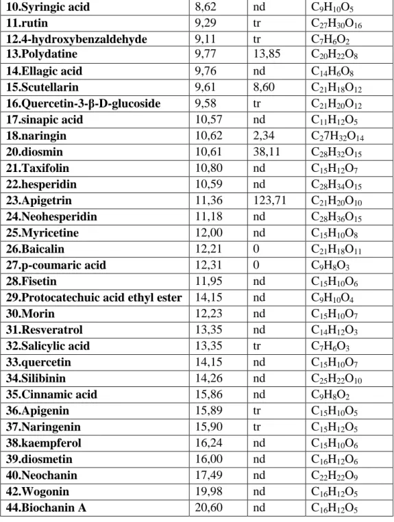
III.3.HPLC analysis of methanolic extracts of studied
plants………...………..79
III.4. Antioxidant activity: In vitro assays…….………88
III.4.1. DPPH scavenging activity of extracts……...…………....88
III.4.2. ABTS scavenging activity of extracts…...………..91
III.4.3. Ferrous ion chelating activity of extracts…………..…....93
III.4.4. Reducing power of plants extracts………96
III.4.5.Antioxidant activity of plants extracts using
β-Carotene bleaching assay………...….99
III.4.6. Antioxidant activity of extracts using Ferric thiocyanate
(FTC) method……….101
III.4.7. Antioxidant activity of extracts using TBA test...…..….103
III.5. Antidiabetic and antioxidant activity :
in vivo assays……….……106
III.5.1.Extracts effect on blood biochemical parameters……....110
III.5.2. Plasma antioxidant capacity using DPPH radical……...113
III.5.3.Assessment of plasma reducing power in treated and
untreated animals……….……..….115
III.5.4. Histopathological examination of Liver, Kidney and
Pancreas………...…….……..116
III.5.5. Effect of different extracts on glucose loaded rats…...122
III.5.6. Body weight alteration………..……..128
III.5.7. Effect of plant extracts on antioxidant parameters in liver
and kidney………..………...129
III.5.7.1. Effect of extracts on catalase activit.y………….………130
III.5.7.2. Effect of extracts on MDA level…..………..……….…...132
III.5.7.3. Effect of extracts on GSH level…..……….…………134
III.5.7.4. Assessment of Total protein……..………..….…137
III.5.8. Determination of diuretic activity of plant extracts...140
Conclusion future considerations………...…..143
References………...……....148
1
List of figures
Fig.1 : Schematic representation of ROS generation. Fig. 2 : Mitochondria Produce ROS.
Fig. 3: Principle reaction catalyzed by xanthine oxidase. Fig.4 : Detoxification of ROS via glutathione-ascorbate cycle. Fig.5 : Structures of phenolic acids
Fig.6 : Subclasses of flavonoids. Fig.7 : Structure of pancrea.
Fig.8 : Effect ofstress ondiabetes.
Fig. 9 : High-resolution model of six insulin molecules assembled in a hexamer. Fig. 10 : Insulin action and diabetes type1 and 2.
Fig.11 : Participation of hyperglycemia in triggering the multiple oxidative stress pathways in the course of diabetes.
Fig.12 : Photograph of Olea europea L.
Fig.13 : Photograph of Trigonella foenum-graecum L.
Fig.14 : Photograph of Eucalyptus globulus L.
Fig. 15: Schematic diagram represents the process of extraction.
Fig. 16: Standard curve of gallic acid for the determination of total polyphenols. Fig.17: Standard curve of quercetin and rutin for the determination of total flavonoids.
Fig.18 : Staistical study by SPPS: results of question 10.
Fig.19. A: HPLC chromatograms of the methanolic extracts of O.europea (MEO)
Fig.19. B: HPLC chromatograms of the methanolic extracts of T.foenum-graecum (MET)
2
Fig.20 : IC50 values of different plants extracts in DPPH assay.
Fig.21 : Comparison between different plant extract and Trolox in ABTS free radical scavenging activity.
Fig.22 : Metal chelating activity of different plant extracts.
Fig.23 : IC50 of different plant extracts in reducing power assay.
Fig.24 : Inhibition percentage of different extracts of plants in β-carotene/ linoleic acid assay after 24h.
Fig.25 : Lipid peroxidation inhibition of extracts using FTC method.
Fig.26 : Malondialdehyde formation pathway from peroxyl radical of triunsaturated C18 fatty acid (a) and formation of TBA chromophore from TBA and malondialdehyde (b).
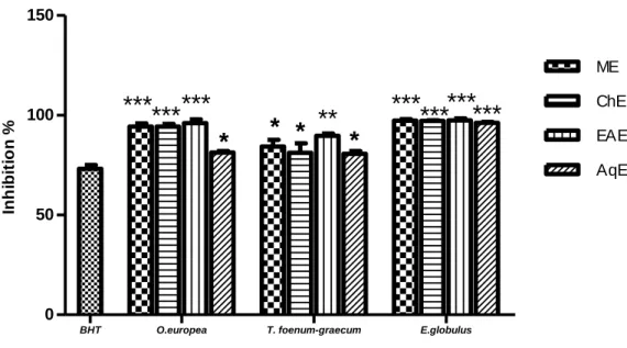
Fig.27: Antioxidant activities of different plant extracts (2 mg/ml) measured by TBA method. Fig.28 : Antioxidant capacity of plant extracts using DPPH radical in plasma.
Fig.29 : Antioxidant capacity of plant extracts using reducing power in plasma.
Fig. 30:Microphotographs of pancreatic islets in rats treated with MEO after 18 days of the treatment. Fig.31: Photomicrographs of liver sections.
Fig.32: Photomicrographs of kidney sections.
Fig.33: Changes in the mean body weight (g).
Fig.34: Effect of methanolic extract of O.europea, T.foenum-graecum, E.globulus on catalase activity in liver (A) and kidney (B) of rats.
Fig.35: Effect of methanolic extract of O.europea, T.foenum-graecum, E.globulus and Glibil on MDA level in liver (A) and kidney (B) of rats.
Fig.36: Effect of methanolic extract of O.europea, T.foenum-graecum and E.globulus on reduced glutathione level in liver (A) and kidney(B) of rats.
3
Fig.37: Effect of methanolic extract of O.europea, T.foenum-graecum and E.globulus on total proteins level in liver (A) and kidney (B) of rats.
4
List of tables
Table 1: Hypoglycimic plants used in the treatement of diabetes by population in Setif region, results of survey.
Table 2: Total polyphenols, flavonoids in plants extracts. Table 3. A: Chromatograph analysis of MEO
Table 3. B: Chromatograph analysis of MET Table 3. C: Chromatograph analysis of MEE
Table 4: Mean values of blood parameters in treated and untreated animals.
Table 5: Biochemical parameters in blood of treated and untreated animals. Table 6: Glucose levels changes in the blood of treated and untreated animals.
5
Introduction
Diabetes is a common disease. World Health Organisation (WHO) estimates that 30 million people had diabetes in 1985, and this number increased to 171 million people in 2000. In that year, an estimated 2.9 million people died of diabeted, representing 5.2% of all deaths, probably the fifth largest cause of mortality in the word (Roglic et al., 2005). It is estimated that in 2030, people with diabetes will reach 366 million, most of them from developing countries, especially among people from45 to 64 years of age (Roglic, 2004).
Diabetes mellitus (DM) is a chronic endocrine disorder of glucose characterized by hyperglycemia resulting from defects in insulin secretion, insulin action, or both (Wild et al., 2004).
The importance of oxidation in the body and in foodstuffs has been widely recognized. Oxidative metabolism is essential for the survival of cells. A side effect of this dependence is the production of free radicals and other reactive oxygen species that cause oxidative changes. There is increasing evidence for the involvement of such species in a variety of normal in vivo regulatory systems (Winrow et al., 1993). When an excess of free radicals is formed, they can overwhelm protective enzymes such as superoxide dismutase, catalase and peroxidase and cause destructive and lethal cellular effects (e.g., apoptosis) by oxidizing membrane lipids, cellular proteins, DNA and enzymes, thus shutting down cellular respiration (Bae et al., 1999 ; Bauer et al.,1999).
But, the chronic hyperglycemia was found to increase the production of free radicals that is associated with long-term damage, dysfunction, and failure of various organs, especially the eyes, kidneys, nerves, heart, and blood vessels (Baynes, 1991 ; Mohamed et al., 1999). Several hypotheses have been reported to explain the genesis of free radicals in diabetes. These include oxidation of glucose, the nonenzymatic and progressive glycation of proteins
6
with consequently increased formation of glucose-derived advanced glycation end products (AGEs) (Booth et al., 1997; Vlassara and Palace, 2001). Evidences indicate that free radicals, membrane lipid peroxidation and protein oxidation are significantly increased in diabetic patients and in experimental diabetic animals (Gallou et al., 1993; Telci et al., 2000).
The method for creating diabetes in animals is injecting drugs such as alloxan or Streptozotocin. These materials inflate and ultimately degenerate the Langerhans islets beta cells (Ikebukuro et al., 2002) where, Streptozotocin or Streptozocin or Izostazin or Zanosar (STZ) is a synthetic antineoplastic agent that is classifically an anti-tumor antibiotic and chemically is related to other nitrosureas used in cancer chemotherapy (Akbarzadeh et al., 2007).
Marles and Farnsworth (1995) listed 1200 species of plants that have been used to treat diabetes worldwide. They mostly belong to the families of Fabaceae, Asteraceae, and
Lamiaceae. A wide array of plant have limitless ability to synthesize substances such as
polyphenols, mainly flavonoids and phenolic acids which exhibit antioxidant properties due their hydrogen-donating and metal-chelating capacities (Weijl et al., 1997) and their possible use in the treatment of non-insulino dependent diabete mellitus (NIDDM) (Ivorra et al., 1988 ; Bailey and Day, 1989; Marles and Farnsworth, 1995).
In this work, a survey of hypoglycemic plants was conducted in Setif region, where Olea
europea, Trigonella foenum-graecum and Eucalyptus globulus presented the higher
proportion. The present study aimed to evaluate the antioxidant activity of the three plants studies, their phenolic contents and the relationship between the oxidative stress and diabete mellitus. It is also aimed to understand of the pathophysiology and natural history of diabetes.
The principal objectives are :
7
- Phytochemical analysis of methanolic extracts of studied plants
- Evaluatation of the antioxidant activity of extracts by different methods in vitro
- Studying the antidiabetic activity of extracts using streptozotocin-induced diabetes in rats
- Determination of biochemical parameters and hematobiochemical in treated and untreated animals
- Evaluatation of plasma antioxidant activity using DPPH assay and reducing power
- Studying the antioxidant properties of plant extracts in vivo (MDA, GSH, catalase activity)
- Studying the possible alterations in tissues (pancrea, liver and kidney) by plant extracts treatement
8
I.1. The implication of the free radicals in the
pathogenecity of diabetes
I.1.1. Oxidative stress
The term “stress” was first used in the biomedical literature as a description of hyperactivity in the hormone system, in particular concerning corticosteroids of the adrenal cortex (Seyle,1936). “Stress” is primarily as a factor causing disease, and even today, as exemplified by this thematic issue, modern stress research is still largely concerned with pathomechanisms of human disease. Today we know that in many of those stress sitations, in fact redox processes play a major role. The concept of physiological stress in general for a long time was ill-defined in physicochemical terms. It took decades before a clearer picture could be established by delineating the molecular mechanisms of stress generation, stress defense and stress signaling (Sies, 1985).
I.1.1.1. Definition
The term “oxidative stress” was coined only 30 years ago (Sies, 1985). The concept is inspired by early publications related to oxygen toxicity, often connected with the problem of aging (Gerschman et al.,1954; Orgel, 1963), the metabolism of oxygen (and other) radicals in biological systems (Harman, 1956), the gradual development of our understanding of mitochondrial physiology (Mitchell,1961; Racker,1977), “mitochondrial” aging research
(Harman,1972 ; Chance and Sies,1979), and the study of redox imbalance in cells and organisms (Schafer and Buettner, 2001). Redox imbalance according to one definition is another name for oxidative stress which is based on the Nernst equation taking into account all the redox couples present in the cell or in the different cellular subcompartments
(Aung-9
Htut et al., 2012). Another more practical and operational definition of oxidative stress is given by Lushchak: “Oxidative stress is a situation when steady-state ROS concentration is transiently or chronically enhanced, disturbing cellular metabolism and its regulation and damaging cellular constituents” (Lushchak, 2014).When, more formation of free radicals or levels of antioxidants are diminished, the cell enters a state called “oxidative stress”(Lian et
al., 2008).
I.1.2.Reactive oxygen speices (ROS)
I.1.2.1.Definition
Free radicals are atoms or groups of atoms containing at least one unpaired electron in their orbital and can be formed when oxygen interacts with certain molecules (Lien et al., 2008). free radical is any chemical species capable of independent existence and possessing one or more unpaired electron, an unpaired electron being one that is alone in an orbital. Radicals, often denoted by the insertion of the superscript dot (.), are generally less stable than non-radicals, although their reactivities vary (Hey and Waters, 1937 ; Moad and Solomon, 1995).
The most popular ROS are superoxide radical (O2-), hydrogen peroxide (H2O2), and (OH-)
originating from one, two or three electron transfers to dioxygen (O2). Under physiological
conditions O2- is not very reactive against the biomolecules of the cell and in aqueous
solutions at neutral or slithly acidic pH disproportionates to H2O2 and O2. H2O2 is relatively
stable and not very reactive, electrically neutral ROS, but is very dangerous because it can pass through cellular membranes and reaches cell compartments far from the site of its formation (Wojktaszek, 1997).
10
Fig.1 : Schematic representation of ROS generation. The single-electron reduction of O2 results in the
generation of the O2 .−. At low pH, dismutation of O2 .− is unavoidable, with one O2.− giving up its
added electron to another O2.− and then with protonation resulting in the generation of H2O2. Again, O2.− can be protonated to form the HO2 −. Additionally, in the presence of transition metals such as
copper and iron, further reactions take place, e.g., through the Haber–Weiss mechanism or the Fenton reaction to give up • OH. O2 − can also react with another very influential signaling-free radical
species, NO−, to give up peroxynitrite (OONO−). (Susinjan, 2015).
I.1.2.2. Sources of ROS
1.Endogenous sources
In chronic infections and inflammation, as well as in other disorders, release of leukocytes and other phagocytic cells readily defends the organism from further injury. The cells do this by releasing free oxidant radicals, and these by-products are generally reactive oxygen species (ROS) such as superoxide anion, hydroxyl radical, nitric oxide, and hydrogen peroxide, which result from cellular redox processes (Ames et al.,1993; Mongelli et al.,1997). At low or moderate concentrations, ROS exert beneficial effects on cellular responses and immune function (Mongelli et al.,1997 ; Wang et al.,1999). At high levels, however, free radicals and oxidants generate oxidative stress, a deleterious process that can damage cell structures, including lipids, proteins, and DNA (Pham-Huy et al., 2008).
11
1.1.Mitochondria as a source of ROS
Mitochondrial energy metabolism is also recognized as the main source of cellular reactive oxygen species (ROS) in most eukaryotic cells (Boveris and Chance, 1973; Boveris et al., 1972 ; Turrens, 2003). However, mitochondria also have the highest antioxidant capacity, making them a player not only as a superoxide anion (O2-) source but also as a cellular redox
sink (Kowaltowski et al., 2009; Peng and Jou, 2010 ; Dikalov, 2011). The initial concept that mitochondrial ROS were essentially an undesirable metabolic by product generated by the mitochondrial respiratory chain has changed. Based on alarge body of experimental evidence, it is now recognized that, under physiological conditions, mitochondrial ROS generation is a continuous and tightly adjusted process required for the regulation of many cellular processes (Hamanaka and Chandel, 2010; Dikalov, 2011; Toledo and Augusto, 2012). In addition to the physiological processes controlled by mitochondrial ROS, a large body of evidence indicates that mitochondrial oxidative imbalance is responsible for the development and progression of a series of abnormalities such as cancer, diabetes, inflammatory diseases, hypertension,neurodegenerative and ischemia-related diseases, as well as aging (Hamanaka and Chandel, 2010 ; Dikalov, 2011).
12
Fig. 2 : Mitochondria Produce ROS (Chandel and Budinger.2007).
1.2. Peroxisomes as a source of ROS and RNS
The term ‘peroxisome’ was introduced by Nobel Laureat Christian de Duve in 1965 to define a cell organelle which contains at least one H2O2-degrading enzyme (Duve, 1965). This
implies that peroxisomes generate ROS as an integral feature of their normal metabolism. This is further exemplified by the fact that peroxisomes in rat liver may be responsible for as much as endogenous stress generators comes from the finding that a long-term administration of peroxisome proliferators to rodents induces oxidative stress in liver cells (Kasai et al., 1989). This is most likely due to the fact that these compounds, which activate the nuclear receptor PPARα (peroxisome proliferator-activated receptor-alpha), lead to many-fold induction of H2O2-producing enzymes without a concomitant increase in catalase activity
(Reddy et al., 1986). Numerous observations indicate that peroxisomes can also protect cells from oxidative stress.For example, the absence of functional peroxisomes causes increased apoptosis in the developing mouse cerebellum (Krysko et al. , 2007) ; human patients suffering from an inherited deficiency of catalase, the most abundant peroxisomal antioxidant
13
enzyme, face an increase risk of developing age-related diseases including diabetes, arthrosclerosis, and cancer (Goth and Eaton, 2000).
1.3. NADPH oxidase as a source of ROS
NAD(P)H oxidase is a membrane-bound enzyme complex which represents a major source of O2•− in the body. It is present in various cells, e.g., the endothelial cells, smooth muscle cells,
fibroblasts, monocytes, and macrophages. Although NAD(P) H oxidases were originally
considered as enzymes expressed only in the phagocytic cells, the recent evidence indicates that there is an entire family of NAD(P) H oxidases (Miyano et al., 2005).
1.4. Prostaglandin H Synthase (PHS) as a source of ROS
Prostaglandin H synthase (PHS), also known as cyclooxygenase (COX), is a heme-containing enzyme that catalyzes the initial steps in the production of prostaglandins and thromboxanes. PHS is a bifunctional enzyme that has COX and peroxidase activities (Kaufmann et al., 1997). The COX component converts arachidonic acid (AA) to the endoperoxide-hydroperoxide prostaglandin G2 (PGG2). The peroxidase component reduces the hydroperoxide to prostaglandin H2, and in the process, a cosubstrate can be oxidized. It is during this step that endogenous compounds or xenobiotics can serve as cosubstrates, which form free radicals that can generate reactive oxygen species (ROS) which oxidatively damage macromolecules such as protein, lipids, and DNA (Marnett, 1990; Wells et al., 2009).
1.5. Cytoplasmic Cytoplasmicsources of ROS and RNS
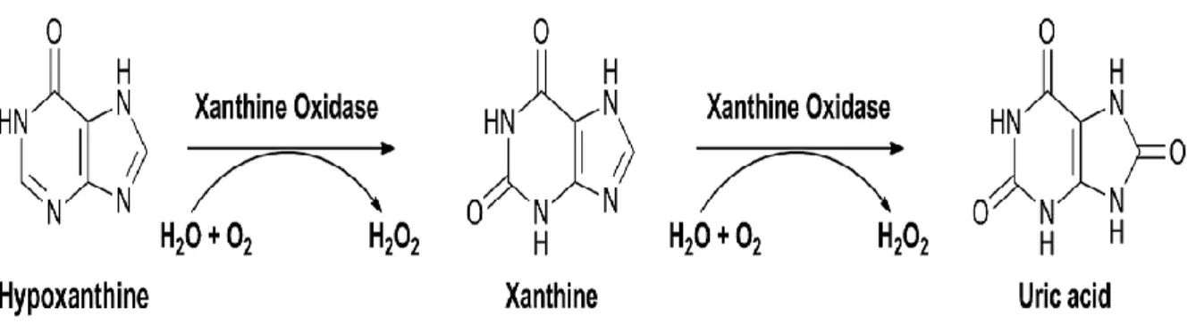
Xanthine oxidoreductase (XOR) is another important enzymatic source of ROS which belongs to metallofl avoprotein family. XOR (EC 1.17.1.4) catalyzes the oxidation of hypoxanthine and xanthine to form uric acid. XOR is shown to exist in two forms: xanthine oxidase (XO) and xanthine dehydrogenase (XDH). The enzyme catalyzes the reduction of O2, leading to the formation of
superoxide (O2•−) and H2O2 ; it is proposed as a central mechanism of oxidative injury (Nishino et al.,
14
Fig. 3: Principle reaction catalyzed by xanthine oxidase (XO) (Rodrigues et al., 2015).
2.Exogenous sources
Free radicals naturally occur in the body as a result of chemical reactions during normal cellular processes. They can also be formed in response to environmental factors such as excess pollution, excessive UV rays, and exposure to cigarette smoke, automo- bile exhaust, and pesticides. Inadequate rest or sleep, inability to manage stress responses, and unhealthy eating habits can also cause free radical damage (Obohb, 2004; Oboha, 2005).
I.1.2.3. ROSs and cellular damage
ROSs are known to damage cellular membranes by inducing lipid peroxidation (Ramadevi and Prasad, 1998). They also can damage DNA, proteins, lipids and chlorophyll (Mittova et
al., 2000). Free radical damage is one of the most prominent causes of devastating diseases
that are responsible for killing many people in the world, such as cardiovascular disease, which can manifest as heart attacks, and cancer (Amic et al., 2003). The aging process has been linked by some researchers to free radical damage in the body (Oboha, 2005). Also,
oxidative stress plays a major role in the development of chronic and degenerative ailments such as cancer, auto-immune disorders, rheumatoid arthritis, cataracts, aging, cardiovascular, and neurodegenerative diseases (Willcox et al., 2004; Pham-Huy et al., 2008).
15
1. Oxidative damage to proteins
Oxidative damage to proteins results in modifications of site-specific amino acid, fragmentation of the peptide chain, aggregation of cross-linked reaction products, altered electrical charge and increased susceptibility to proteolysis ((Farr and Kogama, 1991). Also, oxidative modification of enzymes can have either mild or severe effects on cellular or systemic metabolism, depending on the percentage of molecules that are modified and the chronicity of the modification. Several groups have demonstrated that certain enzymes become oxidatively modified during aging (Oliver et al., 1987). Modification of structural proteins can also lead to a loss of function. For example, when the plasma protein fibrinogen is oxidized either by treatment with an iron/ascorbate radical-generating system or with ionizing radiation, it loses its ability to form a solid clot (Shacter et al., 1995).
2. Oxidative damage to lipids
Formation of peroxides, especially lipid ones, is a consequence of the activation of O2, the
interconversion of reactive species and natural systems protection overcoming. In biological environments, the most favorable substrate for peroxidation is represented by polyunsaturated fatty acids (PUFA), components of cell and subcellular membranes. Peroxidation is a complex process that includes three phases: initiation, propagation, end-decomposition, which interpose, so that only end products can be determined chemically: aldehydes (malondialdehyde), polymerized carbonyl compounds (lipofuscin) (Holley and Cheesman, 1993).
3. Oxidative damage to DNA
Although DNA is a stable and well-protected molecule, ROS can interact with it and cause several types of damage: modification of DNA bases, single- and double-DNA breaks, loss of purines (apurinic sites), damage to the deoxyribose sugar, DNA-protein cross-linkage, and damage to the DNA repair system (Kohen and Nyska, 2002). Of the ROS, the highly reactive
16
OH reacts with DNA by addition to double bonds of DNA bases and by abstraction of an H atom from the methyl group of thymine and each of the CH bonds of 2’-deoxyribose. Addition of OH to the C5-C6 double bond of pyrimidines leads to C5-OH and C6-OH adduct radicals of cytosine and thymine and H atom abstraction from thymine results in the allyl radical. Adduct radicals differ in terms of their redox properties, C5-OH- and C6-OH-adduct radicals of pyrimidines possess reducing and oxidising properties, respectively (Sonntag, 1987, Cooke et al., 2003).
I.1.3. Antioxidant defense system
I.1.3.1. Introduction
Antioxidants are powerful free radical scavengers in the body, while free radicals are highly reactive chemical substances that travel around in the body and cause damage to body cells (Alia et al., 2003). So, antioxidants are the substances that may protect cells from the damage caused by free radicals. Antioxidants interact with and stabilize free radicals and may prevent some of the damage that free radicals might otherwise cause. However, these antioxidants whenever are consumed in large doses can act as prooxidants (Seifried et al., 2007). The antioxidants depending on their source of availability can be endogenous or exogenous in nature. The endogenous antioxidants can either be enzymatic, like superoxide dismutase (SOD), catalase (CAT), glutathione peroxidase (GPx), and glutathione reductase (GRx) (Mates, 2000), or nonenzymatic in nature. The nonenzymatic antioxidants can be further grouped to metabolic antioxidants, such as lipoic acid, glutathione, L-arginine, uric acid, bilirubin (Kohen and Nyska, 2002), and nutrient antioxidants. Some of the nutrient antioxidant can be exogenous in nature as they cannot be produced in the body and must be provided through foods such as vitamin E, vitamin C, carotenoids, and trace elements (Se, Cu, Zn, Mn) (Willett and Macmahon, 1984; Radimer et al., 2004).
17
I.1.3.2. Sources of antioxidants
I.1.2.3.1. Endogenous sources 1. Enzymatic antioxidant
The most important antioxidant enzymes are: superoxide dismutase (SOD Ec 1.15.1.1), catalase (CAT Ec 1.11.1.6), ascorbate peroxidase (apX Ec 1.11.1.11), monodehydroascorbate reductase (MDAR Ec 1.1.5.4), dehydroacscorbate reductase (DHAR EC 1.8.5.1) and glutathione reductase (GR EC 1.6.4.2). At least four of them participate in a highly developed detoxification system named the ascorbateglutathione cycle (halliwell-asada cycle) (knÖrzer
et al., 1996 ; Mittova et al., 2000 ; Morabito and Guerrier, 2000).
Fig.4 : Detoxification of ROS via glutathione-ascorbate cycle (Saruhan et al., 2009) ASC: Ascorbate; APX: Ascorbate peroxidase; GSH Reduced glutathione; GSSG: Oxidized glutathione; GR: Glutathione reductase; DHA Dehydroascorbate; DHAR: Dehydroascorbale reductase; MDHA: Monodehydroascorbale MDHAR: Monodehyroascorbale reductase.
Manganese superoxide dismutase (MnSOD)
Manganese superoxide dismutase (MnSOD, also called SOD2) is a homotetrameric enzyme located in the mitochondrial matrix near the electron transport chain (Fridovich, 1998).Or Manganese superoxide dismutase is a ubiquitous metalloenzyme found in virtually all aerobic organisms from bacteria to humans, and even anaerobes (Ravindranath and Fridovich .,
18
1975). Manganese superoxide dismutase is uniformly distributed throughout the cytoplasm in procaryotic cells (Steinman et al., 1994). So, Manganese superoxide dismutase (MnSOD) is a very important antioxidant enzyme that catalyzes the conversion of superoxide radicals (O2•-)
to hydrogen peroxide and molecular oxygen in the mitochondria (Weisiger and Fridovich, 1973). Under normal physiological conditions, mitochondria are the major source of O2
•-production. Numerous studies have indicated that MnSOD plays an important role in preventing cells from oxidative stress and inhibiting tumorigenicity (Oberley and Buettner, 1979).
Catalase
Catalase is a common enzyme found in nearly all living organisms exposed to oxygen. It catalyzes the decomposition of hydrogen peroxide to water and oxygen (Chelikani et al., 2004). It is a very important enzyme in reproductive reactions. Likewise, catalase has one of the highest turnover numbers of all enzymes; one catalase molecule can convert millions of molecules of hydrogen peroxide to water and oxygen each second (Goodsell, 2004).Catalase can also catalyze the oxidation, by hydrogen peroxide, of various metabolites and toxins, including formaldehyde, formic acid, phenols, acetaldehyde and alcohols. It does so according to the following reaction: H2O2 + H2R → 2H2O + R
The exact mechanism of this reaction is not known. (Murthy et al., 1981).
2. Nonenzymatic antioxidant
Glutathione as an antioxidant
The most important endogenous antioxidant defence systems are composed of the thiol-containing tripeptide glutathione and small thiol-thiol-containing proteins, such as thioredoxin, glutaredoxin, and peroxiredoxin. Of these, glutathione is found at millimolar concentrations in most cells and is the major contributor to the cell’s redox state. Glutathione occurs in cells in
19
both reduced (GSH) and oxidized (GSSG) forms. It may also covalently bind to proteins through glutathionylation (Thomas et al., 1995 ; Huang and Huang, 2002). One important task of cellular glutathione is to scavenge free radicals and peroxides produced during normal cellular respiration, which would otherwise oxidize proteins, lipids and nucleic acids (Hayes and Pulford, 1995 ; Wild and Mulcahy, 2000). GSH reacts non-enzymatically with superoxide, nitric oxide, hydroxyl radical, and peroxynitrite as an antioxidant (Aoyama et al., 2008).
N-acetylcysteine
N- acetylcysteine (NAC) is a metabolite of the sulphur-containing amino acid cysteine. It has the molecular formula HSCH2CH (NHCOCH3) CO2H and formula weight 163.19. In humans
it can be administered orally or by intravenous infusion and can also be inhaled using a nebuliser. NAC exhibits direct and indirect antioxidant properties. Its free thiol group is capable of interacting with the electrophilic groups of ROS (Moldeus et al., 1986 ; Aruoma et
al.,1989). This interaction with ROS leads to intermediate formation of NAC thiol, with NAC
disulphide as a major end product (Cotgreave, 1997). In addition, NAC exerts an indirect antioxidant effect related to its role as a GSH precursor. GSH is a tripeptide made up of glutamic acid, cysteine and glycine. It serves as a central factor in protecting against internal toxic agents (such as cellular aerobic respiration and metabolism of phagocytes) and external agents (such as NO, sulphur oxide and other components of cigarette smoke, and pollution). The sulphydryl group of cysteine neutralises these agents (Moldeus et al., 1986) .
N-acetylcysteine (NAC) is a promising compound to increase GSH synthesis in the brain. NAC stimulates GSH synthesis not only by providing a source of cysteine, but also by activating GR (De Flora et al., 1984). N-acetyl-cysteine (NAC) is an acetylated cysteine residue. An optimal thiol redox state has been demonstrated to be of primary importance if
20
attempting to optimize the protective ability of the cell to oxidative stress. Among the most widely used agents to maintain the cysteine pool is NAC in addition to α-lipoic acid. While other agents have been used, NAC and α-lipoic acid are the most commonly utilized and discussed as a result of their proven safety and efficacy. In addition to the role glutathione and other thiols have on maintaining the cellular redox state, many studies have begun to explore if NAC supplementation can actually improve performance due to its ability to promote a more favorable cellular environment to achieve higher levels of performance (Sen, 2001).
Uric Acid
Uric acid (UA) is an important antioxidant in blood. UA scavenges singlet oxygen, hydroxyl radicals, and peroxynitrite in blood at its physiological concentration (Ames et al., 1981). Uric acid is a final enzymatic product in the degradation of purine nucleosides and free bases in humans. The pathway of purine catabolism in humans is shortest among vertebrates because about 8–20 million years ago during primate evolution the activity of urate oxidase (uricase, an enzyme catalyzing conversion of uric acid to allantoin) was lost in a two-step mutation process (Wu et al., 1992; Oda et al., 2002). In other mammals, the last enzymatic product of purine degradation chain is allantoin, which is excreted in the urine. Lower vertebrates (e.g., fish) have enzymes that further degrade allantoin to allantoic acid and glyoxylic acid and finally to urea. As a consequence, humans have to cope with relatively higher levels of uric acid in the blood (200–400 μM) and are prone to hyperuricemia and gout (Johnson et al., 2005).
I.1.2.3.2. exegenous sources
Vitamins
Ascorbate (Vitamin C) and α-tocopherol (Vitamin E) are also important antioxidants in the brain (Perry et al., 1985; Gilgun-Sherki et al., 2001). The concentration of ascorbate in the
21
human brain ranges from 1 to 2.6 mM (Grunewald, 1993), similar to that the concentration of GSH in the brain. However, humans cannot produce ascorbate and the BBB almost completely blocks ascorbate penetration into the brain (Agus, 1997). Alpha-tocopherol is the most potent antioxidant in the lipid part of the biological membrane (Gilgun-Sherki et al., 2001). However, the α-tocopherol level in the brain is relatively lower than those of ascorbate and GSH (Metcalfe et al., 1989; Grunewald, 1993; Cooper and Kristal, 1997; Gilgun-Sherki
et al., 2001). In addition, the oral administration of α-tocopherol did not increase its
concentration in the central nervous system (CNS) due to its limited penetration of the BBB (Pappert et al.,1996).
Plants phenolic compounds
Plants produce a great variety of organic compounds as a response to environmental stresses like microbial attack, insect/animal predation and ultraviolet radiations. The role of these metabolites is to increase plants resistance to these stresses. They can be classified into three major groups according to their biosynthetic route and structural features: terpenoids, alkaloids, and phenolic compounds.
1. Phenolic acids
All plants produce an amazing diversity of secondary metabolites. One of the most important groups of these metabolites are phenolic acid. Phenolics are characterized by at least one aromatic ring (C6) bearing one or more hydroxyl groups. They are mainly synthetized from cinnamic acid, which is formed from phenylalanine by the action of l-phenyloalanine ammonia-lyase PAL (Ec 4.3.1.5), the branch point enzyme between primary (shikimate pathway) and secondary (phenylopropanoid) metabolism (Dixon and Paiva, 1995). The significance of this route can be supported by the fact that, in normal growth conditions, 20% of carbon fixed by plants flows through this pathway (Diàz et al., 2001).
22
Phenols are divided into several different groups, distinguished by the number of constitutive carbon atoms in conjunction with the structure of the basic phenolic skeleton (simple phenols, benzoic acids, phenylopropanoids and flavonoids) (Harborne, 1964; Rice-Evans et al.,1997; Chaudiere and Ferrari-iliou, 1999 ;).
Hydroxybenzoic Acids Hydroxycinnamic Acids Fig.5 : Structures of phenolic acids (Harborne, 1986).
1.1. Flavonoids
Flavonoids and ubiquinol are considered potential antioxidants, but their role is less certain (Halliwell, 1991). So, flavonoids are secondary plant metabolites, which together with other plant phenols share a common origin : the amino acid phenylalanine (Parr and Bolwell, 2000).
These phenols are derived from a common building block in their carbon skeleton : the phenylpropanoid unit, C6-C3. Biosynthesis according to this pathway produces the large variety of plant phenols: cinnamic acids (C6-C3), benzoic acids (C6-C3, or C6-C1), flavonoids(C6-C3-C6), proanthocyanidins (C6-C3-C6)n, stilbenes(C6-C2-C6), coumarins (C6-C3), lignans (C6-C3-C 3-C6), and lignins (C6-C3)n. Within each family of plant phenols many compounds may exist. Thousand different flavonoids have been described as occurring in plants (Harborne and Baxter, 1999).
23
Fig.6 : Subclasses of flavonoids. Classification is based on variations in the heterocyclic C-ring (Harborne and Baxter, 1999).
1.2. Polyphenols
polyphenols are classified into :
1.2.1. Proanthocyanidin derivatives
These are oligomers containing two to six units of flavan-3-ol or high molecular weight polymer of flavan-3-ol (Haslam, 1998).
1.2.2. Galloyl and hexahydroxydiphenyl ester derivatives
In this class, different gallic and hexadiphenic acid derivatives are present as esters of a polyol (usually D-glucose) at the core of the polyphenolic ester (Haslam, 1998).
1.2.3. Hydroxy cinnamic acid derivatives
These are the condensation oligomers of mono lignols, namely, p-coumaryl alcoholconiferyl alcohol and sinapyl alcohol (Tuckmantel et al., 1999 ; Dean, 2001; Elias,1995). Lignin is one
24
such example formed from the oxidative polymerization of coniferyl alcohol. Lignin is a reticulated polyphenol, having three main functions in plants. They provide mechanical support, play role in conduction of water and also provide protection against biodegradation in plants(Haslam, 1998).
1.2.4. Phloroglucinol derivatives
These oligomers are derived from phloroglucinol subunits. They are formed by oxidative C–C and C–O coupling reactions of phloroglucinol (Haslam, 1998).
1.3. Tannins
Many tannins can be fractionated hydrolytically into their components, for example by treatment with hot water or with tannases, led to the classification of such tannins as ‘hydrolysable tannins’. Non-hydrolysable oligomeric and polymeric proanthocyanidins were
classified as condensed tannins (Würdig and Woller, 1989).
1.3.1. Hydrolysable tannins
the term ‘hydrolysable tannins’ includes both the gallotannins and the ellagitannins (Kashiwada et al., 1992; Weinges and Plieninger, 1999). It should also be mentioned here that there are ellagitannins that are not hydrolysable, because of a further C–C coupling of their polyphenolic residue with the polyol unit, but are nevertheless for historical reasons classified as hydrolysable tannins (Yoshida et al., 1999).
1.3.2. Complex tannins
In 1985 the first tannins were described that contained, in addition to the hexahydroxydiphenoyl (HHDP) units (the characteristic structural element of the monomeric ellagitannins), also C-glycosidic catechin units (Nonaka et al., 1985; Miyamoto et al., 1987) .These tannins were originally classified as ‘non-classified tannins’, because they are only
25
partially hydrolysable due to the C–C coupling of their catechin unit with the glycosidic part (Nishimura et al.,1986). To properly place these ‘non-classified tannins’ in some scheme, the terms ‘complex tannins’ (Haslam, 1989 ; Porter, 1989; Kashiwada et al., 1992). and flavanoellagitannins (Kashiwada et al., 1992; Ferreira and Bekker, 1996) were established over the following years. These examples clearly show that the division of the tannins into two groups, hydrolysable and non-hydrolysable or condensed tannins, (Würdig and Woller, 1989; Griffiths, 1991). cannot do justice to the structural diversity of the tannins.
1.3.3. Condensed tannins
The terms ‘flavanotannins’ or ‘condensed flavanoid tanning substances’ (Weinges and
Plieninger, 1999). that are occasionally found in the literature denote tannins consisting of catechin units. The polymeric flavanotannins, constructed from coupled flavan-3-ol (catechin) units, belong to the condensed tannins (oligomeric and polymeric proanthocyanidins). On the basis of their structural characteristics it is therefore possible to divide the tannins into four major groups: Gallotannins, ellagitannins, complex tannins, and condensed tannins.
(1) Gallotannins are all those tannins in which galloyl units or their meta-depsidic derivatives are bound to diverse polyol-, catechin-, or triterpenoid units.
(2) Ellagitannins are those tannins in which at least two galloyl units are C–C coupled to each other, and do not contain a glycosidically linked catechin unit.
(3) Complex tannins are tannins in which a catechin unit is bound glycosidically to a gallotannin or an ellagitannin unit.
(4) Condensed tannins are all oligomeric and polymeric proanthocyanidins formed by linkage of C-4 of one catechin with C-8 or C-6 of the next monomeric catechin.
26
I.1.3.3. Antioxidant effects of phenolic compounds
Antioxidants may act as physical barriers to prevent ROS generation or ROS access to important biological sites (UV filters, cell membranes); chemical traps/sinks that “absorb” energy and electrons, quenching ROS (carotenoids, anthocyanidins); catalytic systems that neutralize or divert ROS [antioxidant enzymes SOD (superoxide dismutase), catalase, and glutathione peroxidase]; binding/inactivation of metal ions to prevent generation of ROS (ferritin, ceruloplasmin, catechins); and chain-breaking antioxidants which scavenge and destroy ROS (ascorbic acid, tocopherols, uric acid, glutathione, flavonoids) (Karadag et al., 2009).
I.1.3.3.1. Antioxidant activity of flavonoids
The most described property of flavonoids is their capacity to protect the organism against free radicals and oxygenated reactive species (ORS) produced during the metabolism of oxygen.The protective effect of flavonoids is due to several mechanisms such as free radicals trapping, enzymes inhibition and metallic ions chelation. These properties depend on the structure of the flavonoids and the degree of substitution and saturation (Grace, 1994).
*Free radicals trapping
The flavonoids can prevent the damage caused by the free radicals according to various ways; one of them is the direct trapping of the radicals. In this case, the flavonoids are oxidized by the radicals (R) leading to less reactive and more stable species according to the following mechanisms (Halliwell, 1995):
Flavonoid (OH)+ R Flavonoid (O ) + RH
The formed flavonoxy radical (flavonoid (O )) is stabilized by resonance. The non-paired electron can be delocalized on the whole of the aromatic cycle. But, it can continue to evolve