NOTE TO USERS
Page(s) not included in the original manuscript and are unavailable from the author or university. The manuscript
was scanned as received.
ii
This reproduction is the best copy available.
®
UMI
Université de Sherbrooke
Integrin a.6AP4 in colon cancer by
Anders Bondo DYDENSBORG
Département d'anatomie et de biologie cellulaire
Thesis presented to the Faculté de médecine et des sciences de la santé in partial fulfillment of the requirements for a Doctor of Philosophy (Ph.D.) degree in Cell
Biology, Université de Sherbrooke, Sherbrooke, Qc, Canada.
Thesis committee:
1. Dr. Jean-Francois BEAULIEU, Département d'anatomit>-biologie cellulaire, Université de Sherbrooke
2. Dr. Jacques HUOT, Département de médecine, Université Laval 3. Dr. Klaus KLARSKOV, Département de pharmacologie, Université de
Sherbrooke
4. Dre. Nathalie RIV ARD, Département d'anatomie-biologie cellulaire, Université de Sherbrooke.
l+I
Library andArchives Canada Archives Canada Bibliothèque et Published Heritage
Branch Direction du Patrimoine de l'édition 395 Wellington Street
Ottawa ON K1A ON4 Canada
395, rue Wellington Ottawa ON K1A ON4 Canada
NOTICE:
The author has granted a non-exclusive license allowing Library and Archives Canada to reproduce, publish, archive, preserve, conserve, communicate to the public by
telecommunication or on the Internet, loan, distribute and sell theses
worldwide, for commercial or non-commercial purposes, in microform, paper, electronic and/or any other formats.
The author retains copyright ownership and moral rights in this thesis. Neither the thesis nor substantial extracts from it may be printed or otherwise reproduced without the author's permission.
ln compliance with the Canadian Privacy Act some supporting forms may have been removed from this thesis.
While these forms may be included in the document page count,
their removal does not represent any loss of content from the
AVIS:
Your file Votre référence ISBN: 978-0-494-28270-0 Our file Notre référence ISBN: 978-0-494-28270-0
L'auteur a accordé une licence non exclusive permettant
à
la Bibliothèque et Archives Canada de reproduire, publier, archiver,sauvegarder, conserver, transmettre au public par télécommunication ou par l'Internet, prêter, distribuer et vendre des thèses partout dans le monde, à des fins commerciales ou autres, sur support microforme, papier, électronique et/ou autres formats.
L'auteur conserve la propriété du droit d'auteur et des droits moraux qui protège cette thèse. Ni la thèse ni des extraits substantiels de celle-ci ne doivent être imprimés ou autrement reproduits sans son autorisation.
Conformément
à
la loi canadienne sur la protection de la vie privée, quelques formulaires secondaires ont été enlevés de cette thèse. Bien que ces formulaires aient inclus dans la pagination, il n'y aura aucun contenu manquant.Publications
Results from this Ph. D. thesis has been published as articles in the following peer-reviewed joumals:
Ni, H*, Dydensborg, A.B.*, Herring, F.E., Basora, N., Gagné, D., Vachon, P.H., Beaulieu, J-F.
Upregulation of a functional form of the
p4
integrin subunit in colorectal cancers correlates with c-Myc expression. Oncogene, 2005 24:6820-6829.*: Contributed equally to this work.
Dydensborg, A.B., Herring, F.E, Auclair, J., Tremblay, E., Beaulieu, J-F.
Normalizing Genes for Quantitative RT-PCR in Differentiating Human Intestinal Epithelial Cells and Adenocarcinomas of the Colon. American J. of Physiology-GL. 2006 May;290(5):G 1067-74.
Financial Support
1 received studentships from
1. Knud H0jgaards Fond-Grant# 32.739
Table of content
L . lS t o if~' c igures ... v111
...
List of tables ... x
Abbreviations and Glossary ... xi
Rés
umi ...•...••.••.... xiiiSummary ... xvi
I. Introduction ... 1
1.1 The integrin family and the basement membrane ... 1
1.1. l lntegrin functions and structure ... 1
1.2 Integrins a6P 1 and a6P4 ... 4
1.2.1 The a6A and a6B integrin subunits - Structure and ontogeny ... 6
1.2.2 The a6A and a6B integrin subunits - Biochemical differences ... 8
1.2.3 lntegrin a6A and a6B - Alterations of expression ratio ... 10
1.2.4 The a6-containing integrins in carcinomas ... 11
1.2.5 The integrin p4 subunit - Stucture and hemidesmosomes ... 11
1.2.6 lntegrin a6P4 - Functions in normal tissue development ... 14
1.2. 7 a6P4 in carcinoma biology - Angiogenesis, survival & migration ... 15
1.2.8 Carcinoma cell migration, invasion and mobiliz.ation of a6j34 from hemidesmosomes ... 17
1.2.9 a6j34, Receptor tyrosine kinases and lipid rafts ... 18
1.3 The intestine •••••..•.•••••••..•••••.•.••.•••.••••.••.••.••••.•••.•...••...•.•.•••.•••.•••.•..•••••••••••••••••••. 24
1.3.1 The small intestine ... 24
1.3.2 The colon ... 26
1.3.3 lntegrin a6~4 in the small and large intestine ... 26
1.3.4 Integrin a6~4 in colon cancer ... 27
1.4 Normalizing genes for use in mRNA quantification studies •••.•...•..•••.•... 28
1.5 Aim of studies ••••.•.••••••.••••••..•••••••••.••••••••••••••••••.••••••••.•.••••••••.•••..••...•....••••.•..•.•.. 30
IL Materials and Methods .•.••....••..••...•..•.•.•••••••.•••••.•.••••••.•.•••••••.•.••.••••••••••.•••.•••••.••. 32
11.1 Tissues ...•..••.•.•....••...•...•..•...•...•...••.•...•...•.•.•...•.•••••.•• 32
11.2 Antibodies •.••.••.•.•.•...••.•..••...•.•...•...•...•...•....•...•....• 32
11.3 Indirect immunofluorescence •.•..••...•.•••...••..•..•...•..•....•...•.•.•. 33
11.4 Western Blotting ...•...•...•..•.•..•..•..••.•.•.•.•••••.•....•.••...•...•.•....•..•.•••••... 34
11.5 Plasmids and plasmid construction ... 35
11.6 Cell culture .•••.••...•...•.••...•.•...•...••.•.•••.•..••.••...•...•.•..•..••....•.••.••.••••• 3 7 II. 7 cDNA generation and RT-PCR ..••...•.•.•.••..••..•...•..•....•..••..•....•..•..••••. 38
11.8 Real-time quantitative RT-PCR •.••.•.••••••.•.•..••...•...•...•.••...•.•..•.•....••.. 39
11.9 Transfection and luciferase measurement ••...•..••...•...•••...•.. 43
11.10 Data and statistical analyses ..•.••.•....••..•.••••••...•...••.••.•.•.•.•...•...•.•••...•..•..•. 44
111.1.
Investigations of transcript level of MYC and 134 in adenocarcinomas ofthe human colon and their healthy resection margins .•..•..•.•..•.•...•.•...•••..•...• 46
111.1.1. Real-time quantitative RT-PCR analysis of expression levels of transcript ... 46
111.1.2 Correlation between the transcript levels of 134 and MYC ... 48
111.1.3 Functional relationship between MYC and the integrin 134 promoter ... 48
111.1.4 Molecular cloning of MYC and generation of the control 8.MYC ... 50
111.1.5 Response to over expression of MYC in a colon cancer cell line ... 52
111.2 The integrin a.6 splice variants are differentially expressed in the small intestine and a.6A is-upregulated in colon adenocarcinomas and stimulates transcriptional activity of the J3-catenintrCF4 complex ... 54
111.2.1 Differentiation-dependent expression of integrin a.6 splice variants in intestinal cell models ... 54
111.2.2 The A and B variants of the integrin a.6 subunits are differentially expressed along the intestinal crypt-villus axis ... 58
111.2.3 Molecular cloning of the two splice variants of the integrin a6 subunit . 61 111.2.4 The A variant of the integrina6 subunit activates the 13-catenin/TCF4 and MYC transcriptional complexes ... 61
111.2.5 lntegrin a6 is up regulated and undergoes a variant shift in adenocarcinomas of the human colon ... 62
111.3 Identification and validation of nonnalizing genes for usage in mRNA measurements in differentiating human enterocytes and adenocarcinomas of
the human colon ... 67
111.3 .1 Differentiation of hwnan enterocytes ... 67
111.3.2 Differentiation of the intestinal epithelial cell line Caco-2/15 ... 74
111.3.3 Healthy colonie tissue vs. adenocarcinomas of the colon ... 75
IV. Discussion ... 78
IV.l Expression level of (34 and MYC transcripts in carcinomas of the human eolon ... 78
IV.2 a6A and a6B in healthy intestinal cells and adenocarcinomas of the human colon ... 82
IV .3 Role of a6~4 in colon cancer ... 92
IV.4 ldentifying and validating nonnalizing genes for mRNA quantification purposes during the differentiation process of human enterocytes and studies of adenocarcinomas of the human colon ... 94
V. Conclusion and Perspectives ... 99
Acknowledgements ... 103
Ref erence List ... 104
List of Figures
Figure 1: The integrin family ... 3
Figure 2: Integrin activation ... 5
Figure 3: Pre-mRNA splice variants of cx6 ... 7
Figure 4: Hemidesmosomes- Molecular components ... 13
Figure 5: Intracellular domains of J34 ... 19
Figure 6: Intracellular signaling initiated by J34 ... 23
Figure 7: The small intestinal crypt-villus axis ... 25
Figure 8: Up regulation of expression of J34 in colon cancers ... 4 7 Figure 9: Correlation between transcript levels ofMYC and J34 ... .49
Figure 10: M1YC compared to the full length members of the MYC/Max network ... 51
Figure 11: Activation of the J34 promoter by MYC ... 53
Figure 12: ln vitro expression pattern of the two variants of cx6-RT-PCR ... .56
Figure 13: ln vitro expression pattern of the two variants of cx6 -western blots ... 57
Figure 14: ln vivo expression pattern of total cx6, cx6A & cx6B ... 59
Figure 15: ln vivo crypt expression of cx6A & cx6B ... 60
Figure 16 : Impact on cellular functions due to over expression of the A & B variants of cx6 ... 63
Figure 17: Expression level of total cx6, cx6A and cx6B in colon cancer ... 64
Figure 18: Differential expression of integrin cx6 splice variants - human colon cancer cell lines ... 66
Figure 20: Identification of stably expressed normalizing genes- Differentiation .... 70 Figure 21: Identification of stably expressed normalizing genes- Tumors vs. healthy tissue ... 76
List of tables
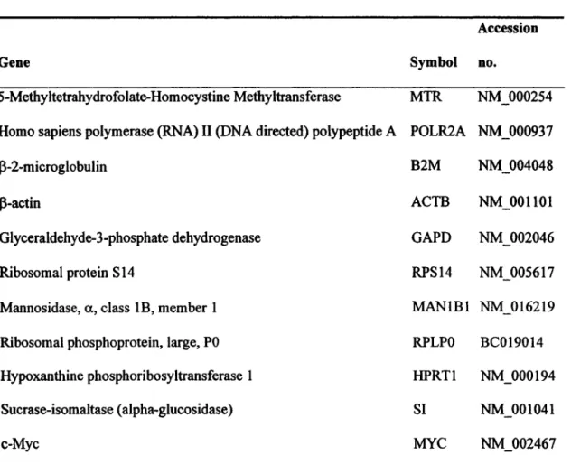
Table 1: Gene names, Symbols and Accession numbers of the genes investigated in relation to normalizing gene identification studies ... .40 Table 2: List of primers used in studies related to a6A, a6B and ~4 ... .41
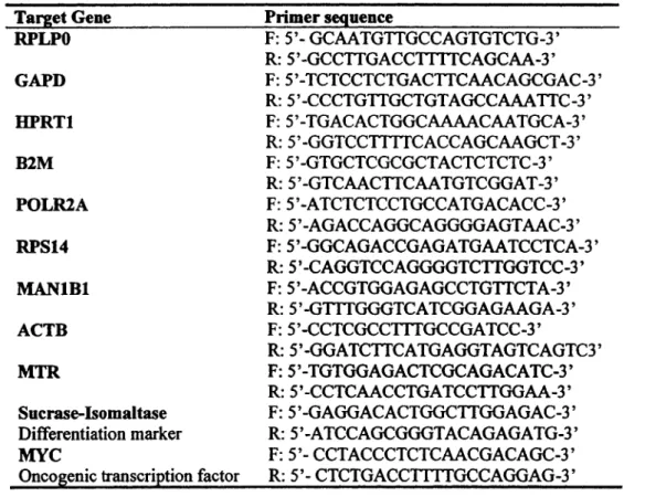
Table 3: List of primers used for identification and validation ofnormalizing genes for studies related to differentiation of enterocytes and adenocarcinoma formation ... .42 Table 4: a6A/a6B ratio modulation in colon cancer ... 64 Table 5: Integrin a6A/a6B transcript ratio ... 66 Table 6: Regression and statistical data of the nine candidate genes during enterocytic differentiation ... 71
Abbreviations and Glossary
a6J34: The human <l6J34 integrin. a6: The human integrin a6 subunit.
(34: The human integrin
f34
subunit. aa: Amino acid.ACt: The difference in C.-value between two given samples.
ALPI: Intestinal Alkaline Phosphatase. Differentiation marker of absorptive enterocytes.
Anoikis: Detachment-induced apoptosis.
bFGF: Fibroblastic Growth Factor 2 (basic). The prototypical member of the FGF-family.
BM: Basement Membrane - sheet of extra cellular matrix proteins upon which epithelial cells resides.
Bp: Base Pair.
cDNA: copy DNA - a DNA strand copied from mRNA (messenger RNA) using reverse transcriptase.
Crvalue: The number of thermal cycles completed to reach a defined threshold of amplification. Inversely related to input cDNA amount.
DPPIV: Dipeptidylpeptidase IV. Differentiation marker of absorptive enterocytes. E-Box: DNA sequence recognized and bound by MYC (Canonical sequence: S' -CA(Cff)GTG-3 ').
Erk: Extracellular receptor kinase - Second messenger kinase of the MAPK family. Part of the mitogenic/proliferative signal transduction pathway.
GOI: Gene Oflnterest.
JNK: Jun Kinase - kinase of the Serffhr protein kinase family.
LPH: Lactase-Phlorizin Hydrolase. Differentiation marker of absorptive enterocytes. MYC: The human c-Myc protein.
PI3-K: Phosphatidylinositol 3'-kinase.
PMA: Phorbol 12-Myristate 13-Acetate. Phorbol ester. Activates protein kinase C (PKC), which subsequently stimulates integrin affinity for the extracellular ligand.
qRT-PCR: Quantitative reverse transcriptase-polymerase chain reaction. Typically performed by real-time monitoring of polymerase chain reaction product fonnation.
RM: Resection margin - the healthy tissue surrounding a tumor. Used as a endogenous control when alterations of gene expression in the primary tumor is investigated.
SI: Sucrase-Isomaltase. Differentiation marker of absorptive enterocytes.
TCF4: T cell factor 4-transcription factor. Is co-activated by (3-catenin.
VEGF:
Vascular endothelial growth factor - Cytokine that mediates numerous functions of endothelial cells. Critical for angiogenesis.L'intégrine a.6Af34 dans le cancer colorectal
Par
Anders Hondo DYDENSBORG
Thèse présentée à la Faculté de médecine et des sciences de la santé en vue de l'obtention du titre de docteur en biologie cellulaire (Ph.D), Université de Sherbrooke,
Sherbrooke, Qc, Canada
Résumé
Les niveaux d'expression de l'intégrine a.6P4 sont reconnus pour être élevés dans un certain nombre de cancers humains. Dans ce contexte pathologique, cette intégrine exerce un rôle stimulateur à plusieurs niveaux de la progression tumorale, tels la promotion de l'invasion et l'activation de voies de signalisation intracellulaires associées à la prolifération et la survie cellulaire. Dans ce travail, les niveaux d'expression de la sous-unité intégrine
P4
dans des adénocarcinomes de colon humain ont été évalués par quantification de l' ARN messager, en comparaison avec les niveaux d'expression deP4
dans les régions saines du colon de chaque patient. Non seulement le messager encodantf34
se trouve surexprimé dans les tissus cancéreux, mais les niveaux d'expression deP4
corrèlent aussi avec l'expression du facteur de transcription oncogénique MYC. Cette corrélation suggère une importance fonctionnelle pour les niveaux d'expression de la sous-unitéP4
compte tenu que lasurexpression de MYC dans une lignée cellulaire de cancer du colon stimule l'activité transcriptionelle d'une variété de constructions du promoteur de p4. Le pré-ARNm de la sous unité intégrine a.6 subit un épissage alternatif conduisant à deux variants différents, soit a6A et a6B. Ceux-ci diffèrent par leurs domaines intracellulaires et leurs propriétés biochimiques. Afin de caractériser le profil d'expression de l'intégrine a6p4 dans les cancers du colon, les niveaux totaux d'expression de a.6 ainsi que ceux des deux variants d'épissage alternatif furent étudiés. Tout comme dans le cas de la sous-unité p4, les niveaux totaux de a.6 sont augmentés dans les cas de cancers comparativement aux marges de résection correspondantes. De manière intéressante, une altération dans les ratios d'expression des deux variants de a.6 a été remarquée dans 81 % des cancers étudiés. Ainsi, ces ratios passent d'une expression prédominante de a6B dans les marges de résection saines, à une expression prédominante de a6A dans les adénocarcinomes primaires. Une telle expression différentielle des deux variants d'épissage alternatif est aussi retrouvée dans l'intestin grêle humain normal. En effet, les cellules cryptales prolifératives expriment le variant a6A, tandis que le compartiment différencié de l'axe crypte-villosité exprime plutôt le variant a6B. L'importance fonctionnelle de ces observations est confirmée par la surexpression des deux variants de a.6, conduisant à une activation différentielle des complexes transcriptionnels P-caténine!fCF4 et MYC, lesquels sont des joueurs importants dans la prolifération des cellules intestinales et la formation des adénocarcinomes. D'autre part, les deux variants de a.6 ne semblent pas affecter la différenciation entérocytaire chez l'humain, tel qu'observé par des mesures de
phlorizine hydrolase, de DPPIV et de la phosphatase alcaline. Finalement, il est de plus en plus clair que les gènes classiques de normalisation utilisés comme référence quantitative, tels que ceux du GAPD et de ~-actine, peuvent présenter une modulation de leur expression à travers divers tissus et au cours de la différenciation cellulaire. Ainsi, des expériences visant à identifier et à valider des gènes de référence utilisés lors de la quantification d' ARN messagers durant les processus cellulaires de différenciation et de progression tumorale au niveau du tube digestif humain, ont été effectuées. Précédemment, des candidats potentiels ont été obtenus par micro-puces d' ADN. Du nombre, deux d'entre eux ont été retenus en parallèle avec 7 gènes de référence couramment utilisés. Des essais RT-PCR en temps réel ont été effectués pour ces gènes, avec des échantillons à différentes étapes de la différenciation entérocytaire humaine, et avec différentes tumeurs primaires et leur marge de résection correspondante. Par des approches statistiques, RPLPO et B2M ont été identifiés comme étant les deux meilleurs gènes de référence à utiliser pour la normalisation de la quantité d' ARN messagers lors d'études faites dans les contextes de différenciation et de cancer respectivement. Ces travaux pourraient servir de paradigme aux études visant à valider d'autres gènes de référence dans des modèles expérimentaux différents.
Integrin a.6Al34 in colon cancer
By
Anders Bondo DYDENSBORG
Thesis presented to the Faculté de médecine et des sciences de la santé in partial fulfillment of the requirements for a Doctor of Philosophy (Ph.D.) degree in Cell
Biology, Université de Sherbrooke, Sherbrooke, Qc, Canada.
Summary
The expression level of the
a6134
integrin has been reported to be elevated in a number of human cancers in accordance with its known stimulatory role in several aspects of cancer progression such as promoting invasion and stimulating intracellular signaling of importance for cellular proliferation and survival. In the present work the expression levels of134
in adenocarcinomas of the human colon were evaluated at the transcript level in comparison with the expression levels in patient matched healthy colonie tissue. Not only were the expression level of the134
subunit found to be up-regulated in cancerous tissue, but levels of expression of134
were found to be closely correlated to levels of expression of those of the oncogenic transcription factor MYC. This correlation was demonstrated to be of functional importance for the expression level of134,
since forced expression of MYC in a colon cancer cell line stimulatedsubunit undergo alternative splicing yielding two different variants: the A and the B variants. Their distinct intracellular domains and their distinct biochemical properties characterize these variants. As part of the characteriz.ation of the expression of the a6J34 integrin in colon cancers, the expression level of total a6 as well as the two different variants were investigated. As for the P4 subunit the total level of a6 were found to be elevated in cancers as compared to their corresponding healthy resection margins. Interestingly, an alteration of the ratio of expression of two splice variants of the a.6 subunit was found to be significantly altered in 81 % of the cancers investigated. Thus, the expression ratio shifted from a predominant expression of a6B in the healthy resection margins to a predominant expression of a6A in the primary adenocarcinomas. Such differential expression of splice variants was also found in the normal human small intestine. Indeed, the proliferative crypts were found to express the a.6A variant, while the differentiated compartments of the crypt-villus axis were found to express the a6B variant. The functional importance of this was confirmed when over-expression of the two different splice variants were demonstrated to differentially activate the J3-cateninffCF4 and MYC transcriptional complexes, two well known players in intestinal proliferation and adenocarcinomas formation. On the other hand the two splice variants were not found to differentially affect differentiation of human enterocytes as assayed by monitoring the transcriptional activity from promoter constructs of sucrase-isomaltase, lactase-phlorizin hydrolase, DPPIV and ALPP. Finally, it has become increasingly clear that the classic normalizing genes such as GAPD and J3-actin
can
display modulated expression levels across tissue types and during cellular differentiation. Therefore,experiments aimed at identifying and validating normalizing genes for transcript measurements during the complex cellular processes of differentiation and cancer progression in the human gut were carried out. Previously generated microarray data were screened for candidate genes of which two were selected for further analyses along with seven commonly used normalizing genes. Real time RT-PCR amplifying the nine putative normalizing genes were performed on various samples representing different differentiation stages of human enterocytes as well as on patient matched primary tumors and healthy resection margins. Using several statistical approaches, RPLPO and B2M were found to be the two best normalizing genes for experiments aimed at assessing transcript during differentiation and in cancers respectively. These studies should serve as a paradigm for other studies aimed at validating other normalizing genes under other experimental settings.
1. Introduction
1.1 The integrin family and the basement membrane
Simple epithelial cells reside on an electron dense protein-structure, termed the basement membrane (BM), separating them from the interstitial matrix (Vracko, 1974). The BM not only serve as a scaffold for proper tissue structure, but also contains positional and behavioral eues for the epithelial cells through its exact molecular composition (fibronectin, laminin(s), collagen(s), vitronectin etc) that is detected by the residing epithelial cells via an assortment of transmembrane receptors. The family of heterodimeric cell-extracellular matrix adhesion receptors termed integrins is one such class of receptors for adhesion to the BM. The spatial eues transmitted to the cell via the integrins puts the cell in an environmental context and as such have a profound impact on diverse cellular processes such as proliferation, differentiation, survival, migration and invasion of both normal cells (van der Plier and Sonnenberg, 2001) and carcinoma cells (Guo and Giancotti, 2004).
1.1.1 lntegrin fonctions and structure
Integrins are composed of an a. and a
13
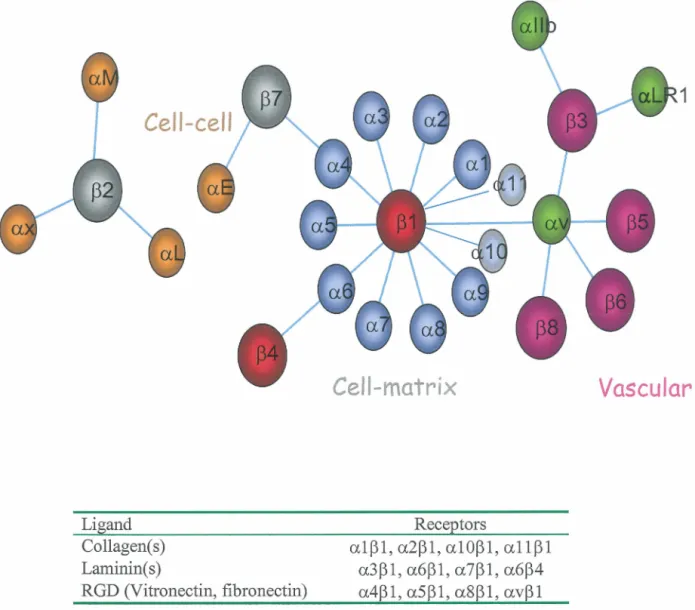
subunit that dimerize to form the functional integrin. Both subunits consist of a single pass transmembrane glycoprotein receptorwith a, generally short (35-50 amino acids), C-terminal intracellular domain. The N-terminal ligand binding extracellular domain varies in size between the two types of subunit; thus, the a. subunit is the largest (roughly 1000 amino acids), white the 13 subunit is the smallest (700 amino acids) (de Melker and Sonnenberg, 1999). 18 a. subunits and 8 13 subunits have been described forming at least 24 functional receptors. These receptors can roughly be divided into sub-classes based upon their cellular function and extracellular ligands: thus integrins containing the 132 subunit are involved in cell-cell interactions white a.Ilb, a.LRl and a.v containing integrins of hematopoitic ce lis mediate contact to vascular ligands (Figure 1) (Beaulieu, 1997). In epithelium throughout the mammalian organism, integrins containing either the 131 or 134 subunits mediate anchorage to the BM. Of these integrins, the a.4, a.5, a.8, or a.v subunits bind the ECM ligands fibronectin and vitronectin via a conserved RGD (Arg-Gly-Asp) motif in the ligand. The common ligand of integrins containing the a.l, a.2, a.10 & a.11 subunits are collagen, white the a.3, a.6 and a.7 containing integrins bind laminins (Beaulieu, 1997; Mercurio, 1995). It should though be noted that both collagen and laminin contain RGD motifs, but that these motifs are inaccessible for binding of the RGD binding class of integrins due to tertiary and quatemary structural masking of the motifs. These so called cryptic sites can however become available for binding following proteolytic processing by matrix metalloproteinases (MMPs) and plasmin resulting in altered biological properties of the matrix thus underscoring both the plasticity and potential complexity of the ECM-cell interaction network (Giannelli et al., 1997; Goldfinger et al., 1998).
The integrin family
Cell-cell
Ligand Collagen(s) Laminin(s) RGD (Vitronectin, fibronectin)Cell-matrix
Receptorsa101, a201, a1001, a1101 a301,a601,a701,a604 a401,a501,a8êl,av01
Vascular
Figure 1: The integrin family. The Integrin family loosely divided according to their functions and ligand(s) (table). Note that other, less common, ligands are likely to exist for all the integrins. For instance the a604 integrin also bind Netrin-1. Modified from Beaulieu, 1997.
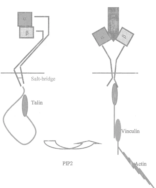
The structural premises of integrin binding to their extracellular ligands and subsequent activation are not completely delineated, though it is known that integrins exist in two confonnational states: a bended in-active state and an extended active state. The activation (affinity) state of the integrins can be modulated by extracellular divalent cations or through complex intracellular processes that are energy-dependent (Hynes, 2002). Briefly, the activation and subsequent binding of the cytoskeletal protein talin to the P-subunit of various integrins (p3 & P 1) lead to activation of the integrin (Tadokoro et al., 2003) through a forced separation of the intracellular domains of the a. and the p subunits. ln this model, this results in a "switch-blade" movement of the integrin stalkes leading to a re-arrangement of the extra-cellular head domains and an unmasking of the ligand binding domains (Figure 2) (Giancotti, 2003).
1.2 Integrins a.6JH and a.6(34
The a.6 subunit dimerizes with either the p 1 or the P4 subunit to fonn two functional integrins a.6p 1 and a.6p4 (Hemler et al., 1989; Tamura et al., 1990) that function as receptors for laminin. In epithelial cells of the skin (Watt, 2002), the mammary gland (Mercurio et al., 2001a) and the intestine (Beaulieu, 1999) and other cell types that express both dimeriz.ation partners, the a.6 subunit preferentially dimerizes with the (34 subunit (Hemler et al., 1989) creating a crucial component of hemidesmosomes
Integrin activation
Salt-bridge
PIP2
Figure 2: Integrin activation. PIP2 (phosphatidylinositol 4,5-bisphophate) binding to talin induces a conformational change that unmasks the integrin
p
subunit binding site within the globular N-terminal domain oftalin. Talin subsequently binds the intracellular domain of thep
subunit resulting in the disruption of the weak (salt bridge) interaction between intra-cellular domains of the a and thep
subunits. The following conformational change is propagated across the plasma membrane leading to unmasking of the integrin-ligand binding1991), peripheral neurons (Hogervorst et al., 1993) and leukocytes (Wei et al., 1997) exclusively express the a6f31 integrin, due to absence of the f34 subunit.
1.2.1 The a6A and a6B integrin subunits - Structure and ontogeny
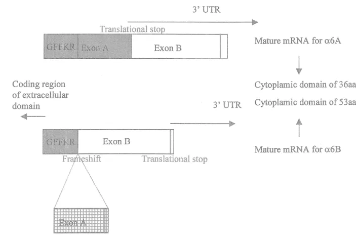
A single exon of the pre-mRNA of the a6 subunit undergoes alternative splicing leading to the formation of two distinct C-tenninal placed intra-cellular domains (Hogervorst et al., 1991; Tamura et al., 1991). Exon A encompasses a translational stop signal that is excluded from the mature mRNA upon out-splicing of exon A from the pre-mRNA leading to both a reading frame shift and a read through into the altematively expressed exon B that harbors a distinct translational stop signal (Figure 3) (de Melker and Sonnenberg, 1999). The two splice variants display dissimilar expression patterns in a tissue-dependent manner (Hogervorst et al., 1993; Tamura et al., 1991) as well as in a temporal manner through out embryo and organ development (Boland, 1995; Tamura et al., 1991; Thorsteinsdottir et al., 1995; Thorsteinsdottir et al., 1999). Thus, the mammary gland, the epidennis and peripheral neurons only express the A variant, while the B variant are exclusive to the nephric epithelium (Hogervorst et al., 1993; Thorsteinsdottir et al., 1995). Other tissue such as the sweat and salivary glands as well as the large intestine have been reported to express both variants (Hogervorst et al., 1993). Undifferentiated murine embryonic stem (ES) cells exclusively express the B variant, but start to express the A variant
PremRNA splice variants of a.6
3' UTR
Mature mRNA for a.6A ExonB
Coding region of extracellular do main
Cytoplamic domain of 36aa 3' UTR Cytoplamic domain of 53aa
...
t
ExonB
Mature mRNA for a.6B Franteshift / .... / " ... ... " · . .... \ ...
Figure 3: Pre-mRNA splice variants of a.6. Following the in virtually all a
integrins conserved five amino acid membrane proximal motif (GFFKR) exon A is alternatively spliced leading to the formation of two different
intra-cellular domains of 36 and 53 amino acids, respectively. Note that the extracellular domain is identical (see text for details).
only express the B variant until (E) 8.5, where the A variant initiates expression in myocardiac cells (Collo et al., 1995; Thorsteinsdottir et al., 1995). Interestingly, the expression level of the A variant forms a gradient from the outer to the inner layers of the myocardium (Collo et al., 1995; Thorsteinsdottir et al., 1995), indicating an involvement in the progression of organogenesis. A similar delayed occurrence of the A variant during embryonic development is seen in the epidermis as well as in the intestinal epithelium, both of which initially express the B variant exclusively (Thorsteinsdottir et al., 1995), while adult epidermis solely expresses the A variant and the adult intestinal epithelium expresses both variants (Hogervorst et al., 1993). The conservation of the two variants in all mammalian species studied (de Melker and Sonnenberg, 1999) further suggest the biological importance of the presence of both variants in the intact organism. Despite this, genome wide creLoxP1 mediated
excision of exon A in mice results in viable and fertile animais that show a very mild phenotype: decreased lymphocyte presence in the lymphnodes and impaired lymphocyte migration through laminin coated transwells (Gimond et al., 1998).
1.2.2 The a6A and a6B integrin subunits - Biochemical differences
Though little biological impact was observed as a consequence of organism wide substitution of the A variant with the B variant, differences in the biochemical properties of the two splice-variants of the integrin a6 subunit are well documented.
subunit) adhere stronger, migrate faster and extend more pseudopodia than cells expressing the a6BP 1 integrin when cultured in normal medium (Shaw and Mercurio, 1994). Furthermore the A variant is a major target for serine phosphorylation following PMA treatment (Delwel et al., 1993; Hogervorst et al., 1993), while the B variant does not appear to react significantly to PMA treatment (Delwel et al., 1993), if at all (Hogervorst et al., 1993). The biological role of this serine phosphorylation is unclear, but likely does not involve inside-out activation of adhesion, since no differences in adhesion of macrophages transfected with the two variants were observed upon PMA treatment (Shaw et al., 1993) and mutation of all the serine residues in the cytoplasmic tail of the A variant to alanine does not change the adhesive response of the cells upon PMA treatment (Shaw and Mercurio, 1993). Rather the differences in serine-phosphorylation are likely to reflect a variant specific difference in capacity to initiate intra-cellular signaling. Indeed, the A variant stimulates tyrosine phosphorylation of the cytoskeletal protein paxilin and two other unidentified proteins (sized 110 and 120 kDa), in contrast to the B variant, when over expressed in a macrophage cell line that were allowed to adhere to laminin (Shaw et al., 1995). Furthermore, cells expressing the A variant phosphorylate the MAP kinases p42/p44 (Erkl/-2) when seeded on laminin, while cells expressing the B variant does not (Ferletta et al., 2003; Wei et al., 1998). In macrophages, this integrin a6AfH-dependent activation of p42/p44 is causal of the superior migratory capacity of a6Af31 expressing cells as compared to a6BP1 expressing cells (Wei et al., 1998).
1.2.3 Integrin a6A and a6B - Alterations of expression ratio
Alterations of the a6A/a6B ratio have been reported to accompany the differentiation of embryonic stem cells (Cooper et al., 1991), embryonic carcinoma cells (Jiang, 1995; Morini, 1997) and lens cells (Walker and Menko, 1999). ln ail cases, differentiation of the cells were accompanied by a down-regulation of expression of the B variant and an up-regulation of the A variant, in agreement with the temporal occurrence of the variants during embryonic development (Thorsteinsdottir et al., 1995). Furthermore, the distribution of the two variants in the retina bas been reported to be compartment specific: the A variant being expressed in the retina surrounding the optic nerves and in the optic nerves themselves, while the B variant being ubiquitously expressed in the retina (de Curtis and Reichardt, 1993 ). Experimental, rather than descriptive, evidence of the distinct cellular roles of the two variants bas though pointed in the direction of the B variant playing a key role in completing differentiation of chondrogenic cells (Segat et al., 2002). Interestingly, the stabilization of the differentiated phenotype was found to be dependent on the presence of both the B and the A variant, indicating that correct cellular function is dependent on the presence of both variants, and that changes in the a6A/a6B ratio rather than an absence or presence of one or the other variant are instrumental in affecting cellular functions (Segat et al., 2002).
1.2.4 The a.6-containing integrins in carcinomas
Studies correlating integrin a6 expression in primary tumors with metastatic capacity and survival of breast cancer patients have indicated a strong negative correlation between integrin a6 expression and medium term (five years) survival (Friedrichs et al., 1995). The effects of the a6-containing integrins in carcinoma biology have been linked to effects mediated by both the a6p 1 and a6P4 integrins (Shaw et al., 1996; Shaw et al., 1997; Wewer et al., 1997). Interestingly, both a6Pl and a6P4 appear to stimulate carcinoma cell survival in a VEGF-dependent manner (Chung et al., 2002; Chung et al., 2004a) indicating a common effector pathway ofthese two integrins.
To date few studies have addressed any differential role of the two splice-variants of the a6 integrin in tumor biology. Studies regarding the expression of the two variants in tumor biology do tend to conclude that increased invasiveness is characterized by alterations in the a6Afa6B ratio compared to non-invasive cells (Tennenbaum et al., 1995), though no impact on survival of patients has been documented as a consequence of alterations of variant-ratio (Friedrichs et al., 1995; Tanaka et al., 2000).
1.2.5 The integrin P4 subunit - Stucture and hemidesmosomes
The integrin
p4
subunit stand out with-in the integrin superfamily due toits large(> 1000 amino acid) intracellular domain, that show no sequence homology to any otherp subunit intracellular domain (Hogervorst et al., 1990). The intracellular domain is composed of two pairs of closely positioned type III fibronectin repeats separated by a connecting segment (CS) (Suzuki and Naitoh, 1990). The p4 subunit is subject to alternative splicing of the pre-mRNA (Clarke et al., 1994; Hogervorst et al., 1990; Niessen et al., 1997; Suzuki and Naitoh, 1990; Tamura et al., 1990; van Leusden et al., 1997) as well as post-translatorial modifications (Basora et al., 1999; Giancotti et al., 1992; Potts et al., 1994) leading to the formation of several structural variants (A-E). The tissue distribution and function(s) ofthese variant forms are somewhat poorly characterized. The P4 subunit only participates in the formation of a single functional integrin: the a6p4 integrin ( a6P4 ).
As opposed to the p 1 and IB containing integrins, a6p4 does not participate in the formation of focal adhesion sites, but rather in adhesion structures termed hemidesmosomes (HD) (Sonnenberg et al., 1991). These anchoring point connects the intermediate filament network of the cells to the underlying BM through a6P4 and the other components of HD: plectin, BP 180, BP 230 (Borradori and Sonnenberg, 1999) and the tetraspan CD151 (Figure 4) (Sterk et al., 2000). The assembly of the HDs is an ordered process in which plectin initiates interactions with the intracellular domain of p4 followed by conformational changes that allow first BP180 and then BP230 to interact with the third and the third and fourth fibronectin repeats respectively (Koster et al., 2004). In cells of simple epithelia such as in the mammary gland (Uematsu et al., 1994) and the intestine (Stutzmann et al., 2000) that do not express BP180 and BP 230, immature hemidesmosomes, termed hemidesmosomes type II (HD Il) are
CD!~
BP180
Plectin
I(eratin
Figure 4: Hemidesmosomes - Molecu.far components, Structure and components of hemidesmosomes. The a6p4 integrin associates with CD 151 through the a6 subunit, while plectin through its N~terminaHy placed Actin Binding Domain (ABD) interacts with the
prolinel330 and 1333 of the first type III fibronectin repeat (FN) and connecting segment (CS) of ~4, leading to an open conformation of the C-terminal portion of
p4.
This openconformation allows BPI 80 to interact with the thlrd FN repeat of p4, while BP230 binds plectin and the J34 subunit through sequences from the connecting segment C-terminally until the fourth F1\l repeat Plectin and BP230 facilitate the interaction ~Nith the keratin network In type II hemidesmosomes BPI BP and CD151 axe not present and the cormection to the keratin network is medfated solely plectin. from Koster et al,
leads to blistering skin diseases, termed epidermolysis bullosa (EB) and epidermolysis bullosa simplex associated with muscular dystrophy (MD-EBS) due to lack of formation of hemidesmosomes (Fontao et al., 2004; Georges-Labouesse et al., 1996; Koster et al., 2001; Niessen et al., 1996; van der Neut et al., 1996).
1.2.6 Integrin a6fi4 - Functions in normal tissue development
fi4 knock-out mice develop normally until birth, but die shortly postnatal due to structural disruption of the epidermis and other squamous epithelia reminiscent of epidermolysis bullosa (van der Neut et al., 1996). The early postnatal death of these animais precluded further investigations of the cellular impact of deletion of a6J34. None the less, it was determined that the knock-out animais did not show any other developmental defects than an absence of hemidesmosomes providing a mechanistic explanation for the fragility of the epidermis (van der Neut et
al.,
1996). Another genetic engineering approach where only the intracellular segment of J34 were deleted confirmed the previous results and further demonstrated the importance of the interactions of the intracellular domain of J34 with the intermediate filament network (Murgia et al., 1998). By performing Cesarean section at El8.5, these authors were able to obtain minimally damaged samples and were thus able to demonstrate proliferative defects in stratified and simple epithelium (epidermis and intestine) as a consequence of impaired signaling mediated by the intracellular domain of J34findings that the intracellular domain of P4 C-tenninal to amino acid 1355 is not needed for interaction with the keratin network (Schaapveld et al., 1998) and that the major tyrosine phoshphorylation sites needed for intracellular signaling are placed in this region (Dans et al., 2001; Shaw, 2001) (see below). Thus, only this C-terminal portion of
p4
(termed the siganlizsation domain) was deleted resulting in viable and fertile off-spring (Nikolopoulos et al., 2005). These animais appear phenotypically normal, but have impaired proliferation in the epidermis due to defective NFKB and ERK. signaling (Nikolopoulos et al., 2005) (see below). Taken together, these studies strongly indicate that the major non-redundant function of the a6P4 integrin in healthy mammals is to provide structural integrity of stratified epithelium.1.2. 7 a6P4 in carcinoma biology - Angiogenesis, survival & migration
An early study reported a correlation between metastatic potential of various clones of murine lung carcinomas and surface expression of a tumor cell associated antigen (Falcioni et al., 1986), that was subsequently demonstrated to be a6P4 (Kennel et al., 1989). Severa! expression studies have accordingly established an enhanced expression of a6P4 in squamous carcinomas (Tennenbaum et al., 1993; Van Waes et al., 1991), thyroid carcinomas (Serini et al., 1996) and bladder carcinomas (Grossman et al., 2000). In carcinomas of the breast, the presence of the a6 subunit has been linked with poor prognoses (Friedrichs et al., 1995) as has the presence of the P4 subunit (Tagliabue et al., 1998). Another less direct line of evidence of a role of a6P4
in carcinoma biology is the findings that laminin5 an important ligand of a6p4 -accumulates along the invasive edges of gastric (Tani et al., 1996), pancreatic (Tani et al., 1997) and colorectal (Lohi et al., 2000) tumors and that the prognostic value of p4 expression in carcinomas of the breast is dependent on the co-expression of laminin-5 (Tagliabue et al., 1998). Furthermore a6P4 presence and laminin-5 production have been identified as an autocrine survival mechanism in breast carcinoma cells (Zahir et al., 2003).
Recently, genetic evidence of the importance of interactions between laminin-5 and a6P4 for the formation of tumors in vivo has emerged: keratinocytes from epidermolysis bullosa patients deficient in laminin-5 or P4 are unable to form tumors in immunosupressed mice, while restoration of expression of either laminin-5 or J34 restore this capacity (Dajee et al., 2003). Though a6P4 was initially thought to be expressed exclusively by epithelial cells (Hemler et al., 1989; Tamura et al., 1990), it is also expressed in endothelial cells (Hiran et al., 2003). This endothelial expression accounts for another, less direct but still crucial, role of a6P4 in carcinoma progression: VEGF- and bFGF-provoked angiogenesis is dependent on the signaling domain of P4 (Nikolopoulos et al., 2004). Thus, carcinoma cell lines implanted in mice genetically engineered to express a signaling-deficient P4 subunit form smaller and considerable less vascularized tumors than corresponding cells implanted in wild type mice in agreement with the dramatically decrease of vasculation of bFGF containing matrigel plugs implanted in mutant mice compared to wild type animais (Nikolopoulos et al., 2004).
At the cellular level, a6J34 is involved in aspects of carcinoma biology in a twofold fashion: it provides physical interaction with the extracellular matrix enabling carcinoma cells to invade and migrate (Chao et al., 1996; Lipscomb et al., 2003; Rabinovitz et al., 2001; Rabinovitz and Mercurio, 1997) and intracellular signaling initiated by a6J34 stimulates invasion (Chao et al., 1996; Rabinovitz and Mercurio, 1997; Shaw et al., 1997) as well as survival (Chung et al., 2002; Lipscomb et al., 2005; Weaver et al., 2002; Zahir et al., 2003).
1.2.8 Carcinoma cell migration, invasion and mobilization of a6J34 from hemidesmosomes
Despite its central role in rigid attachment sites, a6J34 under certain conditions stimulates cellular migration (Mercurio et al., 2001 c ). The first evidence of such arole of a6J34 came from the observation that stable expression of a6J34 in a a6J34-deficient carcinoma cell line leads to enhanced spreading and invasion through matrigel (Chao et al., 1996). The role of a6J34 in facilitating invasion has been documented to be both biochemical through stimulation of intra-cellular signaling of importance for migration (cAMP/RhoA) and invasion (PI3-K/Rac) (O'Connor et al., 2000; O'Connor et al., 1998; Shaw et al., 1997) and mechanical through a direct linkage with F-actin of the myo-actin cytoskeleton in filopodia and lamellae of migrating cells (Rabinovitz et al., 2001; Rabinovitz and Mercurio, 1997). Noticeably, a6J34 has been shown to mediate this invasiveness by excerting traction forces directly on the
BM
and thusphysically creating pores in the BM through which the carcinoma cells potentially can escape (Rabinovitz et al., 2001). a.6134 has also been implicated in activation ofMMP-2 resulting in enzymatic degradation of the underlying BM (Daemi et al., ofMMP-2000). Epidermal Growth Factor (EGF) and signaling mediated through its receptor (EGFR) has been identified as key players in the release of integrin a.6p4 from the physical restraints of hemidesmosomes and its subsequent relocation to the actin network (Mercurio et al., 2001c). The intracellular signaling responsible for this release and relocation involves phosphorylation of serine and tyrosine residues in the intracellular domain of the 134 subunit (Figure 5) (Dans et al., 2001; Mainiero et al., 1996; Rabinovitz et al., 1999) and has been proposed to be dependent on the Src-family kinase Fyn (Mariotti et al., 2001) as well as protein kinase C (Alt et al., 2001; Rabinovitz et al., 1999; Rabinovitz et al., 2004).
1.2.9 a.6134, Receptor tyrosine kinases and lipid rafts
Several studies have demonstrated that a.6134 can initiate signaling in a ligand independent manner (Chao et al., 1996). This apparent conundrum is possibly linked to the ability of a.6134 to physical interact and synergisticly cooperate with receptor tyrosine kinases (RTKs), such as EGF-R (Mainiero et al., 1996; Mariotti et al., 2001; Q.H.Song et al., 2003), the Ron receptor of macrophage stimulating protein (MSP) (Santoro et al., 2003), ErbB2 (Falcioni et al., 1997; Gambaletta et al., 2000) and the
Intracellular domains of j34
Cys 732/736/738/739 & 742 Palmitoylation ~ lipid raft
incorporation
PKC-a/ô phophorylation
~
bindin;~
of 14-3-3 proteins ~ release fromHD and cross talk with a3 f3 l Tyrl526-mediates binding
of the PTB domain of Sh~--i!IH
essential for ERK activation
;.-...
... EGF-R .._ FYN _ . Tyr??? ______ .._
Hemidesmosome~
disassemblyTransmembrane
~ Prol330/1333 Essential for
recruitment of Plectin into HD
} Connecting segment
+---
Tyrl494-Essential for PB-K Activation through IRS-1/2.Figure 5: IntraceHular domains of j34. The intracellular domains of f34 of importance for hemidesmosome assembly, recruitment from hemidesmosomes &
redistribution to lipid rafts as well as intracellular signalization. Pro: Proline, Cys: Cystein, Tyr: Tyrosine, HD: Hemidesmosome, ???: Unknown
Met receptor of hepatocyte growth factor (HGF I scatter factor) (Bertotti et al., 2005; Trusolino et al., 2001 ). Even though the importance of the latter observation of a.6P4 dependent function of Met has been contested (Chung et al., 2004b) there is little doubt that a.6P4 physically interacts with several RTKs and acts in a synergistic fashion to amplify the biochemical consequences of receptor clustering.
A sub-population of a.6p4 is located in lipid rafts - subdomains of the plasma membrane enriched in cholesterol and glycosphingolipids - due to palmitoylation of cysteines in the membrane proximal intracellular domain of P4 (Figure 5) (Gagnoux-Palacios et al., 2003). This localization brings a.6P4 into physical proximity with RTK.s as well as with palmitoylated Src Family Kinases (SFKs) that phosphorylate the intracellular domain of P4 (Mariotti et al., 2001) and thus create binding sites for Shc leading to intracellular signaling (Dans et al., 2001). Intriguingly, several studies report such synergistic cooperation to be independent of ligation of a.6P4 (Bertotti et al., 2005; Gambaletta et al., 2000; Trusolino et al., 2001) in accordance with other studies showing that the presence of a.6P4 in carcinoma cells is sufficient to induce increased migration on non-laminin substrates (Chao et al., 1996; O'Connor et al., 1998). The functional impact of this paradigm is profound since it in theory makes a6P4 capable of initiating intracellular signaling regardless of the composition of the surrounding ECM. Furthermore the cellular function of a.6P4 appears to be altered by these interactions. Thus both EGF and MSP stimulation leads to relocation from stable HD to leading edge lamlepodia followed by increased migration (Dans et al., 2001; Mariotti et al., 2001; Santoro et al., 2003).
1.2.10 Intracellular signaling mediated by the a6J}4 integrin
Two of the principal mediators of intracellular signaling activated by the a.6P4 integrin is
Ras
and Phosphoinostide-30H-kinase (PI3-K) (Mainiero et al., 1997; Shaw et al., 1997). a.6JJ4 specific activation of signaling molecules down stream of Ras and PI3-K include the MAP Kinases Erk and JNK (Mainiero et al., 1997) and AKT (Chung et al., 2002) (Figure 6). This activation ofRas
and PI3-K has been proposed to occur by two not mutually exclusive methods. One proposed model calls for Fyn-( of the Src Family Kinases) mediated tyrosine phosphorylation of the intracellular domain of P4 as a consequence of ligand binding and in response to EGF-R activity (Mariotti et al., 2001) leading to binding of the adaptor protein Shc via a SH2 domain. Shc subsequently recruits Grb2-mSOS that activatesRas
and MAP-Kinase and PI3-K pathways and PI3-K (Dans et al., 2001; Mainiero et al., 1997). An alternative mechanism involves Insulin Receptor Substrate (IRS) 1 and 2 (Shaw, 2001) as signaling intermediates between the intracellular domain of the P4 subunit and PI3-K. a.6JJ4-mediated signaling furthermore leads to phosphorylation and subsequent degradation of the NFKB inhibitor IKBa. resulting in nuclear translocation of NFKB (Figure 6) (Nikolopoulos et al., 2005; Weaver et al., 2002). This activation of NFKB bas been linked with proliferation in normal keratinocytes (Nikolopoulos et al., 2005) and the acquisition of polarity and apoptosis resistance in normal and malign human mammary gland epithelial cells (Weaver et al., 2002). Intriguingly, while a6P4 enhances cellular survival through stimulation of AKT in some cellular settings(Bachelder et al., 1999b ), certain carcinoma cell lines have been demonstrated to undergo growth arrest and apoptosis upon a6B4 overexpression (Bachelder et al., l 999a; Clarke et al., 1995). This a6B4-provoked apoptosis is strongest when the cells are grown under stressfull conditions, such as in serum and/or growth factor free medium or when grown in suspension (Bachelder et al., l 999b) giving weight to the notion that the apoptotic effect reflects a "negative positional eue." The intracellular signaling pathway leading to apoptosis is dependent on functional p53 (Bachelder et al., l 999a) and involves caspase-3 dependent cleavage of AKT (Figure 6) (Bachelder et al., 1999b).
It is of interest to note that a6B4 bas been demonstrated to activate the eukaryotic initiator factor 4E (e1F-4E) in carcinoma cells (Chung et al., 2002). elF-4E is the rate limiting factor of 5'cap dependent translation and is normally inhibited by binding to 4E-BPls (Figure 6). Enhanced activity of eIF-4E is a hallmark of several cancers, including mammary and colorectal cancers, due to its specific effects on the translation of oncogenes such as c-MYC, Cyc/inDJ, P-catenin and VEGF (De Benedetti and Graff, 2004; Kami et al., 2005; Mamane et al., 2004). As such, ligation of a.6~4 has been demonstrated to lead to upregulation of the translation and
expression of VEGF resulting in autocrine survival signaling and greater tolerance to hypoxia in an eIF-4E dependent manner, (Figure 6) (Chung et al., 2002).
Activation of the small GTPases RhoA and Rac leads to migration and invasion respectively (O'Connor et al., 2000; O'Connor et al., 1998; Shaw et al., 1997) (see above 13. 4). a6B4-dependent Rac activity has furthermore been linked to
Intracellular signaling initiated by f}4
...---s==R-
ACTIVATION-IKBa.
l
NFKB
c RAF-1i
Shc-Grb -mSOS IRS-1 &-2 Actin dynamics SURVIVAL 4E-BP1~
1
p70/S6Ki
PROLIFERATION MIGRATION/INVASION eIF-4E 408i i
;"···--···-··· Cyclin D}'illl···-·TRANSLATION __. VEGF
1
,.··-···-···-~
... _.t
t... ... _ ... MYC
Figure 6. Intracellular signaling initiated by a.6f}4 ---. : Stimulation - demonstrated for a.6p4
--i :
Inhibition - demonstrated for a.6P4···~ : Known effect - not demonstrated for a.6f}4
increased NFKB activity resulting in increased survival in breast carcinoma cells (Zahir et al., 2003).
1.3 The intestine
1.3.1 The small intestine
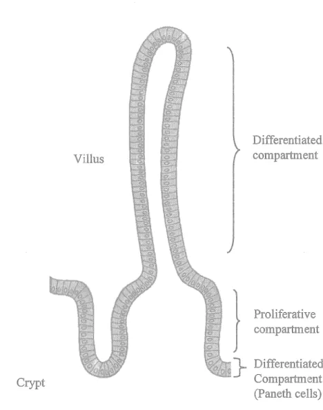
The comparatively simple structural and functional unit of the small intestines makes it an attractive model for the study of epithelial cell maturation and differentiation. lndeed, the crypt-villus axis is divided into two compartments in which the crypts contain the proliferative and poorly differentiated cells, while the villi contain the differentiated enterocytes that rapidly migrate towards the villus tip, from where they are shed into the intestinal lumen, (Figure 7) (Babyatsky and Podolsky, 1995; Beaulieu, 1999). The stem cells reside in the middle to the lower crypt from where they give rise to daughter cells that differentiate into the four principal cell types of the intestinal epithelium: The Paneth cells, which are the only cell type that migrate downwards to the bottom of the crypt, function in intestinal mucosal defense against microbial infection by secreting TNF and cryptidins (intestinal homologues of leukocyte defensin). Goblet cells secrete the continuous layer of mucus that lines the intestine, while enteroendocrine cells secrete gastrointestinal hormones. Finally the absorptive columnar enterocytes function to enzymaticaly degrade and absorb nutrients.
Crypt
The small intestinal Crypt-Villus axis
Villus
l
}
Differentiated compartment Proliferative compartment Diff erentiated Compartment (Paneth cells)Figure 7: The small intestinal Crypt-Villus axis. The functional unit of the small intestine. Intestinal stem cells reside near the bottom of the crypt, from where they give rise to rapidly dividing daughter cells that migrate upwards on the villus until they undergo programmed cell death at the villus tip, from where they are shed into the intestinal lumen. Differentiated Paneth cells reside at the bottom of the crypts. Modified
Positional control of the enterocytes, as well as their subsequent functions is largely controlled by the underlying BM (Beaulieu, 1999) of which specific laminins have been shown to have an instructional effect upon enterocytic cytology (Vachon and Beaulieu, 1995), while analysis of several molecules involved in cell-extracellular matrix (ECM) interactions, including the integrins, during development has revealed distinct patterns of expression along the crypt-villus axis in relation to the differentiation state of the enterocytes (Beaulieu, 1997; Kedinger et al., 1998a; Kedinger et al., 1998b; Teller and Beaulieu, 2001).
1.3.2 The colon
The surface of the adult colon lacks the characteristic villi of the small intestine and thus appears fiat. As in the small intestine, crypts projects away from the lumen. The organization of the crypts is analogous to the small intestine, with undifferentiated and proliferative active cells in the lower half and differentiated cells in the upper half and at the surface epithelium (Babyatsky and Podolsky, 1995).
1.3.3 lntegrin a.6(34 in the small and large intestine
the crypt-villus axis (Beaulieu, 1992). This study did not attempt to differentiate between the A and the B variant of the a.6 subunit and consequently the expression pattern of these two variants in the intestine is not presently reported. An examination of the expression of the 134 subunit using different antibodies revealed a distinct expression pattern along the crypt-villus axis (Basora et al., 1999). While 134 was found ubiquitously at the base of ail epithelial cells from the bottom of the crypt to the tip of the villus it was found to be expressed under two different forms. Antibodies that recognize the final 20 and 31 amino acids of the C-terminal intracellular domain ( ctd) only detected their epitope on the villus, while antibodies raised against extracellular domains of 134 detected their epitope ubiquitously along the crypt-villus axis, thus demonstrating the presence of a ctd-deleted ( ctd-) variant of 134 in the crypts and a full-length ctd+ variant at the villus. The 134ctd- variant was subsequently shown to be cotranslatorially spliced and non-functional for adhesion to laminin-5 (Basora et al., 1999). In the colon a similar differentiation dependent expression pattern of the 134ctd- and 134ctd+ variants have recently been established (Ni et al., 2005).
1.3.4 Integrin a.6f34 in colon cancer
The expression of 134 in colorectal cancers has been the subject of a number of studies that have reported conflicting results (Falcioni et al., 1994; Lohi et al., 2000; Sordat et al., 1998; Stallmach et al., 1992). While ail these studies reported that a.6134 is
expressed at the base of ail epithelia in normal non-malign colonie epithelium, one study (Stallmach et al., 1992) reported a.6P4 to be reduced/lost in carcinomas whereas another (Lohi et al., 2000) reported little alterations of expression and a third (Falcioni et al., 1994) found a heterogenic increase. Finally, a differentiation-dependent expression pattern was identified according to which the expression of a.6P4 is maintained in well differentiated tumors and decreased/lost in moderately and poorly differentiated tumors (Sordat et al., 1998). Recently, we have reported the dual finding that while the p4ctd+ variant is overall upregulated in 22 colon cancers, the p4ctd- variant were ubiquitously lost (Ni et al., 2005) indicating the need of a laminin-5 adhesion competent a.6P4 integrin in colon cancers, in agreement with the above mentioned finding (see l3.3) of requirement of laminin-5/a.6p4 for formation of
squamous cell carcinomas (Dajee et al., 2003).
1.4 Nonnalizing genes for use in mRNA quantification studies
The real-time quantitative reverse transcriptase polymerase chain reaction (real-time qRT-PCR) is rapidly becoming the method of choice for gene expression assessments, because it requires relatively little biological material and is efficient. There are though substantial drawbacks to the utilization of the technique for accurate quantification of transcript levels; the PCR step greatly amplifies the transcript and thus any intrinsic differences in the quantity or quality of input cDNA, or trivial
common way to deal with these difficulties is to express the transcript level of the gene of interest (GOI) nonnalized to another gene commonly tenned housekeeping gene or nonnalizing gene. This method is based on the assumption that the expression level of the nonnalizing gene does not change from sample to sample, i.e. that the fraction of the mRNA pool that are made up by transcript from the nonnalizing gene remains constant from sample to sample.
The differentiation of a tissue (Walters, 2005) or the formation of carcinomas (Boland, 1995) are complex cellular processes and any attempt at assessing gene expression changes using qRT-PCR during these events should have as a prerequisite that the normalizing gene is not affected by the cellular process under investigation. However, inherent difficulties in identifying a "gold standard nonnalizing gene" of cDNA input in qRT-PCR analysis experiments have become apparent with variation across tissue types (Radonic et al., 2004), development stages (Calvo et al., 1997) and different cancer types (Aerts et al., 2004; de Kok et al., 2005) being reported. One present attempt at addressing this problem is to use a weighted average of several nonnalization genes (Pfaffl et al., 2004; Vandesompele et al., 2002). This solution, however, suffers from the drawback that it nonnalizes the GOI against the most common variation of the nonnalizing genes, i.e. variation in the nonnalizing gene(s) are accepted if the majority vary the same way. While the perfect nonnalizing gene may not exist, another more promising strategy is to characterize a single nonnalizing gene for a single tissue type (Schmid et al., 2003), pathology (Dheda et
al.,
2004) or experimental design (Biedennan et al., 2004; Hamalainen et al., 2001; Malarstig et al., 2003; Schmittgen and Zakrrajsek, 2000).1.5 Aim of studies
Sorne of the hallmarks of cancer is i) the ability to sustain non-adherent growth (resistance to anoikis), ii) increased proliferation, iii) increased migratory and invasive potential and iv) increased potential of angiogenesis (Han.ahan and Weinberg, 2000). While some controversy as to the expression pattern of the a6f34 integrin in colorectal cancer exists in the literature (Falcioni et al., 1994; Lohi et al., 2000; Sordat et al., 1998; Stallmach et al., 1992), a6f34 has been demonstrated to play a role in all of these cellular events and it was therefore of pertinent interest to delineate the expression pattern of a6f34 in adenocarcinomas of the human colon.
The first aims of the study was thus to confirm the reported upregulation of expression of the f34 subunit in colon cancers at the transcript level. In order to further characterize the expression of the a6f34 subunit in the human small intestine and in adenocarcinomas of the human colon, the second aim was to delineate the expression pattern of the two splice variants of the a6 subunit in healthy as well as malign tissue. Thirdly, investigations of the differential impact of the two a6 splice variants on colon cancer cells were carried out.
Finally, the literature is providing ample evidence that the choice of normalizing gene can be crucial for the correct interpretation of data related to quantification of steady state levels of mRNA (de Cremoux et al., 2004; Dheda et al., 2004; Hamalainen et al., 2001; Oliveira et al., 1999; Thellin et al., 1999). In order to ensure that the normalizing genes of use in the daily routines of the laboratory were of
experirnental conditions) experirnents to identify and validate candidate genes were carried out.
II. Materials and Methods
11.1
TissuesPrimary tissues of healthy adult ileum were obtained from Quebec Transplant (Quebec, Canada), while fetal tissues (ranging from 16 to 20 weeks of age, postfertili7.ation) were obtained after legal abortion. Both were in accordance with protocols approved by the Institutional Human Research Review Committee for the use of human material. Both adult and fetal speciemens were used for immunofluores cence, while primary extracts of villus cells (fully differentiated enterocytes) were obtained from fetal samples in accordance with a previously published protocol (Perreault and Beaulieu, 1998). Tissue samples of primary adenocarcinomas and healthy resection margins were provided by the Cooperative Human Tissue Network (Midwestem Division, Ohio State University, OH), which is funded by the National Cancer lnstitute. These tissue samples were used only for RNA extraction.
11.2
AntibodiesAn antibody recognizing an extracellular epitope common to both splice variants of integrin a.6 (Mab GoH3) (Sonnenberg et al., 1987) as well as two antododies recognizing integrin a6A (Mab 1359, clone lAlO) and a6B (Mab 1358, clone 684)
of Cell Biology, The Netherlands Cancer Institute, Amsterdam The Netherlands). Subsequently, these antibodies were obtained from Santa Cruz (Santa Cruz, CA) (GoH3) and Chemicon (Temecula, CA, USA) (Mab 1359, clone lAlO and Mab 1358, clone 6B4). Mabs lAlO and 6B4 were used for western blots. For indirect immunofluorescence, 6B4 and GoH3 were used while the Mab lAlO did not yield staining of sufficient quality/strength to allow for conclusive analysis to be performed. A rabbit polyclonal a6A (a6-cytoA) (de Curtis and Reichardt, 1993) antibody was obtained and employed in its place. This antibody was a generous gift from Dr. de Curtis (Department of Molecular Pathology and Medicine, San Raffaele Scientific Institute, Milan, ltaly). To probe for ~-actin, the antibody C4 from Chemicon was employed.
11.3 Indirect immunofluorescence
Cryosections 3µm thick were fixed in 2% paraformaldehyde (a6A) or -20°C ethanol ( a6B). Nonspecific protein-protein interactions were blocked for one hour at room temperature by immersion of slides in 10% goat serum (a6A - lAlO), 2% BSA (a6B - 6B4) or 10% Blotto (a6 - GoH3) followed by incubation with the primary antibodies diluted 1 :200 in their respective blocking solutions for two hours at room temperature. Following extensive washing in PBS, the slides were incubated with FITC conjugated secondary antibodies raised against rabbit and mouse, respectively, for one hour at room temperature before being washed in PBS. The slides were