Characterization of the human Fc gamma RIIB gene promoter: human zinc-finger proteins (ZNF140 and ZNF91) that bind to different regions function as transcription repressors
10
0
0
Texte intégral
(2) 1076 ZNF140 and ZNF91 repress FcγRIIB transcription ocytic cells contain predominantly FcγRIIA transcripts. Further, human epithelial cells and trophoblasts also express all three FcγRII transcripts, predominantly the FcγIIC transcript (14,15). Thus, these findings indicate that different regulatory mechanisms exist among these three FcγRII gene expressions in a variety of cell types. McKenzie and his group (16) characterized the 5⬘ region of the human FcγRIIA gene. They reported that the FcγRIIA gene, which consists of nine exons, has two discrete transcription start sites. One start site was mapped to a 5⬘-untranslated (5⬘-UT) exon ~1 kb 5⬘ to the ATG translation initiation codon and the second start site was mapped near the ATG codon. However, these authors did not report the precise functional characterization of the human FcγRIIA promoter and the molecular mechanism underlying the regulation of the human FcγRII gene expression is still obscure. To unravel the molecular mechanism of the regulation of the human FcγRII gene transcription, we isolated the human FcγRIIB gene promoter and identified two different regionbinding proteins (F2-3 and F4-3 binding proteins) within the promoter region of the human FcγRIIB gene by electrophoretic mobility shift assay (EMSA). We also cloned two different zinc-finger proteins (ZNF140 and ZNF91) that can bind to the F2-3 and F4-3 regions within the human FcγRIIB promoter respectively. These human zinc-finger proteins (ZNF140 and ZNF91) function as repressors for the human FcγRIIB transcription. When they were simultaneously expressed, the two proteins demonstrated an additive suppressive effect. Methods Plasmids and phage library The EMBL3 genomic phage library (kindly provided by the Japanese Cancer Research Resources Bank, Tokyo, Japan) was used for the screening of human FcγRII genomic genes. The pSVOOCAT was obtained from Wako Pure Chemical Industries (Osaka, Japan). The pBluescript II KS⫹ (pBSIIKS⫹), and pCAT-enhancer (pCAT-EN), pGL3-Enhancer (pGL3EN) and pSV-β-galactosidase control vector (pSVβgal) were purchased from Stratagene (La Jolla, CA) and Promega (Madison, WI) respectively. The pCAGGS was kindly supplied by Dr Miyazaki (17) and pcDNA3.1/Hygro(⫹) (pcDNA3.1) was obtained from Invitrogen (Carlsbad, CA). Cloning of the human Fcγ RIIB gene promoter region Four clones were isolated from the EMBL3 phage library using the 32P-labeled 700-bp PstI fragment of the PC23 cDNA as a probe (18) under high-stringent conditions. Construction of deletion mutants using pBSIIKS⫹ plasmid was performed using exonuclease III (Stratagene) according to the manufacturer’s instructions. Construction of plasmids The 1.5-kb EcoRI–NarI fragment of the cloned human FcγRIIB gene was filled in and ligated to SalI linker, and then ligated to the SalI site of pSVOOCAT to give pFcRCAT. The same 1.5-kb fragment was inserted into the SalI sites of pCAT-EN to yield pFcRCAT-EN. The various deletion mutants were prepared using exonuclease III and sequenced, and the. resulting inserts were ligated into the SalI site of pCAT-EN. The pFcR (del-326) CAT-EN was prepared by deleting the –326 to ⫹21 region and inserting into the SalI site of pCATEN. The pFcR-CAT-EN/R was prepared by inserting the 1.5-kb EcoRI–NarI fragment into the SalI site of the pCATEN in a reversed orientation. The pFcR2-154/pGL3EN was prepared by inserting the –154-bp fragment into the SmaI site of the pGL3EN. The full-length ZNF140 (19) and ZNF91 (20) fragments were inserted into EcoRI site of the pcDNA3.1 and pCAGGS respectively. Primer extension Primer extension analysis was performed using a singlestranded synthetic oligonucleotide of sequence 5⬘-GGTAAGAATGACAGGATTCCCAT-3⬘, which is complementary to nucleotides 1–23 of human FcγRIIB cDNA. The primer was labeled with T4 polynucleotide kinase (Takara Shuzo, Tokyo, Japan) and annealed to 5 µg of mRNA in 25 µl of 50 mM Tris–HCl (pH 8.3), 100 mM KCl and 10 mM MgCl2, incubated at 95°C for 5 min, then at 55°C for 60 min and cooled slowly to room temperature. The sample was mixed with 25 µl of 50 mM Tris–HCl (pH 8.3), 100 mM KCl, 10 mM MgCl2, 2 mM dNTP, 20 mM DTT and 200 U of M-MLV reverse transcriptase (Life Technologies, Rockville, MD), and incubated for 60 min at 37°C. The reaction mixture was recovered by ethanol precipitation and loaded onto a polyacrylamide urea gel, followed by autoradiography. Nuclear extracts and EMSA Nuclear extracts were prepared by the method of Dignam et al. (21). EMSA was performed as follows. A fragment (–154 to ⫹21 bp region) and oligonucleotides were end-labeled using [32P]dCTP and Klenow fragment. Nuclear extracts (20 µg) were incubated at 16°C for 20 min with 32P-labeled fragment (~1 ng, 25,000 c.p.m./ng) in 20 µl of binding buffer containing 40 mM Tris–HCl (pH 7.5), 200 mM NaCl, 2 mM DTT, 10% Glycerol, 0.05% NP-40, 5 mM MgCl2, 50 µg/ml poly(dG– dC):poly(dG–dC) (Amersham Pharmacia Biotech, Little Chalfont, UK) and 1 mM EDTA. In our competition studies, a 150 M excess of unlabeled oligonucleotide competitors was added. The DNA–protein complexes were separated in a 4% polyacrylamide gel using a running buffer containing 50 mM Tris–HCl (pH 7.8), 380 mM lysine and 1 mM EDTA. Yeast one-hybrid assay The Matchmaker one-hybrid system from Clontech (Palo Alto, CA) was used according to the manufacturer’s instructions. Briefly, the double-stranded oligonucleotides with the 5⬘-(AAAGGGAGGAGC)⫻4-3⬘ (for F2-3-binding protein) and 5⬘-(AATTTGTTTGCC)⫻3-3⬘ (for F4-3-binding protein) fragments were subcloned into the pHISi-1 and pLacZi. These plasmids (F2-3-EL/pHISi-1 and F2-3-EL/pLacZi for the F2-3binding protein and F4-3-EL/pHISi-1 and F4-3-EL/pLacZi for the F4-3-binding protein) were successively introduced into yeast YM4271, and the appropriate transformants were selected by testing β-galactosidase expression and 3-aminotriazole sensitivity according to the manufacturer’s instructions. These yeast reporter clones were transformed with DNA from the cDNA library (pACT2) made from human placenta cells (Clontech). We screened ~5⫻105 clones in.
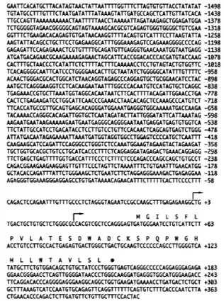
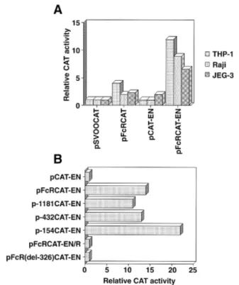
(3) ZNF140 and ZNF91 repress FcγRIIB transcription 1077 each assay. Yeast cells with His⫹ β-gal⫹ phenotypes were selected. The cDNA (prey) plasmids from His⫹ β-gal⫹ yeast colonies were isolated and sequenced. Cell lines The THP-1 human macrophage-like cell line, Raji human Burkitt lymphoma cell line, HSB-2 human T leukemic cell line and JEG-3 human choriocarcinoma cell line were maintained in DMEM medium containing 10% FBS (IBL, Gunma, Japan), 100 µg/ml streptomycin and 100 U/ml penicillin (22–24). Reporter gene assays Cells were transfected with the plasmid by the DEAE–dextran method (Stratagene) or the calcium phosphate method (Stratagene) according to the manufacturer’s instructions. THP-1 and Raji cells (107 cells/tube) were treated with 10 µg of test plasmid in the presence of 250 µg/ml DEAE–dextran sulfate. JEG-3 cells (1.5⫻105 cells/3.5 cm dish) were transfected by the calcium phosphate method. Two days later, cell lysates were prepared and assayed for chloramphenicol acetyl transferase (CAT), luciferase and β-galactosidase activities. The CAT activity was determined by the method described by Gorman et al. (25). After the incubation period, the products were separated from unacetylated chloramphenicol by thin-layer chromatography. The radioactivity was measured using the BioImage analyzer BAS2000 (Fuji Film, Tokyo, Japan). The luciferase and β-galactosidase activities were determined with a Luciferase constant light kit (Roche Diagnostics Japan, Tokyo, Japan) and Galacto-Star (Tropix, Bedford, MA) according to the manufacturer’s instructions. We used the pSVβgal plasmid (0.2 µg) for normalizing transfection efficiencies.. Fig. 1. Nucleotide sequence of the human FcγRIIB 5⬘ flanking region. Two major transcription start sites, as determined by primer extension, are indicated by arrows. The 5⬘ transcription start site is designated as ⫹1. The predicted amino acid sequence, starting at the methionine (M) at ⫹44, is overlined with large letters of the amino acid letter code. A black circle indicates the start of the intron.. Nucleotide accession The nucleotide sequence data reported in this paper will appear in the DDBJ, EMBL and GenBank as nucleotide sequence accession no. D86416. Results Isolation of the 5⬘ flanking region of the human Fcγ RIIB gene and characterization of its promoter region The structure of the human FcγRIIB gene was determined using genomic clones isolated from the EMBL3 Japanese genomic library. Figure 1 shows the 5⬘ flanking sequence of the human FcγRIIB gene. The gene structure of the 5⬘ boundary of the human FcγRIIB in the present study is identical to those reported previously (11,12). By Harr plot analysis, the 5⬘ flanking sequence of the human FcγRIIB gene showed no apparent homology to that of the human FcγRIIA gene reported by McKenzie et al. (16) (data not shown). To determine the transcription start sites, mRNAs from THP-1 and HSB-2 cells that express all three FcγRII transcripts and no FcγRII mRNA respectively were annealed to the primer (complementary to nucleotides 1–23 of the human FcγRIIB structural gene) and then primer extension analysis was performed. We found two major cap sites in human THP-1 cells, while no cap site was noted in HSB-2 cells, as indicated in Fig. 2.. We next determined whether the 5⬘ flanking region of the FcγRIIB gene has promoter activity. As shown in Fig. 3(A), 4.1-, 2.0- and 2.3-fold increases of the CAT activity of cell extracts from THP-1 cells, Raji cells and JEG-3 cells transfected with pFcRCAT were noted as compared with pSVOOCAT. The pFcRCAT-EN produced 12.0-, 8.9- and 3.9-fold increases of the CAT activity in THP-1, Raji and JEG-3 cells respectively over that produced by pCAT-EN (Fig. 3A). Promoter activity was then mapped by comparing CAT expression driven by a series of 5⬘ deletion mutants in THP-1 cells and the results are depicted in Fig. 3(B). The pFcRCAT-EN construct and deletion mutants exhibited significant levels of CAT activity. The pFcRCAT-EN-R and pFcR326CAT-EN, in which the promoter region is in the reverse orientation and the 5⬘ boundary nucleotides (–326 to ⫹21), were internally deleted respectively, did not produce a significant level of CAT activity, as shown in Fig. 3(B). Thus, the 5⬘ boundary for the minimal human FcγRIIB promoter lies between nucleotides –154 and ⫹21. Characterization of nuclear factors bound to the 5⬘ boundary (–148 to –1) of the human Fcγ RIIB gene To examine the DNA-binding factors from various types of cells that bind to the –154 to ⫹1 bp region of the human FcγRIIB gene, EMSA was performed using five oligonucleotides that span –148 to –1 (Fig. 4A). As shown in Fig. 4(C and.
(4) 1078 ZNF140 and ZNF91 repress FcγRIIB transcription. Fig. 2. Mapping of the human FcγRIIB mRNA transcription initiation sites by primer extension analysis. A labeled 23-bp primer complementary to the nucleotides 1–23 of the human FcγRIIB cDNA (structural gene) was hybridized with mRNA from THP-1 human macrophage-like cells (lane 1) or HSB-2 human T leukemic cells (lane 2). Primer was extended by reverse transcriptase, denatured and run on a urea/acrylamide gel. The sequence shown at the left was identified using the same primer except for the absence of the 5⬘ phosphate. Arrows indicate primer elongation stop points.. E), we found that two probes [F2 (–126 to –93) and F4 (–64 to –31)] formed specific bands, because cold F2 and F4 probes inhibited formation of specific bands (bands A, B and C) respectively. The other three probes (F1, F3 and F5) did not reveal any specific band (Fig. 4B, D and F). Since the nuclear factor PU.1 recognized a purine-rich sequence (PU box) (26,27) and the F2 region contained a similar nucleotide sequence, we performed supershift analysis using anti-PU.1 or anti-C/EBPβ antibody. As shown in Fig. 4(G), specific bands A, B and C were not supershifted in the presence of anti-PU.1 or anti-C/EBPβ antibody, although anti-PU.1 and anti-C/EBPβ antibodies could supershift the probes containing PU box derived from human FcγRI gene and CCAAT homology region derived from human IL-6 gene respectively (data not shown). Figure 4(H) shows that F2-binding proteins are different from F4-binding proteins, because the F4 nucleotide could not compete with the 32P-labeled F2 probe. Similarly, the F2 nucleotide did not inhibit complex formation with the 32P-labeled F4 probe (data not shown). To further determine the precise sequences responsible for DNA binding, we next synthesized three overlapping oligonucleotides of the F2 and F4 regions, and performed EMSA (Figs 5 and 6). We found that multiple nuclear factors. Fig. 3. Functional analysis of the promoter region of the human FcγRIIB gene. (A) Various chimeric genes (10 µg) were each transfected into THP-1, Raji and JEG-3, and CAT activity was measured 48 h later. The cell extracts containing 33 µg of protein were incubated with [14C]chloramphenicol at 37°C for 4 h. The substrate converted to the acetylated form was separated by thin-layer chromatography and CAT activities were expressed as percent conversions. Similar results were obtained in three other experiments. (B) Chimeric genes including pFcRCAT-EN, p-1181CAT-EN, p-432CAT-EN, p-154CAT-EN, pFcRCAT-EN-R, pFcR-326CAT-EN and pCAT-EN (10 µg) were each transfected into THP-1 cells. CAT activities were determined using 50 µg of cell extract with 4-h incubation. These data represent one of three experiments performed independently.. bound to the F2-3 (–109 to-93) (Fig. 5C) and F4-3 (–47 to –31) (Fig. 6C) oligonucleotides respectively. No specific bands were noted using the F2-1 (Fig. 5A), F2-2 (Fig. 5B), F4-1 (Fig. 6A) or F4-2 (Fig. 6B) probes. We then introduced mutations in the F2-3 (Fig. 5D) and F4-3 (Fig. 6D) oligonucleotides, and performed EMSA. As shown in Figs 5(E) and 6(E), the F2-related probes (F2-3-1, F2-3-2 and F2-3-3) and F4-related probes (F4-3-1, F4-3-2 and F4-3-3) lost the ability to bind the 32P-labeled F2-3 and F4-3 probes respectively. Thus, these results indicate that the sequences of GGGAGGAGC (–105 to –97) and AATTTGTTTGCC (–47 to –36) within the human FcγRIIB gene promoter are responsible for binding of multiple nuclear factors (Figs 5E and 6E). Cloning of the F2-3 and F4-3 region-binding protein by onehybrid assay To identify nuclear proteins that bind to the F2-3 and F4-3 regions of the human FcγRIIB promoter, we used a one-hybrid assay. As bait sequences, we used (GGGAGGAGC)⫻3 and (AATTTGTTTGCC)⫻4 for F2-3- and F4-3-binding proteins respectively. We screened ~5⫻105 clones of a cDNA library prepared from human placenta in each assay. We isolated.
(5) ZNF140 and ZNF91 repress FcγRIIB transcription 1079. Fig. 4. Multiple nuclear factors from various types of cells bind to the –154 bp 5⬘ flanking region of the human FcγRIIB gene. (A) The DNA sequences of five oligonucleotides used for EMSA. (B–F) Nuclear extracts from THP-1, Raji and JEG-3 cells were mixed with the 32P-labeled probe. An oligonucleotide containing the NF-κB motif was used as an unrelated competitor at a 150 M excess. Bound and unbound probes were separated in native polyacrylamide gel. The arrow indicates the positions of the complex. (G) Nuclear extracts were then prepared, preincubated with antibodies (2 µl/lane) against PU.1 and C/EBPβ, and analyzed by EMSA. The arrow indicates the position of the complex. (H) Nuclear extracts were mixed with the 32P-labeled F2. A 150 M excess of unlabeled oligonucleotide competitors was added. The arrow indicates the position of the complex..
(6) 1080 ZNF140 and ZNF91 repress FcγRIIB transcription. Fig. 5. Analysis of the F2 region-specific EMSA complex. (A–C) Nuclear extracts from THP-1, Raji and JEG-3 cells were mixed with the 32Plabeled probe. An oligonucleotide containing the NF-κB motif was used as an unrelated competitor at a 150 M excess. Bound and unbound probes were separated in native polyacrylamide gel. The arrow indicates the positions of the complex. (D) The DNA sequences of four oligonucleotides used for EMSA. (E) Nuclear extracts were mixed with the 32P-labeled F2-3. A 150 M excess of unlabeled oligonucleotide competitors was added. The arrow indicates the position of the complex.. 12 and 21 His⫹ β-gal ⫹ clones in the F2-3- and F4-3-binding assays respectively. We then isolated and sequenced cDNA (prey) plasmids, and found that plasmids from two clones (F2-3-binding protein) and three clones (F4-3-binding protein) contained partial sequences that are identical to those of the human zinc-finger protein 140 (ZNF140) (19) and zinc-finger protein 91 (ZNF91) (20) respectively. Other clones were not analyzed in the present study. To confirm the binding specificity of the two zinc-finger proteins, we transformed yeasts carrying either F2-3-EL/pHISi-1 and F2-3-EL/pLacZi or F4-3-EL/pHISi-1 and F4-3-EL/pLacZi with pZNF140/pACT2, pZNF91/pACT2 or pACT2. Colonies were tested for β-galactosidase activity. Figure 7(A) shows the typical results. ZNF140/pACT2 and ZNF91/pACT2 could specifically bind to the F2-3 and F4-3 elements respectively in yeast, so that β-galactosidase activity was detected. Conversely, ZNF140/pACT2 and ZNF91/pACT2 did not interact with the F4-3 and F2-3 elements respectively. Figure 7(B) shows that the full lengths of ZNF140 and ZNF91 consisted. of 457 and 1191 amino acid residues respectively. We sequenced the inserts from F2-3- and F4-3-binding clones, and found that the inserts encoded zinc-finger structures of the fragments of ZNF140 and ZNF91 proteins respectively (Fig. 7B, open rectangles). As shown previously (19,20), these transcripts were ubiquitously expressed on various types of cells (data not shown). ZNF140 and ZNF91 function as repressors in –154 human FcγRIIB promoter-mediated luciferase expression Having established that two zinc-finger proteins can specifically bind to the F2-3 and F4-3 region respectively, it is of great interest to determine the function of these proteins. We constructed expression plasmids and transfected them into JEG-3 cells with the reporter plasmid (pFcR2-154/pGL3EN). As shown in Fig. 7(C), when either human ZNF140 or ZNF91 was expressed, both proteins inhibited luciferase activity. Furthermore, the two proteins demonstrated an additive suppressive effect when they were simultaneously expressed..
(7) ZNF140 and ZNF91 repress FcγRIIB transcription 1081. Fig. 6. Analysis of the F4 region-specific EMSA complex. (A–C) Nuclear extracts from THP-1, Raji and JEG-3 cells were mixed with the 32Plabeled probe. An oligonucleotide containing the NF-κB motif was used as an unrelated competitor at a 150 M excess. Bound and unbound probes were separated in native polyacrylamide gel. The arrow indicates the positions of the complex. (D) The DNA sequences of four oligonucleotides used for EMSA. (E) Nuclear extracts were mixed with the 32P-labeled F4-3. A 150 M excess of unlabeled oligonucleotide competitors was added. The arrow indicates the position of the complex.. We also obtained the similar results using human 293T and HepG2 cell lines (data not shown). Discussion In humans, three FcγRII genes (FcγRIIA, FcγRIIB and FcγRIIC) are differentially regulated in a variety of cells. In the present study, we found that the promoter regions of the human FcγRIIA and FcγRIIB genes are different. First, we found that a Harr plot between the FcγRIIA and FcγRIIB promoter sequences indicated no apparent homology (data not shown). Second, two different transcription start sites were found in the human FcγRIIA promoter. One transcription start site is located at a 5⬘-UT exon ~1 kb 5⬘ to the ATG translation initiation codon, while a second start site was mapped near the ATG codon. On the other hand, we found that the single exon continuous with the ATG codon contains at least two transcription start sites in the human FcγRIIB gene, the major one 42 bp 5⬘ to ATG and a minor one 22 bp 5⬘ to ATG in THP-1 cells (Figs 1 and 2). We could not detect any transcription start site 5⬘ to the 42 bp 5⬘ to the ATG codon by primer extension analysis (data not shown), thus indicating that the human FcγRIIB gene does not contain a discrete 5⬘-UT region. Our results are similar to those for the mouse FcγRIIIA gene. (28) and the human FcγRI gene (29–31), both of which have multiple transcription start sites mapped near the ATG codon. Third, we found that various elements found in the FcγRIIA and FcγRIIB gene promoters are not identical. Thus, all of these findings indicate that the expression profiles of the FcγRIIA and FcγRIIB genes are differentially regulated. In the present study, we found that the 5⬘ boundary for the minimal human FcγRIIB promoter lies between nucleotides –154 and ⫹21 in our assay system. This result is similar to those for the human FcγRI (30–34) and mouse FcγRIIIA (28) genes, whose promoters have minimal structures (~150 bp upstream of the cap site) with an IFN-γ-responsive region (GRR) and a PU box, and a PU box and myeloid-restricted region (MRR) respectively. Mutation analysis of the F2-3 and F4-3 regions indicated that the sequences of GGGAGGAGC (–105 to –97) and AATTTGTTTGCC (–47 to –36) within the human FcγRIIB promoter participated in the binding by nuclear factors. The sequence of the F2-3 region is similar to that of the PU.1-responsive element, but the PU.1 nuclear factor did not bind to the F2-3 region, since anti-PU.1 antibody did not supershift (Fig. 4G). This result is in contrast to that for the human FcγRI gene promoter, since the PU.1 was shown to bind to the –107 to –74 region of the human FcγRI promoter (32,33). Thus, these results indicate that the expression.
(8) 1082 ZNF140 and ZNF91 repress FcγRIIB transcription. Fig. 7. Cloning and function of the human ZNF140 and ZNF91. (A) Yeast strains containing F2-3-EL/pLacZi and F2-3-EL/pHISi-1 or F43-EL/pLacZi and F4-3-EL/pHISi-1 were transformed with pACT2, ZNF140/pACT2 or ZNF91/pACT2. The colony was tested for βgalactosidase assay. (B) Structure of the ZNF140 and ZNF91 proteins. Open rectangles indicate the portions of ZNF140 and ZNF91 proteins encoded by the inserts from the F2-3- and F4-3-binding clones identified by a one-hybrid assay. (C) Functional assay of ZNF140 and ZNF91. Ten micrograms of each of pcDNA3.1, pcDNA3.1ZNF140, pCAGGS, pCAGGS-ZNF91 and pFcR2-154/pGL3EN together with 0.2 µg of pSVβgal were transfected into JEG-3 cells. After a 48-h incubation period, cell lysates were prepared and then assayed for luciferase and β-galactosidase activities. Transfection efficiency was normalized by β-galactosidase assay. Data of group 2, 4 and 6 were relative to those of group 1, 3 and 5 respectively. These data represent one of three experiments performed independently.. profiles of the human FcγRIIB and FcγRI genes are also differently regulated. We also isolated the F2-3- and F4-3-binding proteins from the human placenta library by a one-hybrid assay. We found that the human ZNF140 and ZNF91 were the F2-3 and F4-3 region-binding proteins respectively (Fig. 7). These ZNF140 and ZNF91 genes were previously isolated by PCR with zincfinger motifs as primers (19,20,35). In humans, it has been. estimated that there are 300–700 different zinc-finger protein genes and the vast majority of zinc-finger proteins contained a Kruppel-associated box (KRAB), such as the C2H2 type (36,37). These genes are well conserved and were distributed from Drosophila to humans (35,36). Drosophila Kruppel and hunchback ZNF proteins are involved in embryonic pattern formation, while mouse Krox-20 in both patterning of the hindbrain and control of cell proliferation (38,39). Most zincfinger proteins have been shown to be involved in development, but their other functions remain obscure. Vissing and his group reported that ZNF140, ZNF133, ZNF136 and ZNF141— each of which contains a KRAB box—function as transcription repressors when fused to a heterologous DNA-binding domain from the yeast GAL4 protein (40,41). We extended the previous findings by indicating that ZNF140 functioned as a transcription repressor when the human FcγRIIB natural promoter region was connected to the luciferase gene (Fig. 7C). We also found that ZNF91 was also a transcription repressor, although the function of this protein is not yet known. These results indicate that ZNF140 and ZNF91 function as transcription repressors in human FcγRIIB gene expression. Furthermore, T lymphoid cells do not express all three FcγRII transcripts, but high amounts of ZNF91 transcripts are expressed in human T lymphoid cells and T leukemic cell lines (data not shown), HSB-2 and CEM, as found by Bellefroid et al. (20). The fact that ZNF91 functions as a repressor in the FcγRIIB transcription suggests that ZNF91 may repress the FcγRIIB transcription in T lymphoid cells, leading to no expression of the human FcγRIIB. However, we do not yet know the precise mechanisms by which ZNF91 represses transcription. Calame and her group reported that the ZF5 zinc-finger protein activated the HIV-1 long terminal repeat promoter, and repressed the β-actin, c-myc and herpes simplex thymidine kinase promoters (42,43). ZF5 zinc-finger protein thus can both activate and repress in the context of different natural promoters. In the Drosophila system, Sauer and Jackle (44) reported that Kruppel (Kr), required for normal thorax and abdominal development, itself acts as a concentrationdependent positive and negative regulator of transcription. In the present study, we found that multiple nuclear factors could bind to the same F2-3 and F4-3 regions. Thus, other unknown positive factors might regulate the transcription of the human FcγRIIB gene. Alternatively, the ZNF140 and ZNF91 might work as positive regulators at the optimal concentrations, as suggested by Sauer and Jackle (44). Furthermore, THP-1, Raji and JEG-3 cell lysates gave different band patterns in the EMSA assay, indicating that various types of cells might contain different nuclear factors for these regions. Taken collectively, these results indicate that the regulation of the human FcγRIIB gene expression might be complex in various types of cells. Identification of all promoterbinding proteins will resolve these issues.. Acknowledgements We thank T. Iwata and A. Tamori for technical assistance. We also thank D. Mrozek and T. Matsui for their comments. This work was partially supported by Japan Health Sciences Foundation..
(9) ZNF140 and ZNF91 repress FcγRIIB transcription 1083 Abbreviations CAT EMSA FcγRI FcγRII KRAB UT. chloramphenicol acetyl transferase electrophoretic mobility shift assay high-affinity Fc receptor I for IgG low-affinity Fc receptor II for IgG Kruppel-associated box untranslated. References 1 Fanger, M. W., Shen, L., Grazione, R. F. and Guyre, P. M. 1989. Cytotoxicity mediated by human Fc receptors for IgG. Immunol. Today 10:92. 2 Ravetch, J. V. and Kinet, J.-P. 1991. Fc receptors. Annu. Rev. Immunol. 9:457. 3 van de Winkel, J. G. J. and Anderson, C. L. 1991. Biology of human Immunoglobulin G Fc receptors. J. Leuk. Biol. 49:511. 4 Anderson, C. L., Shen, L., Eicher, D. M., Wewers, M. D. and Gill, J. K. 1990. Phagocytosis mediated by three distinct Fγ receptor classes on human leukocytes. J. Exp. Med. 171:1333. 5 Leslie, R. C. Q. 1985. Complex aggregation: a critical event in macrophage handling of soluble immune complexes. Immunol. Today 6:183. 6 Nathan, C. and Cohn, Z. 1980. Role of oxygen-dependent mechanisms in antibody-induced lysis of tumor cells by activated macrophages. J. Exp. Med. 152:198. 7 Kipps, T. J., Parham, P., Punt, J. and Herzenberg, L. A. 1985. Importance of immunoglobulin isotype in human antibodydependent cell-mediated cytotoxicity directed by murine monoclonal antibodies. J. Exp. Med. 161:1. 8 Ferreri, N. R., Howland, W. C. and Spiegelberg, H. L. 1986. Release of leukotrienes C4 and B4 and prostaglandin E2 from human monocytes stimulated with aggregated IgG, IgA, and IgE. J. Immunol. 136:4188. 9 Bich Thuy, L. T. and Revillard, J. P. 1982. Selective suppression of human B lymphocyte differentiation into IgG-producing cells by soluble Fcγ receptors. J. Immunol. 129:150. 10 Yamamoto, K. and Johnston, R., Jr. 1984. Dissociation of phagocytosis from stimulation of the oxidative metabolic burst in macrophages. J. Exp. Med. 159:405. 11 Brooks, D. G., Qiu, W. Q., Luster, A. D. and Ravetch, J. V. 1989. Structure and expression of human IgG FcR II (CD32) Functional heterogeneity is encoded by the alternatively spliced products of multiple genes. J. Exp. Med. 170:1369. 12 Qiu, W. Q., de Bruin, D., Brownstein, B. H., Pearse, R. and Ravetch, J. V. 1990. Organization of the human and mouse lowaffinity FcγR genes: duplication and recombination. Science 248:732. 13 Ravetch, J. V. and Anderson, C. L. 1990. FcγR family: proteins, transcripts, and genes. In Metzger, H., ed., Fc Receptors and the Action of Antibodies, p. 211. American Society for Microbiology, Washington, DC. 14 Cassel, D. L., Keller, M. A., Surrey, S., Schwartz, E., Schreiber, A. D., Rappaport, E. C. and McKenzie, S. E. 1993. Differential expression of FcγRIIA, FcγRIIB and FcγRIIC in hematopoietic cells: analysis of transcripts. Mol. Immunol. 30:451. 15 Stuart, S. G., Simister, N. E., Clarkson, S. B., Kacinski, B. M., Shapiro, M. and Mellman, I. 1989. Human IgG Fc receptor (hFcRII; CD32) exists as multiple isoforms in macrophages, lymphocytes and IgG-transporting placental epithelium. EMBO J. 8:3657. 16 McKenzie, S. E., Keller, M. A., Cassel, D. L., Schreiber, A. D., Schwartz, E., Surrey, S. and Rappaport, E. F. 1992. Characterization of the 5⬘-flanking transcriptional regulatory region of the human Fcγ receptor gene, FcγRIIA. Mol. Immunol. 29:1165. 17 Niwa, H., Yamamura, K. and Miyazaki, J. 1991. Efficient selection for high-expression transfectants with a novel eukaryotic vector. Gene 108:193. 18 Stenegelin, S., Stamenkovic, I. and Seed, B. 1988. Isolation of cDNAs for two distinct human Fc receptors by ligand affinity cloning. EMBO J. 7:1053.. 19 Tommerup, N. and Vissing, H. 1995. Isolation and fine mapping of 16 novel human zinc finger-encoding cDNAs identify putative candidate genes for developmental and malignant disorders. Genomics 27:259. 20 Bellefroid, E. J., Marine, J.-C., Ried, T., Lecocq, P. J., Riviere, M., Amemiya, C., Poncelet, D. A., Coulie, P. G., de Jong, P., Szpirer, C., Ward, D. C. and Martial, J. A. 1993. Clustered organization of homologous KRAB zinc-finger genes with enhanced expression in human T lymphoid cells. EMBO J. 12:1363. 21 Dignam, J. D., Lebovitz, R. M. and Roeder, R. G. 1983. Accurate transcription initiation by RNA polymerase II in a soluble extract from isolated mammalian nuclei. Nucleic Acids Res. 11:1475. 22 Kurata, N., Akiyama, H., Taniyama, T. and Marunouchi, T. 1989. Dose-dependent regulation of macrophage differentiation by mos mRNA in a human monocytic cell line. EMBO J. 8:457. 23 Taniyama, T., Yoshida, K. and Furuta, T. 1988. Demonstration of a novel tumor killing factor secreted from human macrophagemonocyte hybridomas. J. Immunol. 141:4061. 24 Tsuchiya, S., Yamabe, M., Yamaguchi, Y., Kobayashi, Y., Konno, T. and Tada, K. 1980. Establishment and characterization of a human acute monocytic leukemia cell line (THP-1). Int. J. Cancer 26:171. 25 Gorman, C. M., Moffat, L. F. and Howard, B. H. 1982. Recombinant genomes which express chloramphenicol acetyltransferase in mammalian cells. Mol. Cell. Biol. 2:1044. 26 Klemsz, M. J., McKercher, S. R., Celada, A., Beveren, C. V. and Maki, R. A. 1990. The macrophage and B cell-specific transcription factor PU.1 is related to the ets oncoprotein. Cell 61:113. 27 Ray, D., Bosselut, R., Ghysdale, J., Mattei, M.-G., Tavitian, A. and Moreau-Gachelin, F. 1992. Characterization of Spi-B, a transcription factor related to the putative oncoprotein Spi-1/PU.1. Mol. Cell. Biol. 12:4297. 28 Feinman, R., Qiu, W. Q., Pearse, R. N., Nikolajczyk, S., Sen, R., Sheffery, M. and Ravetch, J. V. 1994. PU.1 and an HLH family member contribute to the myeloid-specific transcription of the FcγRIIIA promoter. EMBO J. 13:3852. 29 van de Winkel, J. G. J., Ernst, L. K., Anderson, C. L. and Chiu, I.-M. 1991. Gene organization of the human high affinity receptor for IgG, FcγRI (CD64). J. Biol. Chem. 266:13449. 30 Pearse, R. N., Feinman, R. and Ravetch, J. V. 1991. Characterization of the promoter of the human gene encoding the high-affinity IgG receptor: transcriptional induction by γ-interferon is mediated through common DNA response elements. Proc. Natl Acad. Sci. USA 88:11305. 31 Benech, P. D., Sastry, K., Iyer, R. R., Eichbaum, Q. G., Raveh, D. P. and Ezekowitz, R. A. B. 1992. Definition of interferon γresponse elements in a novel human Fcγ receptor gene (FcγRIb) and characterization of the gene structure. J. Exp. Med. 176:1115. 32 Eichbaum, Q. G., Iyer, R., Raveh, D. P., Mathieu, C. and Ezekowitz, R. A. B. 1994. Restriction of interferon γ responsiveness and basal expression of the myeloid human FcγgR1b gene is mediated by a functional PU.1 site and a transcription initiator consensus. J. Exp. Med. 179:1985. 33 Perez, C., Wietzerbin, J. and Benech, P. D. 1993. Two cis-DNA elements involved in myeloid-cell-specific expression and gamma interferon (IFN-γ) activation of the human high-affinity Fcγ receptor gene: a novel IFN regulatory mechanism. Mol. Cell. Biol. 13:2182. 34 Perez, C., Coeffier, E., Moreau-Gachelin, F., Wietzerbin, J. and Benech, P. D. 1994. Involvement of the transcription factor PU.1/Spi-1 in myeloid cell-restricted expression of an interferoninducible gene encoding the human high-affinity Fcγ receptor. Mol. Cell. Biol. 14:5023. 35 Bellefroid, E. J., Poncelet, D. A., Lecocq, P. J., Revelant, O. and Martial, J. A. 1991. The evolutionarily conserved Kruppelassociated box domain defines a subfamily of eukaryotic multifingered proteins. Proc. Natl Acad. Sci. USA 88:3608. 36 Bellefroid, E. J., Lecocq, P. J., Benhida, A., Poncelet, D. A., Belayew, A. and Martial, J. A. 1989. The human genome contains hundreds of genes coding for finger proteins of the Kruppel type. DNA 8:377..
(10) 1084 ZNF140 and ZNF91 repress FcγRIIB transcription 37 Crossley, P. H. and Little, P. F. R. 1991. A cluster of related zinc finger protein genes is deleted in the mouse embryonic lethal mutation tw18. Proc. Natl Acad. Sci. USA 88:7923. 38 Tautz, D., Lehmann, R., Schnurch, H., Schuh, R., Seifert, E., Kienlin, A., Jones, K. and Jackle, H. 1987. Finger protein of novel structure encoded by hunchback, a second member of the gap class of Drosophila segmentation genes. Nature 327:383. 39 Chavrier, P., Lemaire, P., Revelant, O., Bravo, R. and Charnay, P. 1988. Characterization of a mouse multigene family that encodes zinc finger structure. Mol. Cell. Biol. 8:1319. 40 Margolin, J. F., Friedman, J. R., Meyer, W. K.-H., Vissing, H., Thiesen, H.-J. and Raucher, I. F. J. 1994. Kruppel-associated boxes are potent transcriptional repression domains. Proc. Natl Acad. Sci. USA 91:4509.. 41 Vissing, H., Meyer, W. K., Aagaard, L., Tommerup, N. and Thiesen, H. J. 1995. Repression of transcriptional activity by heterologous KRAB domains present in zinc finger proteins. FEBS Lett. 369:153. 42 Kaplan, J. and Calame, K. 1997. The ZiN/POZ domain of ZF5 is required for both transcriptional activation and repression. Nucleic Acids Res. 25:1108. 43 Numoto, M., Niwa, O., Kaplan, J., Wong, K.-K., Merrell, K., Kamiya, K., Yanagihara, K. and Calame, K. 1993. Transcriptional repressor ZF5 identifies a new conserved domain in zinc finger proteins. Nucleic Acids Res. 21:3767. 44 Sauer, F. and Jackle, H. 1991. Concentration-dependent transcriptional activation or repression by Krupple from a single binding site. Nature 353:563..
(11)
Figure


+3


Documents relatifs
The human thyroglobulin protein contains four major thyroid hormone synthesis sites [21]; An alignment of thyroglobu- lin sequences showed that the zebra finch, zebrafish and
Wang, Tight junction proteins 10 claudin-1 and occludin control hepatitis C virus entry and are downregulated during infection 11 to prevent superinfection, J Virol, 83
On the contrary, in these systems, the virtual peer’s ability to engage in positive social interaction, in the form of building rapport, plays a key role in improving the
of brisding artillery benign gunfire a ratde of sabres shrill whine of shells as into the tattoo arena high above the crowd swings a wild trapeze suspending froms
Keywords: Scheduling, workflows, heterogeneous platforms, period, critical resource, timed Petri
We tested the potential of the existing spectral (RC30, ADS40, SPOT5) and topo-structural (LIDAR) data sets to predict the floristic gradients in a set of seven mire habitats..
Keywords: contingent encounters, corridor conversation, corridor occupation, hospital staff, mobility, multimodal conversation analysis, nurses, outpatient clinic,
Reporter gene assays were performed with extracts from MCF-7 cells that were transiently transfected with a luciferase reporter construct containing two copies of RE2, RE5, RE7,
