Anxiety-like features and spatial memory
problems as a consequence of hippocampal
SV2A expression
Maria Elisa SerranoID1☯*, Odile Bartholome´2☯, Priscilla Van den Ackerveken2,
Andre´ Ferrara3, Bernard Rogister2,4, Alain PlenevauxID1, Ezio Tirelli3
1 GIGA-CRC In vivo imaging, University of Liège, Liège, Belgium, 2 GIGA-Neurosciences, University of Liège, Liège, Belgium, 3 Department of Psychology, University of Liège, Liège, Belgium, 4 Neurology Department, CHU, Academic Hospital, University of Liège, Liège, Belgium
☯These authors contributed equally to this work.
Abstract
The Synaptic Vesicle Protein 2A (SV2A) is a transmembrane protein whose presence is reduced both in animal models and in patients with chronic epilepsy. Besides its implication in the epileptic process, the behavioural consequences of the changes in its expression remain unclear. The purpose of our research is to better understand the possible role(s) of this protein through the phenotype of cKO (Grik4 Cre+/-, SV2A lox/lox) mice, male and female, which present a specific decrease of SV2A expression levels in the hippocampal glutamatergic neurons but without any epileptic seizures. In this study, we compare the cKO mice with cHZ (Grik4 Cre+/-, SV2A lox/+) and WT (Grik4 Cre+/+, SV2A lox/lox) mice through a battery of tests, used to evaluate different features: the anxiety-related features (Elevated Plus Maze), the locomotor activity (Activity Chambers), the contextual fear-related memory (Contextual Fear Conditioning), and the spatial memory (Barnes Maze). Our results showed statistically significant differences in the habituation to a new environment, an increase in the anxiety levels and spatial memory deficit in the cHZ and cKO groups, compared to the WT group. No statistically significant differences due to the genotype appeared in the spontaneous locomotor activity or the fear-linked memory. However, sexual differences were observed in this last feature. These results highlight not only an important role of the SV2A protein in the cognitive and anxiety problems typically encountered in epi-leptic patients, but also a possible role in the symptomatology of other neurodegenerative diseases, such as the Alzheimer’s disease.
Introduction
The SV2 protein family comprises three integral membrane paralogs: SV2A, SV2B, and SV2C. In spite of sharing approximately 60% of their sequences, these three isoforms are implicated in different pathologies, thus suggesting a specific role for each of them [1–4]. Amongst these isoforms, the most studied is the SV2A protein, due to its ubiquitous expression in the brain
a1111111111 a1111111111 a1111111111 a1111111111 a1111111111 OPEN ACCESS
Citation: Serrano ME, Bartholome´ O, Van den
Ackerveken P, Ferrara A, Rogister B, Plenevaux A, et al. (2019) Anxiety-like features and spatial memory problems as a consequence of hippocampal SV2A expression. PLoS ONE 14(6): e0217882.https://doi.org/10.1371/journal. pone.0217882
Editor: Giuseppe Biagini, University of Modena and
Reggio Emilia, ITALY
Received: January 23, 2019 Accepted: May 20, 2019 Published: June 5, 2019
Copyright:© 2019 Serrano et al. This is an open access article distributed under the terms of the
Creative Commons Attribution License, which permits unrestricted use, distribution, and reproduction in any medium, provided the original author and source are credited.
Data Availability Statement: All relevant data are
within the manuscript and its Supporting Information files.
Funding: This work has been supported by Action
de Recherches Concerte´es (ARC, grant number 13/ 17-07) (AP, MES, OB, BR), la Communaute´ Franc¸aise de Belgique (BR) and by the FNRS (BR). AP is research director from Fonds De La Recherche Scientifique -FNRS Belgium. The funders had no role in study design, data collection
and its implication in epileptic disease (see hereunder). Beyond the initial suggestion of its role as a transporter of ions or neurotransmitters [5,6], the SV2A protein seems to act mainly as modulator of the synaptic transmission. The invalidation of this protein does not alter the morphology of the brain, the amount of synapses, or their structure [6,7]. However, its absence reduces the neurotransmission [8,9] and may induce an imbalance between glutamatergic and GABAergic levels [10–13]. Furthermore, three key findings have identified the SV2A protein as an all-important molecule involved in the epileptic process. Accordingly, the authors in [7,14] showed that homozygous SV2A KO mice displayed severe seizures at P7, and died in
status epilepticus two to three weeks after birth. In humans, the homozygous mutation in
SV2A has been also associated with intractable epilepsy, microcephaly, and developmental and growth retardation [15]. Moreover, SV2A has been proved to be the specific molecular target of new antiepileptic drugs, such as the levetiracetam [16] or the brivaracetam [17,18].
Different studies have explored the role of the SV2A protein in epilepsy: in humans, several epileptic disorders are related with a reduction in the amount of SV2A [1,19,20], suggesting a role of this protein in the epileptogenesis and/or the ictiogenesis [12,13,21]. However, the pos-sible implication of SV2A in the cognitive impairment and mood disorders accompanying epileptic disease is still unknown. In light of this, several articles have associated the use of leve-tiracetam with memory and executive function enhancement [22–24]. As for other patholo-gies, the use of this antiepileptic drug has highlighted a pro-cognitive effect as well [25–28], suggesting a role of SV2A in cognition.
The studies carried out with male knockout mice have also provided interesting insights regarding the role of the SV2A protein in cognition. In particular, the phenotyping of the male heterozygous SV2A (+/-) mice revealed no motor differences but rather anxiety-like features in these mice compared with the wild type [29]. Recently, a cKO mouse model (Grik4 Cre+/-, SV2A lox/lox) has been produced and validated by our laboratory using the Cre/loxP
recombi-nation system [30]. This model is characterized by a decrease of the expression of the SV2A protein specifically in the glutamatergic pyramidal neurons of the hippocampus, one of the most affected brain regions in epilepsy [31–34]. Furthermore, this cKO model, in contrast to the SV2A homozygous or heterozygous mice, does not present an epileptic or pro-epileptic phenotype (characterized by a lower epileptic threshold, evaluated with thepentylenetetrazol
test) [30], thus increasing their survival and facilitating their evaluation in different tests [7,14,35].
In this article we evaluate the consequences of the hippocampal downregulation of the SV2A protein in the phenotype ofGrik4 Cre+/-, SV2A lox/lox mice. Using both, males and
females, we studied the possible existence of sex-differences in the cognitive and behavioural processes evaluated, some of them already reported in epileptic patients [36–38]. Thus, the main goal of this research is to unravel the possible role of this protein in the cognitive and behavioural processes associated with the hippocampal structure, offering useful insights into the role of SV2A, not only in the pathology but also in the healthy brain.
Materials and methods
Animals
Homozygous SV2A conditional knockout mice of both sexes (cKO;Grik4 Cre+/-, SV2A lox/ lox), presenting a decrease of SV2A protein expression in excitatory glutamatergic neurons
of the dentate gyrus and the CA3 hippocampal area, were generated by using the Cre/loxP recombination system, following the procedure published in [30]. Their phenotype was com-pared with such of the heterozygous (cHZ;Grik4 Cre+/-, SV2A lox/+) and with the phenotype
and analysis, decision to publish, or preparation of the manuscript.
Competing interests: The authors have declared
that no competing interests exist.
Abbreviations:ηp2, partial eta squared; ACT, Activity chambers; BM, Barnes Maze; CFC, Contextual Fear Conditioning; cHZ mice, Grik4 Cre
+/-, SV2A lox/+; cKO mice, Grik4 Cre+/-, SV2A lox/ lox; CS, Conditioned stimulus; EPM, Elevated Plus
Maze; SV2A, Synaptic Vesicle protein 2A; US, Unconditioned stimulus; WT, Grik4 Cre+/+, SV2A
of wild type mice (WT;Grik4 Cre+/+, SV2A lox/lox). All animals were genotyped by PCR
fol-lowing the protocol described in [30].
A total of 141 mice were employed: 66 males and 69 females. Amongst these animals, 27 males and 36 females were also evaluated using the Barnes Maze test to detect spatial learning or memory problems specifically associated with the hippocampus.
Note that, in principle, for a power of 0.8 and at a critical threshold of 0.05 (alpha), these sample sizes are able to detect medium to very large effects (ηp2> 0.14), but not small effects
which require greater sample sizes [39]. Related calculations were carried out with G�Power software [40].
Experimental design
Two weeks before starting the experiments, six-week old mice belonging to the three geno-types were housed in individual standard transparent poly-carbonate cages (31.5 cm (L)× 15.5 cm (W)× 13 cm (H)) with pine sawdust bedding. During all the experimental procedure, the distance between the cages allowed the animals to maintain visual, olfactory and acoustic inter-actions. Furthermore, tap water and food (standard pellets, Carfil Quality, Oud-Turnhout, Bel-gium) were providedad libitum. The animal room was maintained on a 12:12h dark-light
cycle (lights on at 7:30 am, off at 7:30 pm), at an ambient temperature of 20–24˚C.
When the mice were eight weeks old, the experimental procedure started, performing the tests during the 12h light cycle (between 9 a.m. and 3 p.m.) (seeFig 1). The first day, the anxi-ety-related behaviour was evaluated with the Elevated Plus Maze (EPM). From days four to six, the spontaneous locomotor activity and the habituation to the environment was evaluated with activity chambers (ACT). The contextual fear conditioning memory was assessed with the contextual fear conditioning test (CFC), conducted as follows: on day nine (acquisition trial), mice were placed in the conditioning chamber for the context (conditioned stimulus, CS)–shock (unconditioned stimulus, US) pairing; then, the contextual fear conditioning mem-ory was evaluated one hour, one day, and six days later. Finally, the spatial memmem-ory was assessed two weeks later with the Barnes Maze test (BM) using the following method: (1) the mice were trained during four consecutive days (acquisition trials) to spatially locate a target hole in the Barnes Maze; (2) the spatial memory of the mice was evaluated the following day (probe trial); (3) after two weeks, mice were subject to the same 4-day training in order to assess their memory retention and to determine which level of performance they were able to reach; (4) the first two days after this last training, the cognitive flexibility of mice was tested in a reversal learning procedure. At the end of the experimental procedure (second day of rever-sal learning), animals were sacrificed by cervical dislocation.
Fig 1. Experimental time-line and design. After two weeks of accommodation to the animal laboratory facilities, 69
males and 72 females belonging to the three genotypes (WT, cHZ, and cKO) were evaluated for anxiety-related behaviour (EPM, d1), spontaneous locomotor activity (ACT, from d4 to d6), and contextual fear conditioning memory (CFC, from d9 to d15). Following two weeks of recovery, the spatial memory was assessed (BM, from d29 to d55) with a procedure divided in four parts with a duration of 4 + 1 days, a period of consolidation of two weeks, and 4 + 2 days respectively. Abbreviations: WT:Grik4 Cre+/+, SV2A lox/lox; cHZ: Grik4 Cre+/-, SV2A lox/+; cKO: Grik4 Cre+/-, SV2A lox/lox; EPM: Elevated Plus Maze; ACT: Activity chambers; CFC: Contextual fear conditioning; BM: Barnes
Maze.
The three parts of the experimental process (testing, scoring, and statistical analysis) were blinded: animals were identified as members of A, B, or C groups, without information about the genotype associated to the letters until the generation of results. This course of action elim-inates intentional or subconscious bias which could interfere in the experimental procedure.
The experimental procedures and protocols used in this investigation were reviewed and approved by the Institutional Animal Care and Use Committee of the University of Liege (dos-siers 1258 and 1573), according to the Helsinki declaration, and conducted in accordance with the European guidelines for care of laboratory animals (2010/63/EU). All efforts were made to minimise the number of animals used and their suffering. Moreover, the ARRIVE guidelines (Animal Research Reporting In Vivo Experiments) [41] was followed as closely as possible to confer a minimal intrinsic quality to the study.
Anxiety-related behaviour: EPM
The EPM was used to evaluate fearfulness/anxiety in mice in a single session. This apparatus consists of two open and two closed arms (29 cm (L)× 5 cm (W) × 2.5 cm (H) each one) emerging from a central platform (5 cm× 5 cm) to form a plus shape, which sits 80 cm above the ground level. The floor and walls of the enclosed arms were made of black hard plastic (Forex), while the floor of the open arms was made of grey hard plastic (Forex). The behaviour of the mice was recorded with a webcam (Logitech QuickCam Pro 5000).
For testing, each mouse was placed in the central platform, facing the left open arm, and was allowed to freely explore the maze during five minutes [42]. After each trial the floor was wiped clean with Ethanol 70% and dried.
Two parameters were evaluated: (1) the percentage of entries in the open arms with respect to the total (open / open + closed), and (2) the percentage of time spent in the open arms with respect to the total testing time (5 minutes). An entry was scored when the mouse had entered the arm with all four paws. The natural aversion of rodents for open spaces along with the rela-tionship between anxiogenic drugs and a reduction in the percentage of time spent in the open arms, make both parameters reliable measures of anxiety in mice [43–45]. Data (seeS1 Data) were analysed with a two-way 3× 2 ANOVA, considering the Genotype (3 levels) and the Sex (2 levels) as between-group factors.
Locomotor activity: ACT
The spontaneous locomotor activity and the capacity of habituation to an environment were evaluated in individual activity chambers (Columbus Instrument, Ohio, USA), for three conse-cutive sessions (one per day). Each chamber consisted in an enclosure with PVC opaque black walls and a smooth floor (20.30 cm (L)× 20.30 cm (W) × 20.30 cm (H)) with eight infrared light-beam sensors 1.54 cm above the chamber floor and spaced 2.54 cm apart. The interrup-tion of two consecutive sensors in a chamber was detected by a central computer and mea-sured as a locomotion count by the recording software (Opto-Max Activity Meter, Ohio, USA).
Mice were placed at the middle of the chamber and allowed to freely explore during one hour [42,46]. After each individual session, the floor and the walls of the chambers were cleaned with Ethanol 70% and dried.
The estimation of the travelled distance (in cm) was provided by the recording software for each 60-minute session. Data (seeS2 Data) were assessed with a mixed-model 3× 2 × 3 ANOVA, incorporating theGenotype and the Sex as between-group factors and the repeated
Contextual fear conditioning memory: CFC
The contextual fear conditioning memory was longitudinally assessed at three different time points to detect possible group differences in memory retention. The apparatus used, an automated rodent conditioning system (MED Associates Inc., St. Albans, VT, USA, ENV-307W-TH), consists of two identical conditioning chambers (24 cm (L)× 20 cm (W) × 21.5 cm (H)) each enclosed in a sound-attenuating cubicle with walls and ceiling constructed of clear Plexiglas. The floor of each chamber consisted of 23 stainless steel rods (3 mm in diame-ter, 8 mm apart). The chambers were illuminated by a standard single house light, mounted at the top centre of the right wall. A software program controlled a shock scrambler that delivered the footshock (US) through the floor rods. Stimuli presentation and data recording from both boxes were controlled by a MED-PC program via a specific interface.
The procedure followed was inspired by Kimura’s work [47], with a training session and several sessions to evaluate the CFC memory:
Acquisition trial. The mice were placed in the test chamber (CS), previously cleaned with 70% Ethanol. A lighted up lightbulb indicated the starting of every session, consisting of a pre-shock period of 5 minutes (basal measure) followed by three moderate footpre-shocks (US) of 2 seconds and an intensity of 0.5 mA, administered every 60 seconds. After the last footshock, the mice remained in the chamber for another 60 seconds.
CFC evaluation. The mice were exposed to the CS in three different sessions: 1h, 24h and 6 days after the acquisition trial. The exposition consisted in a 5-minute session without any footshock being delivered.
In all the sessions the parameter analysed was the percentage of freezing time, defining freezing as the total absence of movements (except those related to breathing). This behaviour is considered as part of a defensive mechanism relevant to perception and action preparation in presence of stimulus or situations perceived to be threatening. In humans, there is evidence that the freezing reaction is also present [48,49]. Data (seeS3 Data) were analysed with two mixed-model 3× 2 × 4 ANOVA. The first one served us to analyse the acquisition trial, incor-porating theGenotype and the Sex as between-group factors, and the time pre- (5 minutes)
and post- (1 minute) footshock as a within-subject factor (NumFootshock, 4 levels). With the
second one we analysed the percentage of freezing during the CFC evaluation, using the same between-group factors than during the acquisition, and the time pre- (5 minutes) and post- (1 minute) footshock as a within-subject factor (NumFootshock: 4 levels).
Spatial memory: BM
We employed the BM (Med Associates, Inc., United Kingdom) to study the spatial memory of mice. The apparatus consisted of a white, circular table of 122 cm in diameter, elevated 140 cm above the ground floor, with 18 equally spaced holes of 5 cm in diameter. The aversive stimula-tion was provided by four LED lights surrounding the maze to produce high-intensity lighting in the centre of the maze of 1000 lux. A black acrylic box (escape box) (15 cm (L)× 12.5 cm (W)× 40 cm (H)) was placed under one of the 18 holes (different for each mouse) in order to provide the mice with protection against the light. A metallic ramp facilitated its access. Three black spatial cues (circle, triangle, and cross) with dimensions 210 mm (L) x 297 mm (H), were fixed to the room walls at the BM level (in height), and at 25 cm of the apparatus. A webcam (Logitech QuickCam Pro 5000) connected to a computer was located over the maze to record the mouse performance.
For each trial, mice were deposited into a black box (7 cm (L) x 6 cm (W) x 5 cm (H)) cov-ered with a black cloth and laid in the centre of the maze for 30 seconds. The box was then lifted, and the mouse was allowed to explore the area to find the escape hole. The trial ended
when the mouse completely entered on the metallic ramp (the four paws on the ramp). If the mouse did not enter the escape box within 3 min, it was gently conduced to the target hole. Once inside the box, the entry was shut and the mouse was kept inside during 1 min before it was returned to its home cage. The apparatus was cleaned with 70% ethanol after each trial.
The trials were grouped in four phases (acquisition, probe, long-term acquisition, and reversal learning), following a protocol inspired by Kesby’s work [50,51] and Carrillo-Mora’s advices about spatial memory evaluation [52] (seeS4 Data):
Acquisition trials. Sixteen acquisition trials were performed on four consecutive days (days 1 to 4), with 4× 3-minute trials per day and an inter-trial time ranging from 15 to 20 minutes. Data were analysed using a mixed-model 3× 2 × 4 ANOVA, considering the
Geno-type and the Sex as between-group factors and the Days as a within-subject factor (4 levels).
Probe trial. For the one-day probe trial (day 5), used to assess the spatial memory of mice, the escape box was removed and the spatial memory of mice was tested during 90 seconds. Data were analysed with a two-way 3× 2 ANOVA, with the Genotype and the Sex as between-group factors. We also employed the nonparametric Chi-Square Test of Association to deter-minate if there was a relationship between theGenotype and the Strategy (3 levels) employed to
reach the target.
Long-term acquisition trials. Two weeks after the probe test, mice followed an identical protocol to the acquisition trials (days 19 to 22) to assess the capacity of mice to recover the spatial learning acquired and to determine which level of performance they were able to reach. Data were analysed using a mixed-model 3× 4 ANOVA, considering the Genotype as a between-group factor (3 levels) and theDays as a within-subject factor (4 levels).
Additionally, the first day (day 19) was considered as a measure of thelong-term retention
[53,54], assessed with a two-way 3× 2 ANOVA, with the Genotype and the Sex as between-group factors. The escape box was available during all these trials.
Reversal learning. The two consecutive days after the long-term acquisition trials (days 23 and 24), the cognitive flexibility of mice was tested with the reversal learning procedure. Each day consisted of 4 x 3-min trials identical to the acquisition trials, positioning the escape box on the opposite side of the table. Data from the last day of long-term acquisition (day 25) and the two days of reversal learning were analysed with a mixed-model 3× 2 × 3 ANOVA, considering theGenotype and the Sex as between-group factors and the Days as a
within-sub-ject factor (3 levels).
Three parameters were measured in all the trials: (1) the latency (time in seconds to enter the target box), (2) the number of errors (nose pokes and head deflections over any hole that did not have the target box), and (3) the percentage of use of the spatial strategy (the mouse only visited the escape hole and/or the two next ones).
During the probe trial we also evaluated the time spent by each mouse in the quadrant of the maze that contained the target hole, in the centre, and in the rest of the maze. Furthermore, for each group, we evaluated the preference for the use of three different strategies to find the target hole: the spatial strategy, the serial strategy (systematic search taking it to consecutive holes or to every second hole), and the random strategy (the mouse undertakes an unorga-nized search or searches of separate holes crossing the maze centre).
Data analysis
The statistical analyses were carried out with SPSS (IBM SPSS Statistics 25; USA). GraphPad Prism 5 (GraphPad Software, Inc.; USA) was used to graphically represent the results.
For the experiments, the Levene’s test was used to evaluate the assumption of homogeneity of variance. In the experiments including within-subject measures and following a significant
Mauchly’s test, the Greenhouse–Geisser correction (G.G.) was used to adjust for potential vio-lations of the assumptions of compound symmetry and sphericity. As we have previously detailed in the sections pertaining to each of the tests, data were mainly assessed with two-way ANOVA (EPM; probe trial and long-term retention of BM), mixed-model ANOVA (ACT; CFC; reversal learning, acquisition and long-term acquisition trials of BM), and the chi-square test of association (probe trial of BM). The meaningful between-mean differences were assessed via Tukey’s HSD test (post-hoc) or Bonferroni test derived from the appropriate mean-square error-term. The critical threshold of statistical significance was always p < .05. Partial eta squared (ηp2) are reported as a measure of effect size [39].
Results
Anxiety-related behaviour: EPM
Regarding the percentage of entries, there were no statistically significant differences due to theSex (p > .117). However, there was a main effect of the Genotype (ηp2= 0.085; F2,129= 5.99,
p = .003), and a significant effect of the interactionSex × Genotype (ηp2= 0.047; F2,129= 3.18,
p = .045). Further pairwise comparisons brought out significant differences between WT and cHZ groups (p = .007), and WT and cKO groups (p = .014). When the effect of the interaction
Sex × Genotype was analysed in depth, the results highlighted that these differences were
signif-icant only in the group of males (WT vs cHZ: p = .032; WT vs cKO: p = .001), but not in the group of females (p > .265).
Concerning the percentage of time spent in the open arms, no statistically significant differ-ences were found due to the variablesSex, the Genotype, or the interaction between both (all
p > .145) (seeFig 2).
Locomotor activity: ACT
The statistical analyses did not reveal any significant differences due to theGenotype, the Sex,
or the interactionGenotype × Sex in the distance travelled, with all p > .598.
However, the variableSession (ηp2= 0.509; F2,256= 132.75, p < .001), and the interaction Genotype × Session (ηp2= 0.047; F4,256= 3.13, p = .016) were significant. Further pairwise
comparisons, using the Bonferroni procedure, highlighted differences between the first session and the rest (p < .001) in all the groups. When we analysed the effect of the interaction
Fig 2. Anxiety-related behaviour: EPM. The bar plots represent the Mean and the SEM of the percentage of entries (A) and the percentage of time
(B) in the open arms during the 5-minute trial. Three groups were compared: WT (Grik4 Cre+/+, SV2A lox/lox, n = 45), cHZ (Grik4 Cre+/-, SV2A lox/+, n = 47), and cKO (Grik4 Cre+/-, SV2A lox/lox, n = 43), with Males (n = 22) and females (n = 23). (�) and (��) indicates that both, cHZ and cKO
groups are statistically significant to the WT group, as yielded by a Tukey HSD test taken at p < .05 and p < .01, respectively.
Genotype × Session, we could observe that the second and the third sessions were only
signifi-cant in the WT group, but not in the cHZ (p > .763) or the cKO group (p > .395). This phe-nomenon is more clear in the females than in the males. Nevertheless, the interactions
Sex × Session or Genotype × Sex × Session were not significant (all p > .320). Results are
repre-sented inFig 3.
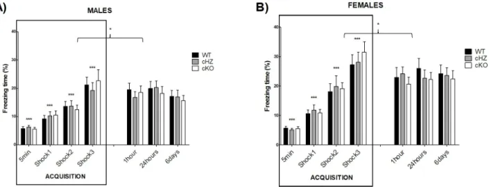
Contextual fear conditioning memory: CFC
Acquisition trial. During the acquisition trial, no statistically significant differences were found due to the variablesGenotype, the interaction Genotype × NumFootshock or the
interac-tionGenotype × Sex (all p > .442).
Nevertheless, there was a main effect of theSex (ηp2= 0.062; F1,130= 8.56, p = .004), a
signif-icant effect of the repeated administration of footshocks (ηp2= 0.526; F2,242= 144.12, p <
.001), and a significant effect of the interactionSex × NumFootshock (ηp2= 0.60; F2,242= 8.36,
p < .001). Bonferroni’s pairwise comparisons evidenced statistically significant differences in the percentage of freezing through the acquisition trial, with a significant increase after each footshock (p < .001). Furthermore, significant differences were found between males and females after the second and the third footshock, with a higher increase in the time of freezing in the females compared to the males.
There were no statistically significant differences due to the interactionGenotype × Sex × NumFootshock, with p = .95.
CFC evaluation. During the CFC evaluation, the cKO group exhibited a lower percentage of freezing time than the WT group, in particular 24 hours and 6 days after the training session, in both sexes. However, the statistical analysis revealed no significant differences due to the
Genotype, the interaction Genotype × Time, or the interaction Genotype × Sex (all p > .586).
Nevertheless, there was a main effect of theSex (ηp2= 0.094; F1,130= 13.39, p < .001), with a
higher percentage of freezing in the group of females. There was also a main effect of theTime
(ηp2= 0.044; F3,335= 5.97, p < .001), with a statistically significant decrease in the percentage
of freezing in the time-points chosen to do the CFC evaluation (p < .001), compared to the freezing displayed by mice after the last footshock.
The interactionsSex × Time and Genotype × Sex × Time were not significant (all p > .293).
The results of both sessions (acquisition trial and CFC evaluation) are represented inFig 4. Fig 3. Locomotor activity: ACT. The lines represent the distance in centimetres travelled by the mice during the 60-minute session (Mean and SEM).
The measure was acquired in three consecutive sessions, comparing the groups WT (Grik4 Cre+/+, SV2A lox/lox), cHZ (Grik4 Cre+/-, SV2A lox/+), and
cKO (Grik4 Cre+/-, SV2A lox/lox). Males (n = 22) are presented in subfigure A, whereas females (n = 23) are shown in subfigure B. (���) indicates that the
corresponding marginal mean (main effect ofSession) is significantly different from the other sessions, as yielded by a Bonferroni test taken at p < .001.
Note that in cHZ and cKO groups no statistically significant differences were found between the second and the third sessions (all p > .395).
Spatial memory: BM
The results of the different trials (acquisition, long-term acquisition and reversal learning) are represented together inFig 5, to better illustrate the continuous learning of mice.
Acquisition trials. There was no significant effect of the variablesGenotype, Sex or the
interaction between both in any of the parameters evaluated (number of errors, latency to reach the target hole or percentage of use of the spatial strategy to reach the target), with all p > .538.
However, the analyses highlighted statistically significant differences in all the measures due to theDays, with a significant decrease in the number of errors (ηp2= 0.096; F2,143= 6.16,
p = .001) and the latency to reach the target (ηp 2
= 0.138; F2,145= 9.26, p < .001), and a
signifi-cant increase in the percentage of use of the spatial strategy (ηp2= 0.333; F2,136= 27.41, p <
.001).
There was no significant effect of the interactionGenotype × Days in the number of errors
or in the latency to reach the target (both p > .661), existing a significant difference between the first and the third days in the three genotypes (all p < .049). However, there was a signifi-cant effect of the interactionGenotype × Days in the percentage of use of the spatial strategy
(ηp2= 0.078; F5,143= 2.32, p = .047). Further Bonferroni’s pairwise comparisons evidenced
sta-tistically significant differences between the first and the third days in the WT (p = .004) and in the cKO groups (p > .001), while in the cHZ group these differences did not appeared as significant until the fourth day of acquisition (p = .002).
The interactionsSex × Days and Genotype × Sex × Days did not appear as significant in any
parameter (all p > .441)
Probe trial. There were no statistically significant differences due to the variables Geno-type, Sex, or the interaction Genotype × Sex neither in the number of errors, the latency or the
strategy employed to reach the target (p > .109).
However, there was a main effect of theGenotype in the percentage of time spend in the
quadrant where the target was situated, withηp2= 0.108; F2,55= 3.32, p < .043. Further
Fig 4. Contextual fear conditioning memory: CFC. The bar plots represent the percentage of freezing (Mean and SEM) during each trial, for the
groups WT (Grik4 Cre+/+, SV2A lox/lox), cHZ (Grik4 Cre+/-, SV2A lox/+), and cKO (Grik4 Cre+/-, SV2A lox/lox). Males (n = 22) are presented in
subfigure A, whereas females (n = 23) are shown in subfigure B. Statistically significant differences were found between sex, with p < .001. (���)
indicates that each marginal mean (given by the main effect of the factor shock), is significantly different from the rest, being the last one (shock 3) the largest, as yielded by Bonferroni tests taken at p < .001. (�) indicates that the marginal mean corresponding to the shock 3 is significantly different
from the marginal means corresponding to the CFC evaluation, as yielded by Bonferroni tests taken at p < .05.
Fig 5. Spatial memory: BM. The lines represent the average per day (4 trials) of the three measured parameters: (1) the number of errors
(A, B), (2) the latency to find the target hole (C, D), and (3) the percentage of use of the spatial strategy (E, F) for Males (n = 9) and Females (n = 12). Three groups were compared (Mean and SEM): WT (Grik4 Cre+/+, SV2A lox/lox), cHZ (Grik4 Cre+/-, SV2A lox/+),
and cKO (Grik4 Cre+/-, SV2A lox/lox). (�) indicates that the corresponding marginal mean (main effect ofDay) is significantly different
from the previous ones, as yielded by Bonferroni test taken at p < .05. (#) indicates that the corresponding marginal mean (main effect of
Genotype) is significantly different between the groups WT and cHZ, as yielded by a Tukey HSD test taken at p < .05. https://doi.org/10.1371/journal.pone.0217882.g005
Bonferroni’s pairwise comparison evidenced a difference between the WT and cKO groups, with the latest spending a less percentage of the time during the probe in the quadrant where the target was situated (p = .034) (seeFig 6E and 6F). Although no statistically significant dif-ferences were found due to the variableSex or the interaction Genotype × Sex (both p > .206),
when both sexes were analysed separately the group of females displayed not only significant differences between the WT and cKO groups (p = .030), but also between the WT and cHZ groups (p = .017).
Fig 6. Spatial memory: BM probe. The bar plots (Mean and SEM) represent the number of errors (A), the latency to find the target (B)
and the percentage of mice using the spatial, serial, and random strategies (C and D), and the percentage of time spent in different positions of the maze (quadrant, others, centre) (E and F), for Males (n = 9) and Females (n = 12). Three groups were compared: WT (Grik4 Cre+/+, SV2A lox/lox), cHZ (Grik4 Cre+/-, SV2A lox/+), and cKO (Grik4 Cre+/-, SV2A lox/lox). (�) indicates that the corresponding marginal
means of both cKO and cHZ groups are significantly different to the WT group (main effect ofGenotype), as yielded by a Tukey HSD test
taken at p < .05.
Additionally, there were no statistically significant differences in the percentage of time spent exploring the rest of the holes or in the percentage of time spent in the center of the maze, neither due to theGenotype, the Sex or the interaction Genotype × Sex (all p > .063).
Long-term retention. The statistical analysis revealed a significant difference due to the
Genotype in the percentage of use of the spatial strategy (ηp2= 0.141; F2,55= 4.50, p = .015),
with statistically significant differences between the WT and the cHZ groups (p = .017). This significant effect of theGenotype was not present in the number of errors or in the latency to
reach the target hole (p < .161). Similarly, there was no significant effect of theSex or the
inter-actionGenotype × Sex in any parameter, with p > .380 in all the cases.
Long-term acquisition trials. The statistical analysis did not show any significant differ-ences due to theGenotype, the Sex or the interaction between both variables in any of the
parameters evaluated (number of errors, latency to reach the target hole or percentage of use of the spatial strategy to reach the target), with p > .538.
However, there were statistically significant differences due to theDays in the number of
errors (ηp2= 0.068; F2,138= 4.24, p = .012), and in the use of the spatial strategy (ηp2= 0.175;
F3,151= 11.46, p < .001). Bonferroni’s pairwise comparison highlighted significant differences
in the number of errors between the last two days (p = .030), and in the use of the spatial strat-egy during the third and the fourth days and the previous ones (both p < .001). There was no effect of theDays in the latency to reach the target hole (p = .294).
The interactionsGenotype × Days and Sex × Days and Genotype × Sex × Days were not
sig-nificant in any of the parameters evaluated, with p > .160 for all.
Reversal learning. The statistical analysis did not reveal any significant differences due to theGenotype, the Sex or the interaction Genotype × Sex in the number of errors, the latency to
reach the target hole, or the percentage of use of the spatial strategy (all p > .084).
However, there were very large statistically significant differences due to theDays in the
number of errors (ηp 2
= 0.519; F2,110= 60.38, p < .001), the latency to reach the target hole (ηp 2
= 0.242; F2,95= 16.60, p < .001), and the use of the spatial strategy (ηp2= 0.693; F2,96= 119.90,
p < .001). In the number of errors and the use of the spatial strategy, these differences were present between all the days (all p < .001). Nevertheless, there were no differences in the latency between the last day of long-term acquisition and the last day of reversal learning (p = .365).
The interactionsGenotype × Days, Sex × Days and Genotype × Sex × Days were not
signifi-cant in any of the parameters evaluated, with all p > .196.
Discussion
During the last few years, the SV2A protein has emerged as a possible key element to under-stand the epileptic disease. Indeed, when theSV2A gene is completely deleted, mice experience
seizures starting seven days after birth and die in status epilepticus around day 15 [7,14]. Fur-thermore, this protein is the molecular target of one of the most prescribed antiepileptic drugs: the levetiracetam [16]. Despite the demonstrated relationship between a decrease in SV2A lev-els in epileptic foci and the presence of brain seizures, the potential implication of this protein in the cognitive problems exhibited by epileptic patients is barely known. The phenotyping of
Grik4 Cre+/-, SV2A lox/lox (cKO) mice, exempt from spontaneous seizures [30], might there-fore help us understand the role of this protein in the cognitive process associated with the hippocampus.
Concerning the spontaneous locomotor activity, our results were similar to those obtained from the heterozygous SV2A mice (+/-) [29], displaying no significant differences attributable to theGenotype. Additionally, through the testing procedure, no overt seizure behaviour was
observed in the cHZ or the cKO groups, which was consistent with the previously published study regarding these mice [30]. However, cKO and cHZ mice presented a significant different capacity of habituation to a new environment, compared to the WT group. Indeed, we noticed an increased behavioural excitability and anxiety in the transgenic mice, reflected in different adverse reactions to the tests:SV2A cKO mice jumped more frequently out of the Barnes maze
than WT mice, and they tried to avoid the footshocks using the chamber walls and the ceiling. Moreover, these mice exhibited an increased aggressiveness after being exposed to the contex-tual fear conditioning test. These emotionally related behaviours have also been reported in chemoconvulsant-induced epileptic models [55].
Due to the link between the hippocampus and the learning and memory [34,56,57], we expected the existence of statistically significant differences between the three groups of mice in the CFC test. Nevertheless, the establishment of a contextual fear conditioning memory was unaffected by the decrease in the SV2A expression. An initial hypothesis is the interference of a memory extinction process, which would disrupt the consolidation and maintenance of the contextual fear conditioning memory. This phenomenon may be caused by a prolonged re-exposure to the context (CS) without receiving the footshocks (US) [58]. However, we did not detect any statistically significant decrease in the percentage of freezing through the CFC eval-uation, disproving this hypothesis. Lamberty et al. [29] obtained equivalent results using the passive avoidance procedure: heterozygous SV2A (+/-) mice had similar results than wild type controls at the end of a multi-trial inhibitory avoidance test, even if they have received foot-shocks along the retention testing. An alternative hypothesis is the existence of compensatory processes, involving the basolateral amygdala complex: even if there is an important role of the hippocampus in the association CS-US [34,56], the amygdala is the main locus in the consoli-dation and maintenance processes of contextual fear memories [59–61]. In this regard, the cKO mice feature a SV2A decrease only in the hippocampal region, expressing normally this protein in the rest of the brain structures, including the amygdala. The interconnections between the amygdaloid complex and the hippocampus, already described by Pitka¨nen’s group [62,63], could compensate the SV2A decrease in the hippocampus, explaining the absence of statistically significant differences between groups. A third hypothesis is the possi-ble existence of a compensation phenomenon: in a review published in 2017 concerning the role of the SV2 family members [2], it was proposed that the SV2B expression in glutamatergic neurons could be compensating the SV2A decrease in those neurons, explaining the lack of other statistically significant differences through the testing process.
Despite the absence of differences due to theGenotype in the CFC test, sex differences were
found in the percentage of freezing time, not only during the acquisition trial, but also during the CFC evaluation. Several studies have explored the underlying causes of these differences, reflecting for example that the dorsal hippocampus is more implicated in males, but there is a preferential recruitment of basal amygdala in the females [64]. Other factors, as the hormones [65], and a different developmental trajectory in the expression of context-mediated freezing [66] also play an important role in the sex differences found in the contextual fear.
On the other hand, the results obtained with the EPM and the BM test highlight statistically significant differences between groups, attributable to theGenotype: in the EPM, cKO and
cHZ groups entered less in the open arms of the EPM, compared with the WT group. This indicator of anxiety was also found by Lamberty et al. in the heterozygous SV2A mice (+/-) [29]. Indeed, the existence of anxiety and mood disorders has been reported not only in animal models of epilepsy [67,68], but also in patients [69–73]. Additionally, it was found a possible influence of the sex in the anxiety level displayed by the different genotypes (interactionSex × Genotype), with significant group differences in males, but not in females. In the literature, the
a few articles describing sex differences in anxiety due to the CB1 and Glu1 receptors [74,75]. Further studies are required to better understand the implication of SV2A in the sexual differ-ences observed in anxiety, since it could have an impact in the choice of the treatment, depend-ing on the sex.
Concerning the BM test, all the genotypes exhibit an equivalent spatial learning through the acquisition trials. However, during the probe test, the cKO group spent less time in the quad-rant where the target was located, indicating a possible spatial memory problem associated with the decrease of SV2A in the hippocampal glutamatergic neurons. In females, also the cHZ group exhibit a worse performance compared with the WT group. Furthermore, during the long-term retention trial (day 19), the cHZ group uses the spatial strategy to reach the target significantly less than the WT group, indicating also in this group possible long-term spatial memory problems. In this line, problems in visuospatial memory, olfactory discrimination, and social recognition have also been reported in animal models of epilepsy [55,67,76]. In these models, the memory impairment is commonly evaluated by means of the Morris water maze test [55,76,77]. However, in our case, its use would not be recommended since, in this test, the learning process and the performance are more affected by a high level of anxiety than in the BM [78,79]. Finally, in clinics it has also been described a comorbidity between epilepsy and both mild cognitive impaired [80] and dementia [81].
Additionally, the BM selected procedure allowed us to assess the cognitive flexibility of mice with a two-day reversal learning trials, carried out during the last two days of the proce-dure. We did not detect statistically significant differences between groups with this comple-mentary analysis, which suggests that the decrease of the SV2A protein in the hippocampus is not interfering in the executive function. However, it is necessary to account for the stress suf-fered by the animals during the previous tests, which might interfere in the reversal learning evaluation [51]. In conclusion, even if this test can provide us with a first approach to the cog-nitive flexibility ofSV2A cKO mice, more accurate analyses with specific behavioural
appara-tus (e.g. nose-poke portals or a touch-sensitive screen) need to be conducted to specifically assess the executive function [82].
All these results suggest that, in the hippocampal area, the deletion of theSV2A gene in one
(cHZ mice) or in both alleles (cKO mice) involves statistically significant changes in cognition, including a cognitive impairment and anxiety-related problems, usually present in the epileptic disease [83–89]. However, further studies in epileptic patients should be conducted to confirm the impact of the variations in SV2A protein levels on the severity of the symptomatology.
Conclusions
The phenotyping ofGrik4 Cre+/-, SV2A lox/lox mice confirms a link between the decrease of
the SV2A protein in the hippocampus, and the memory and anxiety-related problems detected in the chronic epilepsy. These results suggest a possible implication of the SV2A protein in anxiety or memory disorders, such as the post-traumatic stress disorder or the Alzheimer’s disease.
Supporting information
S1 Data. EPM Data. Raw data acquired with the EPM test. The columns represent the number and percentage of entries and the time spent (in seconds) in open and closed arms, for the three groups of mice (WT, cHZ, and cKO). On the right, the descriptive statistics for each measured variable.
S2 Data. ACT Data. Raw data acquired with the ACT test. The columns represent the distance travelled (cm) by the three groups (WT, cHZ, and cKO), in each of the three 60-minute session (day1, day2, day3). On the right, the descriptive statistics.
(XLSX)
S3 Data. CFC Data. Raw data acquired with the CFC test. The columns represent the percent-age of freezing time spent by the three groups (WT, cHZ, and cKO) during the acquisition trial and the CFC evaluation (1h, 24h and 6 days after the acquisition trial). On the right, the descriptive statistics.
(XLSX)
S4 Data. BM Data. Raw data acquired with the BM test. The first sheet corresponds to the acquisition trials (days 1–4), the long-term acquisition trials (days 19–22), and the reversal learning trials (RL1 and RL2). The columns represent the number of errors (errors), latency (latence), and the percentage of use of the spatial strategy (perspat) for the three groups of mice (WT, cHZ, and cKO). The second sheet corresponds to the results obtained during the probe trial by the three groups of mice. Four variables have been analysed: errors, latency, time spent in each part of the maze, and strategy. On the right, the descriptive statistics.
(XLSX)
Acknowledgments
The authors would like to thank Thierry Matonda for his guidance and advices through the interpretation of data. We also would like to thank Miguel Manuel de Villena for the English revision of the manuscript.
Author Contributions
Conceptualization: Maria Elisa Serrano, Andre´ Ferrara, Bernard Rogister, Alain Plenevaux, Ezio Tirelli.
Formal analysis: Maria Elisa Serrano, Odile Bartholome´.
Investigation: Maria Elisa Serrano, Odile Bartholome´, Priscilla Van den Ackerveken. Methodology: Odile Bartholome´, Priscilla Van den Ackerveken, Andre´ Ferrara, Ezio Tirelli. Project administration: Bernard Rogister, Alain Plenevaux, Ezio Tirelli.
Resources: Bernard Rogister, Alain Plenevaux, Ezio Tirelli. Supervision: Bernard Rogister, Alain Plenevaux, Ezio Tirelli. Validation: Odile Bartholome´, Andre´ Ferrara.
Visualization: Maria Elisa Serrano, Andre´ Ferrara. Writing – original draft: Maria Elisa Serrano.
Writing – review & editing: Odile Bartholome´, Priscilla Van den Ackerveken, Andre´ Ferrara, Bernard Rogister, Alain Plenevaux, Ezio Tirelli.
References
1. Crèvecœur J, Kaminski RM, Rogister B, Foerch P, Vandenplas C, Neveux M, et al. Expression pattern of synaptic vesicle protein 2 (SV2) isoforms in patients with temporal lobe epilepsy and hippocampal sclerosis. Neuropathol Appl Neurobiol. 2014; 40: 191–204.https://doi.org/10.1111/nan.12054PMID:
2. Bartholome O, Van den Ackerveken P, Sa´nchez Gil J, de la Brassinne Bonardeaux O, Leprince P, Fran-zen R, et al. Puzzling Out Synaptic Vesicle 2 Family Members Functions. Front Mol Neurosci. 2017;10. PMID:28197071
3. Dardou D, Monlezun S, Foerch P, Courade JP, Cuvelier L, De Ryck M, et al. A role for Sv2c in basal ganglia functions. Brain Res. 2013; 1507: 61–73.https://doi.org/10.1016/j.brainres.2013.02.041PMID:
23458503
4. Clegg N, Ferguson C, True LD, Arnold H, Moorman A, Quinn JE, et al. Molecular characterization of prostatic small-cell neuroendocrine carcinoma. Prostate. 2003; 55: 55–64.https://doi.org/10.1002/pros. 10217PMID:12640661
5. Bajjalieh SM, Frantz GD, Weimann JM, McConnell SK, Scheller RH. Differential expression of synaptic vesicle protein 2 (SV2) isoforms. J Neurosci. 1994; 14: 5223–35. Available:http://www.ncbi.nlm.nih. gov/pubmed/8083732PMID:8083732
6. Janz R, Su¨dhof TC. SV2C is a synaptic vesicle protein with an unusually restricted localization: anatomy of a synaptic vesicle protein family. Neuroscience. 1999; 94: 1279–90. https://doi.org/10.1016/S0306-4522(99)00370-XPMID:10625067
7. Crowder KM, Gunther JM, Jones TA, Hale BD, Zhang HZ, Peterson MR, et al. Abnormal neurotrans-mission in mice lacking synaptic vesicle protein 2A (SV2A). Proc Natl Acad Sci U S A. 1999; 96: 15268– 73.https://doi.org/10.1073/pnas.96.26.15268PMID:10611374
8. Custer KL, Austin NS, Sullivan JM, Bajjalieh SM. Synaptic Vesicle Protein 2 Enhances Release Proba-bility at Quiescent Synapses. J Neurosci. 2006; 26: 1303–1313.https://doi.org/10.1523/JNEUROSCI. 2699-05.2006PMID:16436618
9. Chang W, Su¨dhof TC. SV2 renders primed synaptic vesicles competent for Ca2+ -induced exocytosis. J Neurosci. 2009; 29: 883–97.https://doi.org/10.1523/JNEUROSCI.4521-08.2009PMID:19176798 10. Venkatesan K, Alix P, Marquet A, Doupagne M, Niespodziany I, Rogister B, et al. Altered balance
between excitatory and inhibitory inputs onto CA1 pyramidal neurons from SV2A-deficient but not SV2B-deficient mice. J Neurosci Res. 2012; 90: 2317–2327.https://doi.org/10.1002/jnr.23111PMID:
22847229
11. Tokudome K, Okumura T, Shimizu S, Mashimo T, Takizawa A, Serikawa T, et al. Synaptic vesicle gly-coprotein 2A (SV2A) regulates kindling epileptogenesis via GABAergic neurotransmission. Sci Rep. 2016; 6: 27420.https://doi.org/10.1038/srep27420PMID:27265781
12. Soukupova´ M, Binaschi A, Falcicchia C, Zucchini S, Roncon P, Palma E, et al. Impairment of GABA release in the hippocampus at the time of the first spontaneous seizure in the pilocarpine model of tem-poral lobe epilepsy. Exp Neurol. 2014; 257: 39–49.https://doi.org/10.1016/j.expneurol.2014.04.014
PMID:24768627
13. Soukupova M, Binaschi A, Falcicchia C, Palma E, Roncon P, Zucchini S, et al. Increased extracellular levels of glutamate in the hippocampus of chronically epileptic rats. Neuroscience. 2015; 301: 246–53.
https://doi.org/10.1016/j.neuroscience.2015.06.013PMID:26073699
14. Janz R, Goda Y, Geppert M, Missler M, Su¨ TC, Hughes H. SV2A and SV2B Function as Redundant Ca 2+ Regulators in Neurotransmitter Release. Neuron. 1999; 24: 1003–1016. PMID:10624962
15. Serajee FJ, Huq AM. Homozygous Mutation in Synaptic Vesicle Glycoprotein 2A Gene Results in Intractable Epilepsy, Involuntary Movements, Microcephaly, and Developmental and Growth Retarda-tion. Pediatr Neurol. 2015; 52: 642–6.e1.https://doi.org/10.1016/j.pediatrneurol.2015.02.011PMID:
26002053
16. Lynch BA, Lambeng N, Nocka K, Kensel-Hammes P, Bajjalieh SM, Matagne A, et al. The synaptic vesi-cle protein SV2A is the binding site for the antiepileptic drug levetiracetam. Proc Natl Acad Sci U S A. 2004; 101: 9861–6.https://doi.org/10.1073/pnas.0308208101PMID:15210974
17. Rosenstiel P. Brivaracetam (UCB 34714). Neurotherapeutics. 2007; 4: 84–87.https://doi.org/10.1016/j. nurt.2006.11.004PMID:17199019
18. Klein P, Diaz A, Gasalla T, Whitesides J. A review of the pharmacology and clinical efficacy of brivarace-tam. Clin Pharmacol. 2018; 10: 1–22.https://doi.org/10.2147/CPAA.S114072PMID:29403319 19. Toering ST, Boer K, de Groot M, Troost D, Heimans JJ, Spliet WGM, et al. Expression patterns of
syn-aptic vesicle protein 2A in focal cortical dysplasia and TSC-cortical tubers. Epilepsia. 2009; 50: 1409– 18.https://doi.org/10.1111/j.1528-1167.2008.01955.xPMID:19220410
20. Feng G, Xiao F, Lu Y, Huang Z, Yuan J, Xiao Z, et al. Down-regulation synaptic vesicle protein 2A in the anterior temporal neocortex of patients with intractable epilepsy. J Mol Neurosci. 2009; 39: 354–9.
https://doi.org/10.1007/s12031-009-9288-2PMID:19757204
21. Bragina L, Fattorini G, Giovedı´ S, Melone M, Bosco F, Benfenati F, et al. Analysis of Synaptotagmin, SV2, and Rab3 Expression in Cortical Glutamatergic and GABAergic Axon Terminals. Front Cell Neu-rosci. 2012; 5: 1–9.https://doi.org/10.3389/fncel.2011.00032PMID:22275882
22. Magalhães JC, Gongora M, Vicente R, Bittencourt J, Tanaka G, Velasques B, et al. The influence of levetiracetam in cognitive performance in healthy individuals: neuropsychological, behavioral and electrophysiological approach. Clin Psychopharmacol Neurosci. 2015; 13: 83–93.https://doi.org/10. 9758/cpn.2015.13.1.83PMID:25912541
23. Ge Y-X, Tian X-Z, Lin Y-Y, Liu X-Y. Chronic treatment with levetiracetam reverses deficits in hippocam-pal LTP in vivo in experimental temporal lobe epilepsy rats. Neurosci Lett. 2016; 628: 194–200.https:// doi.org/10.1016/j.neulet.2016.06.043PMID:27345386
24. Celikyurt IK, Ulak G, Mutlu O, Akar FY, Mulayim S, Erden F, et al. Positive impact of levetiracetam on emotional learning and memory in naive mice. Life Sci. 2012; 90: 185–9.https://doi.org/10.1016/j.lfs. 2011.11.003PMID:22119754
25. Bakker A, Krauss GL, Albert MS, Speck CL, Jones LR, Stark CE, et al. Reduction of hippocampal hyperactivity improves cognition in amnestic mild cognitive impairment. Neuron. 2012; 74: 467–74.
https://doi.org/10.1016/j.neuron.2012.03.023PMID:22578498
26. Bakker A, Albert MS, Krauss G, Speck CL, Gallagher M. Response of the medial temporal lobe network in amnestic mild cognitive impairment to therapeutic intervention assessed by fMRI and memory task performance. NeuroImage Clin. 2015; 7: 688–98.https://doi.org/10.1016/j.nicl.2015.02.009PMID:
25844322
27. Koh MT, Shao Y, Rosenzweig-Lipson S, Gallagher M. Treatment with levetiracetam improves cognition in a ketamine rat model of schizophrenia. Schizophr Res. 2017;https://doi.org/10.1016/j.schres.2017. 06.027PMID:28634087
28. Devi L, Ohno M. Effects of levetiracetam, an antiepileptic drug, on memory impairments associated with aging and Alzheimer’s disease in mice. Neurobiol Learn Mem. 2013; 102: 7–11.https://doi.org/10.1016/ j.nlm.2013.02.001PMID:23416036
29. Lamberty Y, Detrait E, Leclercq K, Michel A, De Ryck M. Behavioural phenotyping reveals anxiety-like features of SV2A deficient mice. Behav Brain Res. 2009; 198: 329–33.https://doi.org/10.1016/j.bbr. 2008.11.005PMID:19041904
30. Menten-Dedoyart C, Serrano Navacerrada ME, Bartholome O, Sa´nchez Gil J, Neirinckx V, Wislet S, et al. Development and Validation of a New Mouse Model to Investigate the Role of SV2A in Epilepsy. Schmidt EE, editor. PLoS One. 2016; 11: e0166525.https://doi.org/10.1371/journal.pone.0166525
PMID:27861538
31. van Vliet EA, Aronica E, Redeker S, Boer K, Gorter JA. Decreased expression of synaptic vesicle pro-tein 2A, the binding site for levetiracetam, during epileptogenesis and chronic epilepsy. Epilepsia. 2009; 50: 422–433.https://doi.org/10.1111/j.1528-1167.2008.01727.xPMID:18717715
32. Sendrowski K, Sobaniec W. Hippocampus, hippocampal sclerosis and epilepsy. Pharmacol Reports. 2013; 65: 555–565.https://doi.org/10.1016/S1734-1140(13)71033-8
33. Chatzikonstantinou A. Epilepsy and the hippocampus. Front Neurol Neurosci. 2014; 34: 121–42.
https://doi.org/10.1159/000356435PMID:24777136
34. Chaaya N, Battle AR, Johnson LR. An update on contextual fear memory mechanisms: Transition between Amygdala and Hippocampus. Neurosci Biobehav Rev. 2018; 92: 43–54.https://doi.org/10. 1016/j.neubiorev.2018.05.013PMID:29752958
35. Kaminski RM, Gillard M, Leclercq K, Hanon E, Lorent G, Dassesse D, et al. Proepileptic phenotype of SV2A-deficient mice is associated with reduced anticonvulsant efficacy of levetiracetam. Epilepsia. 2009; 50: 1729–40.https://doi.org/10.1111/j.1528-1167.2009.02089.xPMID:19486357
36. Kotsopoulos IAW, van Merode T, Kessels FGH, de Krom MCTFM, Knottnerus JA. Systematic review and meta-analysis of incidence studies of epilepsy and unprovoked seizures. Epilepsia. 2002; 43: 1402–9. Available:http://www.ncbi.nlm.nih.gov/pubmed/12423392PMID:12423392
37. Savic I. Sex differences in human epilepsy. Exp Neurol. 2014; 259: 38–43.https://doi.org/10.1016/j. expneurol.2014.04.009PMID:24747359
38. Desgent S, Duss S, Sanon NT, Lema P, Le´vesque M, He´bert D, et al. Early-Life Stress Is Associated with Gender-Based Vulnerability to Epileptogenesis in Rat Pups. Baud O, editor. PLoS One. 2012; 7: e42622.https://doi.org/10.1371/journal.pone.0042622PMID:22880055
39. Cohen J. Statistical power analysis for the behavioral sciences. 2nd editio. Lawrence Erlbaum Associ-ates; 1988.
40. Faul F, Erdfelder E, Lang A-G, Buchner A. G*Power 3: a flexible statistical power analysis program for the social, behavioral, and biomedical sciences. Behav Res Methods. 2007; 39: 175–91. Available:
http://www.ncbi.nlm.nih.gov/pubmed/17695343PMID:17695343
41. Kilkenny C, Browne W, Cuthill I, Emerson M, Altman D. Improving bioscience research reporting: The ARRIVE guidelines for reporting animal research. J Pharmacol Pharmacother. 2010; 1: 94.https://doi. org/10.4103/0976-500X.72351PMID:21350617
42. Charlier Y, Brabant C, Serrano ME, Lamberty Y, Tirelli E. The prototypical histamine H3 receptor inverse agonist thioperamide improves multiple aspects of memory processing in an inhibitory avoid-ance task. Behav Brain Res. Elsevier B.V.; 2013; 253: 121–127.https://doi.org/10.1016/j.bbr.2013.07. 016PMID:23867149
43. Lister RG. The use of a plus-maze to measure anxiety in the mouse. Psychopharmacology (Berl). 1987; 92: 180–5. Available:http://www.ncbi.nlm.nih.gov/pubmed/3110839
44. Rodgers RJ, Dalvi A. Anxiety, defence and the elevated plus-maze. Neurosci Biobehav Rev. 1997; 21: 801–10. Available:http://www.ncbi.nlm.nih.gov/pubmed/9415905
45. Berkiks I, Garcia-Segura LM, Nassiri A, Mesfioui A, Ouichou A, Boulbaroud S, et al. The sex differences of the behavior response to early Life immune stimulation: Microglia and astrocytes involvement. Phy-siol Behav. 2019; 199: 386–394.https://doi.org/10.1016/j.physbeh.2018.11.037PMID:30529512 46. Brabant C, Quertemont E, Anaclet C, Lin J-S, Ohtsu H, Tirelli E. The psychostimulant and rewarding
effects of cocaine in histidine decarboxylase knockout mice do not support the hypothesis of an inhibi-tory function of histamine on reward. Psychopharmacology (Berl). 2007; 190: 251–63.https://doi.org/ 10.1007/s00213-006-0603-0PMID:17072589
47. Kimura T, Yamashita S, Nakao S, Park J-M, Murayama M, Mizoroki T, et al. GSK-3beta is required for memory reconsolidation in adult brain. PLoS One. 2008; 3: e3540.https://doi.org/10.1371/journal.pone. 0003540PMID:18958152
48. Rozeske RR, Herry C. Neuronal coding mechanisms mediating fear behavior. Curr Opin Neurobiol. 2018; 52: 60–64.https://doi.org/10.1016/j.conb.2018.04.017PMID:29705550
49. Roelofs K. Freeze for action: neurobiological mechanisms in animal and human freezing. Philos Trans R Soc Lond B Biol Sci. 2017;372.https://doi.org/10.1098/rstb.2016.0206PMID:28242739
50. Kesby JP, Kim JJ, Scadeng M, Woods G, Kado DM, Olefsky JM, et al. Spatial Cognition in Adult and Aged Mice Exposed to High-Fat Diet. PLoS One. 2015; 10: e0140034.https://doi.org/10.1371/journal. pone.0140034PMID:26448649
51. Kesby JP, Markou A, Semenova S, TMARC Group. Effects of HIV/TAT protein expression and chronic selegiline treatment on spatial memory, reversal learning and neurotransmitter levels in mice. Behav Brain Res. 2016; 311: 131–140.https://doi.org/10.1016/j.bbr.2016.05.034PMID:27211061
52. Paul C-M, Magda G, Abel S. Spatial memory: Theoretical basis and comparative review on experimen-tal methods in rodents. Behav Brain Res. 2009; 203: 151–164.https://doi.org/10.1016/j.bbr.2009.05. 022PMID:19467271
53. Sharma S, Rakoczy S, Brown-Borg H. Assessment of spatial memory in mice. Life Sci. 2010; 87: 521– 536.https://doi.org/10.1016/j.lfs.2010.09.004PMID:20837032
54. Zhang J, Ko S-Y, Liao Y, Kwon Y, Jeon SJ, Sohn A, et al. Activation of the dopamine D 1 receptor can extend long-term spatial memory persistence via PKA signaling in mice. Neurobiol Learn Mem. 2018; PMID:2018
55. Minjarez B, Camarena HO, Haramati J, Rodrı´guez-Yañez Y, Mena-Munguı´a S, Buritica´ J, et al. Behav-ioral changes in models of chemoconvulsant-induced epilepsy: A review. Neurosci Biobehav Rev. 2017; 83: 373–380.https://doi.org/10.1016/j.neubiorev.2017.10.016PMID:29107831
56. Anagnostaras SG, Gale GD, Fanselow MS. Hippocampus and contextual fear conditioning: recent con-troversies and advances. Hippocampus. 2001; 11: 8–17.https://doi.org/10.1002/1098-1063(2001) 11:1<8::AID-HIPO1015>3.0.CO;2-7PMID:11261775
57. Sanders MJ, Wiltgen BJ, Fanselow MS. The place of the hippocampus in fear conditioning. Eur J Phar-macol. 2003; 463: 217–23. Available:http://www.ncbi.nlm.nih.gov/pubmed/12600712
58. Isiegas C, Park A, Kandel ER, Abel T, Lattal KM. Transgenic Inhibition of Neuronal Protein Kinase A Activity Facilitates Fear Extinction. J Neurosci. 2006; 26: 12700–12707.https://doi.org/10.1523/ JNEUROSCI.2743-06.2006PMID:17151273
59. Krabbe S, Gru¨ndemann J, Lu¨thi A. Amygdala Inhibitory Circuits Regulate Associative Fear Condition-ing. Biol Psychiatry. 2018; 83: 800–809.https://doi.org/10.1016/j.biopsych.2017.10.006PMID:
29174478
60. Kochli DE, Thompson EC, Fricke EA, Postle AF, Quinn JJ. The amygdala is critical for trace, delay, and contextual fear conditioning. Learn Mem. 2015; 22: 92–100.https://doi.org/10.1101/lm.034918.114
PMID:25593295
61. Wilson YM, Murphy M. A discrete population of neurons in the lateral amygdala is specifically activated by contextual fear conditioning. Learn Mem. 2009; 16: 357–61.https://doi.org/10.1101/lm.1361509
PMID:19470650
62. Pikkarainen M, Ro¨nkko¨ S, Savander V, Insausti R, Pitka¨nen A. Projections from the lateral, basal, and accessory basal nuclei of the amygdala to the hippocampal formation in rat. J Comp Neurol. 1999; 403: 229–60. Available:http://www.ncbi.nlm.nih.gov/pubmed/9886046
63. Pitka¨nen A, Pikkarainen M, Nurminen N, Ylinen A. Reciprocal connections between the amygdala and the hippocampal formation, perirhinal cortex, and postrhinal cortex in rat. A review. Ann N Y Acad Sci. 2000; 911: 369–91. Available:http://www.ncbi.nlm.nih.gov/pubmed/10911886
64. Keiser AA, Turnbull LM, Darian MA, Feldman DE, Song I, Tronson NC. Sex Differences in Context Fear Generalization and Recruitment of Hippocampus and Amygdala during Retrieval. Neuropsychophar-macology. 2017; 42: 397–407.https://doi.org/10.1038/npp.2016.174PMID:27577601
65. Merz CJ, Wolf OT. Sex differences in stress effects on emotional learning. J Neurosci Res. 2017; 95: 93–105.https://doi.org/10.1002/jnr.23811PMID:27870431
66. Colon L, Odynocki N, Santarelli A, Poulos AM. Sexual differentiation of contextual fear responses. Learn Mem. 2018; 25: 230–240.https://doi.org/10.1101/lm.047159.117PMID:29661835
67. Gro¨ticke I, Hoffmann K, Lo¨scher W. Behavioral alterations in the pilocarpine model of temporal lobe epi-lepsy in mice. Exp Neurol. 2007; 207: 329–49.https://doi.org/10.1016/j.expneurol.2007.06.021PMID:
17714705
68. Lopes MW, Lopes SC, Santos DB, Costa AP, Gonc¸alves FM, de Mello N, et al. Time course evaluation of behavioral impairments in the pilocarpine model of epilepsy. Epilepsy Behav. 2016; 55: 92–100.
https://doi.org/10.1016/j.yebeh.2015.12.001PMID:26773677
69. Wiglusz MS, Landowski J, Cubała WJ. Prevalence of anxiety disorders in epilepsy. Epilepsy Behav. 2018; 79: 1–3.https://doi.org/10.1016/j.yebeh.2017.11.025PMID:29223931
70. Jacoby A, Snape D, Lane S, Baker GA. Self-reported anxiety and sleep problems in people with epi-lepsy and their association with quality of life. Epiepi-lepsy Behav. 2015; 43: 149–58.https://doi.org/10. 1016/j.yebeh.2014.09.071PMID:25599986
71. Munger Clary HM. Anxiety and epilepsy: what neurologists and epileptologists should know. Curr Neu-rol Neurosci Rep. 2014; 14: 445.https://doi.org/10.1007/s11910-014-0445-9PMID:24652453 72. Korczyn AD, Schachter SC, Brodie MJ, Dalal SS, Engel J, Guekht A, et al. Epilepsy, cognition, and
neu-ropsychiatry (Epilepsy, Brain, and Mind, part 2). Epilepsy Behav. 2013; 28: 283–302.https://doi.org/10. 1016/j.yebeh.2013.03.012PMID:23764496
73. Elger CE, Helmstaedter C, Kurthen M. Chronic epilepsy and cognition. Lancet Neurol. 2004; 3: 663–72.
https://doi.org/10.1016/S1474-4422(04)00906-8PMID:15488459
74. Xiang X, Huang W, Haile CN, Kosten TA. Hippocampal GluR1 associates with behavior in the elevated plus maze and shows sex differences. Behav Brain Res. 2011; 222: 326–331.https://doi.org/10.1016/j. bbr.2011.03.068PMID:21497621
75. Simone JJ, Baumbach JL, McCormick CM. Sex-specific effects of CB1 receptor antagonism and stress in adolescence on anxiety, corticosterone concentrations, and contextual fear in adulthood in rats. Int J Dev Neurosci. 2018; 69: 119–131.https://doi.org/10.1016/j.ijdevneu.2018.07.011PMID:30063953 76. Majak K, Pitka¨nen A. Do seizures cause irreversible cognitive damage? Evidence from animal studies.
Epilepsy Behav. 2004; 5: 35–44.https://doi.org/10.1016/j.yebeh.2003.11.012
77. Morris R. Developments of a water-maze procedure for studying spatial learning in the rat. J Neurosci Methods. 1984; 11: 47–60. Available:http://www.ncbi.nlm.nih.gov/pubmed/6471907
78. Karabeg MM, Grauthoff S, Kollert SY, Weidner M, Heiming RS, Jansen F, et al. 5-HTT deficiency affects neuroplasticity and increases stress sensitivity resulting in altered spatial learning performance in the Morris water maze but not in the Barnes maze. PLoS One. 2013; 8: e78238.https://doi.org/10. 1371/journal.pone.0078238PMID:24167611
79. Harrison FE, Hosseini AH, McDonald MP. Endogenous anxiety and stress responses in water maze and Barnes maze spatial memory tasks. Behav Brain Res. 2009; 198: 247–51.https://doi.org/10.1016/ j.bbr.2008.10.015PMID:18996418
80. Helmstaedter C, Witt J-A. Epilepsy and cognition—A bidirectional relationship? Seizure. 2017; 49: 83– 89.https://doi.org/10.1016/j.seizure.2017.02.017PMID:28284559
81. Sen A, Capelli V, Husain M. Cognition and dementia in older patients with epilepsy. Brain. 2018; 141: 1592–1608.https://doi.org/10.1093/brain/awy022PMID:29506031
82. Izquierdo A, Brigman JL, Radke AK, Rudebeck PH, Holmes A. The neural basis of reversal learning: An updated perspective. Neuroscience. 2017; 345: 12–26.https://doi.org/10.1016/j.neuroscience.2016.03. 021PMID:26979052
83. Kanner AM, Schachter SC, Barry JJ, Hersdorffer DC, Mula M, Trimble M, et al. Depression and epi-lepsy: Epidemiologic and neurobiologic perspectives that may explain their high comorbid occurrence. Epilepsy Behav. Elsevier Inc.; 2012; 24: 156–168.https://doi.org/10.1016/j.yebeh.2012.01.007PMID:
22632406
84. Bell B, Lin J, Seidenberg M, Hermann B. The neurobiology of cognitive disorders in temporal lobe epi-lepsy. Nat Rev Neurol. 2011;7.
85. McDonald CR, Taylor J, Hamberger M, Helmstaedter C, Hermann BP, Schefft B. Future directions in the neuropsychology of epilepsy. Epilepsy Behav. Elsevier Inc.; 2011; 22: 69–76.https://doi.org/10. 1016/j.yebeh.2011.06.004PMID:21795122
86. Lin JJ, Mula M, Hermann BP. Uncovering the neurobehavioural comorbidities of epilepsy over the life-span. Lancet. 2012; 380: 1180–1192.https://doi.org/10.1016/S0140-6736(12)61455-XPMID:
23021287
87. Hermann B, Seidenberg M, Jones J. The neurobehavioural comorbidities of epilepsy: can a natural his-tory be developed? Lancet Neurol. 2008; 7: 151–160.https://doi.org/10.1016/S1474-4422(08)70018-8
PMID:18207113
88. Kleen JK, Scott RC, Lenck-Santini P-P, Holmes GL. Cognitive and Behavioral Co-Morbidities of Epi-lepsy [Internet]. Jasper’s Basic Mechanisms of the Epilepsies. 2012. Available:http://www.ncbi.nlm.nih. gov/pubmed/22787667
89. Ho¨ller Y, Trinka E. What do temporal lobe epilepsy and progressive mild cognitive impairment have in common? Front Syst Neurosci. 2014; 8: 58.https://doi.org/10.3389/fnsys.2014.00058PMID:24795575