HAL Id: hal-01601710
https://hal.archives-ouvertes.fr/hal-01601710
Submitted on 27 May 2020
HAL is a multi-disciplinary open access archive for the deposit and dissemination of sci-entific research documents, whether they are pub-lished or not. The documents may come from teaching and research institutions in France or abroad, or from public or private research centers.
L’archive ouverte pluridisciplinaire HAL, est destinée au dépôt et à la diffusion de documents scientifiques de niveau recherche, publiés ou non, émanant des établissements d’enseignement et de recherche français ou étrangers, des laboratoires publics ou privés.
The AHP6 cytokinin signaling inhibitor mediates an
auxin-cytokinin crosstalk that regulates the timing of
organ initiation at the shoot apical meristem
Fabrice Besnard, Frédérique Rozier, Teva Vernoux
To cite this version:
Fabrice Besnard, Frédérique Rozier, Teva Vernoux. The AHP6 cytokinin signaling inhibitor mediates an auxin-cytokinin crosstalk that regulates the timing of organ initiation at the shoot apical meristem. Plant Signaling and Behavior, Taylor & Francis, 2014, 9 (4), �10.4161/psb.28788�. �hal-01601710�
Short CommuniCation
Plant Signaling & Behavior 9, e28788; april; © 2014 Landes Bioscience
Short CommuniCation
The spatio-temporal pattern of lateral organ initiation at the shoot apical meristem (SAM), called phyllotaxis, emerges in large part from inhibitory fields centered on organs and generated by polar auxin transport.1 In the Arabidopsis
SAM, polar auxin transport is mostly mediated by the PIN-FORMED1 (PIN1) efflux carriers.2,3,4,5 The spatio-temporal
distribution of the PIN1 pumps suggests that the PIN1 pump network generates local accumulation of auxin that trigger organ initiation and at the same time depletes auxin around the organs, thus generating inhibitory fields by depletion of the morphogenetic molecule controlling organ initiation.2,6,7,8,9
Despite the important biological knowledge on the control of phyllotaxis accumulated in recent years,1 most of the
studies focused on the spatial control of organogenesis and the timing of organ initiation was mostly assumed to be regular, leading to sequential production of organs. However, in a recent publication, we found using live-imaging in the model species Arabidopsis thaliana that the time between consecutive organ initiations or plastochrone is noisy, contrasting with a highly robust relative angle between consecutive organs.10
More precisely, organ initiation was mostly sequential but we observed frequent organ co-initiations, where up to 3 organs
were produced at their expected position but in a very close time window.
We further identified the Arabidopsis Histidine Phosphotransfer Protein 6 (AHP6), an inhibitor of cytokinin (CK) signaling,11 as a negative regulator of organ co-initiations.10
AHP6 gene is specifically expressed in young organs as shown by a pAHP6::GFP transcriptional reporter and whole-mount mRNA
in situ hybridization using colorimetric revelation10 (Fig. 1A).
This expression changes while the organ develops (Fig. 1A) and we proposed that this temporal pattern of transcription is crucial for AHP6 function in plastochrone regulation.10 To
further characterize the dynamics of AHP6 expression, we used our protocol for whole mount mRNA in situ hybridization10
with the tyramide signal amplification fluorescent detection method12 (Rozier et al., in preparation). Combined with
confocal microscopy, the use of a fluorescent detection allows obtaining precise 3D information on the spatial distribution of the AHP6 mRNA in the meristem (Fig. 1B). These results confirm that AHP6 expression is dynamically regulated with a maximum reached in P2 or sometimes P3 primordia, i.e., one to 2 plastochrones after the onset of expression in P1, and then a progressive decrease in the intensity of expression and a
*Correspondence to: Teva Vernoux, Email: teva.vernoux@ens-lyon.fr
Submitted: 03/20/2014; Revised: 04/03/2014; Accepted: 04/04/2014; Published Online: 04/14/2014
Citation: Besnard F, Rozier F, Vernoux T. The AHP6 cytokinin signaling inhibitor mediates an auxin-cytokinin crosstalk that regulates the timing of organ initia-tion at the shoot apical meristem. Plant Signaling & Behavior 2014; 9:e28788; PMID: 24732036; http://dx.doi.org/10.4161/psb.28788
The AHP6 cytokinin signaling inhibitor mediates
an auxin-cytokinin crosstalk that regulates
the timing of organ initiation
at the shoot apical meristem
Fabrice Besnard, Frédérique rozier, and teva Vernoux*
Laboratoire de reproduction et Développement des Plantes; CnrS; inra; EnS Lyon; uCBL; université de Lyon; Lyon, France
Keywords: auxin, phyllotaxis, patterning, cytokinin, plant development, shoot apical meristem
Abbreviations: SAM, shoot apical meristem; AHP6, Arabidopsis Histidine Phosphotransfer Protein 6; CK, cytokinin; STM,
SHOOT-MERISTEMLESS; ARF, Auxin Response Factor; MP, MONOPTEROS
Phyllotaxis, the spatio-temporal pattern of organogenesis at the shoot apical meristem, emerges in large part from inhibitory fields consisting in auxin-depleted areas centered on organs. We recently demonstrated the existence of an additional hormone-based inhibitory field generated by Arabidopsis histidine Phosphotransfer Protein 6 (ahP6), an inhibitor of cytokinin signaling. We have shown that the spatio-temporal distribution of ahP6 in the meristem is essential for optimizing the rhythmicity of organ initiation. here, we further analyzed AHP6 expression using fluorescent whole mount mrna in situ hybridization and demonstrate a precise control of AHP6 level and expression domain over time. While we previously showed a regulation of AHP6 directly downstream of auxin, we show here that AHP6 transcription is unlikely influenced by cytokinin distribution in the meristem. Finally, we provide evidence that cytokinins and auxin might act synergistically during organ initiation, providing a plausible explanation for how ahP6 regulates phyllotaxis.
restriction of the expression to the most central part of lateral organs (Fig. 1B). In addition, it shows that AHP6 is expressed not only in the L1 layer but also in L2 and L3 layers from the onset of expression in P1 primordia onwards (Fig. 1C-D). Note that we
defined as P1 the first primordium where AHP6 expression can be detected as we previously demonstrated that the expression of
pAHP6::GFP is initiated in P1.10
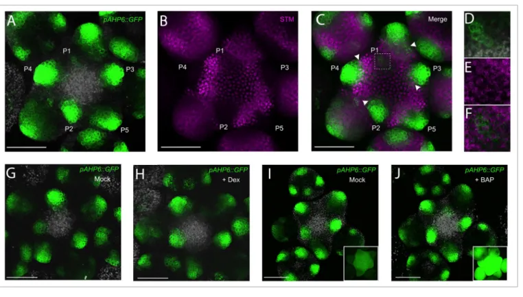
Figure 1. Visualization of the expression pattern of AHP6 in the inflorescence meristem using whole-mount mrna in situ hybridization. (A) representative
AHP6 mrna distribution in an inflorescence meristem detected using colorimetric (nBt/BCiP) revelation (n = 8; view from the top). (B–E) representative AHP6 mrna distribution in an inflorescence meristem detected using tyramide signal amplification fluorescent revelation (n = 7; view from the top).
(B) AHP6 expression pattern in the inflorescence meristem (maximum intensity projection; view from the top). (C–E) transverse optical section in lateral organs as indicated. the organ primordia (P) are numbered from youngest to oldest. Scale bar: 50 μm (a) or 25 μm (B–E; scale is identical in C-E).
Figure 2. AHP6 expression in the meristem is regulated independently of Stm and cytokinins. (A-F) Co-expression analysis of pAHP6::GFP and
pSTM::STM-VENUS (Stm-VEnuS). (A-C): representative meristem (n = 6) expressing pAHP6::GFP (green) and Stm-VEnuS (magenta). (D-E) Close-up of P1 (square region
shown in C). the channels shown corresponds to (A,D) pAHP6::GFP and autofluorescence (gray); (B,E) Stm-VEnuS ; (C,F) pAHP6::GFP and Stm-VEnuS. White arrowheads in (C) indicate organs frontiers where both reporters are co-expressed. (G,H) representative meristems (n = 6) co-expressing pAHP6::GFP and
35S::STM-GR. meristems were observed one day after a mock (G) or a dexamethasone (Dex) treatment (H) as described.24 (I,J) representative pAHP6::GFP
meristems after treatment with a mock (i; n = 3) or a CK solution (1 μm BaP) for 48h (J; n = 4). insets: control plants expressing a cytokinin-inducible
pARR5::GFP reporter treated in parallel with the same solutions (n = 3 for each condition). in (G-J) pAHP6::GFP is in green and autofluorescence in gray. all
This raises the question of which factors could control the dynamic expression of AHP6. In root tissues, strong evidence suggests that AHP6 is regulated directly by auxin13 and we further
supported this regulation by demonstrating that several elements in the AHP6 promoter can be bound by the Auxin Response Factor 5/MONOPTEROS protein, a key transcription factor involved in auxin signaling at the SAM.10 However, we could
also show that AHP6 transcription starts with a delay of one plastochrone after the activation of the synthetic auxin-inducible
DR5::VENUS reporter.10,14 We thus investigated other factors
that could control AHP6 expression. While organ initiation is triggered by auxin, it is also accompanied by a downregulation of the SHOOTMERISTEMLESS (STM) meristem identity gene.14,15 We thus tested a scenario where STM downregulation
during organ initiation could regulate AHP6 expression, either directly or indirectly through a lowering of CK levels as STM activates genes involved in CK biosynthesis.16,17 Co-expression
analysis of a STM-VENUS translational fusion14 and of the
pAHP6::GFP transcriptional reporter did not support this
hypothesis. First, many cells co-express both reporters in organ
frontiers (Fig. 2A-F). Second, although fading away, STM-VENUS protein was still visible in the emerging P1 organ where
AHP6 expression starts (Fig. 2D-F), suggesting that complete
STM downregulation is unlikely a requisite for activation of
AHP6 transcription. Then, neither an inducible mis-expression
of STM in the organs using a 35S::STM-GR line18 (Fig. 2G, H)
nor an exogenous treatment with CK (Fig. 2I, J) could alter the expression of pAHP6::GFP. Taken together, these data suggest that AHP6 is activated independently of STM or CK levels in the SAM and that CK are unlikely involved directly in regulating
AHP6 expression in the shoot, as shown also in the root.13
While expressed specifically in organ primordia in the SAM, AHP6 proteins move outside of the transcription domain of the
AHP6 gene and establish CK signaling inhibitory fields.10 The
timing of AHP6 transcription and the movement of the AHP6 protein result in a differential in AHP6 protein concentration between the 2 earliest organ initia that promotes sequential organ initiation (the one with the lowest AHP6 level/higher cytokinin signaling grows first). However, the mechanism by which a lower AHP6 concentration facilitates organ development
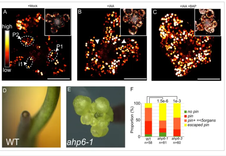
Figure 3. Cytokinin potentiates auxin responses in the Sam. (A-C) Co-treatments with exogenous auxin (iaa) and cytokinin (BaP). representative
images of DR5::VENUS expression in meristems with older primordia digitally removed for clarity (insets: original image, with autofluorescence in Gray) are shown after a mock (a; n = 4), 1 μm iaa (B; n = 7) or 1 μm iaa + 1 μm BaP (C; n = 7) treatment. arrowheads indicate areas in the peripheral zone where there is no Dr5 activation in (B). note the activation of DR5::VENUS throughout the peripheral zone in (C). images were obtained by confocal microscopy and are maximum intensity projections (view from the top). (D-F) organ initiation in 4 week-old nPa-grown plants. Plants were grown as previously described.25 (D) representative Wt. (E) representative ahp6–1 escaped pin inflorescence. (F) Quantification of the phenotype
is still elusive. A plausible hypothesis would be the existence of a synergistic effect of auxin and CK during the earliest steps of organ initiation, as suggested in recent studies.19 Supporting this
possibility, a 12h treatment with both exogenous auxin and CK lead to an expansion of the expression domain of a DR5::GFP reporter,20 when compared with auxin alone, and to an activation
of auxin signaling in most cells at the periphery of the SAM (Fig. 3A-C). These experiments suggest that CK potentiates the response to auxin at the periphery of the SAM i.e., where organ initiation takes place. Since CK signaling activity is elevated in ahp6 mutant SAMs,10 we wondered whether AHP6
loss-of-function could also potentiate auxin responses when auxin is limiting. Indeed, when treated with the auxin transport inhibitor NPA, ahp6 mutants had a significantly higher capacity to produce flowers when the inflorescence stem starts elongating (Fig. 3D-F). These data suggest that the increase in CK signaling activity resulting from the ahp6 mutation can partially rescue the lower organ initiation capacities induced by an inhibition of auxin transport. Although we have not found yet any direct targets responding to the CK signaling increase during organ initiation, we can hypothesize that these targets might act either downstream of or in parallel with auxin during organ initiation.
By analyzing the PIN1 network in ahp6 mutant,10 we have shown
that such targets if they exist are unlikely to act on auxin transport as in roots13,21,22 but could possibly modify auxin concentrations23
and/or the competence of cells to sense and respond to auxin. These different options clearly deserve more investigation in the future. Altogether, our previously published observations and the data presented here converge to indicate that higher CK signaling could potentiate auxin signaling activity during organ initiation, providing a plausible explanation for how a differential in AHP6 concentration regulates the timing of organ initiation.
Disclosure of Potential Conflicts of Interest
No potential conflicts of interest were disclosed.
Acknowledgments
We thank M. Heisler and R. Sablowski for sharing materials; C. Chamot and C. Lionnet (PLATIM, ENS Lyon) for help with confocal microscopy. T.V. was supported by HFSPO CDA 0047/2007 (Human Frontier Science Program Organization), ANR-07-JCJC-0115 (AuxFate; Agence National de la Recherche) and EraSysBio (iSAM) grants; F.B. by a predoctoral grant of the French Ministry of Research.
References
1. Sassi M, Vernoux T. Auxin and self-organization at the shoot apical meristem. J Exp Bot 2013; 64:2579-92; PMID:23585672; http://dx.doi.org/10.1093/ jxb/ert101
2. Reinhardt D, Pesce E-R, Stieger P, Mandel T, Baltensperger K, Bennett M, Traas J, Friml J, Kuhlemeier C. Regulation of phyllotaxis by polar auxin transport. Nature 2003; 426:255-60; PMID:14628043; http://dx.doi.org/10.1038/ nature02081
3. Bainbridge K, Guyomarc’h S, Bayer E, Swarup R, Bennett M, Mandel T, Kuhlemeier C. Auxin influx carriers stabilize phyllotactic patterning. Genes Dev 2008; 22:810-23; PMID:18347099; http://dx.doi. org/10.1101/gad.462608
4. Vernoux T, Kronenberger J, Grandjean O, Laufs P, Traas J. PIN-FORMED 1 regulates cell fate at the periphery of the shoot apical meristem. Development 2000; 127:5157-65; PMID:11060241
5. Okada K, Ueda J, Komaki MK, Bell CJ, Shimura Y. Requirement of the Auxin Polar Transport System in Early Stages of Arabidopsis Floral Bud Formation. Plant Cell 1991; 3:677-84; PMID:12324609 6. Smith RS, Guyomarc’h S, Mandel T, Reinhardt
D, Kuhlemeier C, Prusinkiewicz P. A plausible model of phyllotaxis. Proc Natl Acad Sci U S A 2006; 103:1301-6; PMID:16432192; http://dx.doi. org/10.1073/pnas.0510457103
7. de Reuille PB, Bohn-Courseau I, Ljung K, Morin H, Carraro N, Godin C, Traas J. Computer simulations reveal properties of the cell-cell signaling network at the shoot apex in Arabidopsis. Proc Natl Acad Sci U S A 2006; 103:1627-32; PMID:16432202; http:// dx.doi.org/10.1073/pnas.0510130103
8. Jönsson H, Heisler MG, Shapiro BE, Meyerowitz EM, Mjolsness E. An auxin-driven polarized transport model for phyllotaxis. Proc Natl Acad Sci U S A 2006; 103:1633-8; PMID:16415160; http:// dx.doi.org/10.1073/pnas.0509839103
9. Vernoux T, Brunoud G, Farcot E, Morin V, Van den Daele H, Legrand J, Oliva M, Das P, Larrieu A, Wells D, et al. The auxin signalling network translates dynamic input into robust patterning at the shoot apex. Mol Syst Biol 2011; 7:508; PMID:21734647; http://dx.doi.org/10.1038/msb.2011.39
10. Besnard F, Refahi Y, Morin V, Marteaux B, Brunoud G, Chambrier P, Rozier F, Mirabet V, Legrand J, Lainé S, et al. Cytokinin signalling inhibitory fields provide robustness to phyllotaxis. Nature 2014; 505:417-21; PMID:24336201; http://dx.doi.org/10.1038/ nature12791
11. Mähönen AP, Bishopp A, Higuchi M, Nieminen KM, Kinoshita K, Törmäkangas K, Ikeda Y, Oka A, Kakimoto T, Helariutta Y. Cytokinin signaling and its inhibitor AHP6 regulate cell fate during vascular development. Science 2006; 311:94-8; PMID:16400151; http://dx.doi.org/10.1126/ science.1118875
12. Lauter G, Söll I, Hauptmann G. Multicolor fluorescent in situ hybridization to define abutting and overlapping gene expression in the embryonic zebrafish brain. Neural Dev 2011; 6:10; PMID:21466670; http://dx.doi.org/10.1186/1749-8104-6-10
13. Bishopp A, Help H, El-Showk S, Weijers D, Scheres B, Friml J, Benková E, Mähönen AP, Helariutta Y. A mutually inhibitory interaction between auxin and cytokinin specifies vascular pattern in roots. Curr Biol 2011; 21:917-26; PMID:21620702; http://dx.doi. org/10.1016/j.cub.2011.04.017
14. Heisler MG, Ohno C, Das P, Sieber P, Reddy GV, Long JA, Meyerowitz EM. Patterns of auxin transport and gene expression during primordium development revealed by live imaging of the Arabidopsis inflorescence meristem. Curr Biol 2005; 15:1899-911; PMID:16271866; http://dx.doi.org/10.1016/j. cub.2005.09.052
15. Long JA, Moan EI, Medford JI, Barton MK. A member of the KNOTTED class of homeodomain proteins encoded by the STM gene of Arabidopsis. Nature 1996; 379:66-9; PMID:8538741; http:// dx.doi.org/10.1038/379066a0
16. Yanai O, Shani E, Dolezal K, Tarkowski P, Sablowski R, Sandberg G, Samach A, Ori N. Arabidopsis KNOXI proteins activate cytokinin biosynthesis. Curr Biol 2005; 15:1566-71; PMID:16139212; http://dx.doi. org/10.1016/j.cub.2005.07.060
17. Jasinski S, Piazza P, Craft J, Hay A, Woolley L, Rieu I, Phillips A, Hedden P, Tsiantis M. KNOX action in Arabidopsis is mediated by coordinate regulation of cytokinin and gibberellin activities. Curr Biol 2005; 15:1560-5; PMID:16139211; http://dx.doi. org/10.1016/j.cub.2005.07.023
18. Gallois J-L, Woodward C, Reddy GV, Sablowski R. Combined SHOOT MERISTEMLESS and WUSCHEL trigger ectopic organogenesis in Arabidopsis. Development 2002; 129:3207-17; PMID:12070095
19. Yoshida S, Mandel T, Kuhlemeier C. Stem cell activation by light guides plant organogenesis. Genes Dev 2011; 25:1439-50; PMID:21724835; http:// dx.doi.org/10.1101/gad.631211
20. Benková E, Michniewicz M, Sauer M, Teichmann T, Seifertová D, Jürgens G, Friml J. Local, efflux-dependent auxin gradients as a common module for plant organ formation. Cell 2003; 115:591-602; PMID:14651850; http://dx.doi.org/10.1016/ S0092-8674(03)00924-3
21. Laplaze L, Benkova E, Casimiro I, Maes L, Vanneste S, Swarup R, Weijers D, Calvo V, Parizot B, Herrera-Rodriguez MB, et al. Cytokinins act directly on lateral root founder cells to inhibit root initiation. Plant Cell 2007; 19:3889-900; PMID:18065686; http://dx.doi. org/10.1105/tpc.107.055863
22. Moreira S, Bishopp A, Carvalho H, Campilho A. AHP6 inhibits cytokinin signaling to regulate the orientation of pericycle cell division during lateral root initiation. PLoS One 2013; 8:e56370; PMID:23457561; http:// dx.doi.org/10.1371/journal.pone.0056370
23. Jones B, Gunnerås SA, Petersson SV, Tarkowski P, Graham N, May S, Dolezal K, Sandberg G, Ljung K. Cytokinin regulation of auxin synthesis in Arabidopsis involves a homeostatic feedback loop regulated via auxin and cytokinin signal transduction. Plant Cell 2010; 22:2956-69; PMID:20823193; http://dx.doi. org/10.1105/tpc.110.074856
24. Gómez-Mena C, de Folter S, Costa MMR, Angenent GC, Sablowski R. Transcriptional program controlled by the floral homeotic gene AGAMOUS during early organogenesis. Development 2005; 132:429-38; PMID:15634696; http://dx.doi.org/10.1242/ dev.01600
25. Grandjean O, Vernoux T, Laufs P, Belcram K, Mizukami Y, Traas J. In vivo analysis of cell division, cell growth, and differentiation at the shoot apical meristem in Arabidopsis. Plant Cell 2004; 16:74-87; PMID:14671026; http://dx.doi.org/10.1105/ tpc.017962