Author(s) - Auteur(s) :
Publication date - Date de publication :
Permanent link - Permalien :
Rights / License - Licence de droit d’auteur :
MPV17 does not control cancer cell proliferation
Canonne, Morgane; Wanet, Anaïs; An Nguyen, Thuy Truong ; Khelfi, Alexis; Ayama-Canden,
Sophie; Van Steenbrugge, Martine; Fattaccioli, Antoine; Sokal, Etienne; Najimi, Mustapha;
Arnould, Thierry; Renard, Patricia
Published in: PLoS ONE DOI: 10.1371/journal.pone.0229834 Publication date: 2020 Document Version
Publisher's PDF, also known as Version of record Link to publication
Citation for pulished version (HARVARD):
Canonne, M, Wanet, A, An Nguyen, TT, Khelfi, A, Ayama-Canden, S, Van Steenbrugge, M, Fattaccioli, A, Sokal, E, Najimi, M, Arnould, T & Renard, P 2020, 'MPV17 does not control cancer cell proliferation', PLoS ONE, vol. 15, no. 3, e0229834, pp. 1-18. https://doi.org/10.1371/journal.pone.0229834
General rights
Copyright and moral rights for the publications made accessible in the public portal are retained by the authors and/or other copyright owners and it is a condition of accessing publications that users recognise and abide by the legal requirements associated with these rights. • Users may download and print one copy of any publication from the public portal for the purpose of private study or research. • You may not further distribute the material or use it for any profit-making activity or commercial gain
• You may freely distribute the URL identifying the publication in the public portal ?
Take down policy
1 Laboratory of Biochemistry and Cell Biology (URBC), NAmur Research Institute for LIfe Sciences
(NARILIS), University of Namur (UNamur), Namur, Belgium, 2 Laboratory of Pediatric Hepatology and Cell Therapy, Institut de Recherche Expe´rimentale et Clinique (IREC), Universite´ Catholique de Louvain, Brussels, Belgium
Abstract
MPV17 is described as a mitochondrial inner membrane channel. Although its function remains elusive, mutations in the MPV17 gene result in hepato-cerebral mitochondrial DNA depletion syndrome in humans. In this study, we show that MPV17 silencing does not induce depletion in mitochondrial DNA content in cancer cells. We also show that MPV17 does not control cancer cell proliferation despite the fact that we initially observed a reduced proliferation rate in five MPV17-silenced cancer cell lines with two different shRNAs. How-ever, shRNA-mediated MPV17 knockdown performed in this work provided misguiding results regarding the resulting proliferation phenotype and only a rescue experiment was able to shed definitive light on the implication of MPV17 in cancer cell proliferation. Our results therefore emphasize the caution that is required when scientific conclusions are drawn from a work based on lentiviral vector-based gene silencing and clearly demonstrate the need to systematically perform a rescue experiment in order to ascertain the specific nature of the experimental results.
Introduction
MPV17 is a functionally elusive protein localized in the inner membrane of mitochondrion and for which the encoding gene is located on chromosome 2p23-21 [1] [2] [3] [4]. MPV17 loss-of-function causes a rare autosomal recessive mitochondrial disorder called Mitochon-drial DNA Depletion Syndrome (MDDS) marked by a highly reduced mitochonMitochon-drial DNA (mtDNA) copy number in affected tissues.
To date, there are 100 known individuals affected by one of the 48 describedMPV17 patho-genic variants (approximately half of which are missense) [5]. The vast majority of these patients (96%) suffer from the hepato-cerebral form of MDDS and exhibit a severe mtDNA depletion in the liver (60–99% reduction). This is correlated with a decreased activity of respi-ratory chain complexes. The onset of the disease takes place early in life (neonatal period/ infancy) and condemn the affected individual to a premature death due to liver dysfunction progressing into liver failure. The remaining 4% of the patients suffer from a late-onset ence-phalomyopathic disease with mild or no liver involvement [5].
a1111111111 a1111111111 a1111111111 a1111111111 a1111111111 OPEN ACCESS
Citation: Canonne M, Wanet A, Nguyen TTA, Khelfi
A, Ayama-Canden S, Van Steenbrugge M, et al. (2020) MPV17 does not control cancer cell proliferation. PLoS ONE 15(3): e0229834.https:// doi.org/10.1371/journal.pone.0229834 Editor: Negin P. Martin, National Institute of
Environmental Health Sciences (NIEHS), UNITED STATES
Received: August 1, 2019 Accepted: February 14, 2020 Published: March 10, 2020
Copyright: © 2020 Canonne et al. This is an open
access article distributed under the terms of the
Creative Commons Attribution License, which permits unrestricted use, distribution, and reproduction in any medium, provided the original author and source are credited.
Data Availability Statement: All RNA sequencing
files are available from the Annotare 2.0 database (accession number E-MTAB-8108,https://www. ebi.ac.uk/fg/annotare/).
Funding: The author(s) received no specific
funding for this work.
Competing interests: The authors have declared
MPV17 knockdown reduces the proliferation of NSC34 cells, a mouse motor neuronal cell line [14]. As MPV17 has been implicated in stress response [15] [16] and has been described as a channel with stress-dependent gating properties (oxidative and pH stress,. . .) [7], we then wondered whether MPV17 could have a role in the proliferation of cancer cells or not, as they inherently experience oxidative and metabolic stress.
In order to explore the putative role of MPV17 in the control of cancer cell proliferation, we used a loss-of-function approach. Gene silencing and/or overexpression is generally the first approach in order to investigate gene expression/function and lentiviruses are now widely used to deliver transgenes that integrate into the host genome for gene expression tampering. In this study, while demonstrating that MPV17 does not control neither cell proliferation nor mtDNA content in cancer cells, our experimental results illustrate and emphasize the impor-tance of carrying out a rescue experiment when working with shRNA-mediated knockdown.
Results
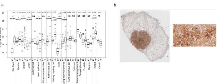
MPV17 is overexpressed in several tumours
Taking advantage of The Genome Cancer Atlas (TGCA) database, we show that the abun-dance ofMPV17 transcript is significantly higher in tumours of 10 different tissues, including liver, bile duct, and colon (Fig 1A). This is confirmed at the protein level by immunohis-tochemistry staining performed on liver tumour biopsies from patients with adenocarcinoma (Fig 1B).
MPV17 silencing is robustly associated with a decreased proliferation rate
in different cancer cell lines
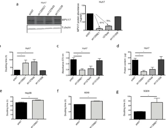
In this study, we first assessed the effects of three commercially available shRNAs targeting MPV17 mRNA: sh129921, sh127649 and sh131038. Using western blot analysis, the efficiency of gene silencing was evaluated by assessing the abundance of MPV17 in Huh7 cells trans-duced with these three shRNA-encoding lentiviral vectors. Both sh129921 and sh127649 led to an efficient knockdown of the gene while sh131038 did not efficiently induceMPV17 silencing (Fig 2A).
We then demonstrated that Huh7 cells silenced forMPV17 with sh129921 and sh127649 displayed a severely decreased proliferation rate, as quantified by three different prolifera-tion assays, namely the doubling time (Fig 2B), the MTT assay (Fig 2C), and the total pro-tein content (Fig 2D). The reduced cell proliferation phenotype was correlated withMPV17
transduced with the vector encoding sh131038, the only shRNA that turned out to be ineffi-cient in the knockdown induction.
To discard the possibility of a putative cell type or cancer type-specific phenotype, we next assessed the impact of sh129921 on the proliferation of two other human hepatoma cell lines, Hep3B and HepG2 cells, and two non-liver cancer cell lines, A549 cells, derived from a human pulmonary adenocarcinoma and SQD9 cells, a human squamous cell carcinoma cell line. Interestingly, Hep3B (Fig 2E), A549 (Fig 2F), SQD9 (Fig 2G) and HepG2 (data not shown) cells transduced with sh129921-encoding vector also displayed a reduced prolifera-tion phenotype.
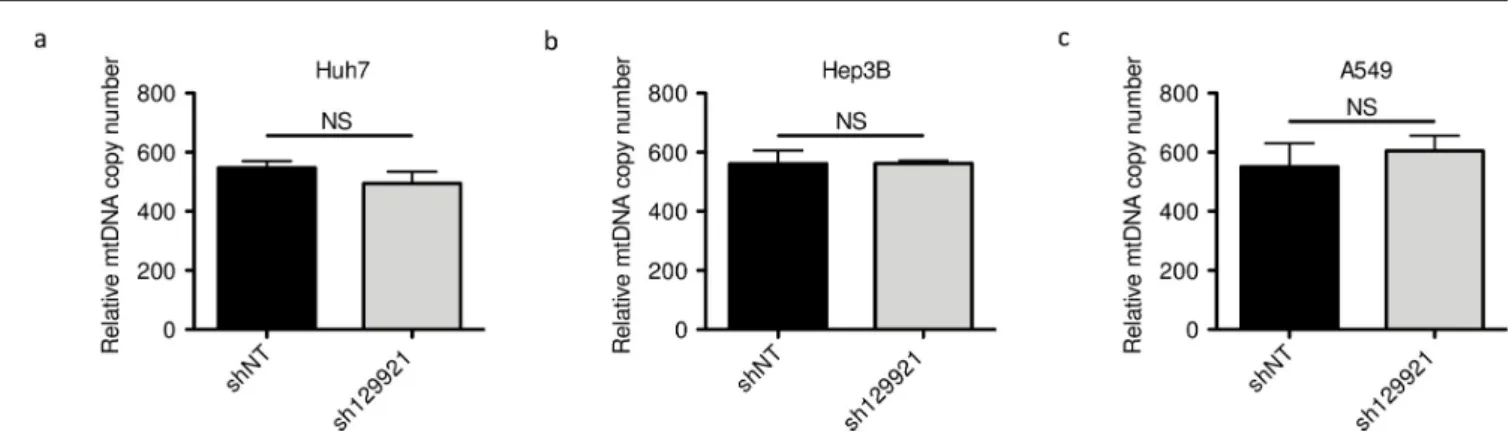
MPV17 silencing is not associated with mtDNA copy number depletion
As MPV17 deficiency is associated with MDDS, we assessed whetherMPV17 silencing was accompanied by depletion in the mtDNA content or not, possibly accounting for the associ-ated decreased proliferation rate. However,MPV17 silencing did not lead to a reduction of mtDNA content in any of the tested cancer cell lines (Fig 3). This result is in agreement with the work of Dalla Rosa and collaborators who showed that proliferating fibroblasts from MPV17-deficient patients do not display any reduced mtDNA content [9]. It is also in accor-dance with the recent work of Alonzo and collaborators who did not find any reduction of mtDNA content inMPV17-silenced HeLa cells [12].
The decreased proliferation rate in
MPV17-silenced cells is associated with
a decrease in the abundance of ATF4
In order to determine the molecular mechanisms underlying the decreased proliferation rate inMPV17-silenced cancer cells, a transcriptomic analysis was performed. The RNA sequencing analysis was performed on Huh7 cells transduced with sh129921 or shNT-encoding vectors. Among the differentially expressed genes, we focused on transcrip-tional regulators potentially responsible for a reduced proliferation capacity. The activat-ing transcription factor 4 (ATF4) frequently upregulated in cancer cells [17], was
Fig 1. Analysis of MPV17 expression in cancer tissues. The Cancer Genome Atlas (TCGA), a public platform allowing the analysis of gene expression
data sets generated by RNA sequencing (http://cancergenome.nih.gov), has been used to determine the expression level ofMPV17 in various tumour tissues (T)versus healthy tissues (N). P values were calculated with the two-tailed Wilcoxon signed rank Test (α = 5%;⇤: p<0.05;⇤⇤: p<0.01;⇤⇤⇤: p<0.001; ⇤⇤⇤⇤: p<0.0001; NS: not significant) (a). Detection of MPV17 by immunohistochemistry in a paraffin-embedded biopsy of a liver adenocarcinoma. A strong signal for MPV17 is associated with the tumour, while the abundance of the protein is low in adjacent normal tissue (b).
downregulated inMPV17-silenced cells (Z-score = -2,792; P = 1,38E-14). It has been shown that ATF4 not only up-regulates the expression of genes encoding actors impli-cated in amino acid import and metabolism [18] [19] [20] but also promotes, indirectly, purine synthesis [21], two essential aspects for cell proliferation. Based on this knowledge, ATF4 was an attractive candidate in the attempt to elucidate the molecular mechanisms underlying the reduced cell proliferation. The reduction ofATF4 transcript abundance was confirmed at the protein level in sh129921 and sh127649-encoding vector transduced Huh7 cells (Fig 4A) as well as in sh129921-encoding vector transduced Hep3B (Fig 4B) and A549 (Fig 4C) cells, when compared with control cells transduced with shNT-con-taining vector.
Altogether, these results seem to strongly support an involvement of MPV17 in cancer cell proliferation asMPV17 silencing was consistently accompanied by a reduction of both cell proliferation rate and ATF4 protein abundance.
Fig 2. Effect of shRNA-mediatedMPV17 knockdown on the proliferation of cancer cell lines. Huh7 cells were
transduced with non-target shRNA lentiviral vectors (shNT) or with shRNA lentiviral vectors targetingMPV17 expression (sh129921, sh127649, sh131038). Hep3B, A549 and SQD9 cells were transduced with lentiviral shNT-encoding vectors or with sh129921-containing vectors. Cells were selected for 6 days with puromycin (2.5μg/mL). Cells were seeded at 8×10³cells/cm2(a, b, c, d, Huh7; g, SQD9), 5×10³cells/cm2(e, Hep3B) and 2.7×10³cells/cm2(f,
A549) and grown for 4 days. MPV17 protein abundance was assessed by western blot analysis (a, Huh7). A representative western blot of 3 independent biological replicates (2 for sh131038) is shown (left) along with the western blot quantification of all the biological replicates (quantification with Image J software, data expressed as relative protein abundance to cells transduced with shNT-encoding vectors, right). Proliferation was then assessed by manual counting to calculate the doubling time (b, Huh7; e, Hep3B; f, A549; g, SQD9), by MTT assay (c, Huh7) and by the total protein content (d, Huh7). Data are presented as mean± S.E.M of 3 independent biological replicates (2 for sh131038). P values were calculated with the one-tailed Mann-Whitney Test (α = 5%;⇤: p<0.05;⇤⇤: p<0.01;⇤⇤⇤: p<0.001). Full blots are presented inS6 Fig.
MPV17 silencing is not always associated with a reduced proliferation
phenotype
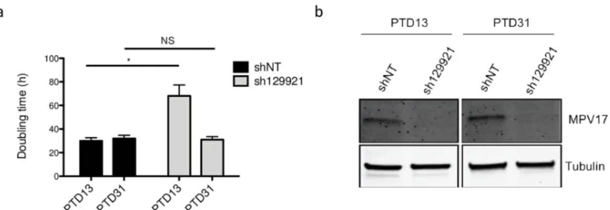
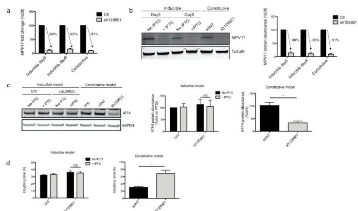
Pursuing our analysis further, we observed that the sh129921-encoding vector transduced Huh7 cells were able to adapt along the passages and progressively restored the proliferation rate (Fig 5A), although MPV17 protein abundance was still strongly reduced (Fig 5B).
As we observed a recovery of the decreased proliferation rate over time, we therefore aimed at generating an IPTG-inducible sh129921 model in Huh7 cells. Strikingly, while we observed a strongMPV17 knockdown in this inducible expression model, with a silencing efficiency
Fig 3. Assessment of mtDNA content inMPV17-silenced cancer cell lines. Huh7 (a), Hep3B (b) and A549 (c) cells were transduced with non-target shRNA
lentiviral vectors (shNT) or with sh129921-containing vectors. Transduced cells were selected for 6 days with puromycin (2.5μg/mL). DNA was then extracted and mtDNA content was assessed by qPCR using NADH dehydrogenase 2 as a specific marker of mtDNA content and beclin for normalization with nuclear DNA. Results are presented as means± S.E.M of 3 independent biological replicates and are expressed in relative copy number to the nuclear DNA. P values were calculated with the one-tailed Mann-Whitney Test (α = 5%; NS;⇤: p<0.05;⇤⇤: p<0.01;⇤⇤⇤: p<0.001).
https://doi.org/10.1371/journal.pone.0229834.g003
Fig 4. ATF4 protein abundance followingMPV17 knockdown in Huh7, Hep3B and A549 cells. Huh7 cells (a) were transduced with non-target
shRNA lentiviral vectors (shNT) or with shRNA lentiviral vectors targetingMPV17 expression (sh129921, sh127649, sh131038). Hep3B (b) and A549 (c) cells were transduced with lentiviral shNT-encoding vectors or with sh129921-containing vectors. Cells were selected for 6 days with puromycin (2.5μg/mL) before assessing ATF4 protein abundance by western blot analysis. For each cell line, a representative western blot of 3 (2 for sh131038) independent biological replicates is shown (left) along with the western blot quantification of all the biological replicates (quantification with Image J software, data expressed as relative protein abundance to untranduced (unt) cells, right). P values were calculated with the one-tailed Mann-Whitney Test (α = 5%;⇤: p<0.05;⇤⇤: p<0.01;⇤⇤⇤: p<0.001). Full blots are presented inS6 Fig.
comparable to the one observed in the constitutive silencing model (Fig 6A and 6B), the cell proliferation rate was unchanged (Fig 6D). Moreover, ATF4 did not display a decreased pro-tein abundance (Fig 6C).
This absence of effect ofMPV17 silencing on the cell proliferation rate in the inducible expression system could have different origins. First, the IPTG molecule itself could have an unexpected effect on Huh7 cells proliferation, even though IPTG is not known to be metabo-lized [22]. Second, the reduced proliferation phenotype in the inducible expression system might need more time to settle down. Indeed, in the constitutive system,MPV17 is silenced for a total of 9 days before assessing the cell proliferation, as opposed to only 5 days in the inducible model (see “Materials and Methods”,S1 Fig). A third difference between the two approaches is that in order to ensure a proper knockdown ofMPV17 in the inducible model, the culture medium was changed every day in order to renew the IPTG, as opposed to every two or three days in the constitutive silencing model. One could thus hypothesize that this daily medium renewal could prevent the settling of the reduced proliferation phenotype by discontinuing the putative intercellular communication. These three hypotheses were tested but failed to explain the different phenotypical outcomes observed for the constitutive or inducible expression models (seeS2andS3Figs).
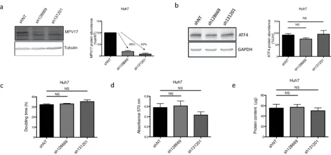
These considerations led us to evaluate on Huh7 cells the effect of two additional commer-cially available shRNAs, sh128669 and sh131201, targeting different regions of theMPV17 transcript (seeS4 Fig). Both shRNAs strongly reduce the abundance of MPV17 protein (Fig 7A) but no significant effect on the proliferation rate was observed (Fig 7C, 7D and 7E). Also, the abundance of ATF4 was not affected byMPV17 knockdown mediated by either sh128669 or sh131201 (Fig 7B). Thus, ATF4 reduced abundance correlates with the proliferation rate but the putative link between the reduced proliferation phenotype (and therefore ATF4) and MPV17 remains to be established.
In conclusion, we found that sh129921, sh127649, sh128669 and sh131201 all led to a strong MPV17 knockdown while the resulting proliferation rates were highly variable. This lack of consistency led us to suspect a putative involvement ofMPV17 isoforms that would be differ-entially affected according to the shRNA used.
Fig 5. Evolution of Huh7 proliferation rate and MPV17 protein abundance in sh129921-mediatedMPV17 knockdown. Huh7 cells
were transduced with non-target shRNA lentiviral vectors (shNT) or with sh129921-containing vectors. Transduced cells were selected for 6 days with puromycin (2.5μg/mL). At 13 and 31 days after the transduction (PTD: post-transduction day), cells were seeded at 8×10³cells/cm2and grown for 4 days. Proliferation was then assessed by manual counting to calculate the doubling time (a) and MPV17
protein abundance was analysed by western blot (b). P values were calculated with the one-tailed Mann-Whitney Test (α = 5%; NS;⇤: p<0.05;⇤⇤: p<0.01;⇤⇤⇤: p<0.001). n = 3. Full blots are presented inS6 Fig.
Therefore, we next interrogated the Genotype-Tissue Expression (GTEx) Consortium (2008, NIH) portal, that inventories the impact of genetic variations on gene expression within major tissues in the human body frompost mortem donors. The GTEx Portal proposes 22 MPV17 isoforms, including two major ones i.e. a short predominant isoform and a long one (seeS5 Fig). This long isoform referenced in RefSeq as NM002437.5 encodes the MPV17 pro-tein that we detected on western blot while the translation of all the otherMPV17 isoforms is not experimentally demonstrated. However, among the 22 isoforms, we observe that all the transcripts targeted by sh129921 (1, 2, 3, 12, 13, 14, 15, 16 and 18) are also targeted by either sh128669 (15, 16) or sh131201 (1, 18) or both (2, 3, 12, 13, 14). The same kind of observation stands for the transcripts targeted by sh127649 (1, 2, 3, 5, 12, 13, 14, 15, 16, 18, 19, 21), provid-ing no clear explanation about the opposite proliferation phenotypes observed for each pair of shRNAs (sh129921/sh127649 versus sh128669/131201) (seeS5 Fig).
Altogether, the absence of effect of the inducible sh129921 on the cell proliferation rate, combined with the observation that, at least, two different shRNAs targetingMPV17 (sh131201 and sh128669) have no effect on the proliferation of Huh7 cells, despite a strong
Fig 6. MPV17 and ATF4 abundances and Huh7 cells proliferation rate following inducible sh129921-mediatedMPV17 knockdown. Huh7 cells were
transduced, or not (Unt), with inducible sh129921 lentiviral vector. Transduced cells were selected for 6 days with puromycin (2.5μg/mL). Cells were then incubated for 5 days in the presence of 0.1 mM of IPTG to induceMPV17 silencing. Cells were seeded at 8×10³cells/cm2and grown for 4 days in daily
renewed medium containing IPTG. RNA was extracted and RT-qPCR was performed to assessMPV17 transcript level (data expressed relatively to respective control (Ctl), n = 3) (a). MPV17 (b) and ATF4 (c) protein abundances were also assessed by western blot analysis after 5 days (MPV17) and 9 days (MPV17; ATF4) of IPTG induction. For each protein, a representative western blot of 3 independent biological replicates is shown (left) along with the western blot quantification of all the biological replicates (quantification with Image J software, data expressed as relative protein abundance to respective controls (Ctl) for MPV17 and untransduced (unt) cells for ATF4, right). Proliferation was then assessed by manual counting to calculate the doubling time (d). As a comparison, we performed at the same time and on the same cells a similar experiment with the constitutive expression of sh129921 (mediating MPV17 knockdown) as described inS1 Fig. P values were calculated with the one-tailed Mann-Whitney Test (α = 5%;⇤: p<0.05;⇤⇤: p<0.01;⇤⇤⇤: p<0.001). Full blots are presented inS6 Fig. n = 3.
decrease in the MPV17 protein abundance, suggest that the decreased proliferation rate of transduced cells observed for two shRNAs (sh129921 and sh127649) is not related to a reduced MPV17 protein abundance. In an attempt to shed light on this question, we eventually per-formed a rescue experiment.
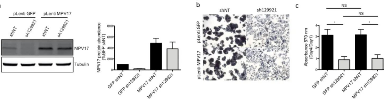
Rescuing MPV17 does not restore the proliferation phenotype. As Huh7 cells could neither be efficiently transfected nor transduced with theMPV17-expressing vector, HepG2 cells were used for the rescue attempt. Indeed, as mentioned before, we also observed the reduced proliferation rate in this hepatoma cell line silenced forMPV17. This cell line turned out to be the most receptive to the lentiviralMPV17-expressing vector. Since a rescue experi-ment consists in re-introducing the shRNA-mediated silenced mRNA, it requires the use of a shRNA targeting the 30UTR of the targeted mRNA. Thus, the reintroduced transcript that
lacks 30UTR is not affected by the shRNA.
We decided to perform MPV17 overexpression before inducing the constitutive knock-down mediated by sh129921. The justification of this chronology resides in the observation that, on the contrary to shNT-encoding vector transduced cells, sh129921-encoding vector transduced cells were not able to stand a second round of transduction due to cell death. This suggests thatMPV17-silenced cells are distressed, in accordance with their decreased prolifera-tion rate. This aspect is therefore not compatible with a short-term assessment of cell
proliferation.
MPV17 knockdown was properly induced in the overexpression control. Indeed, cells dou-ble transduced with pLenti GFP, as a control, and then with pLKO.1 sh129921, displayed a strong reduction of MPV17 abundance accompanied by the decreased proliferation rate (Fig 8A, 8B and 8C). As expected, the reduced proliferation phenotype was absent in pLenti GFP
Fig 7. Effect ofMPV17-targeting sh128669 and sh131201 on Huh7 cells proliferation, MPV17 and ATF4 protein abundances. Cells were
transduced with non-target shRNA lentiviral vectors (shNT) or with shRNA lentiviral vectors targetingMPV17 expression (sh128669 and sh131201). Transduced cells were selected for 6 days with puromycin (2.5μg/mL). Cells were seeded at 8×10³cells/cm2and grown for 4 days. The
abundance of MPV17 (a) and ATF4 (b) proteins was assessed by western blot analysis. For each protein, a representative western blot analysis of 3 independent biological replicates is shown (left) along with the western blots quantification of all the biological replicates (quantification with Image J software, data expressed as relative protein abundance to cells transduced with shNT-encoding vectors (a, MPV17) or untranduced (unt) cells (b, ATF4), right). Proliferation was then assessed by manual counting to calculate the doubling time (c), by MTT assay (d) and by the total protein content (e) and data are presented as mean± S.E.M (3 biological replicates). P values were calculated with the one-tailed Mann-Whitney Test (α = 5%; NS;⇤: p<0.05;⇤⇤: p<0.01;⇤⇤⇤: p<0.001). Full blots are presented inS6 Fig.
and pLKO.1 shNT double-transduced cells (Fig 8B and 8C). Cells double-transduced with pLenti MPV17 and pLKO.1 shNT suitably overexpressed MPV17 (Fig 8A). It is interesting to emphasize the fact that MPV17 overexpression, on its own, has no significant effect on cell pro-liferation (Fig 8B and 8C). This observation can be reconciled with the idea that MPV17 is described as a channel. Thus, the qualitative state of MPV17 (open/closed) would be more rele-vant than its quantitative state. Finally, cells double-transduced with pLenti MPV17 and pLKO.1 sh129921 overexpressed MPV17 but lost the expression of the endogenous protein (Fig 8A). Remarkably, these cells exhibited the reduced proliferation phenotype (Fig 8B and 8C).
Discussion
In this work we studied the putative role of MPV17 in cancer cell proliferation. WhileMPV17 silencing did not lead to depletion of mtDNA content in the tested cancer cell lines, in accor-dance with literature [9] [12], the resulting proliferation phenotypes associated withMPV17 shRNA-mediated knockdown were unsettling and inconclusive, leading us to perform a rescue experiment that eventually excluded a role of MPV17 in cancer cell proliferation. The intrigu-ing and somewhat misleadintrigu-ing results we obtained durintrigu-ing this investigation clearly demon-strate the need to systematically perform a rescue experiment in order to ascertain the specific nature of the experimental results.
In this work, we tested five shRNAs targetingMPV17. The highly variable phenotypical out-comes in Huh7 cells led us to consider a putative contribution ofMPV17 transcript isoforms or the possible existence of an off-target effect. Despite the fact that we did not find any corre-lation between the shRNA-targetedMPV17 transcript isoforms and the proliferation pheno-type, it is not sufficient to preclude this hypothesis as there is no guaranty that theMPV17 transcript isoforms spectrum described in GTEx portal in human healthy liver is conserved in our model.
It is also interesting to note that the predictive alignment (BLAST, ncbi) of sh129921 and sh127649 on the human transcripts did not highlight the putative existence of a shared off-tar-get mRNA. Again, this observation is however not sufficient to exclude a putative off-taroff-tar-get effect as it could be taking place through separate effectors for each shRNA.
Fig 8. Effect of MPV17 rescue on the proliferation ofMPV17-silenced HepG2 cells. Cells were first transduced with pLenti GFP or pLenti MPV17 and
selected for 6 days with puromycin (2.5μg/mL). Cells were then transduced with shNT or sh129921-encoding lentiviral vectors and let to recover for 5 days to allowMPV17 silencing. Cells were then seeded at 1.5×10⁴cells/cm2and grown for 4 days before assessing MPV17 protein abundance (a). A representative
western blot analysis of 3 independent biological replicates is shown (left) along with the western blots quantification of all the biological replicates (quantification with Image J software, data expressed as relative protein abundance to cells transduced with shNT and GFP-encoding vectors, right). Cell proliferation assessment by MTT assay was also performed at day 1 and day 4. The ratio day 4/day 1 is then calculated in order to correct any putative seeding differences that could mask or mislead to a partial phenotype rescue (c). Micrographies were taken at the phase contrast microscope before adding the lysis buffer for the MTT assay (b). Data are presented as mean± S.E.M (3 biological replicates). P values were calculated with the one-tailed Mann-Whitney Test (α = 5%; NS;⇤: p<0.05;⇤⇤: p<0.01;⇤⇤⇤: p<0.001). Full blots are presented inS6 Fig.
Aldrich to ensure a minimum of 70% knockdown in a constitutive model, before switching to an inducible one. This recommendation reflects the risk of obtaining a less potent gene silenc-ing with an inducible vector when compared to a constitutive one due to a lower shRNA expression. In our experimental conditions, the observation that MPV17 knockdown at the protein level was quite comparable in both expression models does not preclude a higher expression of the shRNA in the constitutive expression system, which could either increase the risk of off-targeting or favour a better targeting of some non codingMPV17 transcript isoforms.
While most researchers are confident in their interpretation of a particular gene function when a consistent phenotype is observed with at least two different shRNA sequences targeting a transcript, we and others experienced that this is not sufficient and that a rescue experiment is an all-encompassing insurance of the veracity of the scientific conclusions [24]. However, as properly reported by Peretz and collaborators, “Rescue experiments are a good way to ensure specificity and are being added to an increasing number of studies, although, based on a survey of scientific literature, this is probably limited to less than 0.1% of studies” [24]. Our work stresses the imperative necessity to perform a rescue experiment in each study carrying out shRNA-mediated gene knockdown. Nevertheless, the benefit of a rescue experiment often matches its technical complexity. In terms of limitations while performing a rescue experi-ment, we can mention that the re-expression of the silenced gene, generally at a higher level than the normal endogenous one, might, in itself, lead to undesired effects [24]. In addition, the random integration of the vector can also lead to additional off-target effects. Although the rescue experiment should undeniably be an indispensable control in every experiment based on shRNA-induced knockdown, it is not totally dependable as the absence of rescue of a par-ticular observed phenotype does not necessarily mean that the effect observed with the shRNA was due to off-target effects. Indeed, in ideal circumstances, every splice variant targeted by the shRNA should be restored in the rescue experiment, which might not always be realistic [24]. In this work, the reintroduction of the only known MPV17-coding transcript refuted any role of the protein in cancer cell proliferation.
In conclusion, our work demonstrates that MPV17 does not induce depletion of mtDNA content in cancer cell lines and that MPV17 does not control their cell cycle/proliferation. Importantly, in the future, rescue experiments should be a requirement in any study involving shRNA in order to silence a gene and analyse its subsequent effects on a particular phenotype. While the absence of rescue of the phenotype is not a strict indicator of aspecficity of the results, the restoration of the phenotype surely is a strong argument in favour of the specificity of any shRNA-induced phenotype of interest. Noteworhty, we believe that sh129921 requires further investigation as its robust effect on cell proliferation of different cancer cell lines could
37˚C. SQD9 cells were obtained from UCL (Vanessa Bol, Woluwe, Belgium). Huh7 cells (JCRB0403) were kindly provided by Prof. Sven Diederichs (DKFZ, Heidelberg, Germany). HepG2 (ATCC, HB-8065) were kindly provided by Prof. Luc Bertrand (UCL, Woluwe, Bel-gium). Hep3B were purchased from ATCC (HB-8064). A549 cells (ATCC, CCL-185) were kindly provided by Dr Jacques Piette (ULg, Liege, Belgium). HEK293T were obtained from ATCC (CRL-11268).
Lentivirus production
HEK293T were seeded at 4x10⁶cells in 75 cm2culture flask (Corning, #430641U). The next
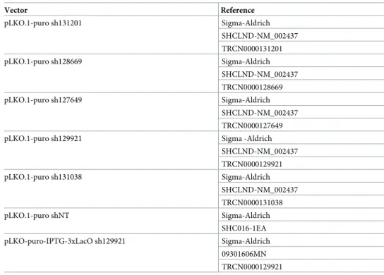
day, the DNA mixture and the lipofectamine solution were prepared separately. The DNA mixture was composed of 0.4μg of the envelope encoding vector pCMV-VSVG (Addgene, #8454), 3.6μg of the packaging vector psPAX2 (Addgene, #12260) and 4 μg of the expressing plasmid (Table 1), in 240μL of 5% opti-MEM (Invitrogen, #31985). The lipofectamine
Table 1. List of plasmids used for MPV17 silencing.
Vector Reference pLKO.1-puro sh131201 Sigma-Aldrich SHCLND-NM_002437 TRCN0000131201 pLKO.1-puro sh128669 Sigma-Aldrich SHCLND-NM_002437 TRCN0000128669 pLKO.1-puro sh127649 Sigma-Aldrich SHCLND-NM_002437 TRCN0000127649 pLKO.1-puro sh129921 Sigma -Aldrich
SHCLND-NM_002437 TRCN0000129921 pLKO.1-puro sh131038 Sigma-Aldrich SHCLND-NM_002437 TRCN0000131038 pLKO.1-puro shNT Sigma-Aldrich SHC016-1EA pLKO-puro-IPTG-3xLacO sh129921 Sigma-Aldrich 09301606MN TRCN0000129921
The vectors backbone and encoding shRNA are specified along with their references.
P8833) at 2.5μg/mL. Experimental timelines of MPV17 silencing with the constitutive and inducible vectors are detailed inS1 Fig. Briefly, for the constitutive silencing ofMPV17, cells were transduced with different shRNA-encoding vectors (pLKO.1-puro shNT, sh127649, sh128669, sh129921, sh131201, sh131038), selected as described above and let to recover for 2 days without puromycin. They were then seeded and allowed to grow for 4 days before assess-ing the proliferation.
For the inducible silencing ofMPV17, cells were transduced or not with pLKO-puro-IPTG-3xLacO encoding sh129921, selected as described above and let to recover for 2 days.MPV17 silencing was then obtained by incubation of the transduced cells in the presence of 0.1 mM of IsoPropyl ß-D-1-ThioGalactopyranoside (IPTG) (Sigma-Aldrich, I6758), renewed daily for 5 days unless stated otherwise. Cells were then seeded and allowed to grow for 4 days in presence of IPTG renewed daily (unless stated otherwise) before assessing the proliferation.
Proliferation assessment
Cell counting. Cells were seeded in 25 cm2culture flask (Corning, #430639) and grown
for 4 days. Cells were rinsed with Phosphate Buffered Saline (PBS) pH 7.4, trypsinized with 0.05% trypsin-EDTA (ThermoFisher, 25300) and centrifuged for 5 min at 200 g. The pellet was resuspended in culture medium and cell suspension density was counted in a Neubauer chamber. Doubling time was calculated as follow: time⇤ln(2) / (ln(number of cells at the end of
the experiment)—ln(number of seeded cells)).
MTT assay. Cells were seeded in 24-well culture plates (Corning, #3524), grown for 4 days and incubated for 1 h with 500μL of tetrazolium dye MTT (Sigma-Aldrich, M2128) (2.5μg/mL in PBS) at 37˚C. Cells were then lysed for 1 h in lysis buffer (9% sodium dodecyl sulphate, 60% N, N-dimethylformamide, pH 4.7), and absorbance was measured with a spec-trophotometer (xMark, Bio-Rad) at 570 nm.
Protein content: Folin protein assay. Cells were seeded in 24-well culture plates (Corn-ing, #3524), grown for 4 days and rinsed twice with PBS. The bovine serum albumin (VWR, 0332) protein standards and samples were incubated 30 min in presence of 200μL of 0.5 M sodium hydroxide, then 10 min with 750μL of a solution A (1.96% sodium carbonate, 0.02% sodium and potassium tartrate and 0.01% copper sulphate) and finally 30 min with 75μL of Folin and Ciocalteu0s phenol reagent (Sigma-Aldrich, F9252) diluted twice in distilled water.
Absorbance was measured with a spectrophotometer (xMark, Bio-Rad) at 740 nm [25].
Cell lysates and pierce protein assay
Cells were seeded in 75 cm2culture flask (Corning, #430641U), rinsed once with PBS and lysed with radioimmunoprecipitation assay buffer (20 mM tris hydroxymethyl, 150 mM
ride acid; pH 6.8, 0.04 M sodium dodecyl sulphate, 0.4 M β-mercaptoethanol, 5% glycerol, 0.15 mM bromophenol blue), boiled for 5 min, resolved on polyacrylamide gel and transferred on a nitrocellulose membrane (Bio-Rad). Membrane was blocked for 1 h at room temperature in Odyssey Blocking Buffer (OBB) (LI-COR, P/N 927) diluted twice in PBS and incubated overnight at 4˚C with the primary antibody diluted in OBB with 0.1% Tween-20 (OBB-T). For MPV17 detection, membrane was treated prior to blocking step with Super Signal Western Blot Enhancer (ThermoFisher, 46640) according to the manufacturer0s recommendations.
The next day, membrane was rinsed 3 x 5 min in PBS with 0.1% Tween-20 (PBS-T), incubated with secondary antibody diluted in OBB-T 0.1% for 1 h at room temperature, rinsed 3 times in PBS-T 0.1%, dried and scanned with the Odyssey Infrared Imager (LI-COR, 9120). For the description of the antibodies used in this study, seeTable 2.
Construct for MPV17 overexpression
MPV17 mRNA was reverse transcribed (Transcriptor First Strand cDNA kit, Roche,
04379012001) using a specific primer (50-AGGTGGAAACGATGGAGTGA-30). A PCR was then
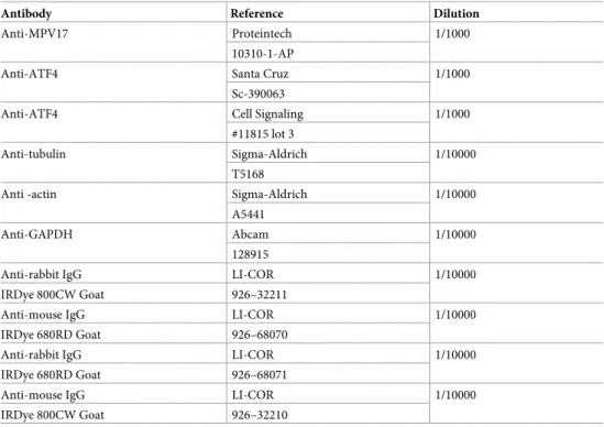
Table 2. List of antibodies and their dilutions used for western blotting.
Antibody Reference Dilution
Anti-MPV17 Proteintech 1/1000
10310-1-AP
Anti-ATF4 Santa Cruz 1/1000
Sc-390063
Anti-ATF4 Cell Signaling 1/1000
#11815 lot 3
Anti-tubulin Sigma-Aldrich 1/10000 T5168
Anti -actin Sigma-Aldrich 1/10000 A5441
Anti-GAPDH Abcam 1/10000
128915
Anti-rabbit IgG LI-COR 1/10000
IRDye 800CW Goat 926–32211
Anti-mouse IgG LI-COR 1/10000
IRDye 680RD Goat 926–68070
Anti-rabbit IgG LI-COR 1/10000
IRDye 680RD Goat 926–68071
Anti-mouse IgG LI-COR 1/10000
IRDye 800CW Goat 926–32210
The antibodies used for the western blot analysis are specified with their reference and working dilutions.
μg/mL). Cells were then trans-duced with PLKO.1-puro vector constitutively encoding sh129921 or shNT (Table 1). Cells were then allowed to recover and generateMPV17-targeting shRNA for 5 days, seeded and allowed to grow for 4 days. Cell proliferation was assessed by MTT assay at day 1 and day 4. To overexpress MPV17, we usedMPV17-silencing and MPV17-overexpression vectors that were both bearing the resistance to puromycin. This obviously constitutes an obstacle in the selec-tion of the cells that are double transduced. However, we decided to proceed further based on the knowledge that the sh129921-encoding vector robustly led to a very efficient transduction rate (nearly 100%), therefore allowing to bypass the need for an ensuing antibiotic selection.
mtDNA content determination
DNA was extracted with the Wizard Genomic DNA Purification Kit (Promega, A1120) according to the manufacturer0s recommendations. qPCR for mtDNA amplification was
per-formed on the gene encoding ND2 (NADH Dehydrogenase 2) using the forward primer50
-TGTTGGTTATACCCTTCCCGTACTA-30and the reverse primer50
-CCTGCAAAGATGGTA-GAGTAGATGA-30. For the normalization with nuclear DNA, the gene encoding Beclin was
amplified with the forward primer50-CCCTCATCACAGGGCTCTCTCCA-30and the reverse
primer50- GGGACTGTAGGCTGGGAACTATGC-30. Real time PCR was performed with
SYBR Select Master Mix (ThermoFisher, 4472908). mtDNA copy number was calculated according to the following formula: 2⇤2^-ΔCt(where ΔCt = Ctmean
ND2–CtmeanBeclin).
RNA extraction and RT-qPCR
RNA was extracted with RNeasy Mini kit (Qiagen, 74104) according to manufacturer0s
recom-mendations and QIAcube (Qiagen). RT was performed with GoScript™ Reverse Transcription Mix (Promega, A2791) according to the manufacturer0s recommendations. qPCR was
per-formed with SYBR Select Master Mix (ThermoFisher, 4472908). We used the 2-ΔΔCtmethod to assess the relative mRNA expression. TheMPV17 primers used in this study are the following: F:GCTCAGGAAGCATGGCACTCT; R: AATGTCACCCAGGCCCATCA.
RNA sequencing
Huh7 cells were transduced with shRNA non-target lentiviral vector (shNT) or with shRNA vector constitutively targetingMPV17 expression (sh129921). RNA quality was analysed with the Bioanalyzer 2100 (Agilent). RNA samples (n = 4) were sent to Genomic Core Leuven (Bel-gium) for RNA sequencing and data were analysed with Ingenuity Pathway Analysis
(QIA-0.5% NGS, overnight at 4˚C. Slices were washed 3 x 3 min in PBS-T 0.05% baths and 3 min in PBS. Slices were then incubated 30 min at room temperature with the secondary antibody EnVision-HRP anti-rabbit (Dako, K400311). Slices were washed 3 x 3 min in PBS-T 0.05% and 3 min in PBS. Slices were then incubated 4 min at room temperature in the DAB solution (Dako, K346811) and washed 5 min in running tap water. Slices were incubated 5 min in Mayer Hematoxyline and washed 5 min in running tap water. Slices were finally incubated 3x 3 min in isopropanol, 3x 3 min in xylene and mounted on coverslip with Entallan glue.
Statistical analysis
Data are expressed as the mean± Standard Error of the Mean (S.E.M). Figure plotting was per-formed with Prism software. Statistical analysis was perper-formed using a one-tailed Mann-Whit-ney Test or a two-tailed Wilcoxon signed rank Test (α = 5%;⇤: p<0.05;⇤⇤: p<0.01;⇤⇤⇤:
p<0.001;⇤⇤⇤⇤: p<0.0001).
Supporting information
S1 Fig. Experimental timelines ofMPV17 silencing in the constitutive and inducible mod-els. Cells were transduced and puromycin-selected with a vector allowing a constitutive (a) or an inducible (b)MPV17 silencing. After 2 days of recovery, cells were either seeded and allowed to grow for 4 days before assessment of proliferation (a) or treated with 0.1 mM of Iso-Propyl ß-D-1-ThioGalactopyranoside (IPTG) for 5 days to induceMPV17 silencing prior to the seeding (b). The proliferation was then assessed after 4 days of growth in presence of IPTG renewed daily.
(PPTX)
S2 Fig. Effect of IPTG on Huh7 cells proliferation following constitutive sh129921-me-diatedMPV17 knockdown. Huh7 cells were transduced with non-target shRNA lentiviral vectors (shNT) or with sh129921-containing vector. Transduced cells were selected for 6 days with puromycin (2.5μg/mL). Cells were seeded at 8×10³cells/cm2and grown for 4 days in the presence or in the absence of 0.1 mM of IPTG. To mimic the conditions found in the inducible model of expression, the IPTG-containing medium was renewed daily. We therefore included a control in which no IPTG was present while the medium was also changed every day (Ctl). Data are presented as mean± S.E.M (3 biological replicates). P values were calculated with the one-tailed Mann-Whitney Test (α = 5%; NS;⇤: p<0.05;⇤⇤: p<0.01;⇤⇤⇤: p<0.001).
(PPTX)
S3 Fig. Effect of the duration of IPTG pre-treatment and of the medium change frequency on Huh7 cells proliferation in response to inducible sh129921-mediatedMPV17 knock-down. Cells were transduced, or not (Unt), with inducible sh129921 lentiviral vectors and selected for 6 days with puromycin (2.5μg/mL). Cells were then incubated for 14 days in the presence of 0.1 mM of IPTG to induceMPV17 silencing and culture medium was changed
sh129921) is indicated above its target site. Each grey box represents an exon ofMPV17 tran-script (NM002437.5). A line indicates the 30UTR and 50UTR ofMPV17 transcript.
(PPTX)
S5 Fig. Localisation of the shRNA-targeted sites on theMPV17 transcript isoforms refer-enced in human liver. The Genotype-Tissue Expression (GTEx) Project was supported by the Common Fund of the Office of the Director of the National Institutes of Health, and by NCI, NHGRI, NHLBI, NIDA, NIMH, and NINDS. The data used for the analysis described in this manuscript and this Fig. were obtained from the GTEx Portal on 02/05/19. We indicated the Sigma Aldrich reference of each shRNA targetingMPV17 transcripts (sh128669, sh131201, sh131038, sh127649 and sh129921) above its targeted site. Each box represents an exon in MPV17 transcript isoforms. The darker the purple, the more abundant the transcript (as referred by Log 10 (TPM)). The 30UTR is located on the left of the image, and the 50UTR on
the right. We added an asterisk (⇤) that indicates both shRNAs providing the reduced
prolifer-ation phenotype, whileø indicates both shRNAs leading to an unchanged proliferation rate. (PPTX)
S6 Fig. Full-length blots of the cropped blots presented in this work. Protein immunostain-ing was performed with secondary antibodies coupled to infrared dyes (IRDye; green: 800nm/ red: 700 nm). a: Full blot of the one presented in Figs2Aand7A; b: Full blot of the one pre-sented in Figs4Aand7B(Anti-ATF4 from Santa Cruz); c: Full blot of the one presented inFig 8A; d: Full blot of the one presented inFig 4C(Anti-ATF4 from Cell Signaling); e: Full blot of the one presented inFig 4B(Anti-ATF4 from Cell Signaling); f: Full blot of the one presented inFig 5B; g: Full blot of the one presented inFig 6B; h and i: Full blots of the ones presented in
S3 Fig; j: Full blot of the one presented inFig 6C(Anti-ATF4 from Santa Cruz). (DOCX)
Acknowledgments
We wish to thank Benjamin LE CALVE for his valuable biological input, the constructive dis-cussions and technical advice. We are grateful to Florian POULAIN for the informative pre-sentation of the GTEx Portal and the help regardingMPV17 isoforms.
Author Contributions
Conceptualization: Morgane Canonne, Thierry Arnould, Patricia Renard. Data curation: Sophie Ayama-Canden.
Visualization: Morgane Canonne.
Writing – original draft: Morgane Canonne.
Writing – review & editing: Anaïs Wanet, Mustapha Najimi, Thierry Arnould, Patricia Renard.
References
1. Karasawa M, Zwacka RM, Reuter A, Fink T, Hsieh CL, Lichter P, et al. The human homolog of the glo-merulosclerosis gene Mpv17: structure and genomic organization. Hum Mol Genet. Oxford University Press; 1993; 2: 1829–1834.https://doi.org/10.1093/hmg/2.11.1829PMID:8281143
2. Spinazzola A, Viscomi C, Fernandez-Vizarra E, Carrara F, D0Adamo P, Calvo S, et al. MPV17 encodes an inner mitochondrial membrane protein and is mutated in infantile hepatic mitochondrial DNA deple-tion. Nat Genet. Nature Publishing Group; 2006; 38: 570–575.https://doi.org/10.1038/ng1765PMID:
16582910
3. Krauss J, Astrinidis P, Astrinides P, Frohnho¨fer HG, Walderich B, Nu¨sslein-Volhard C. transparent, a gene affecting stripe formation in Zebrafish, encodes the mitochondrial protein Mpv17 that is required for iridophore survival. Biol Open. Company of Biologists; 2013; 2: 703–10.https://doi.org/10.1242/bio. 20135132PMID:23862018
4. Trott A, Morano KA. SYM1 Is the Stress-Induced Saccharomyces cerevisiae Ortholog of the Mamma-lian Kidney Disease Gene Mpv17 and Is Required for Ethanol Metabolism and Tolerance during Heat Shock. Eukaryot Cell. American Society for Microbiology (ASM); 2004; 3: 620.https://doi.org/10.1128/ EC.3.3.620-631.2004PMID:15189984
5. El-Hattab AW, Wang J, Dai H, Almannai M, Staufner C, Alfadhel M, et al. MPV17-related mitochondrial DNA maintenance defect: New cases and review of clinical, biochemical, and molecular aspects. Hum Mutat. 2018; 39: 461–470.https://doi.org/10.1002/humu.23387PMID:29282788
6. Lo¨llgen S, Weiher H. The role of the Mpv17 protein mutations of which cause mitochondrial DNA deple-tion syndrome (MDDS): lessons from homologs in different species. Biol Chem. De Gruyter; 2015; 396: 13–25.https://doi.org/10.1515/hsz-2014-0198PMID:25205723
7. Antonenkov VD, Isomursu A, Mennerich D, Vapola MH, Weiher H, Kietzmann T, et al. The Human Mito-chondrial DNA Depletion Syndrome Gene MPV17 Encodes a Non-selective Channel That Modulates Membrane Potential. J Biol Chem. American Society for Biochemistry and Molecular Biology; 2015; 290: 13840–61.https://doi.org/10.1074/jbc.M114.608083PMID:25861990
8. Reinhold R, Kruger V, Meinecke M, Schulz C, Schmidt B, Grunau SD, et al. The Channel-Forming Sym1 Protein Is Transported by the TIM23 Complex in a Presequence-Independent Manner. Mol Cell Biol. 2012; 32: 5009–5021.https://doi.org/10.1128/MCB.00843-12PMID:23045398
9. Dalla Rosa I, Ca´mara Y, Durigon R, Moss CF, Vidoni S, Akman G, et al. MPV17 Loss Causes Deoxynu-cleotide Insufficiency and Slow DNA Replication in Mitochondria. Larsson N-G, editor. PLOS Genet. Public Library of Science; 2016; 12: e1005779.https://doi.org/10.1371/journal.pgen.1005779PMID:
26760297
10. Moss CF, Dalla Rosa I, Hunt LE, Yasukawa T, Young R, Jones AWE, et al. Aberrant ribonucleotide incorporation and multiple deletions in mitochondrial DNA of the murine MPV17 disease model. Nucleic Acids Res. 2017; 45: 12808–12815.https://doi.org/10.1093/nar/gkx1009PMID:29106596
11. Martorano L, Peron M, Laquatra C, Lidron E, Facchinello N, Meneghetti G, et al. The zebrafish ortholo-gue of the human hepatocerebral disease gene MPV17 plays pleiotropic roles in mitochondria. Dis Model Mech. Company of Biologists; 2019;12.https://doi.org/10.1242/dmm.037226PMID:30833296
16. Bottani E, Giordano C, Civiletto G, Di Meo I, Auricchio A, Ciusani E, et al. AAV-mediated liver-specific MPV17 expression restores mtDNA levels and prevents diet-induced liver failure. Mol Ther. American Society of Gene & Cell Therapy; 2014; 22: 10–7.https://doi.org/10.1038/mt.2013.230PMID:24247928 17. Wortel IMN, van der Meer LT, Kilberg MS, van Leeuwen FN. Surviving Stress: Modulation of
ATF4-Mediated Stress Responses in Normal and Malignant Cells. Trends Endocrinol Metab. 2017; 28: 794– 806.https://doi.org/10.1016/j.tem.2017.07.003PMID:28797581
18. Harding HP, Zhang Y, Zeng H, Novoa I, Lu PD, Calfon M, et al. An Integrated Stress Response Regu-lates Amino Acid Metabolism and Resistance to Oxidative Stress. Mol Cell. Cell Press; 2003; 11: 619– 633.https://doi.org/10.1016/s1097-2765(03)00105-9PMID:12667446
19. Malmberg SE, Adams CM. Insulin signaling and the general amino acid control response. Two distinct pathways to amino acid synthesis and uptake. J Biol Chem. American Society for Biochemistry and Molecular Biology; 2008; 283: 19229–34.https://doi.org/10.1074/jbc.M801331200PMID:18480057 20. Wang S, Chen XA, Hu J, Jiang J-K, Li Y, Chan-Salis KY, et al. ATF4 Gene Network Mediates Cellular
Response to the Anticancer PAD Inhibitor YW3-56 in Triple-Negative Breast Cancer Cells. Mol Cancer Ther. NIH Public Access; 2015; 14: 877–88.https://doi.org/10.1158/1535-7163.MCT-14-1093-TPMID:
25612620
21. Ben-Sahra I, Hoxhaj G, Ricoult SJH, Asara JM, Manning BD. mTORC1 induces purine synthesis through control of the mitochondrial tetrahydrofolate cycle. Science. NIH Public Access; 2016; 351: 728–733.https://doi.org/10.1126/science.aad0489PMID:26912861
22. Politi N, Pasotti L, Zucca S, Casanova M, Micoli G, Cusella De Angelis M, et al. Half-life measurements of chemical inducers for recombinant gene expression. J Biol Eng. 2014; 8: 5.https://doi.org/10.1186/ 1754-1611-8-5PMID:24485151
23. RNA Interference and Viruses: Current Innovations and Future Trends—Google Books [Internet]. [cited 27 Feb 2019]. Available:https://books.google.be/books?id=C5TY8W74scIC&pg=PA204&lpg= PA204&dq=inducible+vector+reversible+fine+tuning&source=bl&ots=j8lvhrcKPL&sig= ACfU3U0knMfwhxsLArPPpV4QXORxuiAuHQ&hl=en&sa=X&ved=
2ahUKEwjakOO159vgAhWTQhUIHa0OA4IQ6AEwEHoECAAQAQ#v=onepage&q=inducibl 24. Peretz L, Besser E, Hajbi R, Casden N, Ziv D, Kronenberg N, et al. Combined shRNA over CRISPR/
cas9 as a methodology to detect off-target effects and a potential compensatory mechanism. Sci Rep. Nature Publishing Group; 2018; 8: 93.https://doi.org/10.1038/s41598-017-18551-zPMID:29311693 25. LOWRY OH, ROSEBROUGH NJ, FARR AL, RANDALL RJ. Protein measurement with the Folin phenol
reagent. J Biol Chem. 1951; 193: 265–75. Available:http://www.ncbi.nlm.nih.gov/pubmed/14907713