HAL Id: tel-02613734
https://tel.archives-ouvertes.fr/tel-02613734
Submitted on 20 May 2020
HAL is a multi-disciplinary open access archive for the deposit and dissemination of sci-entific research documents, whether they are pub-lished or not. The documents may come from teaching and research institutions in France or abroad, or from public or private research centers.
L’archive ouverte pluridisciplinaire HAL, est destinée au dépôt et à la diffusion de documents scientifiques de niveau recherche, publiés ou non, émanant des établissements d’enseignement et de recherche français ou étrangers, des laboratoires publics ou privés.
Contribution of the potassium / chloride cotransporter
KCC2 to hippocampal rhythmopathy
Marie Goutierre
To cite this version:
Marie Goutierre. Contribution of the potassium / chloride cotransporter KCC2 to hippocampal rhythmopathy. Neurobiology. Sorbonne Université, 2018. English. �NNT : 2018SORUS600�. �tel-02613734�
Sorbonne Université
Ecole doctorale n°158
- Cerveau Cognition Comportement -
Institut du Fer à Moulin
Equipe « Plasticité des réseaux corticaux et épilepsie »
Contribution of the potassium / chloride cotransporter
KCC2 to hippocampal rhythmopathy.
Implication du transporteur potassium / chlore KCC2 dans la
rythmopathie hippocampique.
Par Marie GOUTIERRE
Thèse de doctorat de Neurosciences
Dirigée par Dr Jean-Christophe PONCER
Présentée et soutenue publiquement le 28 juin 2018
Devant un jury composé de :
Pr Ann LOHOF
Pr Claudio RIVERA
Pr Andrew TREVELYAN
Dr Lisa ROUX
Dr Corentin LE MAGUERESSE
Dr Jean-Christophe PONCER
Présidente du jury
Rapporteur
Rapporteur
Examinateur
Examinateur
Directeur de thèse
i
ABSTRACT
In the CNS, synaptic release of the neurotransmitter GABA is responsible for fast inhibitory transmission. This is predominatly mediated by chloride flow through GABAA receptors. Hence, tight control of chloride homeostasis is critical for maintaing the efficacy of GABAergic transmission. In mature neurons, this is primarily achieved by the activity of the potassium-chloride cotransporter KCC2, which usually acts to extrude intracellular potassium-chloride and potassium ions. Expression of KCC2 however is compromised in numerous neurological disorders including epilepsy, Rett syndrome or neuropathic pain. Subsequent defects in GABA signaling through alteration of transmembrane chloride gradients are thought to underlie the pathological symptoms associated with these conditions. However, KCC2 is also highly expressed in dendritic spines where it plays a major role in controling the efficacy and gating the long-term plasticity of glutamatergic synapses. Remarkably, these functions are independent of chloride transport but instead involve KCC2 interaction with several protein partners. Hence, KCC2 can be classified as a moonlightning protein with multiple functions at excitatory and inhibitory synapses, thereby complicating predictions of the overall effect of its suppression on network activity. During my PhD, I characterized the effects of a chronic downregulation of KCC2 in the rat dentate gyrus at the cellular and synaptic levels as well as on the hippocampal network activity in vivo. Unexpectedly, lack of KCC2 did not impact steady-state GABAergic transmission. In contrast, my work shed light on a novel critical role of KCC2 in controling neuronal excitability through interaction with the leak-potassium channel Task-3 (KCNK9). This in turn alters hippocampal rhythmogenesis independently of GABA signaling. My results thus reveal a novel mechanism through which KCC2 influences neuronal activity separately of its ion transport function. They predict that pathological conditions associated with KCC2 downregulation may be at least partly explained by altered neuronal excitability and point to Task-3 as a new potential therapeutic target in these disorders.
iii
RÉSUMÉ
Dans le système nerveux central, la transmission inhibitrice est principalement assurée par la libération synaptique du neurotransmetteur GABA. La fixation du GABA aux récepteurs GABAA induit en effet un flux entrant d’ions chlorure, résultant en une hyperpolarisation du neurone. Le maintien d’une faible concentration intraneuronale en chlore est donc essentielle à l’action inhibitrice du GABA. Dans les neurones matures, cette fonction est principalement réalisée par l’activité du cotransporteur potassium-chlore KCC2, qui à l’état basal exporte les ions chlorure et potassium vers le milieu extracellulaire. De nombreuses affections neurologiques, telles que l’épilepsie, le syndrome de Rett ou encore les douleurs neuropathiques, sont associées à une diminution de l’expression de KCC2. Un déficit de transmission GABAergique dû à une altération des gradients transmembranaires d’ions chlorure sont généralement invoqués pour rendre compte des symptômes associés à ces affections. Cependant, KCC2 est également fortement exprimé dans les épines dendritiques des neurones corticaux. Sa présence à proximité de la densité postsynaptique influence l’efficacité mais également la plasticité à long terme des synapses glutamatergiques. Ces fonctions inattendues de KCC2 au niveau des synapses excitatrices ne reposent pas sur sa fonction de transport ionique mais impliquent au contraire des interactions avec divers partenaires protéiques. Ainsi, le transporteur KCC2 possède de multiple fonctions et régule différemment les transmissions excitatrice et inhibitrice. Ceci complique la prédiction de l’effet net d’une suppression de KCC2 sur l’activité d’un réseau neuronal dans la pathologie. Durant ma thèse, j’ai caractérisé les effets d’une suppression chronique de KCC2 dans le gyrus dentelé chez le rat adulte au niveau cellulaire, synaptique et de l’activité du réseau hippocampique in vivo. De façon inattendue, j’ai montré que la suppression de KCC2 n’entraînait pas de modifications majeures de la transmission GABAergique. En revanche, j’ai mis en évidence un nouveau mécanisme indépendant du transport ionique par lequel KCC2 contrôle l’excitabilité neuronale et la rythmogénèse hippocampique à travers son interaction avec le canal potassique de fuite Task-3 (KNCK9). Mes résultats prédisent que les déficits associés à une suppression de KCC2 dans la pathologie pourraient être en partie expliqués par cet effet sur l’excitabilité neuronale. Ils suggèrent également que Task-3 pourrait constituer une nouvelle cible thérapeutique dans le traitement des affections impliquant la suppression de KCC2.
v
ACKNOWLEDGMENTS
First of all, I would like to warmly thank all the members of my jury who agreed to review and evaluate my work : Andrew Trevelyan, Claudio Rivera, Lisa Roux, Ann Lohof and Corentin Le Magueresse. I’m looking forward to discussing it with you on the 28th of June.
I also want to thank Clément Léna, Jean-Antoine Girault and Etienne Audinat, who were part of my thesis committee and helped us shape this project through the years.
Mes plus profonds et plus sincères remerciements sont évidemment pour toi, Jean-Christophe.
Tu me pardonneras, je l’espère, mon caractère têtu, ma susceptibilité face aux remarques, mon côté “dernière minute” qui t’a causé bien des stress et une certaine tendance à faire passer les projets des autres avant les miens… En même temps, tu savais déjà à quoi t’en tenir lorsque tu m’as proposé de faire une thèse avec toi !
Ça fait, en effet, maintenant plus que 8 ans qu’on se connaît, 8 années pendant lesquelles tu m’as fait grandir à la fois scientifiquement et humainement.
Merci pour ta disponibilité, ta patience, ta confiance en moi. Ma thèse ne serait pas ce qu’elle est si tu ne m’avais poussé et encouragé comme tu l’as fait ! Au travers de nos (nombreuses) conversations scientifiques, tu m’as appris la rigueur du raisonnement et m’as montré comment on pense et construit un projet de recherche. Au-delà de la science, merci pour ton attention à l’humain et au bien-être de chacun. Combien de fois ne m’as-tu pas dit “fais attention à toi” lorsque tu notais une certaine fatigue ou “est-ce que tout va bien, que puis-je faire pour toi ?” dans des périodes un peu difficiles ! Un merci tout spécial pour ton soutien au cours de ces derniers mois si intenses.
Enfin, merci pour toutes ces discussions extra-scientifiques que nous avons pu avoir ensemble : elles ont été source d’enrichissement pour moi, et j’espère que la réciproque est un peu vraie !
Un petit mot également pour la responsable de la partie Nord de l’équipe. Sabine, merci pour ton enthousiasme scientifique et pour ton attention personnelle à chacun de nous. Scientifiquement, nos intérêts sont pour le moins éloignés mais cela ne nous a jamais empêchés d’échanger sur nos projets et je crois que c’est cette diversité qui fait la richesse de l’équipe.
Quatre années de thèse, c’est du temps partagé avec de nombreux collègues : pauses déjeuners / thé / bière, discussions autours de nos projets (scientifiques et personnels), autant de moments essentiels dans le quotidien.
Un merci particulier pour Quentin : finalement, c’est de ton observation qu’est né tout mon projet donc je te suis sacrément redevable ! Merci aussi à mes premiers voisins de bureau :
vi Martin (le calme et la sérénité incarné), Emmanuel (pour ton accueil et tes partages de chocolat noir) et Eric (l’expert en électrophysiologie).
Merci Ferran et Jessica (la team adénosine) pour votre gentillesse et votre simplicité, et aussi pour avoir su faire découvrir à l’équipe que, non, il n’y a pas que KCC2 dans la vie ! Marion, ma complice des pauses thé, merci pour tous ces temps de partage ensemble. Sana, merci pour ton rire magistral qui a le don de détendre l’atmosphère. Merci aussi pour toutes les conversations scientifiques que nous avons pu avoir et, en particulier, pour m’avoir fait découvrir les subtilités de la biochimie. Clémence, désolée de ne pas avoir été beaucoup plus disponible ces derniers mois pour te guider sur la partie KCC2 / rythmogénèse... Courage pour la fin de thèse, je suis sûre que tu vas gérer, comme d’habitude !
Merci à Yo et Marianne pour leur présence discrète mais toujours bienveillante. Merci Xavier et Floriane pour la bonne humeur que vous avez apportée à l’équipe depuis votre arrivée. A Etienne et Florian qui commencent / vont commencer une thèse (oui, parce que je suis sûre que tu l’auras ta bourse, Florian !), courage pour tout. J’ai une pleine confiance en vous pour mener vos projets avec brio, et pour entretenir l’esprit de l’équipe ! A last word for Manisha, welcome to the team again. I’m happy to leave you all of my Matlab scripts, I know they’re in good hands !
Je ne peux pas oublier non plus ceux qui ne font pas directement partie de l’équipe mais qui ont contribué à rendre ma vie à l’IFM plus facile au quotidien. Je pense en particulier à l’ensemble du pôle laverie (Géraldine, Dominique et Emeline) et administratif (Christine, Jocelyne, Isabelle et Marianne) ainsi qu’à François et Gaël pour leur gestion de l’animalerie en ce moment. Merci également à tous les animaliers du 105, et spécialement Olivier, pour leur disponibilité durant le temps où j’ai travaillé là-bas.
Back to english to thank all the people in the institute who contributed to make these nearly 5 years fly so quickly. First of all, to our long-time neighbors of the first floor, the Mameli team. Manuel, Frank, Anna, Salvatore, Kristina and Massimo : it was amazing knowing you. Your passion and dedication to science really inspired me to give my best in my work and I won’t forget either some of our drinking nights !
More generally, the IFM is a quite special institute in the world of research, filled with people willing to help each other and share more than just work. I cannot cite every member of the IFM but I really want to thank all of you for creating such atmosphere. A special thought to the regular members of the IFM Birretas group !
I was lucky during my PhD to spend some time in the lab of Liset Menendez de La Prida in the Cajal Institute. Thank you Liset, for your warm welcome, your support during my time there and your feedbacks on the in vivo part of my project ! Everybody in the lab made me feel like I belonged to the team but special thanks go to Dani (for teaching me EEG recording but mostly for our long tea-sessions while talking about everything and nothing), Manuel (for sharing such a passion for science, best of luck for your future in the Buzsaki lab !) and François (for taking care of introducing me to Madrid’s nightlife during my first stay there). Pendant ma thèse, j’ai aussi eu l’opportunité d’effectuer une mission à la Cité des Sciences : quelques jours par an pour déconnecter de la vie de laboratoire et apprendre à parler
vii sciences autrement. L’occasion surtout de faire de belles rencontres tant avec les médiateurs (Marlène, Valérie, Graziela, Isabelle, Nadège, Gilles…) qu’avec les autres doctorants (Elodie, Sandra, Batiste et tous les autres...). Au passage, vive la semaine du cerveau et vive le microbiote ;) !!
Je ne peux pas continuer mes remerciements sans un petit mot pour mon laboratoire de M2 et l’ensemble de l’équipe de Michaël Zugaro. Michaël, certes je ne suis pas restée en thèse chez toi (mais on ne va pas revenir encore une fois là-dessus, non ?), mais tu as su me donner le goût pour l’in vivo (et le code Matlab) au point que j’aie eu l’envie d’en rajouter dans mon projet de thèse ! De ces quelques mois au Collège de France, j’ai aussi gardé de belles amitiés. Nicolas, Céline, Virginie, Raly et Marco : merci pour tous ces moments partagés ! La bande ne va pas tarder à se disperser dans le monde (au grand regret des gérants du Village), mais je suis sûre que les liens resteront.
Que ce soit dans mes études, dans mon expérience de scoutisme, pendant mes années en Chine ou durant ma thèse, j’ai eu la chance de rencontrer de nombreuses personnes extraordinaires qui ont contribuées à me faire évoluer et à me donner envie de toujours donner le meilleur de moi-même. Certaines d’entre elles sont devenues de vrais amis et je ne serai pas qui je suis aujourd’hui sans eux. Alors merci Gaëlle, Cécile, Malo, Quentin, Kristell, Bénédicte, Aurélie, Viviane, Pauline, Léa, Mérie… et tous ceux que j’oublie certainement de citer ! Un mot tout spécial pour Quentin : merci d’avoir toujours su si bien me comprendre. Les relations seront forcément un peu différentes à partir de l’an prochain mais elles ne seront pas moins fortes pour autant !
Mon envie de faire de la recherche en biologie est très certainement venue en partie de l’exemple de mon parrain et ma marraine. Merci à eux de m’avoir transmis leur passion. Un immense merci à toute ma famille pour son soutien au cours de ces années de thèse. Maman, je sais l’effort que c’était pour toi de prétendre t’intéresser à la biologie parce que c’est ce que je faisais. Merci pour ça ! Mais surtout merci pour la confiance que toi et papa me faites depuis des années et qui me porte. A mes frères et sœurs, merci pour votre présence et vos encouragements (même si c’était parfois surtout des encouragements à diminuer un peu le rythme !). On n’aime pas trop dire les choses directement dans la famille, mais j’espère que vous savez tous ce que je pense de vous !
Enfin, un grand merci à mes grands-parents, qui sont et ont toujours été des exemples pour moi dans la vie.
Je voudrais enfin terminer avec une pensée pour ceux qui m’ont accompagnée dans la préparation du futur lors de cette fin de thèse. Venceslas, merci pour ton écoute et tes conseils depuis tant d’années, et encore plus particulièrement sur ces derniers mois. A la communauté de Tibériade enfin, merci de m’avoir accueillie comme vous l’avez fait il y a un peu plus d’un an. Merci aussi d’avoir su délicatement vous mettre en retrait lorsqu’il a été nécessaire pour moi de d’abord me consacrer à ma fin de thèse. On se retrouve en octobre pour le début d’une nouvelle aventure !
ix
TABLE OF CONTENTS
ABSTRACT ... i
RÉSUMÉ ... iii
ACKNOWLEDGMENTS ... v
TABLE OF CONTENTS ... ix
TABLE OF FIGURES ... xiii
LIST OF ABBREVIATIONS ... xiv
INTRODUCTION
... 1
I- KCC2, A PROTEIN WITH MULTIPLE FUNCTIONS AT BOTH EXCITATORY AND INHIBITORY SYNAPSES ... 5
1. Expression of KCC2 in the CNS ... 5
a. The CCC family ... 5
b. NKCC1 and KCC2 are the main active CCC in the CNS ... 7
c. Isoforms of KCC2 ... 7
d. Cellular and sub-cellular expression of KCC2 in the CNS ... 8
2. Transport function of KCC2 ... 10
a. Control of the chloride homeostasis : setting the intraneuronal [Cl-] i ... 10
b. KCC2 transport function is critical for hyperpolarizing action of GABA. ... 13
c. Does ECl directly control the polarity of GABA transmission? ... 16
d. Osmotic regulation in neurons ... 17
3. Regulation of KCC2 expression and membrane stability ... 18
a. Age-dependent regulation of KCC2 by the BDNF/TrkB pathway ... 18
b. Activity-dependent regulation by synaptic transmission ... 19
c. Regulation of KCC2 expression through protein interaction ... 22
d. Oligomerization and clustering of KCC2 ... 24
4. Emerging role of KCC2 at glutamatergic synapses ... 25
a. Maturation of dendritic spines ... 25
b. Role of KCC2 at mature glutamatergic synapses ... 26
5. KCC2 in epilepsy ... 28
a. Excitation / inhibition imbalance in epilepsy ... 28
b. KCC2 mutations and epileptogenesis ... 29
c. Chloride homeostasis, GABA transmission and epilepsy ... 32
d. Transport-function of KCC2 and ictogenesis ... 34
e. Is KCC2 downregulated in chronic epilepsy? ... 37
6. Dysregulation of KCC2 expression and function in other pathologies ... 39
a. Spasticity and neuropathic pain ... 39
x
c. Therapeutic approaches in pathologies involving KCC2 ... 41
II- THE HIPPOCAMPUS, A KEY STRUCTURE FOR COGNITION AND ITS INVOLVEMENT IN EPILEPSY ... 43
1. Overview of the hippocampal structure, connectivity and function ... 43
a. Anatomy and cellular physiology of the hippocampus ... 43
b. Hippocampal intrinsic and extrinsic connectivity ... 47
c. Linking physiology, connectivity and behavioral function ... 50
d. Cellular substrate of memory in the hippocampus ... 51
2. Hippocampal oscillations and their behavioral correlates ... 53
a. In vivo electrophysiological recordings : what they are, what they say ... 53
b. Theta rhythm and memory encoding ... 54
c. Sharp-Waves Ripples and memory consolidation ... 57
d. Gamma oscillations : not one but two distinct oscillations... 60
e. Dentate Spikes, the poor parent of hippocampal rhythms ... 62
3. The hippocampus in temporal lobe epilepsy ... 64
a. Cellular alterations of the hippocampus in mTLE ... 64
b. Alterations of theta rhythms in epilepsy ... 66
c. Emergence of novel pathological oscillations around the epileptogenic zone ... 67
III- RATIONALE AND OBJECTIVE OF THE PROJECT ... 71
MATERIALS AND METHODS
... 77
RESULTS
... 85
I- IN VITRO STUDY OF THE IMPACT OF KCC2 SUPPRESSION ON HIPPOCAMPAL RHYTHMOGENESIS ... 85
II- MAIN ARTICLE : KCC2 REGULATES NEURONAL EXCITABILITY AND HIPPOCAMPAL RHYTHMOGENESIS VIA DIRECT INTERACTION WITH TASK-3 CHANNELS ... 89
Preface ... 89 Abstract ... 92 Introduction ... 92 Results ... 94 Discussion ... 100 Figures ... 105 Methods ... 123 References ... 130
III- DOWNSTREAM EFFECTS OF KCC2 SUPPRESSION IN THE DENTATE GYRUS ON HIPPOCAMPAL RHYTHMOGENESIS ...135
DISCUSSION
... 143
I- A NOVEL NON-CANONICAL ROLE OF KCC2 AS A REGULATOR OF TASK-3 ACTIVITY ...143
xi
2. Physiological consequences of KCC2 and Task-3 downregulation ... 147
II- KCC2 AND HIPPOCAMPAL RHYTHMOPATHY : MECHANISMS AND CONSEQUENCES ...149
1. Mechanisms of hippocampal rhythmopathy following KCC2 suppression in the dentate gyrus... 149
2. Functional consequences of these alterations ... 151
III- KCC2 IN EPILEPTOGENESIS ...152
IV- GENERAL CONCLUSION...156
ADDITIONAL PUBLICATION
... 161
xiii
TABLE OF FIGURES
Figure 1 : CCC activity determines chloride flux through GABAARs ...2
Figure 2 : Control of action potential emission ...3
Figure 3 : Phylogeny and structure of the CCC family ...6
Figure 4 : Cellular and subcellular localization of KCC2...9
Figure 5 : KCC2 functions close to its thermodynamic equilibrium ... 11
Figure 6 : Developmental upregulation of KCC2 is critical for GABAergic switch from depolarizing to hyperpolarizing ... 14
Figure 7 : Activity-dependent regulation of KCC2 expression and function ... 21
Figure 8 : KCC2 interactome ... 23
Figure 9 : Impact of KCC2 suppression on mature glutamatergic synapses ... 27
Figure 10 : KCC2 mutations in epilepsy ... 31
Figure 11 : Correlation between KCC2 expression, GABA transmission and interictal activities... 33
Figure 12 : Chloride homeostasis and KCC2 function during ictal phase ... 36
Figure 13 : Therapeutic strategies following KCC2 downregulation ... 42
Figure 14 : Anatomy of the hippocampal formation ... 44
Figure 15 : Main intrinsic circuitry of the hippocampus. Horizontal section schema. ... 47
Figure 16 : Local connectivity in the DG - CA3 region ... 49
Figure 17 : LFP recordings and CSD analysis ... 54
Figure 18 : Theta rhythm in the hippocampus ... 56
Figure 19 : Hippocampal ripples and their function ... 59
Figure 20 : Mechanisms of gamma oscillations and coupling to theta ... 61
Figure 21 : Dentate spikes generation and their relationship to ripples ... 63
Figure 22 : Alterations of hippocampal anatomy in TLE patients ... 66
Figure 23 : Characteristics of fast ripples ... 68
Figure 24 : High variability of rhythmic activity in area CA3 of rat hippocampal slices ... 86
Figure 25 : Pharmacological blockade of KCC2 function does not affect in vitro rhythmogenesis ... 87
Figure 26 : Reduction of slow-gamma power in CA1 following KCC2 suppression in the dentate gyrus... 136
Figure 27 : Suppression of KCC2 in the dentate gyrus alters CA1 ripples ... 137
Figure 28 : Chemogenetic silencing of dentate gyrus neurons does not rescue deficits in gamma and ripples upon KCC2 suppression ... 138
xiv
LIST OF ABBREVIATIONS
4-AP 4 Amino-pyridine
ACSF Artificial Cerebro Spinal Fluid
AMPA α-Amino-3-hydroxy-5-methyl-4-isoxazolepropionic acide
APP Amyloid Precursor Protein
BDNF Brain-derived neurotrophic factor
CA Cornu Ammonis
CCC Chloride co-transporter
CCK Cholecystokinin
CIP1 CCC-interacting protein 1
CKB Brain-type creatine kinase
ClC Chloride channel
CNS Central Nervous System
COPI Coatomer protein I
CREB C-AMP Response Element-binding protein
CSD Current Source Density
CTD C-terminal Domain
DFGABA Driving Force of GABAergic currents
DG Dentate Gyrus
DMSO Dimethyl sulfoxyde
DS Dentate Spike
E/I Excitatory / Inhibitory
EC Entorhinal Cortex
EcoG Electrocorticogram
EEG Electroencephalogram
EGABA Reversal potential of GABAergic currents
Egr Early growth response transcription factor
EPSC Excitatory postsynaptic current
Erk Extracellular signal-regulated kinase
FERM Four.one, Ezrin,Radixin, Moesin
FRS FGF receptor substrate
GABA γ-Amino Butyric Acid
GABAAR Receptor subtype A for GABA
GABAB Receptor subtype B for GABA
GC Granule cell
GDP Giant Depolarization potential
GFP Green Fluorescent Protein
GluK2 Glutamate ionotropic receptor kainate type subunit 2
HEK Human Embryonic Kidney
HPC Hippocampus
ING Interneuron Network of Gamma
xv
K2P Two-pore potassium channel
KCC K+/Cl- co-transporter
KO Knockout
LFP Local Field Potential
LIMK Lin-11, Isl-1 and Mec-3 Kinase
mAchR Metabotropic acetylcholine receptor
MI Modulatory index
MQAE (6-Methoxyquinolinio)acetic acid ethyl ester bromide
mRNA Messenger Ribonucleic Acid
MS-DBB Medial septum – Diagonal band of Broca
Neto2 Neuropilin and tolooid like-2 protein
NKCC Na+/K+/Cl- co-transporter
NMDA N-methyl-D-aspartate
NRSE Neurn-restrictive silencing element
NTD N-etylmaleimide-sensitive factor
OSR Oxidative stress-responsive kinase
P Postnatal day
PAK p21-activated kinase
PING Pyramidal Interneuron Network of Gamma
PKC Protein kinase C
PLCγ Phospholipase C gamma
PP1 Protein phosphatase 1
PTZ Pentylenetetrazole
PV Parvalbumin
Rac Ras-related C3 botulinum toxin substrate
REM Rapid-Eye Movement
SCI Spinal Cord Injury
SE Status Epilepticus
Shc SRC homology 2 domain containing transforming protein
shRNA Short hairpin Ribonucleic Acid
Slc Solute carrier
SOM Somatostatin
SPAK Ste20p-related proline/alanine-rich kinase
SPW-R Sharp-Wave Ripple
Src Proto-oncogene tyrosine-protein kinase
TASK Twik-related Acid-sensitive potassium channel
TBI Traumatic Brain Injury
TLE Temporal Lobe Epilepsy
TM TransMembrane domain
TREK Twik-related potassium channel
TrkB Tropomyosin receptor kinase B
TTX Tetrodotoxin
VGCC Voltage-Gated Calcium Channels
VIP Vasoactive intestinal polypeptide
WNK With No Lysine kinase
1
INTRODUCTION
GENERAL INTRODUCTION
In the central nervous system (CNS), information processing relies on synaptic connections, which enable information transfer between neurons. Following emission of an action potential by the presynaptic neuron, a neurotransmitter is released in the synaptic cleft. Depending on the identity of the neurotransmitter released, the activity of the postsynaptic neurons is either promoted or inhibited. In the mature CNS, excitatory transmission relies mainly on glutamate release while γ-Amino Butyric Acid (GABA) mostly inhibits the postsynaptic neurons.
Both synaptic inhibition and excitation are mediated by charge movements across the plasma membrane. Fast excitatory transmission resulting from glutamate receptor activation is mediated by a net influx of cations (Na+, Ca2+) through the membrane and subsequent
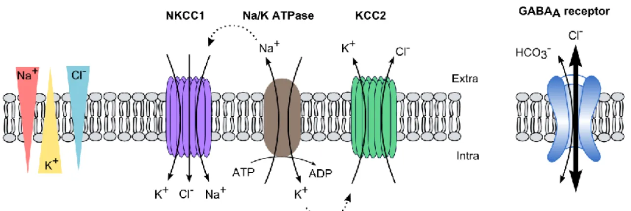
depolarizing excitatory postsynaptic currents (EPSCs). On the other hand, ionotropic receptors subtype A for GABA (GABAAR) possess a mix permeability to chloride and bicarbonate ions with a relative permeability ratio of 4/1 (Kaila and Voipio, 1987). Hence, their activation classically mediates a net hyperpolarizing influx of chloride ions (inhibitory postsynaptic current, IPSC) (Fig 1b and c).
The directionality of ion fluxes through the receptors depends directly on the relationship between their reversal potential (determined by their electrochemical ionic gradient across the membrane of the neuron) and the resting membrane potential (Vm) of the cell. In neurons, the
reversal potential of GABA (EGABA) is close to Vm and small changes in chloride concentration may
be sufficient to reverse the direction of chloride flow through the receptor. Hence, control of chloride homeostasis is crucial for neurons to maintain hyperpolarizing GABA transmission. This is mainly achieved through the combined action of two secondary active transporters, the Na/K/Cl cotransporter NKCC1 and the K/Cl cotransporter KCC2 which, under basal conditions,
2 use the Na+ and K+ electrochemical gradients generated by the Na/K ATPase to respectively load
and extrude chloride ions from the cell (Fig. 1).
Figure 1 : CCC activity determines chloride flux through GABAARs
KCC2 extrudes chloride out of the cell using the electrochemical potassium gradient generated by the Na+/K+ ATPase.
NKCC1, in contrast, transports chloride into neurons using the electrochemical gradient of sodium. Transmembrane chloride gradients and subsequently, the direction of chloride flux through GABAARs depend on the relative activity of the two transporters. In mature neurons, KCC2 activity dominates over NKCC1 leading to a high transmembrane gradient. Hence, GABAARs activation triggers hyperpolarizing influx of chloride ions.
Importantly, emission of action potentials by the postsynaptic neuron does not uniquely depend on the summation of both EPSCs and IPSCs but also on intrinsic properties of the cell such as its membrane potential, its membrane resistance (Rin) or its action potential threshold. Hence,
modifications of both sodium (Ren, 2011) and potassium leak channels influence neuronal excitability by altering both Vm and Rin while voltage-gated sodium channels expression regulate
action potential threshold (Fig 2).
At the network level, neurons tend to fire in a synchronous manner giving rise to oscillations that can be observed in electroencephalogram (EEG). Such neural oscillations, which require a tight control of both excitatory and inhibitory transmission as well as excitability, organize the information into coordinated patterns of activity. Various neural oscillations have been demonstrated as critical for cognitive processes such as perception, memory formation and consolidation but also consciousness. Hence, neural oscillations are believed to represent the intermediate link between cellular activity and behavior.
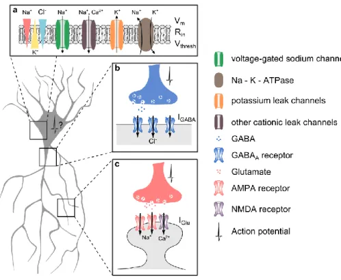
3 Figure 2 : Control of action potential emission
In the CNS, neurons need to constantly integrate stimuli from excitatory and inhibitory synapses. Synaptic GABA release classically triggers hyperpolarizing current (IGABA) mediated by the influx of chloride through GABAARs (b). In
contrast, glutamate mediates depolarizing currents (IGlu) through AMPA and NMDA receptors (c). The emission of an
action potential depends on postsynaptic integration of these synaptic inputs together with intrinsic properties of its membrane such as the membrane resting potential (Vm), the input resistance (Rin) and the action potential threshold
(Vthresh) (a). In a simplistic reduction, the membrane of the neuron can be assimilated to a RC circuit. Hence, according
to Ohm’s law, an action potential will be triggered if Vm - Vthresh = Rin * (IGABA + IGlu)
Altered activity of the K/Cl cotransporter KCC2 has been reported in numerous pathologies including epilepsy and neuropathic pain. Due to the close relationship between KCC2 activity and the polarity of GABAergic transmission, studies of KCC2 in the pathology have often focused on this aspect. However, KCC2 suppression was recently demonstrated to also alter glutamatergic transmission and plasticity through transport-independent mechanisms. Thus, the overall network effect of the loss of KCC2 is not so easy to predict.
During my PhD, I characterized the impact of KCC2 suppression on the hippocampal network. In particular, I focused on how the integration of the diverse cellular and synaptic modifications triggered by this suppression influences hippocampal physiological oscillations and I questioned their ability to favor the emergence of pathological activities. Before presenting these results in the form of a research article at the core of this thesis, I will introduce the function and regulation of KCC2 as well as the cellular and synaptic basis for hippocampal oscillations in health and disease.
5
I-
KCC2, A PROTEIN WITH MULTIPLE FUNCTIONS AT BOTH
EXCITATORY AND INHIBITORY SYNAPSES
1. Expression of KCC2 in the CNS
a. The CCC family
KCC2 is one of the nine members of the cation-chloride cotransporter (CCC) family, encoded by the genes slc12a1-9. Seven of them have been categorized as plasma membrane transporters (Gamba et al., 1993; Gillen et al., 1996; Hartmann and Nothwang, 2014; Hiki et al., 1999; Mercado et al., 2004; Payne et al., 1996). CCCs are widely expressed in all organs. They have been at first largely studied for their role in the control of cell volume upon osmotic challenges in epithelium and kidney in particular (Hoffmann and Dunham, 1995). In the CNS, special attention has been given to CCC function due to their key role in regulation of intraneuronal chloride homeostasis and, consequently, of the polarity of GABAergic transmission (Kaila, 1994; Payne et al., 2003). All CCCs cotransport Na+ and/or K+ ions coupled to chloride and are
characterized by a transport stoichiometry of 1 cation for 1 chloride ion transported. Consequently, transport of chloride through the plasma membrane is accomplished without any net movement of charges across the membrane, which complicates the electrophysiological study of their activity, as we will discuss later (see I.2.a). Finally, all CCCs are secondary active transporters meaning they use the gradients of Na+ and K+ generated by the Na-K-ATPase to
transport chloride.
The family of CCCs has been divided in two main branches (see Gagnon and Delpire, 2013 for review). The first branch corresponds to transporters using Na+ gradient to import chloride into
the cell. Specifically, this branch comprises two Na+-dependent K+/Cl- cotransporters (NKCC1 and
NKCC2) and one Na+-dependent Cl- cotransporter (NCC) which share about 55% conservation in
amino acid sequence. The second branch comprises four K+-dependent Cl- cotransporter (KCC1,
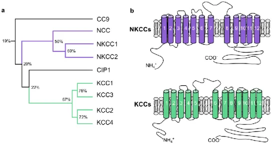
KCC2, KCC3 and KCC4). These transporters use the potassium gradient created by the Na-K-ATPase pump to extrude chloride from the cell and share more than 70% of homology in their sequence. The two remaining members of the CCC family, CIP1 and CC9 are less well characterized. They display relatively low identity with other CCCs (20-25%) and evidence for a chloride transport function is still lacking (Fig. 3A). CIP1 has been suggested to form heterodimers with other CCCs, thereby influencing their transport activity (Caron et al., 2000)
6 while CCC9 has been shown to influence polyamine transport in HEK-293 cells (Daigle et al., 2009). Based on these observations, whether CIP1 and CCC9 belong to the CCC family has been debated (Hartmann and Nothwang, 2014). From an evolutionary perspective, comparing gene sequences of putative CCC from Archeae and Eukaryota taxons suggests the existence of a single ancestral CCC gene in Archea (Hartmann et al., 2014). The diversity in the CCC family among and between species then emerged from consecutive duplication events.
Figure 3 : Phylogeny and structure of the CCC family
(a) Phylogenic tree of the characterized CCC family members. Percent of sequence homology are indicated at each branching. (b) Proposed secondary structure for NKCCs (top) and KCCs (bottom) mainly differ in the position of the long extracellular loop. Adapted from (Gamba 2005)
CCCs are transmembrane proteins with an apparent molecular weight of 120-200 kDa. Even though no crystallographic structure of the CCC has been obtained to date, predictions of the molecular structure with hydrophobicity profiles were made and suggest the presence of 12 transmembrane domains (TM), and an intracellular localization of both N-terminal and C-terminal domains (Gamba 2005; Gerelsaikhan and Turner 2000; Payne et al., 1996). A notable difference between NKCC/NCCs and KCCs is the location of the large extracellular loop which is situated between TM7 and TM8 in NKCC/NCCs and between TM5 and TM6 in KCCs (Gamba 2005, Fig 3B). This extracellular loop contains several glycosylation sites (Hoover et al., 2003) which seem important for correct targeting at the membrane (Weng et al., 2013). A more detailed model of KCC2 structure has been recently proposed (Stödberg et al., 2015) based on its homology with the bacterial amino acid, polyamine, and organocation transporter (ApcT) (Shaffer et al., 2009). This model suggests a key role for the interaction of TM1 and TM6 for the transport function and proposes the putative substrate binding residues to be located in TM8.
7 b. NKCC1 and KCC2 are the main active CCC in the CNS
CCC show differential tissue expression patterns. While NKCC2 and NCC are mostly expressed in the kidney where they regulate salt reabsorption (Kahle et al., 2010; Russell 2000), NKCC1 is ubiquitously expressed (Plotkin et al., 1997). Hence, NKCC1 is the only chloride importer of the CCC family expressed in the CNS.
In contrast, all KCCs are expressed in the CNS, although at various levels. KCC1 and KCC4 exhibit low expression levels in the brain and, consequently, there is no obvious neurological phenotype in mice lacking one of these transporters (Boettger et al., 2002; Rust et al., 2007). Although KCC3 is expressed in the adult brain, its physiological relevance remains largely unknown but has been suggested to mainly contribute to volume homeostasis (Kahle et al., 2015).
Until its recent identification in some pancreatic cells (Kursan et al., 2017), KCC2 was believed to be uniquely expressed in the CNS. In contrast to other KCCs, KCC2 is constitutively active under isotonic conditions (Payne 1997). This feature is conferred by a short amino-acid sequence (1022-1037) called the ISO-domain, which lays in its carboxy-terminal domain (CTD) (Acton et al., 2012; Mercado et al., 2006). Interestingly the ISO-domain-lacking KCC2 transporter could still be activated under hypotonic conditions, indicating that two distinct domains are involved in the activation of KCC2 under isotonic vs. hypotonic conditions.
It should be mentioned that chloride anion exchangers, Na+-dependent anion exchangers as well
as calcium-gated and voltage-gated chloride channels also participate, to some extent, to neuronal chloride homeostasis. For instance, the voltage-gated channel ClC-2 contributes to background chloride conductance and influences GABAAR function (Staley et al., 1996). However, ClC-2 seems to be mostly active following transient chloride loading of the cell rather than in basal conditions (Rinke et al., 2010). Accordingly, ClC-2 knock-out mice do not present deficits of chloride homeostasis (Bösl et al., 2001). Hence, in the rest of this thesis, we will primarily focus on the role of KCC2 in the CNS.
c. Isoforms of KCC2
KCC2 is encoded by the Slc12a5 gene. Two isoforms, KCC2a and KCC2b, were initially identified in the CNS, resulting from two alternative promoters (Uvarov et al., 2007). These isoforms only differ in their amino-terminal domain (NTD) which is 40 amino acids longer in KCC2a than in KCC2b and contains an additional putative Ste20p-related proline/Alanine-rich kinase (SPAK) /
8 oxidative stress-responsive kinase-1 (OSR1) phosphorylation site for KCC2a (de Los Heros et al., 2014). Both variants show similar ion transport properties when expressed in HEK cells (Uvarov et al., 2007) but KCC2a exhibits an increased sensibility to SPAK-dependent regulation due to this additional phosphorylation site (Markkanen et al., 2017). Furthermore, the two isoforms have different subcellular localization (Markkanen et al., 2014), suggesting a contribution of the NTD to the subcellular targeting of the transporter.
Originally, based on a study on its homologous protein KCC1 (Casula et al., 2001), the NTD of KCC2 had been proposed to be essential for the transport function of chloride (Li et al., 2007). Consequently, several groups have used a truncated form of KCC2, KCC2-ΔNTD, as a transport-deficient model in order to study ion-transport independent role of KCC2 (Fiumelli et al., 2013; Horn et al., 2010; Li et al., 2007). However, recent data from the group of Igor Medina in Marseilles revealed that the NTD domain of KCC2 is critical for the membrane insertion of the protein (Friedel et al., 2017). This study suggests KCC2-NTD influences primarily KCC2 trafficking, which is consistent with the difference in subcellular targeting of KCC2a vs. KCC2b.
A third isoform of KCC2 lacking the exon 25, KCC2a-S25, has recently been identified in pancreatic cells (Kursan et al., 2017) but is not expressed in the CNS. This isoform is characterized by the lack of 5 residues immediately after the ISO-domain and its functional properties remain to be determined.
d. Cellular and sub-cellular expression of KCC2 in the CNS
In the CNS, KCC2 expression is entirely neuron-specific due to the presence of neuron-restrictive silencing elements (NRSE), early growth response transcription factor 4 (Egr4) and upstream stimulating factors (USF1 and 2) binding sites in its promoter sequence (Uvarov et al., 2006; Markkanen et al., 2008; Yeo et al., 2009). Expression of KCC2 has been observed throughout the CNS (Fig. 4A,B) including spinal cord (Hübner et al., 2001) cerebellum (Williams et al., 1999), thalamus (Barthó et al., 2004), auditory brainstem (Blaesse et al., 2006) and cortical structures (Gulyás et al., 2001; Rivera et al., 1999). Although KCC2 seems ubiquitously expressed in the brain, differential values of EGABA have been reported depending on brain structures and
neuronal type. This could reflect differential levels of KCC2 expression. For instance, neurons of the reticular thalamic nucleus display low levels of KCC2 compared to adjacent brain structures
9 (Klein et al., 2018). However, changes in EGABA may also reflect changes in the expression of
NKCC1 as well as in the activity of other chloride channels or metabolism of bicarbonate.
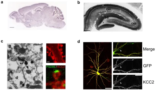
At the subcellular level, KCC2 has been shown to be organized in clusters at the plasma membrane. In 2001, Gulyas and colleagues made the first surprising observation, using immunogold staining and electron microscopy, that KCC2 was highly enriched at the vicinity of excitatory synapses, within dendritic spines (Gulyás et al., 2001, Fig 4C). Data from our group later confirmed that not only is KCC2 enriched in dendritic spines, in close proximity to glutamatergic synapses, but also it seems to be excluded from the postsynaptic density (Chamma et al., 2012; Gauvain et al., 2011). Additionally, and perhaps more expectedly, KCC2 is also enriched near GABAergic synapses (Chamma et al., 2013). Interestingly, KCC2 is completely excluded from the axon, thereby leading to a higher intracellular concentration of chloride in axons compared to the somatodendritic compartment (Price and Trussell 2006, Fig 4D).
Figure 4 : Cellular and subcellular localization of KCC2
(a) In situ hybridization of KCC2 mRNA (Allen Brain Atlas, scale 1mm) indicate KCC2 is expressed in the whole brain (b) Immunohistochemistry revealing KCC2 expression in the hippocampus (Gulyas et al. 2001, scale 0.6mm)
(c) KCC2 is expressed in dendritic spines. Left, electron microscopy revealed KCC2 presence in dendritic spines (Gulyas et al. 2001, scale 0.6µm). Right, immunocytochemistry showed KCC2 forms clusters in dendritic spines (top) and is excluded from the postsynaptic density as detected by PSD95 staining (bottom) (Gauvain et al. 2011, scale 2µm) (d) KCC2 is excluded from the axons (white arrows) as revealed by immunocytochemistry on cultured hippocampal neurons. Scale 40µm for left image and 10µm for insets (Chamma et al. 2013)
As previously mentioned, the subcellular localization of the two isoforms of KCC2 has been recently described. It revealed only partial colocalization of KCC2a and KCC2b in mature neurons
10 (Markkanen et al., 2014). In general, KCC2b is more concentrated than KCC2a at the somatic membrane and the two isoforms are expressed in distinct dendritic compartments. Although the functional relevance of this observation is still unclear, such differences may indicate different roles for the two isoforms in the mature brain.
2. Transport function of KCC2
a. Control of the chloride homeostasis : setting the intraneuronal [Cl-]i
● Thermodynamic considerations about KCC2 activity
Historically, the first observation that intraneuronal concentration of chloride was lower than expected if due to passive distribution only was made in 1988 (Thompson et al. 1988) but the identification of the neuronal chloride extruder KCC2 occurred several years later (Payne et al. 1996).
In neurons, KCC2 operates close to its thermodynamic equilibrium (Payne 1997). Indeed, such equilibrium is obtained when reversal potential for chloride and potassium ions are equal.The reversal potential of an ion is calculated according to the Nernst equation:
Eion = RT/zF * ln([ion]i / [ion]o)
where R represent the ideal gas constant, T the temperature in Kelvin, F the Faraday constant and z the valence of the studied ion.
Hence, the equation ECl = EK is equivalent, after simplification, to [Cl-]o * [K+]o = [Cl-]i * [K+]i
Both extracellular chloride concentration and intracellular potassium can be considered as fixed and close to 140 mM. Such approximations indicate that if [Cl-]
i is higher than [K+]o, KCC2
extrudes both chloride and potassium. Conversely, KCC2 contributes to the influx of both ions when [K+]
o exceeds [Cl-]i. Physiological [Cl-]i has been estimated to vary between 7 and 13 mM in
a computational model (Doyon et al., 2011) while physiological [K+]
o ranges between 2 and 4
mM. However, intense neuronal activation induces a rise in [K+]
o which can reach values up to 8
mM (Branston et al. 1982) hence inducing a reversal in the directionality of KCC2-mediated transport (Fig 5A).
11 Thus, KCC2 is in an interesting position to reduce network excitability by two different means. Indeed, its activity is necessary to cope with the constant chloride load in the neurons due to GABAAR activation enabling the maintenance of a hyperpolarizing inhibitory GABAergic transmission. However, reverse transport through KCC2 also allows to buffer extracellular potassium in case of a transient rise, thereby reducing neuronal excitability (Fig 5B).
Figure 5 : KCC2 functions close to its thermodynamic equilibrium
(a) KCC2 thermodynamic equilibrium is achieved when [Cl-]
o * [K+]o = [Cl-]i * [K+]i. Both [Cl-]o and [K+]I can be considered
constant and close to 140 mM.. Hence, KCC2 reaches its equilibrium when [K+]o = [Cl-]i (as represented by the black
line). Under physiological conditions, the intracellular chloride concentration is higher than the extracellular potassium concentration (green box). Hence, KCC2 classically extrudes chloride and potassium. However, following intense activity, extracellular potassium concentration increases (red box) and KCC2 might reverse if [K+]
o > [Cl-]i.
(b) KCC2 can buffer both intraneuronal chloride following GABAARs activation (top) and extracellular potassium following intense activity (bottom).
● Brief overview of methods for estimating KCC2 activity
Since KCC2 activity is electroneutral, it is impossible to directly measure its activity by classical electrophysiological methods. A typical approach is then to use a radioactive potassium substitute such as 40K+ or more frequently 86Rb+ to evaluate KCC2 transport activity (Gamba
2005; Mount et al. 1998; Payne 1997). In addition to the obvious limitations of using radioactive elements, these techniques only estimate average KCC2 function at the population level and do not permit testing individual cells or subcellular compartments.
In contrast, electrophysiological techniques allow assessing KCC2 function for individual cells by using GABAergic currents as a proxy. Thus, in whole-cell patch clamp experiments, the extrusion capacity of KCC2 may be determined by measuring the recovery rate of GABAergic currents following intense chloride loading in neurons (Staley and Proctor 1999; Pellegrino et al. 2011). Alternatively, KCC2 activity may be derived from the somato-dendritic gradient of EGABA
12 measured while imposing a fixed concentration of chloride through the patch pipette (Gauvain et al., 2011; Kelsch et al., 2001; Khirug et al., 2005). On the other hand, KCC2 activity can also be inferred from measures of steady-state chloride concentration. Since GABAARs are mostly permeable to chloride, measures of EGABA provide a good estimation of the equilibrium potential
of chloride ions and enable evaluation of chloride concentration. To this end, GABAergic currents can be measured in a gramicidin perforated-patch configuration which preserves intraneuronal chloride concentration (Akerman and Cline, 2006; Lee et al., 2011; Woodin et al., 2003).
Electrophysiological techniques of EGABA measurements can be tedious thus preventing screening
of large-scale populations. To counteract this inconvenience, several fluorescent indicators were developed to directly assess chloride concentration. Among these indicators, the organic dye chloride-sensitive MQAE (Doyon et al., 2011; Galeffi et al., 2004) has been largely due for its capacity to cross the plasma membrane. However, MQAE also shows a strong attenuation of its signal over time thus only allowing low frequency of acquisition (Inglefield and Schwartz-Bloom, 1997). Genetic ratiometric CFP - YFP sensors such as Clomeleon and Cl-Sensor do not show such limitation and have alternatively been used to estimate intraneuronal chloride concentration (Chamma et al., 2013; Kuner and Augustine, 2000; Markova et al., 2008). Yet, these ratiometric sensors are sensitive to pH, a parameter that can be affected by KCC2 function, which raises questions concerning the measures obtained by this method.
In conclusion, several techniques exist to estimate KCC2 activity. Each of them has their specific pros and cons and can be used to answer different questions. Hence, measures of chloride concentrations by fluorescence imaging or reversal potential of GABA by electrophysiological techniques inform on steady-state chloride homeostasis. In contrast, estimations of chloride extrusion capacity reflect the ability of the cell to cope with repetitive GABAergic stimulations in a more dynamic way.
● Potential role of impermeant anions for setting chloride concentration
In a much commented publication, the group of Kevin Staley proposed that chloride concentration may not be set by the activity of the CCCs but instead be primarily constrained by the relative concentration of extracellular and intracellular impermeant anions (Glykys et al., 2014). Indeed, impermeant anions may create a gradient of electrical charges across the membrane thereby influencing the transmembrane diffusion of permeable ions such as chloride.
13 Such effect is known as the Gibbs-Donnan effect. The most surprising observation of this study is that pharmacological blockade of the activity of CCCs did not affect chloride concentration when evaluated by chloride imaging, a result in complete contradiction with all prior publications on the topic (see, among many others, Dargaei et al., 2018; Doyon et al., 2011; Dzhala et al., 2005; Gauvain et al., 2011; Rivera et al., 1999; Zhu et al., 2005). Therefore, while impermeant anions may influence intraneuronal chloride concentration, as recently evidenced in reticular thalamic neurons (Klein et al., 2018), twenty years of research on the topic strongly argue for a primary role of CCC activity.
b. KCC2 transport function is critical for hyperpolarizing action of GABA.
● Theoretical models reveal a critical role of KCC2 for maintenance of hyperpolarizing GABA transmission
Given the critical role of KCC2 in chloride homeostasis, it is somehow not surprising that several computational models have revealed a direct relationship between KCC2 transport activity and GABAergic transmission (Doyon et al., 2011; Jedlicka et al., 2010; Lewin et al., 2012).
The main appeal of computational models is that they allow combination of various ions and electrical parameters as well as spatio-temporal integration of the signals to make predictions regarding KCC2 activity and GABA signaling. An interesting discovery of these models is that chloride extrusion capacity was already close to its maximum in neurons since increasing KCC2 activity had very little effects on EGABA (Doyon et al., 2011).
Another consistent finding of all these models is to reveal a differential impact of GABA stimulation depending on the synapse position along the dendritic axis. More precisely, following GABAA receptor activation, chloride accumulates more rapidly in distal compared to proximal compartments. This indicates that distal synapses may be less robust for maintenance of hyperpolarizing GABA transmission when facing intense activation of GABAARs. However, all these models consider KCC2 activity to be equivalent along the dendritic axis. Yet, immunohistochemical studies revealed a higher density of KCC2 in distal dendrites (Báldi et al., 2010). This increased expression of the transporter could allow better coping with chloride accumulation in these compartments.
14 Finally, a mild KCC2 hypofunction was not sufficient to affect steady-state chloride homeostasis (Doyon et al., 2016). However, it resulted in less efficient coping of neurons upon repeated GABAergic stimulations as well as in an alteration of the neural code and information processing. Overall, these data indicate that even in the absence of changes in neuronal chloride concentration, information processing may be affected by both synapse localization and chloride extrusion capacity.
● Developmental switch in the polarity of GABA transmission correlates with KCC2 developmental upregulation
A striking example of the correlation between KCC2 activity and the polarity of GABAAR-mediated currents is observed during development. A few years before KCC2 was even identified, Ben-Ari and collaborators described the presence of giant depolarizing potentials (GDPs) as well as depolarizing responses to GABA in the immature brain (Ben-Ari et al., 1989; Cherubini et al., 1991). Around postnatal day 5 (P5), a switch in the polarity of GABA responses from depolarizing to hyperpolarizing occurs in hippocampal neurons and is closely followed by the disappearance of GDPs. The precise timing of this switch was later refined and placed around P13 in the hippocampus (Khazipov et al., 2004).
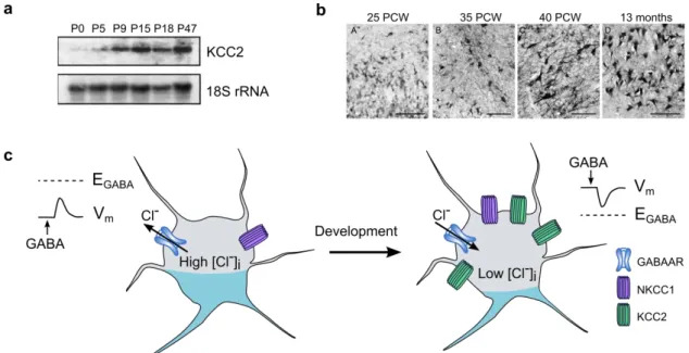
Figure 6 : Developmental upregulation of KCC2 is critical for GABAergic switch from depolarizing to hyperpolarizing
(a) Northern blot experiments revealed upregulation of mRNA levels of KCC2 through development in the rat hippocampus (adapted from Rivera et al. 1999)
(b) KCC2 protein levels are also upregulated in human hippocampus between 25 weeks post conception and 13 months. Scale : 100µm (adapted from Sedmak et al. 2016)
(c) Upregulation of KCC2 through development allow the establishment of a low intraneuronal chloride concentration, thus enabling hyperpolarizing GABA responses
15 Upregulation of KCC2 is observed throughout the forebrain in rodents (Rivera et al., 1999) from P5 - P7 before reaching its maximal level around P15 (Fig 6A). Inhibition of KCC2 expression by incubation of slices with antisense oligodeoxynucleotides against KCC2 mRNA was sufficient to prevent the switch of the polarity of GABAAR-mediated currents. Thus, KCC2 seems to be the main chloride extruder responsible for fast hyperpolarizing GABA transmission (Fig 6C). Several studies later revealed that the precise timing of this upregulation differs depending on brain regions but is highly correlated with the timing of switch of the polarity of GABAAR-mediated currents in each brain region (see Watanabe and Fukuda, 2015 for review). KCC2 developmental expression pattern is not restricted to the rodent brain but is also observed in the human brain between 25 weeks post conception and 13 months (Sedmak et al., 2016, Fig 6B). Similar to the observations made on rodent, the upregulation of KCC2 in the human brain coincides with a transition from depolarizing to hyperpolarizing GABA-mediated currents (Dzhala et al., 2005; Sedmak et al., 2016).
● Physiological relevance of depolarizing GABA in immature brain
In a controversial series of publications, the group of Yuri Zilberter argued that the depolarizing effect of GABA and the observation of GDPs in the immature brain were both experimental artifacts. They suggested that these observations arose from the absence of adequate energy substrates and showed that addition of ketone-bodies (Rheims et al., 2009), lactate or pyruvate (Holmgren et al., 2010) efficiently suppress the GDPs in vitro. Furthermore, slicing itself was suggested to be sufficient to render GABAAR-currents depolarizing at the surface of the slices due to unsatisfied energy requirements (Dzhala et al., 2012). However, the results published by the Zilberter’s group failed to be reproduced by two independent groups (Ruusuvuori et al., 2010; Tyzio et al., 2011) who also demonstrated that actual levels of energy substrates in the pup brain are much lower than those used in the study from the Zilberter’s group. Finally, GDPs have been observed both in intact hippocampal preparation (Khalilov et al., 1999) and in vivo (Sipilä et al., 2006) which refutes the hypothesis of a sole slicing artifact.
Instead, GDPs are thought to be essential for the neurotrophic action of GABA in the immature brain. Indeed, GABAergic synapses are formed and active prior to glutamatergic synapses. Depolarizing currents mediated by GABAARs lead to voltage-gated Ca2+ channels (VGCCs)
activation as well as enhanced recruitment of N-methyl-D-Aspartate receptors (NMDARs) which in turn favors the development of glutamatergic synapses (Akerman and Cline, 2006). Early expression of KCC2 was also proven detrimental for interneuron migration (Bortone and Polleux,
16 2009) due to a lack of recruitment of VGCCs. Thus, KCC2 absence in the developing brain and subsequent depolarizing action of GABA is critical for its correct maturation.
c. Does ECl directly control the polarity of GABA transmission?
So far, I described a straightforward relationship through which KCC2 activity directly controls chloride homeostasis and consequently the reversal potential and polarity of GABAAR-mediated currents. Such affirmation needs to be partly moderated.
First, as I already mentioned, GABAARs also display a lesser conductance to bicarbonate ions. Reversal potential of bicarbonate-mediated currents is around -10 mV, sensibly more depolarized than membrane potential. Hence, activation of GABAARs leads to a depolarizing outward current of bicarbonate. Consequently, regulation of bicarbonate homeostasis may also influence GABA signaling. In particular, carbonic anhydrase, a catalytic enzyme of bicarbonate, is upregulated during development and participates in the regulation of EGABA (Rivera et al., 2005).
This importance of bicarbonate control for GABAergic transmission has been further highlighted in the context of febrile seizures. Indeed, in rat pups, hyperthermia triggers seizures through brain alkalosis, a mechanism which could be mimicked by bicarbonate injection (Schuchmann et al., 2006). In contrast, knock-out animals for the carbonic anhydrase VII show reduced susceptibility to hyperthermic seizures (Ruusuvuori et al., 2013). These seizures were mediated by GABAergic excitation since they were aggravated by diazepam, a positive allosteric modulator of GABAARs.
Second, it is important to distinguish between EGABA and the driving force of GABA (DFGABA). The
latter reflects the difference between the reversal potential of GABAAR-mediated currents and the resting membrane potential and therefore determines the amplitude and polarity of GABAergic currents. Consequently, any changes in Vm will also affect GABA transmission. Thus,
neuronal maturation has been associated with hyperpolarization of their resting membrane potential in hippocampal pyramidal cells (Spigelman et al., 1992), dentate gyrus granule cells (Ambrogini et al., 2004) as well as neurons derived from human stem cells (Johnson et al., 2007) but not striatal neurons (Deng et al., 2004). This indicates that the overall impact of development on GABA transmission may differ depending on cell types. Even more strikingly, Cre-mediated exon excision of KCC2 in the cerebellum modifies DFGABA in Purkinje cells but not in
17 al., 2012). Therefore, as I will show later in this thesis, changes in KCC2 expression may be functionally masked by concomitant changes in resting membrane potential, thereby precluding changes in DFGABA.
Finally, it should be highlighted that depolarizing GABA currents are not necessarily excitatory. Indeed, independently of the polarity of net ion flux through GABAARs, their activation leads to opening of a membrane conductance and therefore decreases the membrane resistance. This in turn hinders subsequent depolarization of the membrane by glutamatergic inputs by a shunting inhibitory effect (Staley and Mody, 1992). In particular, a recent in vivo study demonstrated that GABA transmission was depolarizing but inhibitory due to this shunting effect in neurons from the upper cortical plate during early postnatal development (Kirmse et al., 2015).
d. Osmotic regulation in neurons
In addition to its critical control of chloride homeostasis, KCC2 also plays an essential role in the osmotic regulation of neurons.
KCCs were first described in epithelial and red blood cells as swelling-activated transporters whose activity was critical for facing osmotic challenges (Dunham and Ellory, 1981; Lauf and Theg, 1980; Zeuthen and MacAulay, 2002). This is due to the fact that co-transport of potassium and chloride is indirectly or directly coupled to water transport (MacAulay and Zeuthen, 2010; Zeuthen and MacAulay, 2002). Notably, it has been estimated that up to 500 molecules of water may cross the membrane for each chloride ion transported by KCC2 or NKCC1, probably to partially equilibrate for osmotic changes due to the ion transport (MacAulay and Zeuthen, 2010).
In the CNS, activation of ionotropic GABAAR as well as glutamate receptors leads to net ion influx in the postsynaptic neuron. This influx is accompanied by water fluxes in order to maintain intracellular osmolarity. Thus, intense neuronal activity directly impacts cell volume and can lead to cell swelling and cell death in extreme cases. Since neurons lack aquaporins (Amiry-Moghaddam and Ottersen, 2003), other mechanisms may be necessary for coping with the activity-induced swelling. Hence, digital holographic microscopy revealed an important water influx through both NKCC1 and NMDARs following glutamate application (Jourdain et al., 2011) accompanied by cell swelling. This was counteracted by subsequent swelling-induced activation of KCC2 and water extrusion, which allowed recovery of cell volume. In one study, KCC2
18 transport - function was proven essential to neuronal survival following NMDA-induced excitotoxicity (Pellegrino et al., 2011). Although the authors did not fully explore the mechanisms underlying such neuroprotective effect, the role of KCC2 in coping with activity-induced swelling may be one of them.
Swelling-induced activation of KCC2 is most likely dependent on the inhibition of the With No Lysine Kinase (WNK) as well as SPAK / OSR1 pathway, which results in dephosphorylation of the threonine T906 and T1007 residues (Gagnon et al., 2006; Rinehart et al., 2009). Conversely, extracellular hyperosmotic challenges lead to activation of the WNK, SPAK / OSR1 pathway, phosphorylation of T906 and T1007 residues and inhibition of KCC2 membrane expression. Notably, NKCC1 is also regulated by this pathway in opposite direction thus also participating to the regulation of cell volume following osmotic challenges (Kahle et al., 2010).
In conclusion, KCC2 transport-function is essential in neurons both for maintenance of inhibitory GABAergic transmission and for coping with osmotic challenges associated in particular with synaptic activity. Thus, mechanisms influencing KCC2 expression and membrane stability are likely to also critically impact both of these physiological processes.
3. Regulation of KCC2 expression and membrane stability
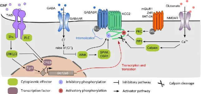
a. Age-dependent regulation of KCC2 by the BDNF/TrkB pathway
The progressive upregulation of KCC2 during development is promoted by brain-derived neurotrophic factor (BDNF) release (Aguado et al., 2003). BDNF binding to the tropomyosin receptor kinase B (TrkB) leads to activation of the extracellular signal regulated kinases 1 and 2 (Erk1/2) pathway via its coupling with the Src homology 2 domain containing transforming protein (Shc) and FGF receptor substrate 2 (FRS-2) (Fig 7). This in turn enhances the activity of the Egr4 transcription factor, which favors the expression of KCC2 (Ludwig et al., 2011). Surprisingly, full knock-out mice for BDNF still present a developmental upregulation of KCC2 (Puskarjov et al., 2015). This could be explained by the presence of compensatory mechanisms to BDNF. Indeed, the neurturin trophic factor can also trigger Egr4 activity thereby regulating KCC2 expression (Ludwig et al., 2011). Interestingly, this upregulation is restricted to the KCC2b isoform (Yeo et al., 2009) resulting in different proportion of the two isoforms in the developing versus mature brain. Hence, whereas KCC2a contributes to about 20-50% of the total KCC2
19 mRNA expression in the neonatal brain, its contribution decreases to 5-10% in the adult brain (Uvarov et al., 2009). Surprisingly, the developmental upregulation of KCC2 also requires prior expression of neuroligin-2 (Sun et al., 2013) although the mechanisms underlying such regulation and whether it involves BDNF or neurturin signaling remains unknown.
Nevertheless, BDNF possesses completely opposite effects on KCC2 expression and membrane stability in the mature brain compared to its role in development. Indeed, BDNF incubation for 2 to 3 hours is sufficient to induce massive downregulation of KCC2 expression and mRNA levels (Rivera et al., 2002). This regulation is dependent on both Shc/FRS-2 and phospholipase Cγ (PLCγ) - cAMP response element-binding protein (CREB) signaling (Rivera et al. 2004). This shift in BDNF / TrkB signaling pathway during maturation could be explained by an age-dependent change of TrkB phosphorylation at its PLCγ binding site (Di Lieto et al., 2012).
Interestingly, BDNF also potentiates the maturation of GABAergic neurons in the developing brain (Yamada et al., 2002) but not at later developmental stages (Mizoguchi et al., 2003). Conversely, in immature but not mature hippocampal neurons, GABA increases BDNF mRNA levels (Berninger et al., 1995), indicating a possible synergistic effect of GABA and BDNF in the developing brain.
b. Activity-dependent regulation by synaptic transmission
● Fast downregulation of KCC2 by excitatory transmission
KCC2 expression and transport-function is rapidly downregulated by neuronal activity under physiological conditions (Kaila et al., 1997; Kitamura et al., 2008; Woodin et al., 2003). This effect was proposed to enable the maintenance of neuronal activity homeostasis (Wang et al., 2006) but also to participate in learning-related functions (Fiumelli and Woodin, 2007).
The group of Mu-Ming Poo was the first to show calcium-dependent downregulation of KCC2 in immature neurons (Woodin et al., 2003). This effect was dependent on both voltage-gated calcium channels (VGCCs) and protein kinase C (PKC) activity (Fiumelli et al., 2005). However, the action of PKC is likely to be indirect since the only known phosphorylation site targeted by PKC is the serine 940 (S940) whose phosphorylation stabilizes KCC2 membrane expression (Lee et al., 2007).