HAL Id: tel-00445703
https://tel.archives-ouvertes.fr/tel-00445703
Submitted on 11 Jan 2010
HAL is a multi-disciplinary open access
archive for the deposit and dissemination of sci-entific research documents, whether they are pub-lished or not. The documents may come from teaching and research institutions in France or
L’archive ouverte pluridisciplinaire HAL, est destinée au dépôt et à la diffusion de documents scientifiques de niveau recherche, publiés ou non, émanant des établissements d’enseignement et de recherche français ou étrangers, des laboratoires
Maurocalcine, un nouveau vecteur de pénétration
cellulaire pour la délivrance cellulaire de composés
imperméables
Narendra Ram Maraheru Sonnappa
To cite this version:
Narendra Ram Maraheru Sonnappa. Maurocalcine, un nouveau vecteur de pénétration cellulaire pour la délivrance cellulaire de composés imperméables. Sciences du Vivant [q-bio]. Université Joseph-Fourier - Grenoble I, 2008. Français. �tel-00445703�
THESE
Pour obtenir le grade de
DOCTEUR DE L’UNIVERSITE JOSEPH FOURIER
Discipline:
NEUROSCIENCESMaurocalcine, un nouveau vecteur de pénétration cellulaire pour la
délivrance cellulaire de composés imperméables
Présenté et soutenu publiquement par,
Narendra RAM
Le 16 Octobre 2008
Devant le jury compose de, Pr. Gilles FAURY, Président Dr. Gilles DIVITA, Rapporteur Dr. Alain JOLIOT, Rapporteur Dr. Jean-Luc COLL, Examinateur Dr. Hervé DARBON, Examinateur Dr. Michel DE WAARD, Directeur
UNIVERSITE JOSEPH FOURIER- GRENOBLE 1
Ecole Doctorale Chimie et Sciences du Vivant
A THESIS SUBMITTED FOR THE DEGREE OF
DOCTOR OF PHILOSOPHY
DISCIPLINE: NEUROSCIENCESFROM THE UNIVERSITY OF JOSEPH FOURIER- GRENOBLE
Maurocalcine is a new cell penetration vector for the in vitro and
in vivo delivery of cell impermeable cargoes
Presented by,
Narendra RAM
On 16th October 2008
Members of the jury,
Pr. Gilles FAURY, President Dr. Gilles DIVITA, Reviewer Dr. Alain JOLIOT, Reviewer Dr. Jean-Luc COLL, Examiner Dr. Hervé DARBON, Examiner Dr. Michel DE WAARD, Director
UNIVERSITY JOSEPH FOURIER- GRENOBLE 1
Doctoral School of Chemistry and Life Sciences
SUMMARY
Maurocalcine (MCa) is a 33 mer peptide toxin initially isolated from the venom of Tunisian scorpion Scorpio maurus palmatus. Since then, it can be produced by chemical synthesis without structural alteration. This peptide triggers interest for three main reasons. First, it has sequence homology with a calcium channel domain involved in excitation–contraction coupling and helps in unravelling the mechanistic basis of Ca2+ mobilization from the sarcoplasmic reticulum. Second, MCa is a powerful activator of the ryanodine receptor, thereby triggering calcium release from intracellular stores. Finally, it is of technological value because of its ability to carry cell-impermeable compounds across the plasma membrane. In this study, I have designed novel more potent analogues of maurocalcine, either by point mutation, or by substitution of cysteine residues, which possess better or equal cell penetration efficiencies, limited cell toxicity and no pharmacological activity on ryanodine receptor. I have analyzed the interaction of these analogues with membrane lipids and glycosaminoglycans and their role in cell penetration. Cell entry pathway of MCa occurs through macropinocytosis, atleast when coupled to streptavidin.
RESUME
La maurocalcine (MCa) est un peptide/toxin de 33 acides aminés initialement isolé du venin d’un scorpion Tunisien Scorpio maurus palmatus. Depuis ce travail pionnier, la molécule a pu être produite par synthèse chimique sans altération structurale. Trois raisons font que ce peptide est particulièrement intéressant. D’une part, il présente une homologie de séquence avec un domaine de canal calcium dépendant du potentiel qui est impliqué dans le couplage excitation-contraction. Cette homologie est utile pour déchiffrer les bases mécanistiques de la mobilisation calcium du réticulum sarcoplasmique. D’autre part, la MCa est un activateur puissant du récepteur à la ryanodine, déclenchant de cette manière la libération de calcium des stocks intracellulaires. Finalement, ce peptide a une valeur technologique en raison de sa capacité à transporter des composés imperméables au travers de la membrane plasmique. Dans ce travail de thèse, j’ai planifié de nouveaux analogues plus efficaces de la maurocalcine, soit par des substitutions uniques d’acides aminés ou en remplaçant l’ensemble des cystéines par des acides aminés isostériques. Ces analogues possèdent des efficacités de pénétration cellulaire équivalente ou supérieure, présentent des toxicités cellulaires limitées et n’ont pas d’activités pharmacologiques sur le récepteur à la ryanodine. J’ai analysé l’interaction de ces analogues avec des lipides membranaires et des glycosaminoglycans de surface, et le rôle de ces composants dans la pénétration cellulaire de la MCa. L’entrée cellulaire de la MCa se produit par macropinocytose quand ce peptide se fixe à la streptavidine.
TABLE OF CONTENTS
I. INTRODUCTION...1
II. REVIEW OF LITERATURE...2
1. Ryanodine receptors ...2
1.1 The mechanics of calcium transport ...2
1.2 Ryanodine receptors: Structure and function...2
1.3 RyR isoforms ...4 1.4 Regulation of RyR ...4 1.4.1 Cytosolic Ca2+...4 1.4.2 ATP and Mg2+...5 1.4.3 Cyclic ADP-ribose ...5 1.4.4 Phosphorylation/dephosphorylation...5 1.4.5 Ryanodine...6 1.4.6 Caffeine ...6 1.4.7 JTV-519...6
1.5 Regulation of RyR by associated proteins ...7
1.5.1 Calmodulin ...7 1.5.2 Calsequestrin (CSQ)...7 1.5.3 FK-506 binding proteins ...8 1.5.4 DHPR-loop peptide ...9 2. Venom toxins ...10 2.1 Introduction...10 2.2 Venom biodiversity...10
2.3 Pharmacology of venom peptides...11
2.3.1 Ion-channel peptides ...11
2.3.2 Venom peptides useful in cardiovascular disease ...11
2.3.3 Venom peptides useful in treating cerebrovascular accident ...12
2.3.4 Venom peptides useful in diabetes...12
2.4 Scorpion toxins ...13
2.4.1 The Scorpions...13
2.4.1.1 Diversity and classification ...13
2.4.2 The scorpion venom ...13
2.4.2.1 Composition and classification...13
2.4.3 Scorpion toxins active on ryanodine receptor ...14
2.4.3.1 Imperatoxin A (IpTxa) ...14
2.4.3.2 Ryanotoxin (RyTx)...15
2.4.3.3 Hemicalcine (HCa) ...15
2.4.3.4 Buthotus judaicus toxin (BjTx) ...15
2.4.3.5 BmK-PL...16 2.4.3.6 Maurocalcine (MCa)...16 3. Membrane transport...20 3.1 Introduction...20 3.2 Endocytosis ...20 3.2.1 Phagocytosis...21 3.2.2 Pinocytosis ...22 3.2.2.1 Macropinocytosis...22
3.2.2.3 Caveolin-mediated uptake ...24
3.2.2.4 Clathrin- and caveolin- independent uptake...26
4. Cell-penetrating peptides...28
4.1 Introduction...28
4.2 Classes of Cell penetrating peptides ...29
4.2.1 Tat-related peptides ...29
4.2.2 Penetratins (Homeodomain-derived peptides) ...30
4.2.3 Transportans ...31
4.2.4 VP22...31
4.2.5 MPG and Pep families...32
4.2.6 Crotamine ...32
4.2.7 Maurocalcine (MCa) ...32
4.2.7.1 Structural evidences...33
4.2.7.2 Functional evidences ...33
4.3 Internalization and intracellular processing of cell penetrating peptides...34
4.3.1 Interaction with the extracellular matrix ...34
4.3.2 Translocation through the cell membrane...35
4.3.3 Endocytosis as a major route of entry of CPPs ...37
4.3.4 Endosomal escape ...38
4.3.5 Nuclear localization...39
4.3.6 Degradation of CPPs within the endosomes or cytoplasm ...39
4.4 Inhibitors of endocytosis...40
4.5 Toxicity of CPPs ...42
5.1 Cargo coupling methods ...43
5.2 Intracellular delivery of different molecules...44
5.2.1 Delivery of peptides ...44
5.2.2 Delivery of proteins...47
5.2.3 Delivery of oligonucleotides ...49
5.2.4 Delivery of imaging agents ...52
5.2.5 Delivery of nanoparticles ...52
III. RESULTS ...54
1. Articles I and II...54
Introduction...55
Conclusion ...56
2. Article III...58
Introduction...59
Conclusion ...60
IV. GENERAL CONCLUSION, DISCUSSION AND FUTURE PROSPECTS ...61
LIST OF FIGURES AND TABLES
FiguresFigure 1: Mammalian striated muscle fiber ...3
Figure 2: Ryanodine receptor...3
Figure 3: RyR Ca2+-release complex ...8
Figure 4: Scorpio maurus palmatus...16
Figure 5: Sequence alignment of peptide A, IpTxa, and MCa...17
Figure 6: Schematic drawing of the ryanodine receptor ...18
Figure 7: Classification system of membrane transport...20
Figure 8: Endocytosis pathways...21
Figure 9: Clathrin-mediated endocytosis ...23
Figure 10: Caveolae ...25
Figure 11: Cellular internalization of penetratin ...36
Figure 12: Cellular uptake of MPG- or Pep- cargo complexes...36
Figure 13: Cargo coupling to CPPs...44
Tables Table 1: Amino acid sequence of CPPs ...30
Table 3: Recent application of CPPs in peptide delivery...46
Table 4: Recent application of CPPs in protein delivery ...48
LIST OF ABREVIATIONS
ACE Angiotensin-converting enzymeAD Alzheimers disease
ADP Adenosine diphosphate
ATP Adenosine-5'-triphosphate
BBB Blood brain barrier BjTx Buthotus judaicus toxin Ca2+ Calcium
CaM Calmodulin
CHO Chinese hamster ovary
CAE Clathrin-mediated endocytosis CPPs Cell-penetrating peptides CRC Calcium release channel CSQ Calsequestrin
DHPR Dihydropyridine receptor EC Excitation-contraction
eNOS Endothelial nitric oxide synthase ER Endoplasmic reticulum
FITC Fluorescein isothiocyanate
FKBP FK binding proteins
GAGs Glycoaminoglycans
GDNF Glial cell derived neurotrophic factor
Gly Glycine
GPI Glycosylphosphatidylinositol HCa Hemicalcine
HSPGs Heparan sulphate proteoglycans IP3Rs Inositol trisphosphate receptors IpTxa Imperatoxin A
K+ Potasium ion
LDH Lactate dehyrogenase MCa Maurocalcine
MRI Magnetic resonance imaging MTPs Membrane transduction peptides MTS Mitochondrial targeting Signal
MTT 3-(4, 5-dimethylthiazol-2-yl)-2, 5-diphenyl-tetrazolium bromide Na+ Sodium ion
NDAP Nicotinamide adenine dinucleotide phosphate
NLS Nuclear localization signal NO Nitric oxide
ONs Oligonucleotides PcTX1 Psalmotoxin 1 PGs Proteoglycans PKA Protein kinase A
PNA Peptide nucleic acid
PTDs Protein transduction domains ROS Reactive oxygen spescies RyR Ryanodine receptor RyTx Ryanotoxin
Amino acids Ala, A Alanine Arg, R Arginine Asn, N Asparagine Asp, D Aspartic acid Cys, C Cysteine Glu, E Glutamic acid Gln, Q Glutamine Gly, G Glycine His, H Histidine Ile, I Isoleucine Leu, L Leucine Lys, K Lysine Met, M Methionine Phe, F Phenylalanine Pro, P Proline Ser, S Serine Thr, T Threonine Trp, W Tryptophan Tyr, Y Tyrosine Val, V Valine
I. INTRODUCTION
The cell membrane, also called plasma membrane, is a fluid mosaic of proteins, carbohydrates and lipids, leading to the formation of an almost impermeable barrier for the movement of hydrophilic molecules into the cell. As a result of its impermeable nature to hydrophilic compounds, the cell membrane also restricts access of many potent pharmaceutical agents to their targets inside the cell. Among the various strategies being used over the past for the delivery of molecules across the cellular membrane, use of cell penetrating peptides as vectors is gaining momentum.
The present thesis concentrates on cell penetrating peptide, namely maurocalcine (MCa), a unique toxin with its pharmacological target in vivo, the ryanodine receptor (RyR) localized inside the cells. Herein, studies have been made to design new analogues of MCa either by point mutation or substitution of cysteine residues, possessing better or equal cell penetration efficiencies, no cell toxicity and no pharmacological activity on RyR. Further more, the mechanistic interaction of MCa with membrane lipids and glycosaminoglycans and the pathway required for the cell translocation have been studied.
II. REVIEW OF LITERATURE
1. Ryanodine receptors
1.1 The mechanics of calcium transport
Calcium is a highly versatile intercellular signal that regulates various biological processes in cells that include contraction, secretion, synaptic transmission, fertilization, proliferation, nuclear pore regulation, metabolism, exocytosis and transcription. Cytosolic Ca2+ levels in cells are modulated by different types of Ca2+ transporters like ion channels, exchangers and pumps (Berridge et al., 2003). In most of the cells, the primary intracellular Ca2+ storage/release organelle is the endoplasmic reticulum (ER). In striated muscles, it is the sarcoplasmic reticulum (SR). The ER and SR contain two multigene families of intracellular Ca2+ -release channel (CRC) proteins, namely the ryanodine receptors (RyRs) and inositol trisphosphate receptors (IP3Rs) that have been extensively characterized over the past. The RyR and IP3R display significant amount of amino acid identity, and this homology is most marked in the sequences that form the channel pores. Though both RyR and IP3R are regulated by calcium, they still have their own distinguishing functional attributes like IP3R activity requires the presence of inositol 1, 4, 5-triphosphate while RyR activity is coupled to a voltage sensor in the plasma membrane of some tissues, such as skeletal muscle.
1.2 Ryanodine receptors: Structure and function
Excitation-contraction (EC) coupling in striated muscle (Figure 1) is the process by which an electrical impulse is transformed into contraction. At the molecular level, EC coupling is the process where membrane depolarization induces conformational changes in dihydropyridine receptor (DHPR) that lead to activation of RyR of SR membrane, which further leads to the release of intracellular calcium ions from SR, which in turn triggers the contraction of myosin and actin filaments within the cell (Proenza et al., 2002; Protasi et al., 2002; Tanabe et al., 1990b).
RyRs (Figure 2) are around 2200-kDa homotetrameric complexes of four 565-kDa subunits, forming a central pore. This channel is localized at the membrane of the ER with the
bulk of its sequence localized in the cytoplasm. They have large N-terminal cytoplasmic domains that modulate the gating of the channel pore located in the C-terminus (Galvan et al., 1999). Further studies have defined three different RyR isoforms.
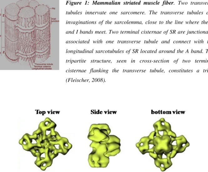
Figure 1: Mammalian striated muscle fiber. Two transverse
tubules innervate one sarcomere. The transverse tubules are invaginations of the sarcolemma, close to the line where the A and I bands meet. Two terminal cisternae of SR are junctionally associated with one transverse tubule and connect with the longitudinal sarcotubules of SR located around the A band. The tripartite structure, seen in cross-section of two terminal cisternae flanking the transverse tubule, constitutes a triad (Fleischer, 2008).
Figure 2: Ryanodine receptor. Three-dimensional surface representation of RyR using
cryo-electron microscopy and image enhancement. Left, transverse tubule face; middle, side view and right, terminal cisternae face (Fleischer, 2008).
1.3 RyR isoforms
Molecular cloning studies have defined three different isoforms of RyR in mammals, namely RyR1, RyR2 and RyR3. All three are encoded by different genes present on different chromosomes (Marks, 1996; Mikami et al., 1989; Takeshima, 1993; Takeshima et al., 1989). In mammalian striated muscles, the expression of the different RyR protein isoforms is tissue specific. RyR1 is the predominant RyR isoform in the skeletal muscle (Coronado et al., 1994; McPherson and Campbell, 1993; Ogawa, 1994). RyR2 is most abundant in cardiac muscles and RyR3 is found in striated muscles but in relatively low levels (Froemming et al., 2000; Sutko et al., 1991). RyR isoforms are also found to be expressed in other tissues. Expression of all the three isoforms of RyR proteins are reported in smooth muscles (Ledbetter et al., 1994; Marks et al., 1989), cerebrum (Furuichi et al., 1994; Hakamata et al., 1992) and cerebellum (Martin et al., 1998).
Non-mammalian skeletal muscles contain nearly equal amounts of two different RyR isoforms, namely -RyR and -RyR which are homologous to mammalian RyR1 and RyR3, respectively (Airey et al., 1993; Lai et al., 1992; Murayama and Ogawa, 1992). In these organisms, both the isoforms are present in approximately equal amounts in contrast to the predominant expression of RyR1 in mammals.
The three RyR isoforms share 70% homology (Nakai et al., 1990). Analysis of the primary amino acid sequences suggests that the membrane-spanning domains of the RyR are clustered near its COOH terminus (Blazev et al., 2001; Zhao et al., 1999) and has also revealed several consensus ligand binding and phosphorylation motifs.
1.4 Regulation of RyR
RyR channels are regulated by various cellular processes, physiological agents, pharmacological drugs and different closely associated proteins that are discussed below.
1.4.1 Cytosolic Ca2+
The influence of Ca2+ on the RyR is quite complex. The Ca2+ activates, inhibits and also conducts through the channel. Activation of RyR occurs at low concentrations of Ca2+ (1-10 µM)
and inhibition at higher concentrations (1-10 mM). RyR channel isoforms do not behave similarly in response to Ca2+ levels (Copello et al., 1997; Laver et al., 1995). RyR1 channel is almost totally inhibited at 1 mM Ca2+ but the other two isoforms, RyR2 and RyR3, require much higher concentrations of Ca2+ (Bull and Marengo, 1993; Chen et al., 1998; Chu et al., 1993). The physiological role of these very high levels of Ca2+ inhibition is not very well understood.
1.4.2 ATP and Mg2+
Cytosolic Mg2+ is a potent inhibitor of RyR (Copello et al., 2002; Laver et al., 1997; Smith et al., 1985), whereas ATP is an effective activator of RyR channel (Sonnleitner et al., 1997; Xu et al., 1996). Again the action of ATP and Mg2+ on RyR channel isoforms is specific. RyR1 is much more sensitive to ATP and Mg2+ than RyR2 and RyR3 (Jeyakumar et al., 1998). The action of Mg2+ is quite complicated since it may compete with Ca2+ both at the activation site (Laver et al., 1997) and also at the Ca2+ inhibition site (Copello et al., 2002), thereby shifting the Ca2+ sensitivity of the channel. In the presence of ATP and Mg2+, RyR1 requires less Ca2+ to activate than in their absence (Takeshima et al., 1998).
1.4.3 Cyclic ADP-ribose
Cyclic ADP-ribose (cADPR) is a metabolite of NADP that dramatically activates mammalian RyR channels (Meszaros et al., 1993). However, studies later suggested only minor effects of cADPR on RyR (Sitsapesan and Williams, 1995) and in some cases, no impact was reported (Fruen et al., 1994). Studies also suggested that two closely associated proteins, calmodulin and FK-506 binding protein (Lee, 1997), are required for the effect of cADPR on RyR. cADPR, in some cases, also appeared to activate the Ca2+-ATPase, which indirectly activated the RyR channel (Lukyanenko et al., 2001). Overall, the direct action of cADPR on RyR channels seems to be controversial.
1.4.4 Phosphorylation/dephosphorylation
RyR is a macromolecular complex that contains many consensus phosphorylation sites. The effect of exogenously applied kinases and phosphatases on RyR channel has been reported (Hain et al., 1995; Lokuta et al., 1995; Marx et al., 2000). Protein kinase A (PKA) activates RyR
increases depolarization-induced Ca2+ release from SR vesicles (Igami et al., 1999). Reports suggest that RyR2 is also activated by PKA (Hain et al., 1995), whereas others suggest that PKA destabilizes RyR2 channel opening through phosphorylation-induced dissociation of FKBP12.6 protein (Marx et al., 2000). The differences in these finding would probably result from different experimental setup altogether.
1.4.5 Ryanodine
Ryanodine is a plant alkaloid found naturally in the stem and roots of Ryania speciosa (Elison and Jenden, 1967; Jenden and Fairhurst, 1969). Stepwise studies made over a period of time with respect to ryanodine include : (i) purification of ryanodine binding protein (Campbell et al., 1987; Fleischer et al., 1985; Hymel et al., 1988), (ii) visualization of purified tetrameric complex through electron microscopy (Inui et al., 1987; Lai et al., 1988) and (iii) incorporation of the ryanodine binding receptor into lipid bilayers, demonstrated that this receptor is an ion channel (Imagawa et al., 1987). All these initial findings demonstrated that the receptor is the SR Ca2+ release channel in striated muscles (Fill and Coronado, 1988; Fleischer and Inui, 1989). Ryanodine, as an alkaloid, binds with high affinity to RyR (Fabiato, 1985; Frank and Sleator, 1975). High affinity [3H] -ryanodine turned out to be usefull in identifying different isoforms of RyR (Sutko et al., 1997).
1.4.6 Caffeine
Caffeine is one among the group of stimulants called methylxanthine, or xanthine that occur naturally in plants. It is known to activate Ca2+ release through the activation of RyR at millimolar concentrations. It is also reported that caffeine increases the sensitivity of endogenous RyR to modulators, such as Ca2+ and ATP, leading to increased channel activity (Rousseau et al., 1988).
1.4.7 JTV-519
JTV-519, also known as K201, is a 1 , 4-benzothiazepine derivative that shares a high degree of structural similarity with the voltage-dependent L-type Ca2+ channel blocker diltiazem (Tse et al., 2001). K201 inhibits diastolic SR Ca2+ leak and increases calstabin (FKBP) binding to
and anti-arrhythmic properties because of its ability to inhibit Ca2+ leak (Inagaki et al., 2000). Further more, studies have proposed that K201 inhibits SR Ca2+ leak by restoring the binding of FKBP12.6 to RyR2. The exact role of FKBP12.6 is not very well understood, but it is believed to stabilize the closed state of RyR2 channel (Kohno et al., 2003).
1.5 Regulation of RyR by associated proteins
There are large numbers of different proteins that are associated with RyR channels and also these play a role in the modulation of this channel. However not all these proteins have been thoroughly studied. A brief summary of how some of the proteins interact and modulate the RyR channel is summarized below.
1.5.1 Calmodulin
Calmodulin (CaM) is a ubiquitous, calcium-binding protein that can bind to and regulate a multitude of different protein targets, thereby affecting many different cellular functions. CaM was the first protein that was found to interact with single RyR channels in lipid bilayers (Smith et al., 1989). At low Ca2+ levels, CaM activates both RyR1 and RyR2 channels, but in the presence of high Ca2+ levels, it inhibits these channels (Fruen et al., 1994; Tripathy et al., 1995). As far as reports with RyR2 are concerned, CaM has always an inhibitory effect (Fruen et al., 1994). This modulation of RyR channel involves direct interaction of CaM-RyR and is not dependent on ATP (Smith et al., 1989). CaM also appears to bind to voltage-dependent Ca2+ channels of the plasma membrane, the dihydropyridine receptor (DHPR) of skeletal muscles, and has a series of structural consequences between DHPR and RyR1 interactions (Hamilton et al., 2000).
1.5.2 Calsequestrin (CSQ)
Calsequestrin is the major calcium sequestering protein in the SR and it forms a
quaternary complex with the RyR. It has been suggested that Ca2+ and pH-dependent conformational changes in calsequestrin modulate RyR channel activity (Hidalgo et al., 1996).
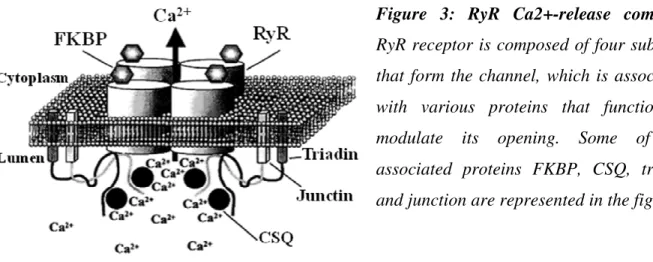
Calsequestrin is known to be located in the direct vicinity of RyR (Franzini-Armstrong et al., 1987), binds to triadin and junctin (Figure 3) and forms a quaternary complex that controls Ca2+
calsequestrin on RyR. Lipid bilayer studies suggest that calsequestrin activates the RyR channel (Kawasaki and Kasai, 1994) but there is one study mentioning that calsequestrin inhibits the RyR channel (Beard et al., 2002). Thus, the role of calsequestrin on RyR channel needs further more studies.
Figure 3: RyR Ca2+-release complex.
RyR receptor is composed of four subunits that form the channel, which is associated with various proteins that function to modulate its opening. Some of the associated proteins FKBP, CSQ, triadin and junction are represented in the figure.
1.5.3 FK-506 binding proteins
FK-506 is an immunosuppressive drug that binds to FK-506 binding protein (FKBP). Depending on the molecular mass, FKBP members are named as FKBP12 and FKBP12.6. FKBP12 also known as calstabin 1 is a 108 amino acid protein that shares 85% sequence identity with FKBP12.6, (calstabin 2). Both FKBP12 and FKBP12.6 associate with RyR1, RyR2 and RyR3 proteins in apparent stoichiometric proportions (Figure 3) and thus there are four FKBPs bound to each RyR channel complex (Timerman et al., 1993; Timerman et al., 1996). Reports suggest that removal of FKBP12 from RyR1 channel activates the channel (Ahern et al., 1994; Barg et al., 1997) but the impact of FKBP on RyR2 is not very well understood. Some suggest that removal of FKBP12.6 from RyR2 channel activates the channel (Xiao et al., 1997) and other report that FKBP12.6 removal has no impact on the RyR2 channel (Barg et al., 1997). The FKBP12-binding domain of RyR1 is mapped to its central regulatory domain between amino acids 2401-2840 (Bultynck et al., 2001), whereby the valylprolyl residue was shown to be critical
for establishing high affinity FKBP12 interaction (Gaburjakova et al., 2001). Though the interaction of FKBPs with RyR2 and RyR3 (Murayama et al., 1999) have been demonstrated, their interaction sites have not yet been determined and characterized very well. Some of the controversial reports regarding FKBP modulation of RyR1 channel indicate that the removal of FKBP12 leads to uncoupling of DHPR-RyR1 interaction suggesting that FKBP12 may be a physical coupler between RyR1 and DHPR (Lamb and Stephenson, 1996).
1.5.4 DHPR-loop peptide
The functional interaction of the voltage-dependent Ca2+ channel of the plasma membrane, the dihydropyridine receptor (DHPR) with RyR in striated muscles, is commonly thought to produce Excitation-Contraction (EC) coupling (Tanabe et al., 1990a; Tanabe et al., 1993). Depolarization of the plasma membrane induces conformational changes in DHPR that leads to activation of RyR channel in the SR membrane. This activation of RyR leads to massive Ca2+ release from the SR which in turn initiates contraction. The interaction of DHPR-RyR is tissue specific. In skeletal muscles, the interaction is thought to be a physical link between two proteins, whereas in cardiac muscles, it is the influx of Ca2+ through muscle that activates the RyR2 channel. The cytoplasmic II-III loop of skeletal DHPR 1-subunit is required for the functional interaction of DHPR with RyR1 (Tanabe et al., 1990a). Peptide fragments that correspond to the II-III loop activate single RyR channel function and interestingly, different regions of the peptide have different actions on RyR1 channel (Lu et al., 1994). Peptide A corresponds to a functional domain of the II-III loop that is known to activate RyR1 channel (Dulhunty et al., 1999), whereas peptide C, another domain of the same II-III loop blocks the activating action of peptide A (Gurrola et al., 1999). In one of controversial report, using a chimeric DHPR, lacking the peptide A region, expressed in myotubes lacking endogenous DHPR, normal EC coupling was observed (Grabner et al., 1999). Structural similarities of some of the reported scorpion peptide toxins with peptide A and their ability to bind and activate RyR (Samso et al., 1999), is altogether opening a new insight in the study of domain A and scorpion toxins which is discussed in detail in the following chapter.
2. Venom toxins
2.1 IntroductionToxins found in venoms are often around 10- to 70-mer peptides with high density of disulphide bonds. Often people name them as toxin peptides, but it would be more appropriate to designate them as small proteins, since they possesses secondary structures like helices, -sheets and turns.
Toxins are usually evolved as part of defensive or prey capture strategies. As a consequence, they predominantly act on key physiological systems. The major systems that are targeted by the toxins are the neuromuscular system, the neuronal system and the cardiovascular system. Within these systems, toxins target a considerable number of macromolecules, including the ion channels, hormone receptors, enzymes and transporters. However, toxins possess a number of basic characteristics that we would like drugs to possess which include potency and specificity. Thus, toxins have become subject of interest as they form an invaluable source of pharmacological agents.
2.2 Venom biodiversity
Venomous animals have evolved a large array of peptide toxins, many of which are bioactive. Their small size, relative ease of synthesis, structural stability and target specificity make them important pharmacological probes. It is estimated that more than 50,000 conopeptides from cone snails exist, of which less than 0.1% have been characterized pharmacologically. The ones which are characterized are found to be active on diverse range of ion channels and receptors associated with pain signaling pathways (Lewis and Garcia, 2003). Similarly, considering the diversity of venom peptides found in spiders, snakes, sea anemones and scorpions, it requires lots of work to characterize them pharmacologically. Traditionally, assay-directed fractionation was used to identify peptides of interest present amongst the large number of related molecules in the venom. Recent developments of novel high-throughput assays for the diversity of targets, along with improved separation and sequencing capabilities has simplified peptide isolation and characterization (Lewis, 2000; Lewis et al., 2000; Olivera et al., 1984).
Furthermore these peptides are characterized across multiple targets both in vitro and in vivo using chemically synthesized peptides.
2.3 Pharmacology of venom peptides 2.3.1 Ion-channel peptides
Several venom peptides have been characterized over the past for their pharmacological targets or activity. A wide range of different voltage-sensitive calcium channel peptides are characterized from the venom of cone snails (Olivera et al., 1987), spiders (Mintz et al., 1991; Newcomb et al., 1998) and snakes (de Weille et al., 1991). The -conotoxins are a class of small peptides that inhibit nicotinic acetylcholine receptors similar to snake -neurotoxins (McIntosh et al., 1999) and are being used as analgesics (Sandall et al., 2003). Voltage-sensitive sodium channels are crucial for the functioning of the nervous system and it is not surprising that a number of venom peptides from spider (Nicholson et al., 1994; Omecinsky et al., 1996), sea anemone (Vincent et al., 1980), coral (Gonoi et al., 1986), scorpions (Possani et al., 1999) and cone snails (Cruz et al., 1985; Fainzilber et al., 1994) have evolved to target these channels. Psalmotoxin 1 (PcTX1) is one such example that is a potentand specific blocker of proton-gated sodium channel. It was isolated from the venom of the South American tarantula Psalmopoeus cambridgei (Escoubas et al., 2000). Potassium channels are a large and diverse family of proteins that are implicated in the regulation of many cellular functions. Of the several potassium-channel-blocking peptides identified so far, only few have shown promising results in vivo. Multiple sclerosis is a disease of the central nervous system and is characterized by disseminated patches of demyelination in the brain and spinal cord which results in multiple and varied neurological disorders. ShK, a 35-residue polypeptide toxin from sea anemone Stichodactyla helianthus, is known to block voltage-gated Kv1.3 potassium channels and has been found to be effective against multiple sclerosis (Suarez-Kurtz et al., 1999).
2.3.2 Venom peptides useful in cardiovascular disease
Several venom peptides are being used against cardiovascular diseases, one such example is captoprill, an antihypertensive agent that essentially inhibits angiotensin-converting enzyme (ACE). ACE is a vasoconstrictor associated with hypertension and an essential enzyme for the
production of angiotensin (Dei Cas et al., 2003). Another classical example that is in preclinical development as a novel anti-bleeding agent for use in open heart surgery is Textilinin, a novel antifibrinolytic serine protease inhibitor from the venom of common brown snake (Filippovich et al., 2002).
2.3.3 Venom peptides useful in treating cerebrovascular accident
A stroke, or cerebrovascular accident (CVA), occurs when blood supply to part of the brain is disrupted, causing brain cells to die. When blood flow to the brain is impaired, oxygen and glucose cannot be delivered to the brain. Ancrod from the venom of Malayan pit viper and batroxobin from the venom of Bothrops atrox moojeni reduces this neurological deficits by enzymatically cleaving the blood fibrinogen (Bell, 1997) when used in the early stages of stroke (Samsa et al., 2002).
2.3.4 Venom peptides useful in diabetes
Diabetes mellitus, is a syndrome characterized by disordered metabolism and abnormally high blood sugar resulting from insufficient levels of the insulin hormone. Exendin-4 peptide from the venom of Heloderma suspectum, is presently in phase III clinical trials for the treatment of type 2 diabetes (Eng et al., 1992) and this peptide shares structural homology with another toxin namely -latrotoxin, from the black widow spider that has been found to be useful in the treatment of alzheimers disease (Perry and Greig, 2002). Paralleling to these latest developments, further applications regarding toxins are emerging that are of technological nature and are discussed in the later chapters.
2.4 Scorpion toxins 2.4.1 The Scorpions
2.4.1.1 Diversity and classification
Scorpions are one of the most ancient groups of animals on earth with about more than 400 millions of years of evolution and approximately 1,500 different species. During this lengthy evolutionary period, they have largely preserved their morphology and have adapted to survive in a wide variety of habitats including tropical forests, rain forests, grasslands, temperate forests, deserts and even snow covered mountains. Scorpions belonging to family Buthidae are represented by the genera Androctonus, Buthus, Mesobuthus, Buthotus, Parabuthus and Leirus located in North Africa, Asia and India. Centruroides are located in USA, Mexico, and Central America. Tityus are found in South America and Uroplectes in Africa (Debont et al., 1998; Hancock, 2001).
2.4.2 The scorpion venom
2.4.2.1 Composition and classification
Scorpions are interesting organisms because of the fact that their venoms contain molecules of medical importance. Advanced methods of fractionation, chromatography and peptide sequencing have made it possible to characterize the venom components. The basic steps involved in the characterization of venom is the identification of peptide toxins, analyses of their structure, function and targets (Favreau et al., 2006). Scorpion venoms contain a variety of biologically active components that include enzymes, peptides, nucleotides, lipids, mucoproteins and biogenic amines. The best studied components are the polypeptides that are active on the ion channels and receptors (Catterall, 1980; Garcia et al., 1997; Valdivia et al., 1992). Some of the criteria’s that are being used to classify the scorpion toxins are based on their molecular size as long chain toxins and short chain toxins; according to activity on distinct animals as mammal-specific toxins, insect-mammal-specific toxins and crustaceans-mammal-specific toxins; based on the mechanism of action as neurotoxins and cytotoxins; and toxins with disulphide bridges and without disulphide bridges. Toxins with disulphide bridges are usually active on ion channels. Those without
the action on ion channels, four different families of scorpion venom toxins have been defined namely: Na+- channel toxins (Catterall, 1980), K+-channel toxins (Carbone et al., 1982; Miller et al., 1985), Cl--channel toxins (DeBin et al., 1993) and Ca2+-channel toxins (Valdivia et al., 1992). Na+- channel toxins belong to long chain scorpion toxins consisting of 60-80 amino acids with four disulphide bridges. To date, around 230 primary structures of Na+- channel scorpion toxins have been determined (Srinivasan et al., 2002). K+-channel toxins are composed of 20-70 amino acids and generally compacted by three disulphide bridges. Based on their molecular size and location of cysteine residues, K+-channel scorpion toxins are further classified as -KTx, -KTx, and -KTx (Tytgat et al., 1999). Further few short-chains K+-channel scorpion toxins from the venoms of Heterometrus fulvipes are designated as kappa-KTx because of the presence of a novel bihelical scaffold (Chagot et al., 2005). Maurotoxin (MTX), a 34-residue toxin initially isolated fromthe venom of the scorpion Scorpio maurus palmatus is found to be active on several K+ -channel targets namely, apamin-sensitive small conductance Ca2+-activated K+ (SK) channels (Kharrat et al., 1996; Kharrat et al., 1997), intermediate conductance Ca2+-activated K+ (IK) channels (Castle et al., 2003), and several types of voltage-gated Kv channels (Shaker B and Kv1.2) (Carlier et al., 2000). MTX does not block the Kv1.1 channel type, whereas it is moderately active on Kv1.3(Fajloun et al., 2000b). Cl- channel scorpion toxins are usually low molecular weight peptides consisting of 35-38 amino acids with four disulphide bridges. One remarkable Cl- channel scorpion toxin is chlorotoxin. It has been purified from Leiurus quinquestriatus and blocks the channel in the epithelium of mouse. It binds specifically to Cl- channels in glia cells. Kurtoxin from Parabuthus transvaalicus binds with high affinity and inhibits Ca2+-channel by modifying the voltage-dependent gating (Lopez-Gonzalez et al., 2003). Many of the scorpion toxins are active on intracellular calcium release channel, the ryanodine receptor which is discussed below in detail.
2.4.3 Scorpion toxins active on ryanodine receptor 2.4.3.1 Imperatoxin A (IpTxa)
IpTxa, a 33- amino acid peptide was the first discovered member of an ever growing family of scorpion toxins active on the RyR and initially purified from Pandinus imperator (Valdivia et al., 1992). This peptide has three cysteine residues that stabilize its three-dimensional
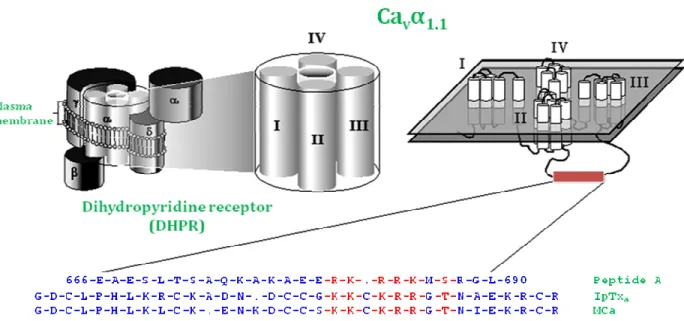
structure by forming disulphide bridges (Zamudio et al., 1997). IpTxa, was found to increase [3H]-ryanodine binding on skeletal muscle RyR, but not on the cardiac RyR, although it induces the appearance of long-lived subconductance states in both isoforms (Tripathy et al., 1998). IpTxa shares similar structural and functional properties with peptide A (Figure 5) Later on, it was showed that peptide A of the II-III loop of DHPR (Glu666-Leu690) binds to the same RyR1 site as IpTxa. Using [125I]-labelled IpTxa, itwas found that IpTxa binds on RyR1 with nanomolar affinity, whereas peptide A interacts at micromolar concentrations (Gurrola et al., 1999).
2.4.3.2 Ryanotoxin (RyTx)
RyTx is a 11.4-kDa peptide from the venom of the scorpion Buthotus judiacus that induces changes in ryanodine receptors of rabbit SR. RyTx was found to increase the binding affinity of [3H]-ryanodine in a reversible manner with a 50% effective dose at 0.16 µM. Results also suggested that binding sites for ryanotoxin and ryanodine were different and thought to be useful in identifying domains coupling the ryanodine receptor to the voltage sensor, or domains affecting the gating and conductance of the ryanodine receptor channel (Morrissette et al., 1996). 2.4.3.3 Hemicalcine (HCa)
HCa is a 33-mer peptide toxin isolated from Hemiscorpius lepturus, which is the most dangerous scorpion of Khuzestan, the south-west, hot and humid province of Iran. Of all scorpion stings, 10–15% during the hot season and almost all cases during the winter are due to H. lepturus. These observations are based on a sample of 2534 patients who brought a scorpion specimen to a medical centre while seeking treatment (Radmanesh, 1990). This peptide is active on ryanodine sensitive Ca2+ channels, as it increases [3H]-ryanodine binding on RyR1 and triggers Ca2+ release from SR. It shares 85 and 91% sequence identity with four other scorpion toxins active on RyR, namely maurocalcine, imperatoxin A, opicalcine 1 and opicalcine 2 (Shahbazzadeh et al., 2007).
2.4.3.4 Buthotus judaicus toxin (BjTx)
Buthotus judaicus toxin 1 (BjTx-1) and toxin 2 (BjTx-2) are twonovel peptides purified from the venom of the scorpion Buthotus judaicus and found to be active on RyR. Their amino
and Ile in BjTx-2. Despite slight differences in EC50, both toxins increase the binding of [3 H]-ryanodineto RyR1 at micromolar concentrationsbut had no effect on RyR2. Three-dimensional structural modeling reveals a cluster of positively charged residues (Lys11 to Lys16) as a prominent structural motif of both BjTx-1 and BjTx-2 (Zhu et al., 2004).
2.4.3.5 BmK-PL
BmK-PL is a peptide toxin isolated from the venom of Chinese scorpion Buthus martensi Karsch. This toxin was found to stimulate Ca2+-release channel activity of SR by an indirect mechanism, that does not involve direct interaction of the toxin with RyR, but possibly by binding to an associated protein such as triadin (Kuniyasu et al., 1999).
2.4.3.6 Maurocalcine (MCa)
MCa is a 33 amino acid residue peptide toxinisolated from the scorpion Scorpio maurus palmatus. Three similar peptides have been isolated or cloned from the venoms of different scorpions: IpTxa (El-Hayek and Ikemoto, 1998), that shares 82% sequence identity with MCa, and both opicalcine 1 and 2 (Zhu et al., 2003), from the scorpion Opistophthalmus carinatus, that show 91% and 88% sequence identities with MCa, respectively.
A B
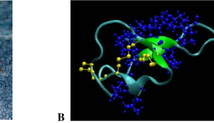
Figure 4: (A) Scorpio maurus palmatus (Shachak and Brand, 1983),(B) 3-D structure of MCa.
The structure was drawn by VMD1.8.6 software. Blue represents the basic amino acids and yellow the disulfide bridges.
Figure 5: Sequence alignment of peptide A, IpTxa, and MCa. Schematic representation of the
DHPR complex illustrating the subunit composition. MCa has sequence similarities with domain A of the II–III loop from the Cav 1.1 subunit of DHPR and with IpTxa, another scorpion toxin (Esteve et al., 2003)
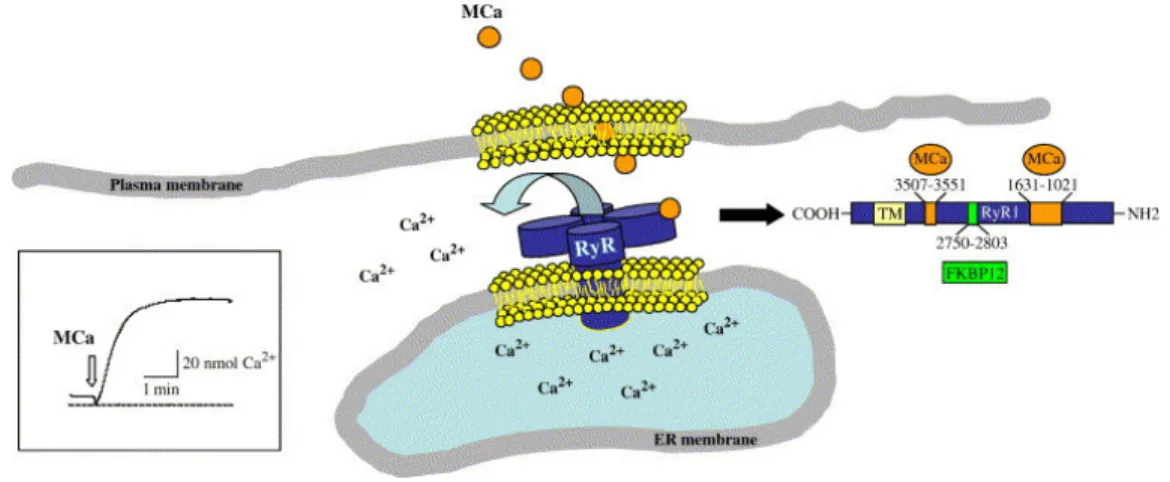
MCa triggers interest for three main reasons. Firstly, MCa is a powerful activator of the ryanodine receptor (RyR) (Figure 6), thereby triggering calcium release from intracellular stores (Chen et al., 2003; Esteve et al., 2003). MCa binds with nanomolar affinity onto RyR1, a calcium channel from the sarcoplasmic reticulum (SR), and generates greater channel opening probability interspersed with long-lasting openings in a mode of sub-conductance state (Fajloun et al., 2000a). Using a set of RyR1 fragments, two discrete domains of RyR1 responsible for its interaction with MCa were identified. MCa was found to bind to two discrete RyR1 regions encompassing residues 1021-1631 and 3201-3661, respectively. The second site was further restricted to amino acids 3351-3507. (Altafaj et al., 2005). MCa also interacts directly with RyR2 with an apparent affinity of 150 nM and found to bind to two domains of RyR2, which are homologous with those previously identified on RyR1. The effect of MCa binding to RyR2 was evaluated by [3H]-ryanodine binding experiments, Ca2+ release measurements from cardiac
sarcoplasmic reticulum vesicles and single-channel recordings, showing that MCa has no effect on the open probability or on the RyR2 channel conductance level (Altafaj et al., 2007).
Figure 6: Schematic drawing of the ryanodine receptor. RyR is a calcium channel localized in
the membrane of the endoplasmic reticulum. Its function is to produce calcium release from this internal calcium stock. MCa has a well identified binding site on RyR that is localized on the cytoplasmic side of the channel. Binding of MCa to its site on RyR triggers calcium release from endoplasmic reticulum vesicles and Ca2+release can be measured by the change in fluorescence intensity of a calcium indicato,r as shown here (Boisseau et al., 2006).
Secondly, MCa has a unique sequence homology with a cytoplasmic domain of the pore-forming subunit of the skeletal muscle DHPR (Figure 5). This homology implicates a DHPR region that is well known for its involvement in the mechanical coupling between the DHPR and RyR1, a process whereby a modification in membrane potential is sensed by the DHPR, transmitted to RyR, and produces internal calcium release followed by muscle contraction. It is therefore expected that a close examination of the cellular effects of MCa on the process of excitation-contraction coupling may reveal intimate details of the mechanistic aspects linking the functioning of the DHPR to that of RyR. In that sense, a role of domain A in the termination of calcium release upon membrane repolarisation has been proposed through the use of MCa (Pouvreau et al., 2006).
Third, MCa appears unique in the field of scorpion toxins for its ability to cross the plasma membrane; this raises considerable technological interest in the peptide and is discussed in detail in the later chapter.
3. Membrane transport
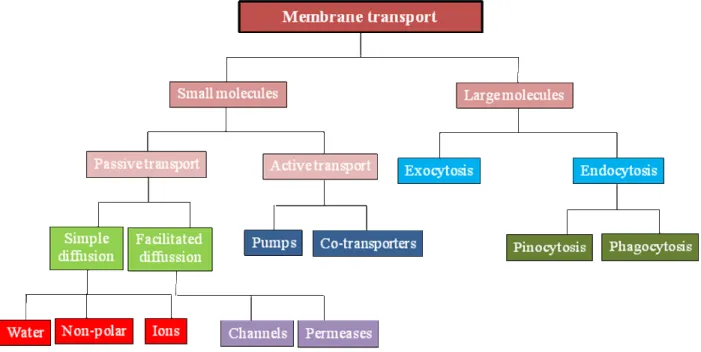
3.1 IntroductionThe Plasma membrane is a dynamic structure that separates the chemically distinct intracellular cytoplasm from the extracellular environment and also regulates the entry and exit of small and large molecules. Small molecules such as amino acids, sugars, ions and nutrients can cross the plasma membrane through the action of integral membrane protein pumps or channels (Figure 7). For the uptake of larger molecules, cells have developed a process termed as endocytosis.
Figure 7: Classification system of membrane transport.
3.2 Endocytosis
Endocytosis is a basic cellular process that is used by cells to internalize a variety of molecules. Macromolecules are carried into the cells by vesicles formed by the invagination of the membrane. These vesicles are pinched-off into the cytoplasm. Since the macromolecules can be quite diverse, understanding the different pathways that mediate their internalization and how these pathways are regulated is important for many areas of cell and developmental biology.
Endocytosis occurs by multiple mechanisms that fall into two broad categories, phagocytosis or “cell eating” and pinocytosis or “cell drinking” (Figure 8).
Figure 8: Endocytosis pathways. Large particles are taken up by phagocytosis, whereas fluid
uptake occurs by macropinocytosis. Both processes are triggered by actin-remodelling. Depending on various factors, particles can be taken up by many other pathways that are dependent on clathrin or caveolin or independent of both (Mayor and Pagano, 2007).
3.2.1 Phagocytosis
Phagocytosis is a process in mammals that is driven by specialized cells, including macrophages, monocytes and neutrophils to clear large pathogens such as bacteria and cellular debris (Aderem and Underhill, 1999). This process is regulated by specific cell-surface receptors and signaling cascades. Particle internalization is initiated by the interaction of specific receptors on the surface of the phagocyte, leading to the polymerization of the actin and internalization of the particle (Hall and Nobes, 2000). Recent advances have highlighted the significance of phagocytic receptors, in particular the fibronectin receptor and other integrins (Blystone et al., 1994). The mechanism underlying actin assembly remains obscure. Recent advances suggest that
phosphatidylinositol 3-kinase and protein kinase C have a role in actin polymerization (Fallman et al., 1992; Panayotou and Waterfield, 1993; Zamudio et al., 1997). Further more, it is not clear if actin polymerization alone can drive particle internalization or if it requires molecular motors. Recently it has been shown that myosin II accumulates in macrophages and neutrophils during phagocytosis, implying that it might act as mechanical motor (Stendahl et al., 1980; Valerius et al., 1981). After internalization, the actin-based machinery is shed from the phagosome and the infectious agents are destroyed with the help of various acids, free oxygen radicals and acid hydrolases (Russell, 1995a; Russell, 1995b). There are different modes of phagocytosis which are basically determined by the particle to be ingested and the receptor that recognizes the particle. 3.2.2 Pinocytosis
Pinocytosis is a process of intracellular accumulation of molecules either by nonspecific binding of the solutes to the cell membrane or by binding to specific high-affinity receptors. During this process, there is invagination of the plasma membrane. This process is also referred to as cellular drinking, since the substances around the area of invagination are dissolved in water and ingested into the cell
3.2.2.1 Macropinocytosis
Macropinocytosis defines a series of events leading to extensive reorganization of plasma membrane. Unlike phagocytosis, macropinocytosis starts with the reorganization of actin, but here the protrusions do not climb up, but instead they collapse and fuse with the plasma membrane to generate large endocytic vesicles, called macropinosomes (Conner and Schmid, 2003). Macropinosomes are relatively large vesicles as compared to other pinocytotic vesicles and provide efficient route for non-selective endocytosis of solute macromolecules. Rab proteins are small GTPases that control multiple membrane trafficking events during the process of macropinocytosis. To date around twelve Rab members have been located around the endocytotic structures. Of these, Rab5 controls several processes including invagination, endosomal fusion, signaling and motility (Stenmark et al., 1994). ADP-ribosylation factors (ARFs) are also small GTPases that function in membrane traffic. One variant ARF6 in conjugation with actin cytoskeleton, exchange factors and activators, help in regulating macropinocytosis, cell adhesion
and migration (Donaldson, 2003; Sabe, 2003). Also possible involvement of rafts in macropinosome formation has been suggested (Watarai et al., 2002). However, the fate of the macropinosome is being debated since studies suggest either fusion with lysosomes (Racoosin and Swanson, 1993) or with each other (Hewlett et al., 1994) and this perhaps depends on the cell type used (Swanson and Watts, 1995).
3.2.2.2 Clathrin-mediated endocytosis (CAE)
Clathrin-mediated endocytosis was previously referred to as receptor mediated endocytosis but, nowadays it is well established that most pinocytic pathways also involve specific receptor-ligand interactions. The uptake of transferrin and its receptors is a classical example of CAE (Hanover et al., 1984; van Dam and Stoorvogel, 2002). CAE is important for intercellular communication during tissue and organ development and throughout the life of an organism as it modulates signal transduction (Di Fiore and De Camilli, 2001; Seto et al., 2002). CAE of membrane pumps that controls the transport of ions across the plasma membrane in neurons helps in controlling synaptic transmission and may have a role in learning and memory (Beattie et al., 2000). It is also involved in recycling vesicle proteins (Takei et al., 1996).
Figure 9: Clathrin-mediated endocytosis. (1) Assembly of clathrin triskelions into polygonal
lattice with the help of adaptor proteins; (2) Deformation of plasma membrane into coated pits; (3) Recruitment of dynamin to the neck of coated pits; (4) Pinching off of the coated pit; (5) Recycling of clathrin triskelions, adaptor proteins and dynamin (Conner and Schmid, 2003).
CAE involves interaction of multi-functional adaptor proteins with plasma membrane, clathrin and several accessory proteins and phosphoinositides. It occurs at specialized structures called coated pits, which are formed by the assembly of cytosolic coat proteins, the main assembly unit being clathrin. Assembly unit of clathrin called triskelion, is a three-legged structure consisting of three heavy and three light chains (Greene et al., 2000; Schmid, 1997). Under non-physiological conditions, clathrin triskelions self-assemble into closed polygonal cages, however in physiological conditions, it requires adaptor proteins. Two classes of adaptor proteins have been identified based on their ability to assemble clathrin, namely monomeric assembly protein AP180 and heterotetrameric adaptor protein (AP1-4) (Kirchhausen, 1999; Robinson and Bonifacino, 2001). Complex formation between AP180 and AP2 gives greater clathrin assembly activity suggesting synergistic effects between them in clathrin assembly (Hao et al., 1999). AP2 basically consists of two domains namely, core and ear. The Core consists of two large structurally related subunits called - and 2- adaptins, a medium subunit, µ2, and a small subunit, 2. Ears are formed by the carboxy termini of - and 2- adaptins, respectively (Collins et al., 2002). Together, the coat proteins, clathrin, AP2 and AP180 are involved to select the cargo and form coated pits. These coated pits are eventually pinched off from the plasma membrane, a process regulated by dynamin. Dynamin is a large-molecular-mass protein with GTPase activity. It has an N-terminal GTPase domain involved in binding and hydrolysis of GTP. The central region specifically binds to phosphatidylinositol-4, 5-bisphosphate and the C-terminal region is involved in dynamin oligomerization and self assembly (McNiven et al., 2000; Muhlberg et al., 1997; Schmid et al., 1998; Wigge and McMahon, 1998). After the endocytic vesicle is pinched off, clathrin is recycled back to the plasma membrane and reused, whereas the endocytic vesicle is targeted to the early endosome (Figure 9).
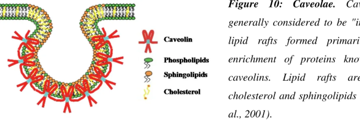
3.2.2.3 Caveolin-mediated uptake
Caveolae are flask-shaped invaginations of the plasma membrane and are characterized by the presence of caveolin in many cell types (Figure 10). Caveolin is a hairpin-like integral membrane protein of 21 kDa. Caveolins are palmitoylated in the C-terminal segment (Dietzen et al., 1995), phosphorylated on tyrosine residues (Glenney, 1989), bind to cholesterol (Murata et al., 1995) and they form dimmers and oligomers (Monier et al., 1995). Caveolins are essential for
the formation and stability of caveolae. In the absence of caveolins, no caveolae are seen and when expressed in cells lacking caveolae, they induce caveolar formation (Fra et al., 1995). Basically three isoforms of caveolins have been identified; Caveolin-1 and 2 are often seen in majority of differentiated cells, whereas caveolin 3 is localized in skeletal and cardiac muscles (Way and Parton, 1995).
Figure 10: Caveolae. Caveolae are
generally considered to be "invaginated" lipid rafts formed primarily due to enrichment of proteins known as the caveolins. Lipid rafts are rich in cholesterol and sphingolipids (Galbiati et al., 2001).
The lipid composition of the caveolae corresponds to that of lipid rafts rich in cholesterol and sphingolipids. They are essential for the formation and stability of caveolae (Brown and London, 1998; Simons and Toomre, 2000). Removal of cholesterol from the plasma membrane, leads to the disappearance of caveolae. Thus caveolae can also defined as caveolin-containing plasma membrane invaginations rich in lipid rafts (Rothberg et al., 1992). In addition caveolae also contains dynamin which is localized to the neck of flask-shaped caveolar vesicles (Henley et al., 1998; Oh et al., 1998). As in case of clathrin-mediated endocytosis, dynamin is involved in pinching off the caveolar vesicles from the plasma membrane (De Camilli et al., 1995; Sever et al., 2000).
Recent works have confirmed the caveolar entry of cholera toxin (Montesano et al., 1982), folic acid (Rothberg et al., 1990), serum albumin (Schnitzer et al., 1994) and alkaline phosphatase (Parton et al., 1994). Simian virus 40 (SV40) is one of the most exclusively studied ligands for caveolar endocytosis (Pelkmans et al., 2001; Roy et al., 1999). SV40 has several advantages as model ligand since the particle is well characterized in terms of composition and structure. SV40 after binding to the plasma membrane through the major histocompatibility
(MHC) class I antigen (Breau et al., 1992), diffuses laterally until it gets trapped in caveolae (Pelkmans et al., 2001). Lateron, virus containing caveolae, which are relatively smaller in diameter compared to virus-free caveolae (Stang et al., 1997), pinch off from the plasma membrane and move as caveolin-coated endocytic vesicles in the cytoplasm. Virus-free caveolae do not internalize. Some of the other events triggered during this process include tyrosine phosphorylation (Pelkmans et al., 2002), recruitment of actin and formation of actin tails (Chen and Norkin, 1999). But most of these events are transient; once SV40 is internalized, phosohotyrosines disappear and actin cytoskeleton returns to a normal pattern.
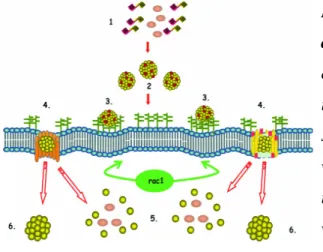
3.2.2.4 Clathrin- and caveolin- independent uptake
The mechanisms of endocytosis governed independent of caveolae and clathrin are very poorly understood. Each of these endocytosis processes fulfills unique functions in cell by the transport of various cargoes that include ligands, fluid, adhesion molecules and toxins. Depending on the type of cargo molecules to be transported, there are different types of processes involved in the formation of vesicles, pinching-off of the vesicle from the plasma membrane and in the regulation of entry of the cargo (Figure??). Again intracellular destiny in each case is not identical.
Rafts are small structures that diffuse freely on the surface of the cell (Edidin, 2001). They have unique lipid compositions that provide a physical basis for selectivity of membrane proteins and glycolipids (Anderson and Jacobson, 2002). Since rafts are small, they can be captured and internalized within an endocytic vesicle. Caveolae represents just one type of lipid rafts rich in cholesterol and sphingolipids. Based on ferro-fluid purification method, recently an endosomal protein, namely Flotillin-1 was identified. Flotillin-1 was found to reside in a specific population of endocytic intermediates and these intermediates accumulated both glycosylphosphatidylinositol (GPI)-linked proteins and cholera toxin B subunit. It was found that flotillin-1 small interfering RNA (siRNA) inhibited both clathrin-independent uptake of cholera toxin and endocytosis of a GPI-linked protein (Glebov et al., 2006). It has been reported that glycosylphosphatidylinositol-anchored proteins (GPI-Aps) have been endocytised to a recycling endosomal compartment but not to the Golgi through a clathrin- and caveolae- independent pathway (Sabharanjak and Mayor, 2004).
With ultrastructural and biochemical experiments, it was shown that clathrin-independent endocytosis of IL2 receptors exists constitutively in lymphocytes and is associated with detergent-resistant membrane domains (Lamaze et al., 2001). Thus recent identification of cargo molecule specific pathways helps in defining various mechanisms of endocytosis that have been broadly classified as independent of clathrin and caveolae uptake.
4. Cell-penetrating peptides
4.1 IntroductionCell-penetrating peptides (CPPs) also known as protein transduction domains (PTDs) or membrane transduction peptides (MTPs) are usually 7 to 33 amino acid long peptides capable of translocating into cells. These peptides are classified either as cationic or amphipathic peptides based on their sequence. Cationic CPPs contain clusters of primarily arginine residues. Studies evolving in the field of CPPs have mainly focused on three important aspects, namely: i) Defining the structural properties of CPPs that afford the capability to translocate the cell membrane, ii) Elucidating the translocation mechanism of these peptides which remains a controversial topic and iii) Exploiting the ability of the CPPs to deliver a wide range of impermeable molecules across the cellular membrane.
In a non-exhaustive list of CPPs, one point to be noticed is the lack of sequence homology, but again they possess some common functional features. Structure based studies indicate an important role of basic amino acids in transduction (Mi et al., 2000; Mitchell et al., 2000). In addition, the spatial separation of hydrophobic and positively charged residues appears important for defining good transducers (Oehlke et al., 1998; Scheller et al., 1999). Generally, CPPs lack cell selectivity. They can enter into numerous cell types though the quantitative comparison of the efficacy of penetration in various cell types for different CPPs would reveal some differences. A recent systematic evaluation of transduction of fluorescent oligo-arginines has showed highly variable differences of penetration between D- and L- forms of peptides (Tunnemann et al., 2007).
Cell penetration of CPPs does not require any specific membrane receptor for their translocation but this does not mean that they do not interact with any membrane components. Two types of cell surface components have been shown to interact with CPPs, glycoaminoglycans (GAGs) and the negatively charged lipids (Console et al., 2003; Magzoub et al., 2002) and the basis of these interactions were shown to be electrostatic (Ziegler and Seelig, 2004). One more assumption which is being hotly debated is that CPPs do not require metabolic energy for cell entry (Thoren et al., 2000). Evidence for this comes from experiments in which
CPP cell entry was preserved even at 4°C (Esteve et al., 2005) or in the presence of metabolic inhibitors (Vives et al., 2003). However several mechanisms of penetration have been proposed (Patel et al., 2007). In one process, penetration involves a reorganization of the plasma membrane which allows the peptide to move from the extracellular face of the membrane to the intracellular one (Thoren et al., 2004). This mechanism allows for the delivery of the peptide freely into the cytoplasm (Vasir and Labhasetwar, 2007). In another process, CPPs may follow any endocytotic pathway, a process which requires energy (Ross and Murphy, 2004). The type of endocytosis namely macropinocytosis, clathrin- or caveolin-dependent, clathrin- and caveolin-independent endocytosis depends on the cell type, CPP sequence and nature of cargo. Various experimental strategies are therefore pursued to favor the leak of CPP and cargo from the endosomes to the cytoplasm like addition of the lysosomotropic reagent chloroquine (Turner et al., 2005b).
4.2 Classes of Cell penetrating peptides 4.2.1 Tat-related peptides
The HIV-1 Tat is a protein composed of 86 amino acids that binds to the trans-acting response element (TAR) of the viral RNA to transactivate the viral promoter (Frankel and Young, 1998). In the late 1980s, energy-independent translocation of the Tat protein was reported (Frankel and Pabo, 1988; Green and Loewenstein, 1988). In 1994, it was reported that chemical conjugation of the Tat protein (residues 1-72 and 37-72) to other proteins enabled the conjugates to be efficiently internalized into cells (Fawell et al., 1994). Further studies suggested that the arginine-rich segment in the Tat protein (residues 48-60) is a critical component of translocation (Vives et al., 1997). Genetically engineered fusion proteins of Tat fused with cargo proteins, were produced in E. coli, purified and purified fusion proteins were shown to be efficiently internalized into the cells (Wadia and Dowdy, 2002; Wadia and Dowdy, 2003). Successful in vivo delivery of Tat- -galactosidae fusion protein to various organs in mice after intraperitoneal injection of fusion protein was reported (Schwarze et al., 1999). Tat fusion proteins have been used to treat mouse models of cancer and inflammation (Snyder and Dowdy, 2005; Wadia and Dowdy, 2005).
Tat has also been used to deliver phage encapsulated DNA to cells, and liposome encapsulated DNA for gene expression in mice (Eguchi et al., 2001; Glover et al., 2005). Efficiency of transduction of Tat depends on the cargoes being used and this principle is clearly demonstrated by difficulties in transducing large cargoes like nucleic acids (Fischer et al., 2005; Meade and Dowdy, 2007). An interesting finding based on the studies with Tat suggested that the cell fixation process has a considerable effect on the cellular localization of the internalized peptide. In unfixed cells, the internalized peptide was seen in punctuate structures within the cytosol, whereas in fixed cells, the punctuate structures were lost and the peptide was found diffused throughout the cytosol and in the nucleus (Richard et al., 2003).
Cell penetrating peptide Amino acid sequence
Tat49-57 RKKRRQRRR Penetratin RQIKIWFQNRRMKWKK Transportan GWTLNSAGYLLGKINLKALAALAKKIL Pep-1 KETWWETWWTEWSQPKKKRKV MPG GALFLGFLGAAGSTMGAWSQPKKKRKV Polyarginines (R9) RRRRRRRRR Crotamine YKQCHKKGGHCFPKEKICLPPSSDFGKMDCRWRWKCCKKGSG
Table 1: Amino acid sequence of CPPs.
4.2.2 Penetratins (Homeodomain-derived peptides)
Homeodomain proteins belong to the class of transcription factors that bind to DNA through specific sequence of 60 amino acids called the homeodomain. It consists of three -helices, with one turn between helices 2 and 3. It was first found that the 60 amino acid long polypeptide was able to cross the plasma membrane of the differentiated neurons (Joliot et al., 1991a). Several mutants of homeodomains were analysed for the cell penetration and it was found that with deletion of two hydrophobic residues at positions 48 and 49 within the third -helix, the peptide lost its ability to cross the plasma membrane (Joliot et al., 1991b). Further more, a synthetic peptide of 16 residues corresponding to amino acids 43-58 of the homeodomain called as penetratin was confirmed to possess the translocation properties of the entire homeodomain (Derossi et al., 1994). Penetratin is claimed to be the first reported CPP. It is characterized by a high content in basic residues and hydrophobic residues mostly tryptophanes