HAL Id: tel-02457099
https://tel.archives-ouvertes.fr/tel-02457099
Submitted on 27 Jan 2020HAL is a multi-disciplinary open access
archive for the deposit and dissemination of sci-entific research documents, whether they are pub-lished or not. The documents may come from teaching and research institutions in France or abroad, or from public or private research centers.
L’archive ouverte pluridisciplinaire HAL, est destinée au dépôt et à la diffusion de documents scientifiques de niveau recherche, publiés ou non, émanant des établissements d’enseignement et de recherche français ou étrangers, des laboratoires publics ou privés.
Role of the interaction between endoplasmic reticulum
and mitochondria in endothelial dysfunction induced by
human microparticles
Zainab Safiedeen
To cite this version:
Zainab Safiedeen. Role of the interaction between endoplasmic reticulum and mitochondria in en-dothelial dysfunction induced by human microparticles. Immunology. Université d’Angers; Université libannaise de Beyrouth, 2016. English. �NNT : 2016ANGE0063�. �tel-02457099�
Zainab SAFIEDEEN
THESE EN COTUTELLE
Mémoire présenté en vue de l’obtention du
grade de Docteur de l'Université d'Angers
sous le sceau de l’Université Bretagne Loire
École doctorale : Ecole Doctorale Biologie Santé
Discipline : Aspects moléculaires et cellulaires de la biologie
Spécialité : Biologie cellulaire
Unité de recherche :INSERM 1063 SOPAM : « Stress Oxydant et Pathologies Métaboliques »
ET
L’Université Libanaise
Ecole Doctorale des Sciences et Technologie
Spécialité : Immunologie Moléculaire
Soutenue le 26 septembre 2016
Thèse N° : 108480
Rôle de l'interaction entre le réticulum endoplasmique et
les mitochondries dans la dysfonction endothéliale induite
par des microparticules humaines
JURY
Rapporteurs : Florence PINET, Directeur de Recherche, Université de Lille Paul MULDER,Professeur d’Université,Université de Rouen
Examinateurs : Bernard MULLER, Professeur d’Université,Université de Bordeaux
Kazem ZIBARA, Professeur d’Université,Université Libanaise
Directeur de Thèse : Carmen MARTINEZ, Directeur de Recherche,Université d’Angers
1
Table of Materials
Acknowledgements ... 3
Publications & communications ... 7
Abbreviations... 8
List of Figures... 10
List of Tables... 10
Introduction ... 11
A- M
ETABOLIC SYNDROME... 11
a) Definition & clinical outcomes ... 11
b) Metabolic syndrome (MetS) criteria... 13
B- M
ICROPARTICLES... 15
a) Definition of MPs... 15
b) Characteristics of MPs ... 17
1. Structure, origine and types... 17
2. Composition ... 19
3. Mechanisms of MP production ... 19
4. Mechanisms of MP interaction with target cells... 20
c) Role of MetS in the liberation of MPs ... 22
C- C
ARDIOVASCULAR DISEASES AS CONSEQUENCES OFM
ETS ... 22
a) Epidemiology ... 22
b) CVDs and MetS ... 23
c) Endothelial dysfunction ... 23
1. Definition ... 24
2. Endothelial dysfunction and MetS ... 24
3. MPs: players in endothelial dysfunction ... 25
4. Neutral sphingomyelinases: another worker in endothelial dysfunction ... 26
D- E
NDOPLASMICR
ETICULUMS
TRESS... 27
a) Endoplasmic reticulum (ER) histology and function... 27
b) ER stress: the unfolded protein response ... 27
c) Endoplasmic reticulum stress: its role in diseases and a novel target for therapy ... 30
d) ER stress is involved in MetS and endothelial dysfunction... 32
2
1. Mitochondria histology and function ... 32
2. Mitochondria and mitochondrial reactive oxygen species as mediators of metabolic
alterations and endothelial dysfunction... 33
3. ER and mitochondrial interaction ... 35
4. ER and oxidative stress dialogue ... 36
Aim of the study... 37
Article ... 38
Discussion & Conclusion ... 39
Review ... 43
Publication ... 44
3
A
CKNOWLEDGEMENTS
I would like first to express my praise and thanks to Allaah for being with me all the way.
I would like to express my gratitude and respect for the members of the jury.
For Mr Professor Paul MULDER and Mrs Doctor Florence PINET for giving me the honor of
being the rapporteurs of this thesis. For Professor Bernard MULLER and Professor Kazem
ZIBARA who agreed to examine this work.
The greatest thank of all goes to Doctor Carmen MARTINEZ, my thesis director. Words
cannot explain my deep gratitude. Working with you is one of the good things that can happen
in someone’s life, and I was lucky enough to have that. Carmen is the symbol of hard work,
cleverness, intelligence, ingenuity, regularity, patience, diplomacy, management, and
positivity. She is the kind of a person that makes you want to work more, give more, her wide
knowledge forces you to advance. Your confidence in me encouraged me to give my best in
order to never let you down. You were a director, a teacher, a motivator and a lot more.
Thanks for all the expertise and skills you provided me with. Thanks for all the corrections
you did and the time you spent on the reports, articles, thesis (mainly in my language). You
were always my hope, with you I knew that I will always arrive.
I would like to thank Doctor Kazem ZIBARA who helped me to get to where and what I am
now. All his directions through the earlier years made me a better student. Huge thanks for
Doctor Mohamad EZZ-EDDINE for having confidence in me and having me as his thesis
student.
I want to particularly thank Mr Doctor Ramaroson ANDRIANTSITOHAINA, the director of
SOPAM 1063 unit, for accepting me as a member in his team, for all his guidance, and pieces
of advice and the constructive criticism he provided me with, these were pushing me to
improve and advance.
I would like to thank Doctor Gilles SIMARD and Doctor Pascal REYNIER for giving me the
gratification of working with them.
4
I would like to thank Doctor Denis LEIBER and Doctor Soazig LE LAY for their advice and
their support particularly during Monday meetings. And I would like to thank Doctor
LEIBER for his participation in my article and for his ideas and help.
Lots of gratitude and respect to Doctor Raffaella, and Gregory. You were beyond every
achievement and success; until I started to believe that you have a magic touch and it is
sufficient that you take a look or say something and things will work out immediately. You
were always there whenever I needed an opinion, or help and most importantly for the
calculations of concentrations. You are people of expert, wide knowledge, and humble who
give from their hearts without waiting a reward.
A huge deep thank for Doctor Nadia and Doctor Liliana. When I arrived here I was
determined not to get attached, however, god offered me two sisters. With you I never felt that
I am alone, with you I never had the impression that I could break down, or I would fall. I
always felt that you were my guardian angels, who are always there to protect me. Thanks for
the support, defense, trust and confidence that was mutual between us, the nice words and the
kind messages, the hope you plant in me every time something bad arrives. For the late chat
evenings, and the phone calls every day of the week. Spending these days with you was a
pleasure. Moments with you are worth life time.
An ultimate thanks for the girls that I had the chance to share my almost three years with
Patricia, Marine, Doctor Emilie, and earlier Catherine. Actually I owe you so much; you have
lots of credit on me that at a certain moment I stopped counting. You were generous enough
to help me in every aspect of my life, filling administrative papers, exchanging science,
encouraging me, hearing me, sharing with me my good and bad moments, laughing and
crying with me, supporting me, I never felt that there is anything that I cannot share it with
you.
My lovely, cute Marine, thanks a lot for all the times I disturbed you when you are eating to
help in my mails, and also for your constructive opinions. You have never let me down.
I would like to thank particularly the amazing, adorable and loving Patricia for having the
patience of teaching me French and correcting my phrases every time without harassing.
When I first arrived here I was zero in French, now I am better and she is the number one
reason beyond that. Moreover, thanks for helping me in ideas for the schemas and taking care
5
of all the details to have a well-organized, neat manuscript as it was yours. These are the least
things that I could mention. For sure, I could not do it without you.
I thank very warmly Doctor Luisa and Doctor Vanessa, for all the good times we spent
together, for every moment you lifted me up when I was down, for all the support, for the nice
messages, for all the nice outings and lunches.
I would like to pass my greatest esteems to Mireille WERTHEIMER and Catherine BRIAND
for all the care they took of me, they made me feel like the spoiled child.
Obviously, this work would not have been possible without all the members of the 1063 unit,
the past and present, particularly Ousama, Edward, Simon, Camille, Maeva, Mehdi, Mathieu,
Doctor Emiliane, Sameul, Eid, Doctor Marion, Doctor Audrey, Doctor Manolo, Doctor
Gaceb, Doctor Sylvain and Doctor Wojciech.
In this laboratory I found my second family, a family that embraced me, that stuck by myside,
where all the members are special and you cannot help falling for them, you cannot get mad
or sad from, and makes you start thinking how you are going to live your next days without
them. However family stays in heart, no matter how far we go they will always be there.
An extraordinary thank for my friends Mariam, Rasha, Zaher and Abdalla. I really could not
imagine living in Angers without you. You were the preferable thing in whole France. And
for Zahraa, Hussein and Rokaya, even though we were in two different continents, you were
always here in every step of the way.
A special thanks for the best friend ever Doctor Martin SERRANO SANCHEZ who is always
there for me whenever I need him.
Finally, this thesis is dedicated for my dad Mr Jaafar SAFIEDEEN, mom Mrs Afaf
SAFIEDEEN and my sisters Maya, Dina, Aya, Hiba, Tala. You are the main reason of every
success in my life. You were the force that made me continue. The idea of seeing that sparkle
of happiness in your eyes was the engine that keeps pushing me. The ultimate goal of my life
is to make you always proud. I hope one day I will arrive there.
6
To my very precious parents
To my five sisters
To my family
To my friends
7
P
UBLICATIONS
&
COMMUNICATIONS
Published Articles:
Safiedeen Z, Andriantsitohaina R, Martinez MC. Dialogue between endoplasmic reticulum
and mitochondria as a key actor of vascular dysfunction associated to metabolic disorders. Int
J Biochem Cell Biol. 2016; 77(Pt A):10-14.
Submitted Articles:
Safiedeen Z
, Rodríguez-Gómez I, Vergori L, Soleti R, Vaithilingam D, Douma I, Agouni A,
Leiber D, Dubois S, Simard G, Zibara K, Andriantsitohaina R, Martinez MC. Endoplasmic
reticulum cross-talk with mitochondria mediates human microparticle-induced endothelial
dysfunction. Submitted in Antioxid Redox Signal.
Chao de la Barca JM, Simard G, Amati-Bonneau P, Safiedeen Z, Prunier-Mirebeau D,
Chupin, Cédric Gadras S, Tessier L, Gueguen N, Chevrollier A, Desquiret-Dumas V, Ferré
M, Bris C, Nzoughet JK, Bocca C, Leruez S, Verny C, Miléa D, Bonneau D, Lenaers G,
Martinez MC, Procaccio V, Reynier P. Metabolomics uncovers endoplasmic reticulum stress
in Leber’s hereditary optic neuropathy. Submitted in Brain.
Poster communications:
Safiedeen Z
, Rodriguez-Gomez I, Soleti R, Vaithilingam D, Agouni A, Andriantsitohaina R,
Zibara K, Martinez MC. Microparticles from apoptotic T lymphocytes induce endothelial
dysfunction through induction of endoplasmic reticulum stress. Arch Cardiovasc Dis Suppl
2015; 7149.
Safiedeen Z, Vergori L, Rodriguez-Gomez I, Leiber D, Dubois S, Zibara K,
Andriantsitohaina R, Martinez MC. Mitochondrial and cytosolic reactive oxygen species and
endoplasmic reticulum stress mediate human microparticle-induced endothelial dysfunction.
Arch Cardiovasc Dis Suppl 2016.
Andriantsitohaina R, Safiedeen Z, Soleti R, Vergori L, Rodriguez-Gomez I, Leiber D, Dubois
S, Simard G, Zibara K, Martinez MC. Obligatory role of mitochondrial cross-talk with
endoplasmic reticulum in the regulation of oxidative stress leading to endothelial dysfunction
by human microparticles. J Extracell Ves 2016; 5:31552.
8
A
BBREVIATIONS
4-
PBA:
4-phenylbutyrate
AACE:
American Association of Clinical Endocrinologists
ATF4: activating transcription factor 4
ATF6: activating transcription factor 6
BH4: Tetrahydrobiopterin
BIP: immunoglobulin heavy chain-binding protein
BP: blood pressure
CEACAM: carcinoembryonic antigen-related cell adhesion molecule
CHOP: C/EBP homologous protein
cROS: cytosolic ROS
CRP: C-reactive protein
CVDs: cardiovascular diseases
EGIR: European Group for Study of IR
eIF2α: eukaryotic initiation factor 2α
EMPs: Endothelial-derived MPs
eNOS: endothelial NO synthase
ER: endoplasmic reticulum
ERAD: ER-associated protein degradation
EVs: extracellular vesicles
GRP75: glucose regulated protein 75
GRP78: glucose regulated protein
HDL: high-density lipoprotein
HDLc: HDL cholesterol
ICAM-1: intercellular adhesion molecule
IDF: International Diabetes Federeation
ILs: interleukins
IP3R: inositol triphosphate receptor
IRE1: inositol requiring kinase 1
LDL: low density lipoprotein
LDLR: low-density lipoprotein receptor
LMPs: Leukocyte-derived MPs
9
MAMs: mitochondria-associated membranes
MCU: mitochondrial Ca
2+uniport
MetS MPs: metabolic syndrome microparticles
MetS: metabolic syndrome
Mfn1: mitofusin-1
Mfn2: mitofusin-2
MMPs: Monocyte-derived MPs
MPs: microparticles
mROS: mitochondrial ROS
NCEP ATPIII: National Cholesterol Education Program Adult Treatment Expert Panel III
NO: nitric oxide
NSMases: neutral sphingomyelinases
OXPHOS: oxidative phosphorylation
PECAM: platelet endothelial cell adhesion molecule
PERK: protein kinase (PKR)-like endoplasmic reticulum kinase
PMPs: platelet-derived MPs
PS: phosphatidylserine
ROS: reactive oxygen species
SMases: sphingomyelinases
T2DM: type 2 diabetes mellitus
TNF-α: tumor necrosis factor alpha
TRAIL: tumor necrosis factor (TNF)-related apoptosis inducing ligand
Tudca:
tauroursodeoxycholic acid
UPR: unfolded protein response
VDAC: voltage-dependent anion channel
WHO: The World Health Organization
XBP1: X boxbinding protein 1
10
L
IST OF
F
IGURES
Figure 1. Metabolic syndrome criteria leading to cardiovascular diseases. ... 14
Figure 2. Biogenesis of extracellular vesicles and content of MPs... 16
Figure 3.Schematic representation of the panel of molecules transmitted by MPs. ... 19
Figure 4. Mechanisms of MPs formation and their mode of interaction with target cell. ... 21
Figure 5. Schematic representation of MPs- induced endothelial dysfunction. ... 26
Figure 6. Overview of the three signaling branches of the ER stress response, the UPR... 29
Figure 7. Uncoupling the endothelial NO synthase (eNOS); the mechanism through which
ROS participates in endothelial dysfunction and subsequent vascular diseases. ... 34
Figure 8. Mitochondria-associated endoplasmic reticulum complex... 35
Figure 9. The pathways taken by apoptotic T MPs and MetS MPs in inducing endothelial
dysfunction. ... 42
L
IST OF
T
ABLES
Table 1. Criteria set out for the diagnosis of MetS according to a number of influential
associations... 12
Table 2. Types of microparticles, composition, and role in disease progression. ... 18
Table 3. Human diseases linked to ER stress ... 30
11
I
NTRODUCTION
A- M
ETABOLIC SYNDROME
a) Definition & clinical outcomes
Metabolic syndrome (MetS) is a rising and a master clinical concern emerging worldwide
nowadays, due to the easy life style, lack of exercise and increasing obesity. For this, it stood
out as a concept rather than a diagnosis
(1). It was first defined as a cluster of physiological
risk factors that are present in higher deal than predictable conditions
(2). In 1975, Haller and
Hanefeld characterized the term MetS as the multiplex risk factors, when combined all
together culminate in different outcomes including type 2 diabetes mellitus (T2DM) and
cardiovascular diseases (CVDs), and consequently increasing the rate of mortality
(3). Later,
MetS was defined as a unified group of physiological, biochemical, clinical and metabolic
factors that directly link to increased risk of atherosclerotic CVD and all reasons of mortality.
Also, it was defined as a state of chronic low grade inflammation that emerges as a result of
environmental and genetic factors
(4).
The World Health Organization (WHO)
(5), the European Group for Study of IR (EGIR)
(6),
the National Cholesterol Education Program Adult Treatment Expert Panel III (NCEP
ATPIII)
(7), the American Association of Clinical Endocrinologists (AACE)
(8), and the
International Diabetes Federation (IDF)
(9), identified components of the MetS that relate to
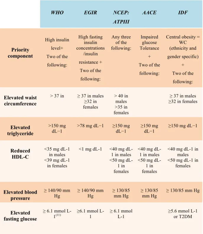
increased consequential risk of CVD. As detailed in Table 1, each of these groups has
developed their own (granted, typically overlapping) criteria for defining MetS, with regard to
component’s priority and thresholds for each of the variables of MetS
(10)in order to
accurately identify people at a higher than average risk of developing CVDs.
12
Table 1. Criteria set out for the diagnosis of MetS according to a number of influential associations
WHO
EGIR
NCEP:
ATPIII
AACE
IDF
Priority
component
High insulin level+ Two of the following: High fasting insulin concentrations /insulin resistance + Two of the following: Any three of the following: Impaired glucose Tolerance + Two of the following: Central obesity = WC (ethnicity and gender specific) + Two of the following:Elevated waist
circumference
> 37 in ≥ 37 in males ≥32 in females > 40 in males >35 in females ≥ 37 in males ≥32 in femalesElevated
triglyceride
>150 mg dL−1 >78 mg dL−1 ≥150 mg dL−1 ≥150 mg dL−1 ≥150 mg dL−1Reduced
HDL-C
<35 mg dL-1 in males <39 mg dL-1 in females <1 mg dL-1 <40 mg dL-1 in males <50 mg dL-1 in females <40 mg dL-1 in males <50 mg dL-1 in females <40 mg dL-1 in males <50 mg dL-1 in femalesElevated blood
pressure
≥ 140/90 mm Hg ≥ 140/90 mm Hg ≥ 130/85 mm Hg ≥ 130/85 mm Hg ≥ 130/85 mm HgElevated
fasting glucose
≥ 6.1 mmol L-1(11) ≥6.1 mmol L-1 ≥ 6.1 mmol L-1 ≥5.6 mmol L-1 or T2DM13
b) Metabolic syndrome (MetS) criteria
In addition to the criteria proposed by the different organizations (Table 1), other additional
factors such as pro-inflammatory (high levels of C-reactive protein (CRP)) and prothrombic
(enhanced coagulation, platelet dysfunction) states, physical inactivity, smoking, and aging
characterize MetS. All of which are considered underlying, major, emerging risk factors for
CVD
(12). However, only five components are used for defining metabolic subjects. Therefore,
MetS is defined as a condition where an individual presents at least 3 of the following criteria
(i) abdominal obesity (precisely waist circumference), (ii) increased fasting glucose, (iii) high
blood pressure, (iv) increased plasma triglyceride, and (v) reduced high-density lipoprotein
(HDL) cholesterol concentrations
(13)(Figure 1).
Obesity: The prevalence of MetS is directly correlated with the development of obesity
(14).
Evidence refers that MetS starts with excess central adiposity
(15). It is mostly believed that it
is a result of excess triglycerides in adipose tissue
(16), which is a result of a major nutrient
consumption than that demanded for normal metabolism
(17). A person is considered obese
when the body mass index is higher than 30 kg/m
2(18).
Abdominal obesity is the strongest form associated with MetS. It is expressed clinically as
increased waist circumference
(12). Increased abdominal fat mass also known as
“apple-shaped” model showed to be directly linked to MetS development. More than that,
intra-abdominal fat is well known nowadays as an active endocrine organ responsible for secreting
a wide spectrum of cytokines like interleukins (ILs), tumor necrosis factor alpha (TNF-α), all
of which that act on increasing metabolic disorders
(19). Interestingly, obesity is considered in
all MetS definitions, for example, most obese people have postprandial hyperinsulinemia and
relatively low insulin sensitivity
(20).
Hyperglycemia: Most persons with the MetS have high levels of plasma glucose
(21). The
primary cause of hyperglycemia in patients with MetS is insulin resistance
(22), characterized
by high plasma insulin concentration that is unable to decrease plasma glucose
(23). The
factors participating to insulin resistance are complex, however, high concentrations of free
fatty acid represents a key event. This also explains the interaction between different MetS
criteria
(19). Moreover, it is considered as the combining mechanism underlying the clustering
of metabolic abnormalities
(24), and majority of persons with MetS exhibit insulin resistance
(23). For this, ATP III considers insulin resistance as one of the MetS components
(12).
Dyslipidemia: Dyslipidemia is characterized by high levels of triglycerides
(19)and low
concentrations of high density lipoprotein (HDL) cholesterol
(25). Also, MetS cases are
correlated with atherogenic dyslipidemia. High levels of low density lipoprotein (LDL)
15
Recently, new elements emerged as effectors in metabolic and cardiovascular pathologies.
Among these are the microparticles (MPs).
B- M
ICROPARTICLES
Cell-cell cross-talk is an essential element of development, homeostasis maintenance and cell
defense. Far from the classical signaling through cell-cell contact and soluble factors,
intracellular signaling via extracellular vesicles (EVs) emerges as a mode of communication
with an ability to deliver various messages. EVs comprise exosomes, MPs and apoptotic
body.
(29). Among these types of EVs, only the effects of MPs on MetS have been described in
the literature.
a) Definition of MPs
MPs are small membrane vesicles, heterogeneous in size with less than 1 µm in diameter,
released from cells in response to activation or apoptosis
(30). These vesicles are produced
from the blebbing of the plasma membrane
(31).
MPs differ from exosomes and apoptotic bodies by the mechanism implicated in their
formation and by their size and composition. In fact, exosomes are assumed to be a
homogenous population of vesicles of 0.02-0.1 µm size. They are formed within the
endosome by invagination of the limiting membranes, resulting in the formation of
multivesicular bodies. Subsequently, multivesicular bodies fuse with the plasma membrane
and release exosomes into the extracellular environment
(32).Apoptotic bodies are large
vesicles (0.5 - 4 µm), formed during programmed cell death following cell fragmentation.
These vesicles have a high content of cytoplasmic and nuclear elements (proteins and nucleic
acids)
(33)(Fig 2).
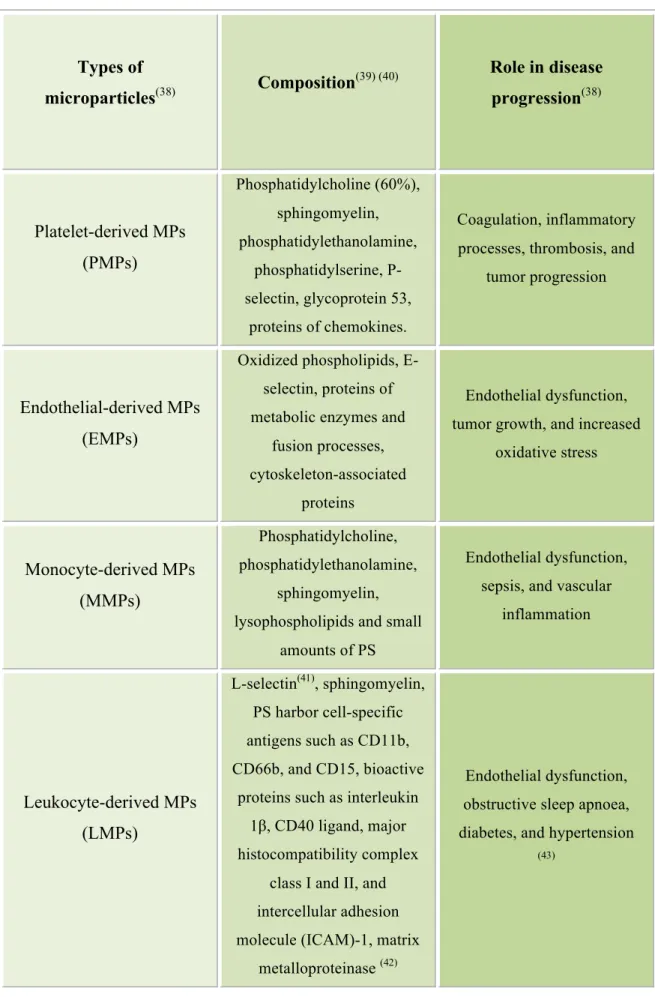
17
b) Characteristics of MPs
1. Structure, origine and types
MPs emerging from the cell share the same cytoplasm, nucleic acid, and cytoskeleton residues
that the mother cell. They are surrounded by a phospholipid bilayer associated with proteins
such as receptors, ligands, selectins and integrins. As a consequence of cell activation or
apoptosis, which result in MP formation, the negatively charged phospholipids such as the
phosphatidylserine (PS) becomes in the outer leaflet of the membrane, resulting in MPs
baring PS in their membranes
(35).
Based on their specific surface proteins, it was found that MPs are released from cells of
vascular wall (such as endothelial cells and smooth muscle cells) or from circulating cells
(such as platelets, erythrocytes, T and B cells, monocytes) and tumor cells
(36). However,
platelet-derived MPs (PMPs) represent ~70-90% of circulating MPs
(30). In addition, MPs can
be found in various biofluids (blood, urine, ...)
(37).
18
Table 2. Types of microparticles, composition, and role in disease progression.
Types of
microparticles
(38)Composition
(39) (40)Role in disease
progression
(38)Platelet-derived MPs
(PMPs)
Phosphatidylcholine (60%), sphingomyelin, phosphatidylethanolamine, phosphatidylserine, P-selectin, glycoprotein 53, proteins of chemokines. Coagulation, inflammatory processes, thrombosis, andtumor progression
Endothelial-derived MPs
(EMPs)
Oxidized phospholipids, E-selectin, proteins of metabolic enzymes and
fusion processes, cytoskeleton-associated
proteins
Endothelial dysfunction, tumor growth, and increased
oxidative stress
Monocyte-derived MPs
(MMPs)
Phosphatidylcholine, phosphatidylethanolamine, sphingomyelin, lysophospholipids and smallamounts of PS
Endothelial dysfunction, sepsis, and vascular
inflammation
Leukocyte-derived MPs
(LMPs)
L-selectin(41), sphingomyelin, PS harbor cell-specific antigens such as CD11b, CD66b, and CD15, bioactiveproteins such as interleukin 1β, CD40 ligand, major histocompatibility complex
class I and II, and intercellular adhesion molecule (ICAM)-1, matrix
metalloproteinase (42)
Endothelial dysfunction, obstructive sleep apnoea, diabetes, and hypertension
20
(protein kinase C, tyrosine protein kinases) and phosphatase inhibition, resulting eventually in
cytoskeleton blebbing
(48). During apoptosis, membrane blebbing and MP formation results
from different signaling apoptotic pathways such as ROCK1 and caspase 3, and the tumor
necrosis factor (TNF)-related apoptosis inducing ligand (TRAIL) and its receptor TRAIL-R2.
Moreover, their quantity can vary based on their mode of formation (Fig 4)
(49). Moreover,
oxidative stress and inflammation are possible key factors in promoting MP generation
(50).
4. Mechanisms of MP interaction with target cells
MPs can communicate with target cells by different mechanisms. 1- By interacting with the
ligands present on the surface of target cells, MPs activate cascade signaling. For example, we
have previously shown that MPs isolated from MetS patients baring Fas ligand interacts with
Fas receptor present on vascular smooth muscle cells causing vascular hyporeactivity
(51); 2-
MPs can transfer part of their components without fusion with target cells. Indeed, it has been
shown that the chemokine receptor CCR5 was transferred to mononuclear CCR5
-cells after
their incubation with MPs baring the concerned receptor
(52); 3- MPs can transfer proteins,
mRNA, miRNA, and bioactive lipids by fusing with target cells. For example, the fusion of
MPs derived from mature macrophages with the monocyte plasma membrane permits their
differentiation into macrophages through the transfer of miR-223 carried by the MPs
(53); 4-
Internalization of MPs within the target cells. We have shown that internalization of
lymphocytic T-MPs protects endothelial cells from apoptosis, partially by decreasing
cytoplasmic reactive oxygen species (ROS) and by transferring antioxidant enzymes such as
the superoxide dismutase
(54)(Fig 4).
22
c) Role of MetS in the liberation of MPs
Previous studies have shown an increase in the levels of circulating MPs in MetS subjects in
comparison to healthy ones. Indeed, levels of MPs from pro-coagulant (Annexin V
+),
platelets, erythrocytes and endothelial cells are increased in individuals with MetS versus
healthy subjects
(56).
Arteaga and colleagues
(57)have reported marked elevation of EMPs
levels in patients with MetS compared to controls. Also, it has been shown that the
postprandial state after a high-fat meal induces the release of EMPs and that the magnitude of
postprandial triglyceridemia correlates with the postprandial increase in circulating EMP
levels
(58). In addition, Chironi and colleagues
(59)have shown that, in patients with MetS,
leukocyte-derived MP levels are higher than that in those free of such syndrome. Moreover, in
the overall study population, leukocyte-derived MP levels increase gradually in parallel with
the number of components of MetS. Finally, Helal et al.
(50), revealed the association of MPs
with the components of the MetS, and their deleterious impact on atherothrombosis. Indeed,
the relationship between MP subpopulations and hyperglycemia, hypertension, the extent of
lipidemia and the level of HDL cholesterol have been reported mainly in advanced stages of
cardiovascular disorders. Therefore, it could be speculated that MPs are possible biomarkers
for the identification of those subjects with MetS and an elevated risk of developing
cardiovascular complications
(50).
C- C
ARDIOVASCULAR
DISEASES
AS
CONSEQUENCES OF
M
ET
S
a) Epidemiology
Cardiovascular pathologies comprise stroke, arterial fibrillation, sudden cardiac arrest,
coronary heart disease, and heart failure
(60). All of which remain the major cause of death
among Europeans and worldwide (29.6% of all deaths), and despite recent decrease in
mortality rates in many countries, it is still responsible for over 4 million deaths per year,
close to half of all deaths in Europe. More than all communicable, maternal, neonatal and
nutritional disorders double the number of deaths caused by cancers. Referring to an
epidemiological overview done on 2014, which based on a number of European and
international data sources, CVDs continue to cause a much greater mortality burden among
Europeans than any other disease. Overall, CVDs caused 51% of deaths among women and
23
international data sources, CVDs continue to cause a much greater mortality burden among
Europeans than any other disease. Overall, CVDs caused 51% of deaths among women and
42% among men in the last year of data, compared with 19 and 23%, respectively, for all
cancers
(61). A statistical update done by the American Heart Association in 2015 showed that
CVDs prevalence was higher in children and young adults, in comparison to middle-aged and
older adults. These risk factors were referred to the poor lifestyle behaviors, lifestyle-related
risk factors (tobacco, high fat diets, ...) and physical inactivity
(60).
b) CVDs and MetS
MetS is a major cause of CVDs. Several studies have shown that the risk of CVDs is
correlated with the number of MetS components, as the risk increases with the increase in the
number of MetS components
(19). Also, patients with coronary heart disease or those who have
experienced a stroke usually display more than one major metabolic criterion
(60). This is
agreement with the fact that the rate of ischemic stroke is higher in MetS patients compared to
non-metabolic ones
(62). Moreover, the Third National Health and Nutrition Examination
Survey has reported that the increased risk of myocardial infraction is correlated with the
presence of MetS
(63). Lately, it has been estimated that increased vasoconstrictive substances
and increased prothrombotic adipokines takes charge of increased CVD incidence, both of
which are associated with MetS
(23). Also, the assembly of hypertension and obesity is also
known to cause higher rate of morbidity and mortality associated with CVDs
(64). The
atherogenic profile which consists of increased triglycerides and LDL, and decreased HDL,
increases the risk of CVDs. Furthermore, diabetic hyperlipidemia or hyperglycemia
accelerates atherogenesis
(65). Finally, components of MetS may be involved, directly or
indirectly, in endothelial dysfunction which is associated with atherosclerosis initiation and
progression
(66), a marker of cardiovascular outcomes
(67).
c) Endothelial dysfunction
Several epidemiological studies have reported that endothelial dysfunction is associated with
cardiovascular risk factors. Endothelial dysfunction is a systemic phenomenon associated with
vascular inflammation, lipid deposition, thrombosis
(68)and represents an early step in the
development of atherosclerosis, a potential lead to CVD development
(67). Therefore,
endothelial dysfunction represents a primary disturbance in cardiovascular events.
24
1. Definition
Endothelial dysfunction is used to describe abnormal function and alterations of the
endothelium, shifting its function toward vasoconstriction instead of vasodilation,
pro-inflammatory state and changes in its prothrombotic and proliferative properties
(69). In other
words, endothelial dysfunction results from the imbalance between vasodilatory substances
such as nitric oxide (NO), vasoconstrictive ones like endothelin-1 and prothrombotic factors
like the plasminogen activator inhibitor-1
(23). Thus, during endothelial dysfunction, a
decrease in NO bioavailability
(68)an enhancement of ROS liberation, activation of
inflammation and alteration of barrier function can be observed
(70).
2. Endothelial dysfunction and MetS
Individuals with MetS exhibit higher degree of endothelial dysfunction
(4). Each
component of the MetS has been reported to impair endothelial function, and its severity was
associated with the number of those components
(15). Ahirwar et al
(23)showed that NO levels
in the plasma were lower in MetS patients compared to control ones, reflecting endothelial
dysfunction in MetS. Visceral fat tissue drives proatherogenic adipokines production, which
in turn contributes in increased oxidative stress and chronic inflammation, both of which
affect endothelial function
(15). More precisely, the association of increased visceral obesity
and other metabolic perturbations impaired NO biodisposability, causing endothelial
dysfunction
(4). Decreased NO bioavailabilty due to elevated oxygen species release, may be
the major reason of endothelial dysfunction in obesity
(15). On the one hand, insulin resistance
may also impair NO bioavailability
(4). Insulin signaling
is important to endothelial cells as it
stimulates the production of NO from the endothelium leading to vasodilation. In cases of
insulin resistance, the vasoconstrictor endothelin-1 is released from vascular endothelium.
This imbalance between vasoconstrictor and vasodilator actions of insulin, under insulin
resistance conditions, represents an important factor in the vascular pathophysiology of
insulin resistance and endothelial dysfunction
(71). On the other hand, the PI3K/Akt cascade of
the insulin signaling pathway is decreased in insulin resistance leading to endothelial damage
(72). This
emerges insulin resistance as a potential link between MetS and endothelial
dysfunction
(15).
It has been also demonstrated that some metabolic components cause
endothelial dysfunction by boosting several hormones, inflammatory cytokines and molecules
(4). Furthermore, elevated high sensitive CRP levels, which are a feature of MetS, are
associated with reduced basal and stimulated NO release from endothelial cells through
various mechanisms such as insulin resistance. Moreover, high leukocyte count may
25
contribute to endothelial dysfunction, through inflammatory cytokines and cytotoxic products
secretion
(15). Also, it has been shown that hypertension is associated with decreased
endothelial-dependent relaxation and reduced endothelial NO synthase (eNOS)
phosphorylation
(73).
3. MPs: players in endothelial dysfunction
Due to their localization in the blood stream, circulating MPs have a major role in interaction
with circulating cells or components of the vessel wall including the endothelium, considered
as the primary target for cardiovascular risk factor
(31). Thus, increased levels of circulating
MPs released from platelets, leukocytes, erythrocytes and endothelial cells, have been
described in diseases associated with cardiovascular complications
(50). Moreover, EMPs bear
molecules able to initiate coagulation, induce monocyte adhesion, activate neutrophils, and
affect vasodilation, antithrombotic and antiadhesive properties of the vascular wall
(31). For all
these reasons, MPs can be largely related to pathogenesis of various CVDs mainly launched
by endothelial dysfunction
(38). Interestingly, increased number of circulating EMPs is found
in cases of endothelial cell damage and dysfunction. In addition, EMPs can aggravate
endothelial dysfunction. Indeed, EMPs impair endothelium-dependent relaxation and NO
release in rat aorta through superoxide anion production
(74). Apoptotic endothelial MPs
originating from damaged endothelial cells are considered as markers of endothelial cell
injury and vascular aging
(75). In addition, circulating MPs, rich in endothelial and platelet
surface markers, from patients with acute myocardial infarction cause severe endothelial
dysfunction in healthy blood vessels by affecting the endothelial NO transduction pathway,
leading to decreased relaxations to acetylcholine in rat aortic rings
(76).
More than that, in vitro apoptotic T lymphocyte-derived MPs, at concentrations that can be
reached in circulating blood in pathological disorders, impair endothelium-dependent
relaxation in conductance and small resistance arteries in response to agonists and shear
pressure, respectively. In addition, these T lymphocyte-derived MPs affect vascular
contraction in mouse aorta by acting directly on smooth muscle cells
(77). Finally, these MPs
induce endothelial dysfunction similar to that of circulating MPs, where they decreased NO
production and increased ROS production by a mechanism depending on xanthine oxidase
activity in endothelial cells. These effects were associated with a reduction of eNOS activity
(Fig 5)
(78). A possible mechanism through which these MPs interact with endothelial cells is
through low-density lipoprotein receptor (LDLR)-mediated endocytosis
(79).
27
sphingomyelinase (NSMase) plays a critical role in the development of cardiac failure,
atherosclerosis, and decline in vasomotion
(80). ROS production is one mechanism through
which NSMases affects the cardiovascular system. NSMase activates hypoxia-induced
phosphorylation of NADPH oxidase (p47
phox) and ROS formation in vascular smooth muscle
cells
(81); with ROS production playing an important role in the development of CVDs
(82).
Activation of NSMase in arteries from aging rats results in a decrease in eNOS
phosphorylation and activation, leading to a loss of vasomotor function
(83). Furthermore,
supplementation of isolated aortic rings with lipoic acid reversed age-related loss of
endothelial glutathione, leading to reduced NSMase activation and ceramide level in the
endothelium and improved endothelial NO-dependent vasomotor function
(84).
D- E
NDOPLASMIC
R
ETICULUM
S
TRESS
a) Endoplasmic reticulum (ER) histology and function
The endoplasmic reticulum (ER) is a continuous membrane network in the cytosol
(85). In
eukaryotic cells, the ER exhibits four major physiological functions: (i) it is the site of the
synthesis of membrane and secretory proteins; (ii) it is the place of membrane and secretory
protein folding into their native conformation, where these nascent proteins undergo
post-translational modifications;(iii) the ER is the main site of Ca
2+storage which is involved in
intracellular cascade signaling; and (iv) the ER membrane is implicated in the biosynthesis of
lipids and sterols
(86). Normal ER functioning can be disrupted when the stream of nascent,
unfolded polypeptide chain surpasses the folding capacity of the ER
(87).
b) ER stress: the unfolded protein response
More than that the disruption of any ER processes, increased oxidative stress, or the activation
of SMases through the Fas receptor engagement
(88), cause ER stress and the subsequent ER
stress response termed the unfolded protein response (UPR). The UPR expresses the
ER-to-nucleus signaling cascades, to reestablish ER homeostasis. Homeostasis is restored by
increasing molecular chaperones expression and ER foldases, or by stimulating phospholipid
synthesis and ER-associated protein degradation (ERAD) and autophagy, selectively
degrading mRNAs encoding secretory proteins, activating an antioxidant response, and
attenuating general translation and transcription of genes encoding secretory proteins. Also,
the UPR activates inflammatory and apoptotic signaling pathways
(89).
28
The UPR is initiated upon the recognition of unfolded proteins by chaperones, which are
normally involved in every aspect of ER quality control
(90). One of the best characterized ER
chaperone proteins is the glucose regulated protein (GRP78) commonly referred to as BIP, the
immunoglobulin heavy chain-binding protein
(91). BIP belongs to Hsp70 family ATPase
involved in numerous functions, including translocating nascent polypeptides, facilitating de
novo protein folding and assembly, targeting misfolded proteins to ERAD machinery, and
maintaining Ca
2+homeostasis
(90). BIP binds transiently to newly-synthesized proteins in the
ER and more permanently to misfolded, underglycosylated or unassembled proteins avoiding
their transport from the ER
(92). The prolonged interaction of the folding protein with the
chaperone machinery
(87), sequesters BIP away from the ER luminal domain of the three ER
resident transmembrane proteins: the inositol requiring enzyme 1 (IRE1), and double-stranded
RNA-activated protein kinase (PKR)-like endoplasmic reticulum kinase (PERK), the
activating transcription factor 6α (ATF6α),leading to their activation, where normally they are
kept in an inactive form through their interaction with ER luminal chaperones
(93).
Inositol requiring enzyme 1 (IRE1): is a bifunctional enzyme with serine/ threonine protein
kinase and endoribonuclease (RNase) activity in its cytosolic domain. It is also a type I
transmembrane protein that senses ER stress by its N-terminal luminal domain. Release from
suppression by GRP78 triggers its homodimerization and autophosphorylation as part of the
activation process. Activated IRE1 cleaves a 26-base fragment from the mRNA encoding X
boxbinding protein 1 (XBP1), resulting in spliced XBP1s and translation of a potent
transcription factor controlling the expression of genes involved in ERAD and protein folding,
as well as other genes implicated in directing the synthesis of phospholipids that are required
for the expansion of ER membranes during ER stress
(94).
PKR-like ER kinase (PERK): is a type I ER transmembrane kinase. When activated by ER
stress, PERK homodimerizes and autophosphorylates and then directly phosphorylates Ser51
on the alpha subunit of eukaryotic initiation factor 2 (eIF2α). Phosphorylated eIF2α prevents
the formation of ribosomal initiation complexes leading to global mRNA translational
attenuation
(95). Thereby decreasing protein influx to the ER in support of resolving the
cytotoxic threat from accumulated misfolded proteins
(96).
On the other hand, some mRNAs
require eIF2α phosphorylation for translation such as the mRNA encoding activating
transcription factor 4 (ATF4). ATF4 is a b-ZIP transcription factor that regulates several UPR
target genes including those involved in ER stress mediated apoptosis such as C/EBP
homologous protein (CHOP)
(95).
30
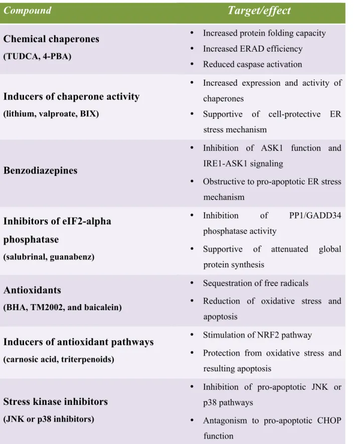
c) Endoplasmic reticulum stress: its role in diseases and a novel target for
therapy
A large number of human diseases has been associated with ER protein-folding defects and
ER stress/UPR (see Table 3) and therefore is being recognized as an emerging target for
therapy. For that different therapeutic purposes including chemical compounds have been
proposed in order to alleviate ER stress (see Table 4)
(97)Table 3. Human diseases linked to ER stress
Disease
Linkage to ER stress
Cancer
• Tumor-specific
microenvironment
activates ER stress
• Cancer cells display chronic display of ER
stress markers
• Knockdown of GRP78 or of CHOP
affects chemosensitivity
Parkinson’s disease
• Parkin expression impacts ER stress
• ATF4 leads to increase in parkin
expression
Atherosclerosis
• Oxidized lipids induce ER stress
• Hyperhomocysteinemia induces ER stress
• Cholesterol loading induces ER
stress-mediated cell death
• Reduced plaque necrosis in mice lacking
CHOP
Heart disease
• ER stress contributes to cardiomyocyte
apoptosis
• Activation of ER stress in infarcted mouse
heart
• GRP78 and GRP94 protect against
ischemic injury
HBV and HCV infection
• HBV induces GRP78 and GRP94
• HCV suppresses IRE1/XBP1 pathway
Type 2 Diabetes
• Obesity induces ER stress
• Hyperlipidemia
and
hyperglycemia
induce ER stress
• Free fatty acids (palmitate) induce beta
cell apoptosis
• Deletion of CHOP improves beta cell
function and survival
31
Table 4. Compounds with potency to ameliorate ER stress.
Compound
Target/effect
Chemical chaperones
(TUDCA, 4-PBA)
• Increased protein folding capacity
• Increased ERAD efficiency
• Reduced caspase activation
Inducers of chaperone activity
(lithium, valproate, BIX)
• Increased expression and activity of
chaperones
• Supportive of cell-protective ER
stress mechanism
Benzodiazepines
• Inhibition of ASK1 function and
IRE1-ASK1 signaling
• Obstructive to pro-apoptotic ER stress
mechanism
Inhibitors of eIF2-alpha
phosphatase
(salubrinal, guanabenz)
• Inhibition
of
PP1/GADD34
phosphatase activity
• Supportive of attenuated global
protein synthesis
Antioxidants
(BHA, TM2002, and baicalein)
• Sequestration of free radicals
• Reduction of oxidative stress and
apoptosis
Inducers of antioxidant pathways
(carnosic acid, triterpenoids)
• Stimulation of NRF2 pathway
• Protection from oxidative stress and
resulting apoptosis
Stress kinase inhibitors
(JNK or p38 inhibitors)
• Inhibition of pro-apoptotic JNK or
p38 pathways
• Antagonism to pro-apoptotic CHOP
function
32
d) ER stress is involved in MetS and endothelial dysfunction
Studies on obese rodents show that UPR markers, such as PERK, eIF2α, and BIP are
overexpressed in their livers, and adipose tissues, compared to control ones
(98). Investigations
made on obese persons presented the activation of ATF6, eIF2α, IRE1 and XBP1, markers of
stress
(99). Also, the induction of the ER stress and the UPR is associated with the
dysregulation of lipid and lipoprotein metabolism in MetS
(100). These data reveals the
implication of ER stress in MetS through different mechanisms. For example, increased levels
of cholesterol and saturated fatty acids present in obese individuals decrease the fluidity of ER
membrane, leading to the inhibition of the SERCA Ca
2+-ATPases, depletion of ER luminal
Ca
2+stores, inhibition of ER-resident molecular chaperones, and the accumulation of
unfolded proteins in the ER
(101). From the other side, experiments show the involvement of
ER stress in endothelial dysfunction. Endothelial cells from athero-susceptible regions in
normal swine aorta exhibited elevated UPR components, ATF6, IRE1, and XBP1
(102). This
effect entangles different mechanisms. Kassan et al
(73)showed an improvement in the
endothelium-dependent relaxation in the aortas of angiotensin II- induced hypertensive mice
upon their treatment with tauroursodeoxycholic acid (Tudca) or 4-phenylbutyrate
(4-
PBA),
ER stress inhibitors (mentioned in Table 4), increased eNOS phosphorylation demonstrating
that ER stress is an important factor in vascular endothelial dysfunction in hypertension.
Another study targeting ER stress-induced endothelial dysfunction in bovine aortic
endothelial cells, showed that oxidized and glycated LDL induce oxidative stress in
endothelial cells, which in turn induces the UPR through a mechanism involving disturbed ER
Ca
2+metabolism, resulting in endothelial dysfunction
(103). Galán et al
(104)showed that ER
stress activation by tunicamycin, led to vascular endothelial dysfunction through p38 MAPK
dependent mechanism, reduced ERK1/2 phosphorylation and oxidative stress in endothelial
cells from coronary arteries.
e) ER stress partners in crime: mitochondria and oxidative stress
1. Mitochondria histology and function
Mitochondria are double-membraned intracellular organelles that are responsible of energy
production for vital metabolic reactions and cellular homeostasis
(105). Mitochondria are also
implicated in main cellular functions such as Ca
2+homeostasis, heme-biosynthesis, steroid
hormone biosynthesis, nutrient metabolism, ammonia removal, and activation of signaling
pathways
(106)involved in innate immunity, autophagy and cell death
(107), ROS and NO
33
production
(108). Indispensable for this crucial role of mitochondria is the presence of the
mitochondrial respiratory chain and the oxidative phosphorylation (OXPHOS) that transduce
electron transport into energy generation in the form of ATP upon ADP phosphorylation
(105).
However, the primary factor that initiates the dysfunction of mitochondria has been proposed
to be the defects in OXPHOS which can further enhance the production of ROS
(109).
2. Mitochondria and mitochondrial reactive oxygen species as mediators of
metabolic alterations and endothelial dysfunction
Endothelial cells have low mitochondria content compared to cardiomyocytes and
hepatocytes; however, mitochondrial dynamics act as a pivotal orchestrator of endothelial cell
homeostasis under normal conditions. Damage of mitochondrial dynamics and biogenesis
participates in endothelial dysfunction and diverse vascular diseases
(110). In endothelial cells,
where anaerobic glycolysis covers the majority of the cytosolic energy demand, mitochondrial
functions are shifted towards signaling phenomena rather than ATP production
(111). Similarly,
the intracellular distribution of mitochondria differs depending on the type of endothelial cell
and reflects their important signaling role in the regulation of cell-specific processes such as
ROS-dependent gene expression or modulation of local Ca
2+concentrations and signaling
(112), and mostly its communication with other cellular organelle ,especially the ER
(113).
Furthermore, mitochondrial morphology is dynamic and sensitive to metabolic alterations
(114). Metabolic status can dramatically affect the form and function of mitochondria, which
consequently influences the organ function
(115). Mitochondrial fragmentation and the
subsequent loss of mitochondrial networks have been described in endothelial cells from
diabetic patients as well as in cultured human aortic endothelial cells exposed to high glucose
concentration
(116). Altered mitochondrial membrane potential is an important factor that
triggers excess mitochondrial ROS (mROS) production in the setting of risk factors, including
aging, hypercholesterolemia, hyperglycemia, smoking, and hypoxia. Different sources of
mROS have been identified in endothelial cells one such example are complexes I and III of
the mitochondrial respiratory chain
(117).
Clinical investigations suggest that many vascular diseases are accompanied with elevated
mROS levels. One typical mechanism by which mROS participates in endothelial dysfunction
and subsequent vascular diseases is by uncoupling the mitochondrial eNOS (Fig 7). As a
consequence of eNOS uncoupling, NO production is reduced and the pre-existing oxidative
stress is enhanced, which contribute significantly to endothelial dysfunction and vascular
diseases
(118).
36
Furthermore, recent data showed that during ER stress, the PERK signaling pathway regulates
mitochondrial function
(126). PERK has been proposed to interact with mitochondrial Mfn2,
indicating its role in stabilizing ER-mitochondrial contacts
(127). Consistent with this, genetic
depletion of PERK disturbs ER morphology and reduces the number of ER-mitochondrial
contacts
(128). PERK deficient cells show defects in regulating electron transport chain activity
reflected by increased basal and maximal respiration
(127). Moreover, PERK signaling
regulates mitochondrial proteostasis and function during ER stress that can lead to
mitochondrial dysfunction and promote cellular pathology. In addition, rapid induction of ER
stress response with tunicamycin induces changes in MAM indicating that ER stress regulates
the dialogue with mitochondria
(126). In fact, mitochondrial dysfunction has been directly
linked to the ER stress response in liver
(129). Finally, Lu et al
(130)have found that high-fat diet
rich in saturated fatty acid palmitate induces swollen mitochondria and extends ER in
endothelial cells of mouse aorta.
4. ER and oxidative stress dialogue
Increased ROS production is associated with the overexpression of ER stress markers, the
perturbations in the ER-mitochondria cross talk and endothelial dysfunction. This was
demonstrated by He X et al
(131)in endothelial cells exposed to hypoxia/ reoxygenation injury.
Moreover, in the case of hypertension, ER stress induction along with increased oxidative
stress and decreased antioxidant activity, were beyond endothelial dysfunction occurrence in
methionine-enriched rats
(132). Moreover, in hypertension, ER stress was found to induce
endothelial dysfunction through increasing NADPH oxidase activity
(104). On the other hand,
Li and colleagues
(133)found that high concentrations of uric acid, being a major determinant
of total antioxidant capacity of plasma, induced intracellular ROS accumulation, likely
triggering ER stress, all of which led to decreased eNOS activity and endothelial dysfunction
induction.
These data illustrate the collaborating role of ER, mitochondria and oxidative stress in
inducing endothelial dysfunction in metabolic disorders.
37
A
IM OF THE STUDY
In this work, we aimed to study the mechanism through which the two types of MPs, MPs
obtained by in vitro apoptotic treatment of T lymphocytes and MPs from MetS patients,
induce endothelial dysfunction. Referring to the literature, we proposed the ER stress as a
possible candidate associated with such model. For this, we tracked the UPR pathways and
studied the evolution of its components upon the treatment of human aortic endothelial cells
with MPs in the presence or absence of the ER stress inhibitor Tudca. Furthermore, we issued
to evaluate the implication of oxidative stress in our studies. For that, we checked the
production of cytosolic and mitochondrial ROS at different times. Moreover, several
inhibitors of ROS sources (inhibitors of NADPH oxidase, xanthine oxidase, mitochondrial
complex I inhibitor, and eNOS inhibitor) and the mitochondrial ROS scavenger, were used to
identify ROS origin. Furthermore, we attempted to see the possible perturbations occurring in
the mitochondria and its possible participation in decreasing NO bioavailability and therefore
inducing endothelial dysfunction. Besides, we tested the expression of the two ingredients
Mfn2 and VDAC1of the ER and mitochondrial interactions, the MAMs. In addition, we
pursued to figure out the probable interactions occurring between MPs and endothelial cells.
Thus, we investigated the engagement of Fas/FasL and LDL receptor in our model by
neutralizing the Fas L present on MPs from one side and LDL receptor found on endothelial
cells. To finalize the pathway, we tried to realize the influenced piece of Fas and LDL
receptors activation. For that purpose, we examined neutral SMase implication through
targeting neutral SMase expression by siRNA and checking its consequence effect.
38
C
O
N
FID
EN
TIA
L.
Fo
r Pe
er
R
ev
ie
w
O
nly
Safiedeen 1ORIGINAL RESEARCH COMMUNICATION
Temporal cross-talk between endoplasmic reticulum and mitochondria regulates oxidative stress and mediates microparticle-induced endothelial dysfunction
Zainab Safiedeen1,2, Isabel Rodríguez-Gómez1, Luisa Vergori1, Raffaella Soleti1, Dayannath Vaithilingam1, Imene Douma1, Abdelali Agouni3, Denis Leiber1, Séverine Dubois1,4, Gilles Simard1,4, Kazem Zibara2,5, Ramaroson Andriantsitohaina1,4, and M. Carmen Martínez1,4
1
INSERM U1063, Stress Oxydant et Pathologies Métaboliques, Université d’Angers; Angers, France
2ER045, Laboratory of Stem Cells, PRASE, DSST, Lebanese University, Beirut, Lebanon, 3
University of Surrey, Faculty of Health and Medical Sciences, Guildford, United Kingdom
4
Centre Hospitalo-Universitaire d’Angers, Angers, France
5
Biology Department, Faculty of Sciences-I, Lebanese University, Beirut, Lebanon
Present address of A. Agouni: Qatar University, College of Pharmacy, Doha, Qatar.
Corresponding author: M.C. Martinez, INSERM U1063, Stress Oxydant et Pathologies
Métaboliques, Institut de Biologie en Santé, 4 rue Larrey, F-49933 Angers, France. Phone: +33 2 44 68 85 79. E-mail: [email protected]
Abbreviated title: Unfolded protein response/microvesicles Word count: 5998
Reference numbers: 34 Greyscale illustrations: 2 Color illustrations: 6 (online)
Page 1 of 47
Please destroy all records after use for peer review. Mary Ann Liebert Inc., 140 Huguenot Street, New Rochelle, NY 10801 Antioxidants & Redox Signaling
1 2 3 4 5 6 7 8 9 10 11 12 13 14 15 16 17 18 19 20 21 22 23 24 25 26 27 28 29 30 31 32 33 34 35 36 37 38 39 40 41 42 43 44 45 46 47 48 49 50 51 52 53 54 55 56 57 58 59 60