HAL Id: inserm-02483109
https://www.hal.inserm.fr/inserm-02483109
Submitted on 18 Feb 2020
HAL is a multi-disciplinary open access
archive for the deposit and dissemination of
sci-entific research documents, whether they are
pub-lished or not. The documents may come from
teaching and research institutions in France or
abroad, or from public or private research centers.
L’archive ouverte pluridisciplinaire HAL, est
destinée au dépôt et à la diffusion de documents
scientifiques de niveau recherche, publiés ou non,
émanant des établissements d’enseignement et de
recherche français ou étrangers, des laboratoires
publics ou privés.
inflammatory endothelial microvesicles that act as
autocrine effectors of endothelial dysfunction
Isaac Maximiliano Bugueno, Fatiha Zobairi El-Ghazouani, Fareeha Batool,
Hanine El Itawi, Eduardo Anglés-Cano, Nadia Benkirane-Jessel, Florence
Toti, Olivier Huck
To cite this version:
Isaac Maximiliano Bugueno, Fatiha Zobairi El-Ghazouani, Fareeha Batool, Hanine El Itawi, Eduardo
Anglés-Cano, et al.. Porphyromonas gingivalis triggers the shedding of inflammatory endothelial
microvesicles that act as autocrine effectors of endothelial dysfunction. Scientific Reports, Nature
Publishing Group, 2020, 10 (1), pp.1778. �10.1038/s41598-020-58374-z�. �inserm-02483109�
Porphyromonas gingivalis triggers
the shedding of inflammatory
endothelial microvesicles that act
as autocrine effectors of endothelial
dysfunction
Isaac Maximiliano Bugueno
1, Fatiha Zobairi el-Ghazouani
1, Fareeha Batool
1, Hanine el itawi
1,
Eduardo Anglès-cano
2, Nadia Benkirane-Jessel
1, Florence toti
1,4& Olivier Huck
1,3*A link between periodontitis and atherothrombosis has been highlighted. The aim of this study was to determine the influence of Porphyromonas gingivalis on endothelial microvesicles (EMVpg) shedding and
their contribution to endothelial inflammation. Endothelial cells (EC) were infected with P. gingivalis (MOI = 100) for 24 h. EMVpg were isolated and their concentration was evaluated by prothrombinase
assay. EMVpg were significantly increased in comparison with EMVctrl shedded by unstimulated
cells. While EMVctrl from untreated EC had no effect, whereas, the proportion of apoptotic EC was
increased by 30 nM EMVpg and viability was decreased down to 25%, a value elicited by P. gingivalis
alone. Moreover, high concentration of EMVpg (30 nM) induced a pro-inflammatory and pro-oxidative
cell response including up-regulation of TNF-α, IL-6 and IL-8 as well as an altered expression of iNOS and eNOS at both mRNA and protein level. An increase of VCAM-1 and ICAM-1 mRNA expression (4.5 folds and 3 folds respectively (p < 0.05 vs untreated) was also observed after EMVpg (30 nM)
stimulation whereas P. gingivalis infection was less effective, suggesting a specific triggering by EMVpg.
Kinasome analysis demonstrated the specific effect induced by EMVpg on main pro-inflammatory
pathways including JNK/AKT and STAT. EMVpg are effective pro-inflammatory effectors that may
have detrimental effect on vascular homeostasis and should be considered as potential autocrine and paracrine effectors involved in the link between periodontitis and atherothrombosis.
Periodontitis is an infectious inflammatory disease associated with soft tissue inflammation and destruction of the tooth-supporting tissues characterized by increased periodontal pocket depth, gingival bleeding and clinical attachment loss. Such disease impairs oral health related quality of life and is considered the main cause of dental mobility and tooth loss1. Periodontitis affects a large proportion of the worldwide population and prevalence of
its severe forms is estimated to be around 11% with a peak incidence around 38 years of age2. The development of
periodontitis is associated with the establishment of a dysbiosis characterized by the predominance of anaerobic species, including Porphyromonas gingivalis (P. gingivalis), and an imbalanced host-immune response inducing periodontal tissue destruction3.
During the last decades, periodontal diseases were associated with various chronic diseases such as diabetes, rheumatoid arthritis, adverse pregnancy outcomes and cardiovascular diseases, suggesting a systemic impact4–6
with an enhanced proportion of worsened cardiovascular outcomes7–9. Indeed, patients with periodontitis are
more prone to endothelial dysfunction, aneurysmal disease progression10, coronary artery narrowing, and an
increased all-cause and cardio-vascular related mortality11,12. Moreover, in a clinical trial, intensive periodontal
1INSERM (French National Institute of Health and Medical Research), UMR 1260, Regenerative Nanomedicine, Fédération de Médecine Translationnelle de Strasbourg (FMTS), Strasbourg, France. 2Université de Paris, Innovative Therapies in Haemostasis, INSERM UMR_S 1140, F-75006, Paris, France. 3Université de Strasbourg, Faculté de Chirurgie-dentaire, 8 rue Sainte-Elisabeth, 67000, Strasbourg, France. 4Faculté de Pharmacie, Université de Strasbourg, 8 rue Sainte-Elisabeth, 67000, Strasbourg, France. *email: o.huck@unistra.fr
treatment of patients with severe periodontitis, improved at 6 months the endothelial dysfunction, a well-known marker of vascular injury7 thereby strengthening the causative link.
Among the proposed biological mechanisms3,5,10,13, the impact of oral and more specifically periodontal
bacteria, on arterial homeostasis14 is suggested by their eventual dissemination in blood flow from the
perio-dontal pocket. Interestingly, they were detected in atheromatous plaques as well as in the wall of healthy ves-sels in patients suffering from mild to severe periodontitis15,16. P. gingivalis, an anaerobic bacteria considered
as a keystone periodontal pathogen, was demonstrated, in vitro17–20 and in vivo21,22 as a pro-inflammatory and
pro-atheromatous mediator. Indeed, P. gingivalis exhibits a large number of virulence factors such as lipopolysac-charide (Pg-LPS), fimbriae or gingipaïns, contributing to the modulation of the innate immune response mainly through activation of Toll-Like-Receptors (TLRs)–dependent pathways1,5,17,18. Nevertheless, infection by P.
gin-givalis enhances endothelial inflammation or cell death in response to either low-density lipoprotein cholesterol
(LDL) or pro-inflammatory cytokines (TNF-α), two circulating mediators associated with elevated cardiovascu-lar risk, thus, highlighting a potential impact in the development of atherothrombosis18. However, the hypothesis
of a detrimental effect solely induced by direct infection remains controversial as clinical trials assessing the preventative effect of antibiotic therapy did not show significant benefit in the secondary prevention of cardi-ovascular acute events in patients with history of myocardial infarction or myocardial ischemia23. Recently, the
CANTOS trial pointed out inflammation as a key driver of atherothrombosis in patients with pro-inflammatory background. However, anti-interleukin-1β (IL-1β) antibodies although improving the cardiovascular outcomes failed to reduce cardiovascular mortality24. The eventual vascular impact of a variety of inflammatory
media-tors acting as autocrine or paracrine cellular effecmedia-tors has been proposed25,26. Amongst them, microvesicles of
endothelial origin (EMV) shed from the inflamed endothelium in response to infection could be the possible missing link between the infection-driven and the pro-thrombotic vascular responses.
Microvesicles (MV), also termed microparticles, are plasma membrane vesicles ranging from 50 nm to 1 µm released from stimulated cells. They contain a variety of active molecules such as lipids, enzymes, receptors and microRNAs. One characteristic feature of MV is that they expose phosphatidylserine (PhtdSer), an anionic phos-pholipid translocated from the inner to the outer leaflet of the plasma membrane. In addition, membrane proteins at the surface of the mother cell allow the characterization of their cell origin in body fluids. Circulating procoag-ulant EMV have been demonstrated as relevant biomarkers of vascular insult of atherothrombotic, inflammatory or mechanical origin, including ischemia reperfusion. Regardless of their cell origin, circulating MV emerge as the new actors of cellular crosstalk acting as procoagulant, pro-inflammatory, apoptotic or senescent pathogenic messengers under pathological conditions. Verily, the initial cellular stress at the origin of the MV shedding appears of relevance in the induction of a specific cell dysfunction27. In the context of infectious disease, the
shedding of CD105+EMV has been proven to have a prognosis value in sepsis-induced coagulopathy28, whereas
circulating MV of platelet and leukocyte origin released upon inflammation favor the recruitment of leukocytes at the surface of the inflamed endothelium29,30.
This study aims to determine in vitro the influence of P. gingivalis infection on EMV shedding (EMVPg) and to
evaluate an eventual autocrine action of EMVPg as noxious effectors possibly contributing to the dissemination of
endothelial cell inflammatory responses and dysfunction.
Materials and Methods
Cell culture.
Human umbilical vein endothelial cells (EC) (HUVEC, PromoCell, Heidelberg, Germany) were cultured in EGM2 medium (Promocell, Heidelberg, Germany) supplemented with 10% Fetal Bovine Serum at 37 °C in a humidified atmosphere with 5% CO2. Culture medium was changed each 3 days and no antibiotics wereadded to the medium.
Endothelial cell infection by P. gingivalis.
P. gingivalis strain 33277 (ATCC, Manassas, VA, USA)was cultured under strict anaerobic conditions at 37 °C in brain-heart infusion medium (Sigma-Aldrich, Saint-Quentin Fallavier, France) supplemented with hemin (5 μg/ml) and menadione (1 μg/ml) (Sigma-Aldrich). Bacteria were collected and counted as previously described31. Twenty-four hours before infection, 2 × 10⁵ EC/
ml were seeded per well in a 24-well plate. EC were washed twice with PBS before infection with P. gingivalis at a multiplicity of infection (MOI) of 100. After 2 h of infection, medium was removed and infected EC were washed three times with PBS to remove non-adherent and external bacteria. Then, metronidazole (200 μg/mL) was added for 1 h to kill external bacteria and, after washing, 1 mL of fresh medium was added in each well. For compara-tive purposes, in some experiments, EC were stimulated with P. gingivalis ultrapure lipopolysaccharide (Pg-LPS) (1 μg/ml) (InvivoGen, San Diego, CA, USA) for 24 h.
Measurement of EMV released in EC supernatant.
After 24 h, supernatants from LPS-treated or P.gingivalis-infected EC were collected under sterile conditions. Detached EC and debris were discarded by low
speed centrifugation (300 g; 15 min). EMV were then concentrated after two successive centrifugations (14000 g; 1 h at 4 °C), collected in 400 to 600 μl of HBSS (Hanks’ Balanced Salt solution) (Sigma-Aldrich) and stored at 4 °C. EMV concentration was measured by prothrombinase assay of the MV ubiquitous Phtdser exposure as previously described. Briefly, EMV were captured onto insolubilized Annexin-V, a protein with high affinity for Phtdser, using streptavidin-coated microtitration plates (Roche Diagnostics, Germany). After three washings, EMV were measured by prothrombinase assay in which blood clotting factors and calcium concentrations ensure that the PhtdSer borne by EMV is the rate-limiting parameter in the generation of soluble thrombin from prothrombin. After 10 min of incubation at 37 °C with human FXa (106 pM, Hyphen Biomed, France), Factor Va (FVa, 250 pM, Sekuisu, USA), prothrombin (FII, 3.5 µM, Hyphen BioMed, Paris, France) and CaCl2, thrombin generation was
assessed in a multiplate spectrophotometer at 405 nm (Versamax, Molecular Devices, Wokingam Berkshire, UK) using a chromogenic thrombin substrate (PNAPEP0216 1.52 mM, Cryopep, Montpellier, France). Results were
expressed as nanomolar PhtdSer equivalents (nM PhtdSer eq.) by reference to a standard curve established using liposomes of known composition and concentration32.
Treatment of EC with EMV.
EC were seeded in a 24-wells plate at 2 × 10⁵ cells/well. After 24 h, attached ECwere washed and incubated with 5 to 30 nM of EMVCtrl (unstimulated EC) or EMVPg according to the experiment.
Measurement of cell metabolic activity.
Cell metabolic activity was determined using colorimetric AlamarBlue test (Life Technologies, Saint-Aubin, France) as described previously and according to manufac-turer’s instructions33. Briefly, after 24 h of stimulation with EMV or P. gingivalis, 300 μl of cell supernatants weretransferred to 96-well plates and optical density was measured at 570 and 595 nm in order to determine the per-centage of AlamarBlue reduction.
Determination of the cell viability.
The cellular viability was evaluated using a fluorescence-based LIVE/ DEAD®
assay (LIVE/DEAD®
Cell Imaging Kit, Molecular Probes™
, Invitrogen) after 24 h of either infection or EMV stimulation. Cells were washed twice with PBS before staining. The staining solution consisted of 0.5 μL/mL calcein AM reagent and 2 μL/mL EthD-1 reagent mixed in 2 mL PBS. Samples were incubated for 10 min before analysis using a 10x and 20x objective lens of a fluorescence microscope (Olympus BX53F, Tokyo, Japan), filters for fluorescein and Texas Red for calcein and EthD-1 staining and digital CCD color imaging system (Microscope Digital Camera DP72; CellSens Entry®
, Olympus, Tokyo, Japan).Measurement of cell apoptosis and necrosis.
Annexin V-propidium iodide double staining was per-formed on washed trypsinized EC using the Annexin-V-FLUOS Staining Kit (Roche Applied Science, Meylan, France) according to manufacturer’s instructions. For each condition, a total of 1 × 10⁴ isolated cells wereana-lyzed by flow cytometry using a BDTM LSR II. The percentage of positive cells for IPPE (necrosis), Annexin-VFITC
and IPPE (late apoptosis), Annexin-V Positive Cells VFITC (apoptosis), and unlabeled (viable cells) was determined
from quadrant analysis.
RNA isolation, reverse transcription and quantitative real-time PCR analysis.
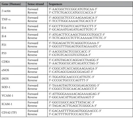
Total RNAs from samples were extracted using Tri reagent (Sigma-Aldrich) according to the manufacturer’s instructions. The total RNA concentration was quantified by spectrophotometry (NanoDrop 1000, Fischer Scientific, Illkirch, France) at 230 nm. Reverse transcription was performed using the iScript Reverse Transcription Supermix kit (Biorad, Miltry-Mory, France) according to the manufacturer’s instructions. qPCR was performed on the cDNA samples and gene expression was further analyzed using the CFX ConnectTM Real-Time PCR Detection System (Biorad,Miltry-Mory, France). Amplification reactions have been performed using iTAq Universal SYBR Green Supermix (Biorad, Miltry-Mory, France). β-actin was used as endogenous RNA control (housekeeping gene) in all samples. Primers related to β-actin, TNF-α, Il-6, Il-8, P21, P53, CDK4, eNOS, iNOS, SOD-1, VCAM-1, ICAM-1 and tissu-lar factor (TF) were synthesized (ThermoFischer, Saint-Aubin, France) (Table 1). Expression level was calculated after normalization to the housekeeping gene expression.
Gene (Human) Sense strand Sequences
β-actin ForwardReverse 5′-AACGGCTCCGGCATGTGCAA-3′5′-CTTCTGACCCATGCCCACCA-3′ TNF-α ForwardReverse 5′-AGGCGCTCCCCAAGAAGACA-3′5′-TCCTTGGCAAAACTGCACCT-3′ Il-6 ForwardReverse 5′-GCCTTCGGTCCAGTTGCCTT-3′5′-GCAGAATGAGATGAGTTGTC-3′ Il-8 ForwardReverse 5′-ATGACTTCCAAGCTGGCCGTGGCT-3′5′-TCTCAGCCCTCTTCAAAAACTTCTC-3′ P21 ForwardReverse 5′-TGGAGACTCTCAGGGTCGAAA-3′5′-GGCGTTTGGAGTGGTAGAAATC-3′ P53 ForwardReverse 5′-AACGGTACTCCGCCACC-3′5′-CGTGTCACCGTCGTGGA-3′ CDK4 ForwardReverse 5′-CATGTAGACCAGGACCTAAGG-3′5′-AACTGGCGCATCAGATCCTAG-3′ eNOS ForwardReverse 5′-CGGCATCACCAGGAAGAAGA-3′5′-CATGAGCGAGGCGGAGAT-3′ iNOS ForwardReverse 5′-TGGATGCAACCCCATTGTC-3′5′-CCCGCTGCCCCAGTTT-3′ SOD-1 ForwardReverse 5′-TAAAGTAGTCGCGGAGACGGG-3′5′-CGGCCTCGCAACACAAGCCT-3′ VCAM-1 ForwardReverse 5′-ATTGGGAAAAACAGAAAAGAG-3′5′-GGCAACATTGACATAAAGT-3′ ICAM-1 ForwardReverse 5′-GGCCGGCCAGCTTATACAC-3′5′-TAGACACTTGAGCTCGGGCA-3′ CD142 (TF) ForwardReverse 5′-GACAATTTTGGAGTGGGAACCC-3′5′-CACTTTTGTTCCCACCTG-3′
Proteome profile array of phospho-kinases.
Total protein extraction and purification were performed from cell lysates as previously described20. Briefly, cells were lysed for 5 min on ice in 200 μl of ice-cold RIPA buffer(65 mM Tris–HCl, pH 7.4, 150 mM NaCl, and 0.5% sodium deoxycholate) supplemented with phosphatase inhib-itor cocktails I and II and a protease inhibinhib-itor cocktail (Sigma, Darmstadt, Germany). Then, standardized con-centration of total proteins was subjected to a Proteome Profiler Human Phospho-Kinase Array (R&D Systems, Lille, France) following manufacturer’s instructions. The density of spots, corresponding to protein activation, was measured by MyImageTM Analysis Software 2.0 (Thermofisher) for each molecule and each condition.
Western blotting.
Briefly, cells were lysed for 5 min on ice in 200 μl of ice-cold RIPA buffer (65 mM Tris– HCl, pH 7.4, 150 mM NaCl, and 0.5% sodium deoxycholate) supplemented with phosphatase inhibitor cocktails I and II and a protease inhibitor cocktail (Sigma, Darmstadt, Germany). Lysates were centrifuged at 10,000 g at 4 °C for 10 min, supernatants were collected for quantification using the Bradford protein assay (Bio-Rad, Hercules, CA, USA) and 20 μg of cell lysates were loaded on a 12% SDS-PAGE for each condition. Antibodies against human eNOS (1/1000, mouse IgG, BD Biosciences, Le Pont de Claix, France), P21 (1/1000, rabbit IgG, Abcam, Paris, France), P53 (1/1000, rabbit IgG, Santa Cruz Biotechnology, Heidelberg, Germany), VCAM (1/500, rabbit IgG, Abcam, Paris, France), ICAM-1 (1/1000, mouse IgG, Life technologies, Courtaboeuf, France), CDK4 (1/1000, rabbit IgG, Life technologies), SOD-1 (1/1000, rabbit IgG, Life technologies), iNOS (1/1000, rabbit IgG, Life technologies) and against β-actin (1/2000, mouse IgG) from Santa Cruz Biotechnology (Heidelberg, Germany) were used for immunolabelling. Secondary alkaline phosphatase conjugated antibodies (anti-mouse (1/3000) or anti-rabbit (1/5000)) were purchased from Bethyl Laboratories (Montgomery, Texas, USA).Enzyme-linked immunosorbent assay.
TNF-α secreted in the cell supernatant was assessed by a sand-wich enzyme-linked immunosorbent assay (ELISA). Briefly, a goat anti-Human TNF-α antibody, (PeproTech, Rocky Hill, NJ, USA) was coated on a multi-well plate (4 μg/mL). Then, supernatants were added overnight at 4 °C. After washing with PBS, TNF-α was detected using a biotinylated goat anti-Human TNF-α (PeproTech). After 3 washing steps to discard unbound antibodies, HRP conjugated streptavidin (mix of solution A + B, as manufacturer instructions) was added and incubated for 20 to 30 minutes at room temperature. Finally, 100 μL of the stop solution (horseradish peroxidase and TMB substrate Solution, SS04, Life Technologies, Saint-Aubin, France) was added to each well for 5 to 30 min and then OD was measured by spectrophotometer (450 nm). The concentration of TNF-α was calculated by reference to a standard curve obtained with recombinant human TNF-α (PeproTech).Statistical analysis.
Statistical analysis was performed using pair-wise Anova test and post-hoc Tukey’s test. Statistical significance level was considered for p < 0.05. Data were analyzed using PRISM 6.0 (GraphPad, La Jolla, CA, USA). All experiments have been performed at least three times from three different EC culture batches (biological and technical replicates).Results
Endothelial microvesicles generated in response to P. gingivalis are pro-apoptotic
endothe-lial effectors.
P. gingivalis infection (MOI:100) led to a significant 2.8 fold in the shedding of EC after 24 h(infected vs. untreated, p < 0.05) (Fig. 1A) whereas the ultrapure Pg-LPS alone (1 μg/ml) had no significant effect (Fig. 1A). Interestingly, no MV shedding was detected in P. gingivalis culture supernatant (Supplementary Informations Fig. 1). The putative cytotoxic effect of EMVPg shed after P. gingivalis infection and of EMVCtrl shed
from unstimulated cells was assessed after 24 h of incubation with the endothelial monolayer. While no cytotoxic effect has been observed for EMVCtrl, increasing concentrations of EMVPg (5 to 30 nM) led to a significant 10%
and 25% concentration-dependent reduction of cell viability and survival respectively (Fig. 1B–D, p < 0.05 vs untreated cells), values measured after 30 nM EMVPg treatment reaching the range of those elicited by P. gingivalis
alone (MOI:100). A nearly 2-fold enhancement in the proportion of EC showing late apoptosis was observed in response to P. gingivalis infection or to 20–30 nM EMVPg by flow cytometry and fluorescence microscopy (P.
gingivalis-triggered 1.7-fold fold vs untreated; 20 nM EMVPg -induced 1.6-fold vs untreated and 30 nM EMVPg
-induced 1.5-fold vs untreated), while, the proportion of EC with early apoptosis was unchanged at 20 nM and drastically increased by 30 nM EMVPg suggesting a concentration threshold (Fig. 1E).
EMV
pgswitch the mRNA and protein expression profiles of endothelial cells to a
characteris-tic pro-inflammatory pattern.
The potential pro-inflammatory P. gingivalis or EMVPg -induced actionwas first assessed by the analysis of mRNA transcripts following a 24 h incubation with 5–30 nM EMVPg. An
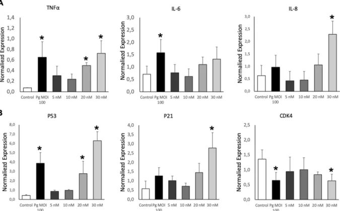
up-regulation in the expression of pro-inflammatory TNF-α, IL-6 and IL-8 cytokines mRNA was detected, reaching similar TNF-α expression after either P. gingivalis infection or 30 nM EMVPg (6.5 folds P. gingivalis or
EMVPg -induced vs untreated) (Fig. 2A). IL-6 mRNA expression was enhanced by 2.2 fold (P. gingivalis -induced
vs. untreated; p < 0.05) whereas, IL-8 mRNA was highly and solely enhanced by 30 nM EMVPg by 6-fold. This
pro-inflammatory switch was further confirmed by ELISA assay. Increased TNF-α concentrations after P.
gingiv-alis infection (2.3 fold; p < 0.05) and EMVPg stimulation (2 folds; p < 0.05) were measured after 24 h of incubation
(Fig. 3A). Interestingly, proteome analysis showed that several inflammation-related pathways are modulated following P. gingivalis or EMVPg stimulation (Fig. 4). Interestingly, not only did the JNK/AKT but also STAT were
differentially up-regulated following endothelial infection or stimulation.
Because leukocyte recruitment at the surface of the inflamed endothelium is one of the initial steps for athero-thrombosis34, expression of endothelial VCAM-1 and ICAM-1 adhesion molecules that favor leukocyte
attach-ment was also assessed. After 24 h incubation, 30 nM EMVPg significantly augmented both VCAM-1 and ICAM-1
mRNA expression (4.5 folds and 3 folds respectively (p < 0.05 vs untreated) whereas P. gingivalis infection was less effective at the chosen concentration, suggesting a specific triggering by EMVPg as compared to the above
pro-inflammatory cytokines induction (Fig. 5A). Nevertheless, infection by P. gingivalis, significantly increased the expression of tissue factor (TF), the cellular initiator of the coagulation cascade at the endothelial surface, and also a pro-inflammatory and pro-apoptotic inducer35 after 24 h. Interestingly, EMV
Pg incubation led to a
concentration-dependent expression of TF mRNA, reaching values prompted by P. gingivalis infection (Fig. 5A).
Figure 1. P. gingivalis promotes EMV shedding and alters endothelial cell viability. (A) The generation of EMV
from naïve EC (control) and following 24 h of infection with P. gingivalis (Pg) (MOI = 100) or stimulation with
Pg-LPS (1μg/ml) was measured in the supernatant by prothrombinase assay. Concentrations are represented by
mean +/− SD from 3 independent experiments; *p < 0.05 vs control (unstimulated EC). (B) Metabolic activity of EC infected with Pg (MOI = 100) or exposed to EMVCtrl (upper panel) and EMVPg (lower panel) (5, 10, 20
and 30 nM) for 24 h measured by AlamarBlue assay. Results are expressed as mean +/− SD from 3 independent experiments; *p < 0.05 vs control (unstimulated EC). (C) Live-Dead assay to evaluate the ratio of live EC versus dead EC in cells exposed to P. gingivalis (Pg) (MOI:100) and EMVPg (5, 10, 20 and 30 nM) for 24 h. Results are
expressed as percentage of live and dead EC (mean +/−SD). (D) Immunofluorescence imaging of live-dead staining (green: live EC, red: dead EC) for each condition after 24 hours of exposure. (E) Evaluation of type of cell death by flow cytometry after P. gingivalis infection and EMVPg (20 and 30 nM) exposure for 24 h. EC were
labelled with Annexin-V FITC and propidium iodide (IPPE). All data were expressed as mean ± SD. *(p < 0.05 vs untreated cells).
EMV
pgmodulate endothelial oxidative stress and promote endothelial dysfunction.
Becausenitric oxide is one of the major vasoprotectors released by endothelial cells under physiological conditions and a very diffusible mediator of vaso-relaxation, we investigated whether the expression of NO synthases would be altered by EMVPg. In this context, under conditions mimicking cardiovascular pathological conditions, like
cytokine treatment or angiotensin II-induced endothelial dysfunction, iNOS, the inducible form of NO synthase (iNOS) is up-regulated, whereas, the constitutive eNOS expression remains unchanged36. Herein, 24 h after P.
gingivalis infection or 20–30 nM EMVPg EC treatment, the expression of eNOS was significantly reduced at both
transcriptional and protein levels (2.1 folds; p < 0.05) (Figs. 3B, 4, 5B). At contrary, increased iNOS expression was induced by P. gingivalis and EMVPg and enhanced by 2 and 5.8 folds respectively (p < 0.05 vs. untreated),
thereby indicating endothelial dysfunction, eventually altered vascular function and possible eNOS uncoupling37.
The expression of endothelial superoxide dismutase (SOD-1), an enzyme that rapidly inactivates superoxide ani-ons, was also triggered by P. gingivalis and 30 nM EMVPg that both induced a 400% mRNA rise after 24 h,
empha-sizing the importance of cytoprotective signaling pathways in the endothelial response to major oxidative stress (Figs. 3B, 5B).
Because oxidative stress and the control of cell cycle are closely related, we also investigated whether P.
gingiv-alis infection or EMVPg could also trigger regulators of the cell cycle. Indeed, both endothelial treatments targeted
the oncosuppressor P53 and its downstream partner P21, a cyclin-dependent kinase inhibitor 1 and an inducer of senescence with a significant increase in P21 and P53 mRNA levels (4.7 and 14.3 folds respectively) (Fig. 2B). Moreover, the P53 up-regulation was confirmed by western-blot and proteomic analysis (Figs. 3B, 4). Conversely, a significant 2.3-fold decrease in the mRNA and protein expression of the cyclin-dependent kinase CDK4 was evidenced (p < 0.05 vs untreated) (Figs. 2B, 3B).
Altogether, these data strongly suggest that endothelial dysfunction in response to P. gingivalis infection or EMVPg stimulation is associated with oxidative stress and modulation of the cell cycle in a redox-dependent
man-ner as reported in human and murine endothelial cells36,38.
Discussion
This study demonstrates the autocrine pro-inflammatory properties of EMVPg shed from EC in response to P.
gingivalis infection. Results indicate that after 24 h of incubation, the shed EMVPg appear as key mediators of
endothelial dysfunction, switching the cytoprotective phenotype of endothelial cells toward a pro-inflammatory, pro-coagulant, pro-apoptotic and, eventually, a pro-senescent phenotype. All such phenotypes are a hallmark of sustained periodontitis in periodontal vascularization and atherothrombosis progression in large arteries.
Figure 2. EMVPg trigger inflammatory endothelial response. (A) The mRNA expression of inflammatory
markers TNF-α, IL-6 and IL-8 in EC exposed to P. gingivalis (Pg) (MOI:100) and EMVPg (5, 10, 20 and 30 nM)
for 24 h was measured by qRT-PCR. (B) The gene expression of cell cyle related markers p53, p21 and CDK4 EC in EC exposed to P. gingivalis (Pg) (MOI:100) and EMVPg (5, 10, 20 and 30 nM) for 24 h. All data were expressed
as mean ± SD from 3 independent experiments. *p < 0.05 between cells infected or stimulated against control (unstimulated cells).
Furthermore, most of the cell responses investigated were of comparable severity whether initiated by P. gingivalis or by high concentration of EMVPg.
Because MV are procoagulant owing to PhtdSer exposure and the eventual presence of TF when shed from monocytes, neutrophils and endothelial cells, their impact as pathogenic effectors in acute or chronic cardiovas-cular diseases and associated disorders such as myocardial infarction, atrial fibrillation, unstable angina, type-2 diabetes and their accumulation in the arteriosclerotic plaque has been extensively studied in relationship with the vascular inflammatory responses27. In blood, MV constitute a dynamic storage pool of vascular effectors,
whereby, their cellular origin and concentration characterizing the severity of the disease or of organ damage. Of note, EMV, even circulating as a small proportion of the vascular pool were found of prognostic value in organ and cellular graft rejection39, pulmonary hypertension40, sepsis-induced coagulopathy41 and cardiovascular
diseases42–45.
Strikingly, reports concerning infection-related MV shedding and how both host and pathogens produce MV are scarce. For instance, virulence factors, like LMP1 from the replicating Epstein-Barr virus, can be embedded in MV released from the infected cell and further spread virus through paracrine interactions with yet non-infected
Figure 3. EMVPg induce pro-inflammatory and pro-oxidative protein expression similar to P. gingivalis
infection. (A) TNF-α secretion in supernatant by EC in response to P. gingivalis (Pg) (MOI:100) and EMVPg (5,
20 and 30 nM) for 24 h was measured by ELISA. (B) Intra-cellular protein expression of eNOS, P21, ICAM-1, CDK4, P53, VCAM, iNOS and SOD-1 in response to P. gingivalis (Pg) (MOI:100) and EMVPg (5, 20 and 30 nM)
for 24 h was evaluated by Western Blot. All data were expressed as mean ± SD from 3 independent experiments and normalized against internal control β-actin. *p < 0.05 between cells infected or stimulated against control (unstimulated cells).
target cells. Similarly, human immunodeficiency virus also promotes MV shedding to transfer CCR5, its co-receptor on macrophages, to non-exposing target cells, consequently, promoting its own spreading46. MV
hijacking by pathogens could also be the causative of link between periodontitis and cardiovascular disease, as strongly supported by our data and previous reports47. In congruence with our data, Chlamydia pneumoniae was
also detected within atheromatous plaques and demonstrated to up-regulate the endothelial expression of the TF within 24 h together with the release of TF+-EMV that persisted for 1 week48. Most interestingly, we identified
via proteomic and transcriptomic assays that the EMV shedding in response to P. gingivalis was associated with characteristic prerequisite pathways of the cytoskeleton proteolytic cleavage and plasma membrane remodeling preceding the MV release from the inflamed or stimulated endothelium38. Indeed, the P38 MAP kinase pathway
that promotes the release of pro-inflammatory MV from human aortic endothelial cells in response to TNF-α49
was activated in our infection- driven model of pathogenic MV generation.
Figure 4. Exposure to EMVPg modulates significantly inflammatory pathways related to kinases activation.
Analysis of kinases activation induced by P. gingivalis infection (Pg) (MOI:100) and EMVPg (30 nM) for 24 h
evaluated by phospho-kinase array. The density of spots was measured by MyImageTM Analysis Softwate 2.0
(Thermofisher) for each molecule and each condition. (A) Graphical representation of the kinases expression in untreated EC, in EC following P. gingivalis infection (Pg) (MOI:100) and EMVPg (30 nM) stimulation for 24 h.
In this study, we emphasized the role of EMVPg as systemic pro-inflammatory and pro-oxidant effectors of
endothelial responses mediated by P. gingivalis. In our in vitro MV-mediated model, EMVPg at 30 nM acted as
cytotoxic endothelial effectors to an extent that was similar to the sole P. gingivalis infection (MOI = 100), via the activation of inflammatory pathways evidenced through Kinasome analysis. The up-regulation of TNF-α and IL-6 proinflammatory cytokines and their secretion eventually associated to an uncontrolled oxidative stress via the down-regulation of eNOS and the up-regulation of iNOS suggest NO synthase uncoupling. Therefore, our data altogether strengthen, the hypothesis of an EMV mediated P. gingivalis systemic response. This observation is strongly suggestive of a noxious role of EMVPg, eventually disseminated in blood flow to target healthy endothelial
cells, even under conditions where its pathogenic inducer P. gingivalis would remain sequestered in the perio-dontal tissues by cells of the innate immune response50. Endothelial inflammation via TNF-α, IL-1β, or IL-6
pro-inflammatory cytokines favors leukocyte recruitment, endothelial dysfunction and activation of blood coag-ulation making interleukin-driven proatherothrombotic processes a potential pharmacological target explored in several clinical trials51,52. In view of the recent anti-interleukin therapy trials, that failed to demonstrate a benefit
on cardiovascular outcome, circulating EMV released upon pathogen infection appear to be another target since EMV are able to concentrate at endothelial sites where flow disturbance favors enhanced interactions with MV, namely at artery branches prone to the development of atherotrombosis plaques. It was reported that MV seques-tered in plaques act as paracrine endothelial up-regulators of pro-inflammatory ICAM-1 or VCAM-1 prompting leukocyte recruitment in the growing plaque43,53. Whether the plaque of patients subjected to chronic exposure
of EMV released from distant endothelial infected sites undergo exaggerated endothelial inflammation remains to be demonstrated in specific animal models of atherothrombosis and in clinical trials. Nevertheless, we also showed that EMVPg triggered inflammation-related pathways. Indeed, the modulation of upstream NF-KB
acti-vators but also of transcription factors such as STAT confirmed their pro-inflammatory effect, as STAT pathway is a major signaling route converting the cytokine signal into gene expression programs regulating the proliferation and differentiation of the immune cell54.
P. gingivalis was previously demonstrated as a specific TLR4-dependent endothelial actor in pro-inflammatory
TNF-α and IL-6 secretion following endothelial invasion or via its virulence factors such as LPS55,56. Moreover,
several studies demonstrated the importance of NF-KB pathways in P. gingivalis elicited inflammation in several
cell types57–60. In a previous report, we were able to demonstrate that TLR4+MV generated by Pseudomonas were
true mediators of NF-KB-dependent signaling in TLR4−HEK engineered cells via IKB phosphorylation61. In the
present study, our data confirm and extend these observations, by showing that EMV released upon P. gingivalis infection are autocrine endothelial effectors possibly contributing to plaque endothelium inflammation and ero-sion62. Such effect appears specific to the EMV shedding trigger as only EMV
Pg were able to induce a significant
endothelial cell response while none was measured with EMVCtrl. This observation highlights the need of deep
Figure 5. EMVPg induce expression of atherothrombosis and oxidative stress markers expression. (A) The
mRNA expression of endothelial markers VCAM, ICAM and Tissular factor (TF) in EC exposed to P. gingivalis (Pg) (MOI:100) and EMVPg (5, 10, 20 and 30 nM) for 24 h was measured by qRT-PCR. (B) The gene expression
of oxidative stress markers eNOS, iNOS and SOD-1 in EC exposed to P. gingivalis (Pg) (MOI:100) and EMVPg
(5, 10, 20 and 30 nM) for 24 h. All data were expressed as mean ± SD from 3 independent experiments. *p < 0.05 between cells infected or stimulated against control (unstimulated cells).
analysis of the EMVPg content, including mRNA, miRNA and proteins, as it was shown that such content is highly
influenced by both triggers; the cellular environment and pre-existing conditions63,64. It also paves the way of
future clinical trials aiming to determine MV phenotype in the context of variable periodontitis severity and P.
gingivalis infection.
Accumulation of ROS and endothelial redox imbalance have been widely demonstrated in the atherothrom-botic plaque in vitro, in animal models and in human vascular tissues43,65. Oxidative stress is up-regulated
upon infection by Escherichia coli or Salmonella typhimurium66–68. Conversely, in vitro models indicate that
eNOS endothelial activity is reduced upon TNF-α or LPS incubation69 whereas experimental bacterial
men-ingitis in engineered mice with iNOS invalidation support the hypothesis of a dual endothelial iNOS and eNOS up-regulation upon comparison with wild type individuals70. Here, EMV
Pg triggered oxidative stress via altered
the expression of iNOS and eNOS at mRNA and protein levels, data altogether supporting eNOS uncoupling owing to major ROS accumulation and consecutive endothelial dysfunction, together with blunted cytoprotective signaling pathways against excessive inflammatory and pro-thrombotic responses. Interestingly, circulating MV in patients with coronary artery disease are elevated in comparison to that with healthy individuals and contain reduced functional eNOS. Furthermore, it was also demonstrated that a proportion of EMV circulating in such patients reduce the availability of NO in coronary artery endothelial cells and down regulate its expression71.
Further studies are required to characterize a specific sub-population of endothelial MV eventually responsi-ble for the systemic dissemination of the pathogen-driven inflammatory and pro-oxidant signals to uninfected endothelial cells and possibly contributing to endothelial dysfunction in patients with cardiovascular disease.
In this study, P. gingivalis strain ATCC 33277 was used as a pathogenic model. It should be mentioned that this strain is not the most virulent and future studies should determine the pro-inflammatory impact of others strains or mutants lacking fimbriae or gingipaïns as it was demonstrated a significant impact of such virulence factors on experimental outcomes in different cell types72,73. Future studies should also determine the precise composition
of EMVPg as their content is clearly influenced by the quantity and type of cellular stress trigger. Indeed, a specific
insight should be made on mRNA, miRNA, proteins and membrane receptors64,74,75.
In conclusion, P. gingivalis-induced EMV are effective pro-inflammatory effectors potentially involved in car-diovascular disease worsening. Nevertheless, autocrine and paracrine actions of EMV need further characteri-zation of embedded noxious content, and of their cellular and subcellular molecular partners. Further studies are needed to understand the impact of sustained yet low bacterial or virulence factors as well as low grade cytokine dissemination. A specific emphasis should be done on more relevant in vitro models such as human aortic endothelial cells and in vivo. Causative links between periodontitis and cardiovascular diseases should be investigated to decipher the systemic routes of the periodontal pathogenic signal dissemination. Local EMV released at distance from the thrombogenic plaque are a good candidate.
Received: 29 September 2019; Accepted: 13 January 2020; Published: xx xx xxxx
References
1. Hajishengallis, G. & Lamont, R. J. Breaking bad: Manipulation of the host response by Porphyromonas gingivalis. Eur. J. Immunol.
44, 328–338 (2014).
2. Kassebaum, N. J. et al. Global burden of severe periodontitis in 1990–2010: a systematic review and meta-regression. J. Dent. Res. 93, 1045–1053 (2014).
3. Hajishengallis, G. Periodontitis: from microbial immune subversion to systemic inflammation. Nat. Rev. Immunol. 15, 30–44 (2015). 4. Linden, G. J., Lyons, A. & Scannapieco, F. A. Periodontal systemic associations: review of the evidence. J. Clin. Periodontol. 40, 8–19
(2014).
5. Huck, O., Elkaim, R., Davideau, J. L. & Tenenbaum, H. Evaluating periodontal risk for patients at risk of or suffering from atherosclerosis: recent biological hypotheses and therapeutic consequences. Arch. Cardiovasc. Dis. 104, 352–358 (2011).
6. Lockhart, P. B. et al. Periodontal disease and atherosclerotic vascular disease: does the evidence support an independent association?: a scientific statement from the American Heart Association. Circulation 125, 2520–2544 (2012).
7. Tonetti, M. S. et al. Treatment of Periodontitis and Endothelial Function. N. Engl. J. Med. 356, 911–920 (2007). 8. Pothineni, N. V. K. et al. Infections, atherosclerosis, and coronary heart disease. Eur. Heart J. 38, 3195–3201 (2017).
9. Rosenfeld, M. E. & Campbell, L. A. Pathogens and atherosclerosis: update on the potential contribution of multiple infectious organisms to the pathogenesis of atherosclerosis. Thromb. Haemost. 106, 858–867 (2011).
10. Salhi, L. et al. Can Periodontitis Influence the Progression of Abdominal Aortic Aneurysm? A Systematic Review. Angiology 70, 479–491 (2019).
11. Xu, F. & Lu, B. Prospective association of periodontal disease with cardiovascular and all-cause mortality: NHANES III follow-up study. Atherosclerosis 218, 536–542 (2011).
12. Buhlin, K. et al. Periodontitis is associated with angiographically verified coronary artery disease. J. Clin. Periodontol. 38(11), 1007–14 (2011).
13. Liljestrand, J. M. et al. Immunologic burden links periodontitis to acute coronary syndrome. Atherosclerosis 268, 177–184 (2018). 14. Zelkha, S. A., Freilich, R. W. & Amar, S. Periodontal innate immune mechanisms relevant to atherosclerosis and obesity. Periodontol.
2000(54), 207–221 (2010).
15. Elkaïm, R. et al. Prevalence of periodontal pathogens in subgingival lesions, atherosclerotic plaques and healthy blood vessels: a preliminary study. J. Periodontal Res. 43, 224–231 (2008).
16. Amar, S. & Engelke, M. Periodontal Innate Immune Mechanisms Relevant to Atherosclerosis. Mol. Oral Microbiol. 30, 171–185 (2015).
17. Huck, O., Elkaim, R., Davideau, J. L. & Tenenbaum, H. Porphyromonas gingivalis-impaired innate immune response via NLRP3 proteolysis in endothelial cells. Innate Immun. 21, 65–72 (2015).
18. Bugueno, I. M. et al. Porphyromonas gingivalis Differentially Modulates Cell Death Profile in Ox-LDL and TNF-α Pre-Treated Endothelial Cells. PLoS ONE 11 (2016).
19. Huck, O. et al. Identification and Characterization of MicroRNA Differentially Expressed in Macrophages Exposed to Porphyromonas gingivalis Infection. Infect. Immun. 23;85(3) (2017).
20. Bugueno, I. M., Batool, F., Korah, L., Benkirane-Jessel, N. & Huck, O. Porphyromonas gingivalis Differentially Modulates Apoptosome Apoptotic Peptidase Activating Factor 1 in Epithelial Cells and Fibroblasts. Am. J. Pathol. 188, 404–416 (2018).
21. Velsko, I. M. et al. Active invasion of oral and aortic tissues by Porphyromonas gingivalis in mice causally links periodontitis and atherosclerosis. PloS One 9, e97811 (2014).
22. Li, L., Messas, E., Batista, E. L., Levin, R. A. & Amar, S. Porphyromonas gingivalis infection accelerates the progression of atherosclerosis in a heterozygous apolipoprotein E-deficient murine model. Circulation 105, 861–867 (2002).
23. Song, Z., Brassard, P. & Brophy, J. M. A meta-analysis of antibiotic use for the secondary prevention of cardiovascular diseases. Can.
J. Cardiol. 24, 391–395 (2008).
24. Lutgen, E., Atzler, D., Döring, Y., Duchene, J., Steffens Weber, C. Immunotherapy for cardiovascular disease. Eur. Heart J., https:// doi.org/10.1093/eurheartj/ehz283 (2019).
25. Welsh, P., Grassia, G., Botha, S., Sattar, N. & Maffia, P. Targeting inflammation to reduce cardiovascular disease risk: a realistic clinical prospect? Br. J. Pharmacol. 174, 3898–3913 (2017).
26. Van Tassell, B. W., Toldo, S., Mezzaroma, E. & Abbate, A. Targeting Interleukin-1 in Heart Disease. Circulation 128, 1910–1923 (2013).
27. Ridger, V. C. et al. Microvesicles in vascular homeostasis and diseases. Position Paper of the European Society of Cardiology (ESC) Working Group on Atherosclerosis and Vascular Biology. Thromb. Haemost. 117, 1296–1316 (2017).
28. Meziani, F., Delabranche, X., Asfar, P. & Toti, F. Bench-to-bedside review: circulating microparticles–a new player in sepsis? Crit.
Care Lond. Engl. 14, 236 (2010).
29. Barry, O. P., Praticò, D., Savani, R. C. & Fitzgeral, G. A. Modulation of monocyte-endothelial cell interactions by platelet microparticles. J. Clin. Invest. 102, 136–144 (1998).
30. Mause, S. F., Von Hundelshausen, Zernecke, A., Koene, R. R. & Weber, C. Platelet microparticles: a transcellular delivery system for RANTES promoting monocyte recruitment on endothelium. Arterioscler. Thromb. Vasc. Biol. 25, 1512–1518 (2005).
31. Huck, O., Elkaim, R., Davideau, J. L. & Tenenbaum, H. Porphyromonas gingivalis and its lipopolysaccharide differentially regulate the expression of cathepsin B in endothelial cells. Mol. Oral Microbiol. 27, 137–148 (2012).
32. Jy, W. et al. Measuring circulating cell-derived microparticles. J. Thromb. Haemost. 2, 1842–1851 (2004).
33. Keller, L. et al. Nanoengineered implant as a new platform for regenerative nanomedicine using 3D well-organized human cell spheroids. Int. J. Nanomedicine 12, 447–457 (2017).
34. May, A. E. et al. Platelet-Leukocyte Interactions in Inflammation and Atherothrombosis. Semin. Thromb. Hemost. 33, 123–127 (2007).
35. Cimmino, G. & Cirillo, P. Tissue factor: newer concepts in thrombosis and its role beyond thrombosis and hemostasis. Cardiovasc.
Diagn. Ther. 8, 581–593 (2018).
36. Khemais-Benkhiat, S. et al. The Redox-sensitive Induction of the Local Angiotensin System Promotes Both Premature and Replicative Endothelial Senescence: Preventive Effect of a Standardized Crataegus Extract. J. Gerontol. A. Biol. Sci. Med. Sci. 71, 1581–1590 (2016).
37. Bhagat, K., Hingorani, A. D., Palacios, M., Charles, I. G. & Vallance, P. Cytokine-induced venodilatation in humans in vivo: eNOS masquerading as iNOS. Cardiovasc. Res. 41, 754–764 (1999).
38. Burger, D. et al. Endothelial microparticle formation by angiotensin II is mediated via Ang II receptor type I/NADPH oxidase/Rho kinase pathways targeted to lipid rafts. Arterioscler. Thromb. Vasc. Biol. 31, 1898–1907 (2011).
39. Morel, O. et al. Endothelial cell activation contributes to the release of procoagulant microparticles during acute cardiac allograft rejection. J. Heart Lung Transplant. 27, 38–45 (2008).
40. Bakouboula, B. et al. Procoagulant membrane microparticles correlate with the severity of pulmonary arterial hypertension. Am. J.
Respir. Crit. Care Med. 177, 536–543 (2008).
41. Delabranche, X., Helms, J. & Meziani, F. Immunohaemostasis: a new view on haemostasis during sepsis. Ann. Intensive Care 7 (2017).
42. Amabile, N. et al. Association of circulating endothelial microparticles with cardiometabolic risk factors in the Framingham Heart Study. Eur. Heart J. 35, 2972–2979 (2014).
43. Rautou, P. E. et al. Microparticles from human atherosclerotic plaques promote endothelial ICAM-1-dependent monocyte adhesion and transendothelial migration. Circ. Res. 108, 335–343 (2011).
44. Yin, M., Loyer, X. & Boulanger, C. M. Extracellular vesicles as new pharmacological targets to treat atherosclerosis. Eur. J. Pharmacol.
763, 90–103 (2015).
45. Boulanger, C. M. & Dignat-George, F. Microparticles: an introduction. Arterioscler. Thromb. Vasc. Biol. 31, 2–3 (2011).
46. Mack, M. et al. Transfer of the chemokine receptor CCR5 between cells by membrane-derived microparticles: a mechanism for cellular human immunodeficiency virus 1 infection. Nat. Med. 6, 769–775 (2000).
47. Lai, F. W., Lichty, B. D. & Bowdish, D. M. E. Microvesicles: ubiquitous contributors to infection and immunity. J. Leukoc. Biol. 97, 237–245 (2015).
48. Ettelaie, C., Collier, M. E. W., James, N. J. & Li, C. Induction of tissue factor expression and release as microparticles in ECV304 cell line by Chlamydia pneumoniae infection. Atherosclerosis 190, 343–351 (2007).
49. Curtis, A. M. et al. p38 mitogen-activated protein kinase targets the production of proinflammatory endothelial microparticles. J.
Thromb. Haemost. 7, 701–709 (2009).
50. Cooper, P. R., Palmer, L. J. & Chapple, I. L. C. Neutrophil extracellular traps as a new paradigm in innate immunity: friend or foe?
Periodontol. 2000(63), 165–197 (2013).
51. Ridker, P. M. Anticytokine Agents: Targeting Interleukin Signaling Pathways for the Treatment of Atherothrombosis. Circ. Res. 124, 437–450 (2019).
52. Ridker, P. M. Clinician’s Guide to Reducing Inflammation to Reduce Atherothrombotic Risk: JACC Review Topic of the Week. J. Am.
Coll. Cardiol. 72, 3320–3331 (2018).
53. Leroyer, A. S. et al. Cellular origins and thrombogenic activity of microparticles isolated from human atherosclerotic plaques. J. Am.
Coll. Cardiol. 49, 772–777 (2007).
54. Pfitzner, E., Kliem, S., Baus, D. & Litterst, C. M. The role of STATs in inflammation and inflammatory diseases. Curr. Pharm. Des. 10, 2839–2850 (2004).
55. Kocgozlu, L., Elkaim, R., Tenenbaum, H. & Werner, S. Variable cell responses to P. gingivalis lipopolysaccharide. J. Dent. Res. 88, 741–745 (2009).
56. Ho, Y. S. et al. Porphyromonas gingivalis fimbriae-dependent interleukin-6 autocrine regulation by increase of gp130 in endothelial cells. J. Periodontal Res. 44, 550–556 (2009).
57. Huck, O. et al. Reduction of Articular and Systemic Inflammation by Kava-241 in Porphyromonas gingivalis-induced Arthritis Murine Model. Infect. Immun., https://doi.org/10.1128/IAI.00356-18 (2018).
58. Singh, S. P., Huck, O., Abraham, N. G. & Amar, S. Kavain Reduces Porphyromonas gingivalis-Induced Adipocyte Inflammation: Role of PGC-1α Signaling. J. Immunol. 201, 1491–1499 (2018).
59. Yuan, H., Gupte, R., Zelkh, S. & Amar, S. Receptor activator of nuclear factor kappa B ligand antagonists inhibit tissue inflammation and bone loss in experimental periodontitis. J. Clin. Periodontol. 38, 1029–1036 (2011).
60. Wan, M., Liu, J. & Ouyang, X. Nucleotide-binding oligomerization domain 1 regulates Porphyromonas gingivalis-induced vascular cell adhesion molecule 1 and intercellular adhesion molecule 1 expression in endothelial cells through NF-κB pathway. J. Periodontal
61. Constantinescu, A. A. et al. Exocrine cell-derived microparticles in response to lipopolysaccharide promote endocrine dysfunction in cystic fibrosis. J. Cyst. Fibros. 13, 219–226 (2014).
62. Hansson, G. K., Libby, P. & Tabas, I. Inflammation and plaque vulnerability. J. Intern. Med. 278, 483–493 (2015).
63. Giannella, A. et al. Circulating levels and characterization of microparticles in patients with different degrees of glucose tolerance.
Cardiovasc. Diabetol. 16, 118 (2017).
64. Enjeti, A. K., Ariyarajah, A., D’Crus, A., Seldon, M. & Lincz, L. F. Circulating microvesicle number, function and small RNA content vary with age, gender, smoking status, lipid and hormone profiles. Thromb. Res. 156, 65–72 (2017).
65. Loyer, X., Vion, A. C., Tedgui, A. & Boulanger, C. M. Microvesicles as cell-cell messengers in cardiovascular diseases. Circ. Res. 114, 345–353 (2014).
66. Chiang, S. M. & Schellhorn, H. E. Regulators of oxidative stress response genes in Escherichia coli and their functional conservation in bacteria. Arch. Biochem. Biophys. 525, 161–169 (2012).
67. Fu, J. et al. Salmonella proteomics under oxidative stress reveals coordinated regulation of antioxidant defense with iron metabolism and bacterial virulence. J. Proteomics 157, 52–58 (2017).
68. Farr, S. B. & Kogoma, T. Oxidative stress responses in Escherichia coli and Salmonella typhimurium. Microbiol. Rev. 55, 561–585 (1991).
69. Cardaropoli, S., Silvagno, F., Morra, E., Pescarmona, G. P. & Todros, T. Infectious and inflammatory stimuli decrease endothelial nitric oxide synthase activity in vitro. J. Hypertens. 21, 2103–2110 (2003).
70. Winkler, F., Koedel, U., Kastenbauer, S. & Pfister, H. W. Differential expression of nitric oxide synthases in bacterial meningitis: role of the inducible isoform for blood-brain barrier breakdown. J. Infect. Dis. 183, 1749–1759 (2001).
71. Abbas, M. et al. Endothelial Microparticles From Acute Coronary Syndrome Patients Induce Premature Coronary Artery Endothelial Cell Aging and Thrombogenicity: Role of the Ang II/AT1 Receptor/NADPH Oxidase-Mediated Activation of MAPKs and PI3-Kinase Pathways. Circulation 135, 280–296 (2017).
72. Jayaprakash, K., Demirel, I., Khalaf, H. & Bengtsson, T. Porphyromonas gingivalis-induced inflammatory responses in THP1 cells are altered by native and modified low-density lipoproteins in a strain-dependent manner. APMIS. 126, 667–677 (2018). 73. Rodrigues, P. H. et al. Porphyromonas gingivalis strain specific interactions with human coronary artery endothelial cells: a
comparative study. PLoS One. 7, e52606 (2012).
74. Madkhali, Y. et al. The Ratio of Factor VIIa:Tissue Factor Content within Microvesicles Determines the Differential Influence on Endothelial Cells. TH Open. 3, e132–e145 (2019).
75. Pasquier, J. et al. Circulating microparticles in acute diabetic Charcot foot exhibit a high content of inflammatory cytokines, and support monocyte-to-osteoclast cell induction. Sci Rep 7, 16450 (2017).
Acknowledgements
This work was partly funded by Agence Nationale pour la Recherche (ANR ENDOPAROMP ANR-17-CE17-0024-01 and ANR COCERP ANR-16-CE29-0009-03).
Author contributions
I.M. Bugueno, F. Zobairi El-Ghazouani, E. Anglès-Cano, F. Toti, O. Huck contributed to conception and design, data acquisition, analysis and interpretation, drafted and critically revised the manuscript. F. Batool, H. El Itawi, N. Benkirane-Jessel contributed to data analysis and critically revised the manuscript. All authors gave final approval and agree to be accountable for all aspects of the work.
Competing interests
The authors declare no competing interests.
Additional information
Supplementary information is available for this paper at https://doi.org/10.1038/s41598-020-58374-z.
Correspondence and requests for materials should be addressed to O.H. Reprints and permissions information is available at www.nature.com/reprints.
Publisher’s note Springer Nature remains neutral with regard to jurisdictional claims in published maps and
institutional affiliations.
Open Access This article is licensed under a Creative Commons Attribution 4.0 International
License, which permits use, sharing, adaptation, distribution and reproduction in any medium or format, as long as you give appropriate credit to the original author(s) and the source, provide a link to the Cre-ative Commons license, and indicate if changes were made. The images or other third party material in this article are included in the article’s Creative Commons license, unless indicated otherwise in a credit line to the material. If material is not included in the article’s Creative Commons license and your intended use is not per-mitted by statutory regulation or exceeds the perper-mitted use, you will need to obtain permission directly from the copyright holder. To view a copy of this license, visit http://creativecommons.org/licenses/by/4.0/.