Bacterial Adhesion in Structured Environments
MASSACHUSETTS INSThTUTE OF TECFN! : Yby
2 0
14
Ronn S. Friedlander
LIBRAF RiE
B.S. Biomedical Engineering and Ecology & Natural Resources, Rutgers University, 2008M.Phil. Clinical Biochemistry, University of Cambridge, 2009
SUBMITTED TO THE DIVISION OF HEALTH SCIENCES AND TECHNOLOGY IN PARTIAL FULFILLMENT OF THE REQUIREMENTS FOR THE DEGREE OF
DOCTOR OF PHILOSOPHY IN
MEDICAL ENGINEERING AND MEDICAL PHYSICS AT THE
MASSACHUSETTS INSTITUTE OF TECHNOLOGY SEPTEMBER 2014
2014 Massachusetts Institute of Technology. All rights reserved.
Signature redacted
Signature of Author:
Harvard-MIT Program in Health Sciences and Technology August 29, 2014
Signature redacted
Certified by:
Joanna Aizenberg, Ph.D. Amy Smith Berylson Professor of Materials Science,
Professor of Chemistry and Chemical Biology, Harvard School of Engineering and Applied Sciences
Thesis Supervisor
Signature redacted
Accepted by:_
Emery N. Brown, M.D., Ph.D. Director, Harvard-MIT Program in Health Sciences and Technology Professor of Computational Neuroscience and Health Sciences and Technology
Bacterial Adhesion in Structured Environments
by
Ronn S. Friedlander
Submitted to the Harvard-MIT program in Health Sciences and Technology on August 29, 2014 in partial fulfillment of the requirements for the degree of
Doctor of Philosophy in Medical Engineering and Medical Physics ABSTRACT
Biofilms-surface-bound communities of microbes-are a major medical concern, as they can be sources of infection that are difficult to eradicate. Their formation starts with the attachment of bacteria to available surfaces-often implantable biomaterials. The development of materials that prevent bacterial adhesion is therefore of paramount importance, and it requires a thorough understanding of the materials and bacterial surface properties that enable adhesive interactions. We herein design model surfaces and examine the interplay between micro-scale geometry, surface energy and bacterial surface properties with respect to adhesion, with the ultimate goal of understanding bacterial adhesion in structured environments, and establishing principles for design of novel surfaces that effectively repel bacteria.
We first study adhesion of Escherichia coli to engineered surfaces possessing superficially unfavorable geometries. We show that cells can overcome geometric constraints with the aid of flagella, which are able to reach between narrow crevices, thus improving adhesion and expanding the range of surfaces to which cells can adhere. We examine binding of purified flagella to abiotic surfaces by means of quartz crystal microbalance (QCM) and show that flagella bind preferentially to hydrophobic surfaces, yet they do not appreciably bind to hydrophilic surfaces. Using mutant strains, we investigate the role of flagella in surface attachment of live cells and demonstrate that flagellated cells adhere best to hydrophobic substrates; however flagella may impede cell adhesion to hydrophilic surfaces.
To further explore hydrophilic, structured environments with physiological relevance, we examine mucin-a natural hydrogel that typically harbors microbes in animals, while protecting the host. We purify mucins and use them in their native, three-dimensional configuration to probe bacterial swimming behavior and surface attachment in their presence. We demonstrate that mucins maintain-and possibly enhance-swimming ability for E. coli and Pseudomonas aeruginosa, and show that they greatly reduce adhesion to underlying substrates.
Finally, we build on our established design principles and construct anti-adhesive surfaces by combining hydrophilic chemistries with topographic features smaller than cellular dimensions. This work suggests a path toward anti-adhesive materials that may be optimized for mechanical robustness, longevity and specific environments of application.
Thesis supervisor: Joanna Aizenberg, Ph.D.
Title: Amy Smith Berylson Professor of Materials Science, Professor of Chemistry and Chemical Biology, Harvard School of Engineering and Applied Sciences
Acknowledgments
When I reflect on the journey I took to get here, I must thank Ashley Winter for showing me the path of biomedical engineering, which combined my love of biological systems with the rigor of engineering principles. Despite our distance in time and space from those early college days, we remain close friends. There was a point in college when I knew I could cut it at a place like MIT. I would say the encouragement and guidance of Charlie Roth at Rutgers was what enabled my confidence to grow; it got me to that point where I could challenge myself to attend the excellent grad programs at Cambridge and MIT/Harvard.
My time in the HST program has been amazing and I must thank those HMS professors who stick out in my mind as making my medical classes invigorating: Rick Mitchell, Shiv Pillai and David Housman. Beyond the classes at HMS and MIT, I was lucky enough to join the lab of Joanna Aizenberg. I am privileged to have spent the last four years in Joanna's lab, working with some of the most creative and intelligent minds I have ever encountered. Joanna took me in as a young student, gave me the tools and freedom to follow my own research path, and enabled me to mature as a scientist. I continue to be impressed by Joanna's ability to drill down to the important points of a research project and very rapidly ask the most poignant questions. I hope some of this has rubbed off on me.
I owe much of my success to my wonderful colleagues who have been friends and collaborators throughout my time at HST. Mike Bucaro was my first mentor in the Aizenberg lab and also made sure everyone was socializing. I won't forget how important he was in helping me find my place in the lab. I soon found Hera Vlamakis, who helped me learn some genetic techniques and was always there to read over and help me with my manuscripts. I am very appreciative of Karen Fahrner for all her wisdom and help as we figured out how to purify flagella. Making the right connections has been so crucial for this research project, due to the numerous academic fields involved. I cannot overstate how important the BASF biofilms group was for helping me to make the right contacts and foster discussions about bacterial behavior among a highly interdisciplinary group of scientists who all wanted to help each other solve problems. Of course, having funding from BASF, the NSF and the Cystic Fibrosis Foundation has enabled much of this work to take place.
Hardly an easy journey, carrying out my PhD work was one of the most challenging tasks I have ever taken on. I could not have done it without mentorship and the support of friends. Research discussions with Katharina Ribbeck were so helpful in building my confidence in the various projects I undertook. I cannot thank her enough for those supportive conversations and collaborations. Having Roberto Kolter as a mentor and thesis committee member was very helpful and enlightening. Also, engaging with other microbiologists and friends through MSI was extremely enjoyable and helpful. Outside of school, I continue to have the constant support of my friends: Alex Bruno, who can always be counted on for a trivia night outing; Jake Brukhman, who always challenges my assumptions and inspires creativity; Eyal Wellisch, who has been there for me since I was 2 years old; my roommates Ben Holmes, Christine Platzek, Dan Rassi and Will Silversmith, who are always around and supportive in things ranging from video gaming to building beverage empires-they help me keep things interesting.
Finally, I thank my family. They made me who I am today. They gave me everything I needed to succeed and the encouragement and tools to be independent. Sandy and my Dad have always encouraged me and been there for me. My mom has been so supportive of me in all that I do and has always inspired me to be curious and question everything. I will continue to do that.
Table of Contents
Abstract 3
Acknowlegments 5
Chapter 1: Background 8
Clinical Impact of Bacterial Biofilms 8
Prevention of Bacterial Adhesion to Biomaterials 10
Topographical and Geometrical Approaches to Biofilm Control 13
Thesis Organization 16
Bibliography 18
Chapter 2: The role of flagella in adhesion of Escherichia coli to structured substrates 24
Introduction 24
Results 25
Discussion and Conclusion 38
Experimental Section 44
Bibliography 51
Chapter 3: Flagellar adhesion to abiotic surfaces 54
Introduction 54
Results and Discussion 57
Experimental Section 69
Bibliography 74
Chapter 4: Antiadhesive properties of three-dimensional mucin gels 79
Introduction 79
Results and Discussion 80
Bibliography 103
Chapter 5: Paths for future study 107
Section 1: Escherichia coli flagellar diversity and its relation to surface adhesion 107
Introduction 107
Preliminary Results and Discussion 110
Future Work 117
Experimental Section 119
Section 2: Structured, coated surfaces for the prevention of bacterial adhesion: 127 explorations and future work
Introduction 127
Preliminary Results and Discussion 129
Future Work 138
Experimental Section 141
Bibliography 143
Chapter 6: Perspective: topographically patterned surfaces for the prevention of 146 bacterial adhesion
Introduction 146
Bacterial Responses to Surface Topography 147
Superhydrophobicity as an Antiadhesive Strategy 150
Prospects for Topographical Anti-fouling Substrates 154
Closing Remarks 157
Chapter 1
BackgroundCLINICAL IMPACT OF BACTERIAL BIOFILMS
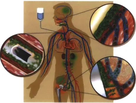
Bacterial infection is a ubiquitous cause of morbidity and mortality that places a considerable burden on our healthcare system. Nearly two million healthcare associated infections (HAIs) per year cost U.S. hospitals between $28 and $45 billion [1]. Of these, ~450,000 are catheter-associated urinary tract infections and 92,000 are central line associated bloodstream infections [1]. In 2002, urinary tract and bloodstream HAIs alone (often via implantation of inert biomaterials) resulted in 13,088 and 30,665 deaths, respectively [2]. In the most insidious HAIs, the contributing bacteria exist in a sessile, protective state called a biofilm. These biofilms form on the surfaces of medical implant materials, such as catheters, heart valves and prostheses, to name only a few [3, 4].
Figure 1.1. Schematic of the steps of bioflim formation. The biofilm life cycle illustrated in three steps: initial attachment events, the growth of complex biofilms, and detachment events by clumps of bacteria or so-called 'seeding dispersal.' Source: Center for Biofilm Engineering, Bozeman, MT.
The standard model for the progression of biofilm development (Figure 1.1) starts with planktonic microbes adhering reversibly to a surface coated with a conditioning film created by
components of the aqueous environment (blood, interstitial fluid, growth medium, etc.) [5, 6]. Once the initial colonizers are present, they can serve to nucleate microcolonies. As these microcolonies reach a threshold size, the microbes begin to secrete matrix and irreversibly attach to a surface [7, 8]. The NIH estimates that 80% of persistent bacterial infections are in the form of biofilms (Figure 1.2) [9, 10]. The overall heterogeneity of gene expression, structure, chemistry and material properties of mature biofilms allows at least parts of them to survive a broad spectrum of assaults [11]. Whether by gene transfer or by antibiotic-induced population bottlenecks that allow resistant 'persister cells' to repopulate biofilms [12], antibiotic susceptibility is greatly reduced in these communities. An early demonstration of this showed Pseudomonas aeruginosa biofilms to be viable after treatment with tobramycin concentrations in excess of twenty times the concentration necessary to completely kill planktonic cells of the same strain [13]. There have since been numerous demonstrations of this phenomenon with other species and antibiotics [10, 14-16]. Biofilm populations are extremely dense compared with disseminated swimming bacterial populations. Recent studies show density-dependent antibiotic resistance effects that do not occur in relatively sparse planktonic populations [17, 18]. The matrix enclosure, which can be composed of polysaccharides, protein fibers, surface organelles, and/or nucleic acids, reduces the distance and rate at which antimicrobials can diffuse into the biofilm [9, 19], thus making it very difficult to destroy a biofilm with drug treatments alone. Finally, the sub-lethal doses of antibiotics that do reach the deeper levels of a biofilm will introduce strong selective pressure for resistance [14, 20]. The prevalence and variety of antibiotic-resistant bacterial species is ever increasing, yet the number of antibiotic FDA-approved new molecular entities has steadily dwindled over the past 25 years [21].
Figure 1.2. Bioflim implant infections. Schematic depiction of implants that can become infected with biofilms and seed new infections in the body. These include catheters, dental implants and hip implants. Source: Center for Biofilm Engineering, Bozeman, MT.
PREVENTION OF BACTERIAL ADHESION TO BIOMATERIALS
Bacterial surface adhesion is a ubiquitous phenomenon in the natural world, and as the first step in biofilm formation, it enables microbes to survive changing environments, chemical and physical assaults and depletion of resources [22, 23]. Due to the survival advantages imparted by the biofilm lifestyle, pristine, unoccupied surfaces provide high-value real estate to bacteria, particularly when conditions favor a survival mode, rather than a proliferative one [9]. Unfortunately, numerous materials applications require the maintenance of pristine surfaces in bacteria-rich environments. Beyond biomedical materials, these also include remote optical sensors and marine vessels, to name a few [8, 9, 24, 25]. These surfaces become exposed to bacteria during use and are ultimately fouled by adhering microbes. Once these microbes progress to mature biofilms, they become difficult or impossible to remove [26]. Researchers
have therefore focused on preventing bacterial adhesion to indwelling medical devices and this has become an important, interdisciplinary field of research.
Two primary methods are currently practiced to reduce bacterial colonization of indwelling materials, both of which involve antimicrobial agents. One common approach is to administer intravenous and topical prophylactic agents before and during implant procedures and to continue an antibiotic regimen for a period after the procedure. This is practiced in conjunction with joint replacement procedures as well as with implantation of some dental prostheses [27]. The result of this antibiotic regimen, combined with strict aseptic techniques and reduced operating times, is a reduction in overall postoperative infection rates. However, prophylaxis has had only weak evidence of efficacy when used for urinary catheterization [28]. Of course, the administration of prophylactic antibiotics will increase the rate of development of resistance. Orthopedic practitioners already debate whether this practice should be continued due to the increased emergence of antibiotic resistant bacteria [29, 30].
Another strategy that has emerged relies on coating or bonding devices with antimicrobial agents to deter biofilm formation. Silicone shunts impregnated with various antibiotics (rifampicin, clindamycin) significantly reduced the survival of Staphylococcus epidermidis in vitro [31]. A clinical study of cefazolin-bonded venous and arterial catheters showed a reduction in infection (usually S. epidermidis) from 14% of control catheters to 2% of antibiotic-bonded catheters [32]. For urinary catheters, silver alloy coatings have been used as antimicrobials and have been shown to reduce infection, especially during the first week of implantation [33]. These reductions have been modest, with most studies reporting <10% reduction in risk of infection (though one study reported a risk reduction of 32%) [34]. Medicated vascular catheters still have infection rates between 1.2 and 4.8 cases per 1000
catheter days, which is high considering that many patients are catheterized for more than a week at a time [35].
A less popular, but intriguing strategy for biofilm prevention is the probiotic approach. Strong evidence suggests that the presence of some beneficial bacteria in the body can act to inhibit the growth of pathogenic microbes [36, 37]. Furthermore, researchers have shown that specific actions, such as eating yogurt to prevent colonization of laryngeal implants, can reduce incidence of infection [37]. One study demonstrated that the presence of S. aureus in human nares could be eliminated by artificial implantation of Corynebacteria [38]. In an ongoing phase I clinical trial, non-pathogenic E. coli are being used to pre-coat catheters, so as to prevent adhesion of pathogens [39, 40].
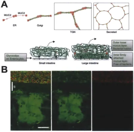
A number of promising coatings based on hydrophilic polymers have been shown to reduce bacterial adhesion. These include but are not limited to: polyethylene glycol (PEG) [41-43], chitosan [44, 45], heparin [[41-43], and purified mucin [46]. Mucins are particularly interesting materials as they form the major medium for host-microbe interactions. In vitro studies of antifouling mucin coatings have demonstrated varying results in performance. Due to the complexity of these molecules, it is likely that their structural integrity is critical for their antimicrobial properties. Mucins are large glycoproteins with the majority of their molecular weight comprised of oligosaccharides. They are secreted by goblet cells as packets that become hydrated, expand and are crosslinked to form a structured network (Figure 1.3) [47, 48]. It is possible that the structure of this network is related to their ability to regulate the large bacterial populations that reside adjacent to host epithelia (e.g. in the colon). Different methods of
preparation and surface tethering utilized in previous studies are perhaps causes for conflicting findings.
A
EA GOO ~
X
TGN Secreted
B
Figure 1.3. Mucins harbor bacteria but protect the host. (A) Schematic outline of the Muc2 mucin and its formation of mucus in the small and large intestine. Assembly of the Muc2 mucin (protein core red) into dimeric forms in the endoplasmic reticulum (ER), 0-glycosylation (green) in the Golgi apparatus, formation of trimeric forms in the trans Golgi network (TGN) and a schematic picture of the secreted Muc2 polymer in a structured network [49]. (B) Combined Muc2 immunostaining (green) and FISH analysis using the general bacterial probe EUB338-Alexa Fluor 555 (red) of distal colon shows Muc2-positive goblet cells and an inner stratified (s) mucus layer on the epithelium. This layer is devoid of bacteria, which can only be detected in the outer mucus layer. The inner mucus generates a spatial separation between the cells and the microflora (Scale bar: 20 im) [50].
TOPOGRAPHICAL AND GEOMETRICAL APPROACHES TO BIOFILM CONTROL Given the diversity of bacterial biofilm communities and the ubiquity of conditioning films (which may mask surface chemistry), a more general, physical approach should be taken to prevent adhesion. In the fields of cell biology and tissue engineering, surface architecture has been studied and manipulated extensively in order to achieve a measure of control over tissue
cell behavior [51-55]. Only recently have these techniques been applied for the control of biofilm formation [56].
There is strong evidence that bacteria can respond quite dramatically to surface patterns. Takeuchi et al. showed that E. coli could be formed into coil shapes by allowing them to grow within toroid trenches [57]. Work by Diaz et al. showed that nano-scale trenches reduce migration of surface-bound Psuedomonas fluorescens [58]. A study from our lab demonstrated that E. coli, P. aeruginosa and Bacillus subtilis can be patterned in a controllable manner using nano-scale post arrays on a similar size scale to the bacteria. The spacing of the posts can be adjusted to achieve different, predictable patterns [59]. Furthermore, when the posts are sufficiently close together, the bacteria will arrange themselves in a random orientation at the tips of the posts, and not at the base of the substrate.
Micro- and nano-topographies have been used in several studies to determine whether they can influence bacterial adhesion. Campoccia et al. compared adhesion of S. aureus on an array of 100 nm diameter, 160 nm tall posts spaced 200 nm apart to adhesion on flat surfaces, and was unable to observe a significant influence of this topography [60]. Another study looked at adhesion of multiple cocci and rod-shaped bacteria to surfaces with circular indentations between 0.2 pm and 2 gm; this study showed differential adhesion based on both species and feature size [61]. For the most part, bacteria were retained within the indentations except in the smallest feature sizes. In those cases, the adhesion was similar to adhesion on flat surfaces. One interesting study used shark skin-inspired micro-scale patterns [62] in poly(dimethyl siloxane) (PDMS) elastomer to reduce biofilm formation by S. aureus (Figure 1.4) [63, 64]. This same material was originally optimized for reduction of biofouling by zoospores, however it appears to show efficacy in reducing biofilms as well [63, 65].
Figure 1.4. Shark skin-inspired surfaces for prevention of biofouling. Clockwise from top: Scales from the spinner shark, SharkletTM film, S. aureus biofilm on flat PDMS (day 14 of culture), and on SharkletTm PDMS (day 14 of culture). Figure adapted from Chung et al. and Scardino et al.
Among the studies of bacteria-structure interactions, there are few that address the interplay of structure and chemistry outside the context of superhydrophobic materials. On a microscopic scale, the interfacial features confronting microbes are structural as well as chemical. For example, the cilia and microvilli lining some mammalian epithelial surfaces also have a glycocalyx and secreted mucins that overlay the sub-micron surface structure. Bioinspired antifouling materials have yet to replicate this complexity. Therefore, we aimed to examine the interplay between micro-scale geometry, surface energy and bacterial surface properties with respect to adhesion, with the ultimate goal of understanding bacterial adhesion in structured environments, and establishing principles for design of novel surfaces that effectively repel bacteria.
THESIS ORGANIZATION
The organization of this thesis is designed to reflect the process of discovery during my doctoral work. Each chapter of the thesis serves to ask (and in many cases answer) an important question regarding microbial behavior in structured environments. As in my research, the research findings in one chapter typically motivate the subject of the next. Experiments at the beginning of my studies led me to examine adhesion of E. coli to patterned PDMS substrates. Initial analysis hinted at the importance of bacterial appendages, which led me on the path of research described herein.
Chapter 2 examines adhesion of E. coli to structured surfaces (Figure 1.5). The surfaces are designed to determine how cells are able to adhere when they cannot fit between micro-structures. We reveal the role of flagella in this process and then further probe their contribution using more directed approaches. By generating mutant strains lacking flagella, we demonstrate the relative contribution of flagella in the adhesive process. We also generate substrates with variable surface feature spacing to determine when cells benefit most from using their surface appendages. We show that flagella improve adhesion to patterned PDMS substrates. Specifically, flagella are able to reach in between crevices where the cell body cannot fit, thus improving adhesion and expanding the range of surfaces to which E. coli can adhere.
Understanding the role of flagella in adhesion of bacteria to patterned substrates raises the question of what substrate properties enable nonspecific flagellar surface attachment. Chapter 3 explores this question using self assembled monolayers (SAMs) to generate chemically similar, well defined surfaces that vary in hydrophobicity. Using purified flagella and quartz crystal microbalance (QCM), we examine binding of flagella to abiotic surfaces. We show that flagella bind preferentially to hydrophobic surfaces, and do not appreciably bind to hydrophilic surfaces.
Using SAMs and cells with or without flagella, we also demonstrate that flagellated cells adhere best to hydrophobic substrates, but cells without flagella actually adhere better to hydrophilic substrates than those with flagella, indicating that flagella may impede cell adhesion to hydrophilic surfaces.
In order to further explore hydrophilic, structured environments, Chapter 4 examines mucin-a natural hydrogel that typically harbors microbes in animal hosts, while keeping them in check. We purify mucins and use them in their native, three-dimensional gel configuration to probe bacterial swimming behavior and surface attachment in their presence. We demonstrate that swimming behavior is kept intact-and possibly enhanced-in E. coli and P. aeruginosa in the presence of mucin. Furthermore, mucin gels greatly reduce adhesion to underlying glass substrates. Cells that are immotile, however, form suspended clumps or flocs that are antibiotic resistant, analogous to those seen in the lungs of cystic fibrosis patients.
In Chapter 5, we begin to examine areas for future study, and present preliminary findings. Part 1 looks at flagellar diversity in E. coli across different strains and uses this as a model to ask whether flagellar adhesion to hydrophobic substrates is a more general phenomenon. We clone several flagella genes from various E. coli strains, corresponding to different flagellar morphotypes, into the same background strain. Then, we purify these flagella and examine their adhesion to SAMs in QCM experiments. Part 2 focuses on generating new substrates using the knowledge gained in the previous chapters. We generate inverse opal monolayers with structure on the micron scale. We begin to examine pore size and various coatings including mucin and PEG to determine their effects on bacterial adhesion. Parts 1 and 2 of Chapter 5 establish clear lines of inquiry for future studies. This work and preliminary results pave the way for additional research in bacterial adhesion to structured surfaces.
Finally, Chapter 6 examines our findings in the context of existing research. Here, I offer a broader perspective on the research field and extract themes from this thesis and other works in the field. I suggest some approaches that take into account these themes, building on existing strategies and offering new ones, for the improved design of antifouling surfaces.
Figure 1.5. E. coli on a topographical substrate. False-colored scanning electron micrograph of E. coli cells that are adherent to a patterned PDMS substrate. Area shown is where the patterned region meets a flat, unpatterned region. Note the prominence of extracellular fibers
(colored yellow)-an abundance of flagella. Coloring courtesy of Michael Bucaro. BIBLIOGRAPHY
1. Scott R, Detection C (2008) The Direct medical costs of healthcare-associated infections in U.S. hospitals and the benefits of prevention.
2. Klevens R, Edwards, JR, Richards C, et al. (2007) Estimating health care-associated infections and deaths in US hospitals, 2002. Public Health Rep 122:160.
3. Donlan R, Costerton J (2002) Biofilms: survival mechanisms of clinically relevant microorganisms. Clin Microbiol Rev 15:167-193.
Biofilms, Infection, and Antimicrobial Therapy 39.
5. Donlan R (2002) Biofilms: microbial life on surfaces. Emerg Infect Dis 8:881-890.
6. Loeb G, Neihof R (1975) Marine conditioning films. Adv Inorg Chem 145:319-335.
7. Bos R, van der Mei HC, Busscher HJ (1999) Physico-chemistry of initial microbial adhesive interactions - its mechanisms and methods for study. FEMS Microbiol Rev 23:179-230. 8. Characklis W (1981) Bioengineering report: Fouling biofilm development: A process analysis.
Biotechnol Bioeng 23:1923-1960.
9. Costerton JW, Stewart PS, Greenberg EP (1999) Bacterial biofilms: a common cause of persistent infections. Science 284:1318-1322.
10. Davies D (2003) Understanding biofilm resistance to antibacterial agents. Nat Rev Drug Discov 2:114-122.
11. Stewart P, Franklin M (2008) Physiological heterogeneity in biofilms. Nat Rev Microbiol 6:199-210.
12. Lewis K, Spoering AL, Kaldalu N, et al. 12 Persisters: Specialized Cells Responsible for Biofilm Tolerance to Antimicrobial Agents. Biofilms, Infection 241.
13. Nickel J, Ruseska I, Wright J, Costerton J (1985) Tobramycin resistance of Pseudomonas aeruginosa cells growing as a biofilm on urinary catheter material. Antimicrob Agents Chemother 27:619-624.
14. Gilbert P, Das J, Foley 1 (1997) Biofilm susceptibility to antimicrobials. Adv Dent Res 11:160-167.
15. Ito A, Taniuchi A, May T, et al. (2009) Increased Antibiotic Resistance of Escherichia coli in Mature Biofilms. Appl Environ Microbiol 75:4093-4100.
16. Hoiby N, Bjarnsholt T, Givskov M, et al. (2010) Antibiotic resistance of bacterial biofilms. Int J Antimicrob Agents 35:322-332.
17. Connell J, Wessel A, Parsek M, et al. (2010) Probing prokaryotic social behaviors with bacterial "lobster traps."MBio 1:
18. Butler M, Wang
Q,
Harshey R (2010) Cell density and mobility protect swarming bacteria against antibiotics. Proceedings of the National Academy of Sciences 107:3776-3781. 19. Epstein A, Pokroy B, Seminara A, Aizenberg J (2011) Bacterial biofilm shows persistentresistance to liquid wetting and gas penetration. Proc Natl Acad Sci U S A 108:995-1000. 20. Kohanski M, Depristo M, Collins J (2010) Sublethal antibiotic treatment leads to multidrug
21. Spellberg B, Guidos R, Gilbert D, et al. (2008) The epidemic of antibiotic-resistant
infections: a call to action for the medical community from the Infectious Diseases Society of America. Clin Infect Dis 46:155-164.
22. Hall-Stoodley L, Costerton J, Stoodley P (2004) Bacterial biofilms: from the Natural environment to infectious diseases. Nat Rev Microbiol 2:95-108.
23. Costerton J, Cheng K, Geesey G, et al. (1987) Bacterial biofilms in nature and disease. Annu Rev Microbiol 41:435-464.
24. Delauney L, Compere C, Lehaitre M (2010) Biofouling protection for marine environmental sensors. Ocean Sci 6:503-511.
25. Manov DVM, Chang GC, Dickey TD (2004) Methods for Reducing Biofouling of Moored
Optical Sensors. Journal of atmospheric and oceanic technology 21:958-968.
26. Dunne W (2002) Bacterial adhesion: seen any good biofilms lately? Clin Microbiol Rev
15:155.
27. Hanssen A, Osmon D (1999) The use of prophylactic antimicrobial agents during and after hip arthroplasty. Clin Orthop Relat Res 124-138.
28. Scott B (2010) Clinical and cost effectiveness of urethral catheterisation: a review. J Perioper Pract 20:235-240.
29. Hanssen A (2004) Prophylactic use of antibiotic bone cement: an emerging standard--in opposition. J Arthroplasty 19:73-77.
30. Edgeworth J (2009) Intravascular catheter infections. J Hosp Infect 73:323-330.
31. Bayston R, Ashraf W, Bhundia C (2004) Mode of action of an antimicrobial biomaterial for use in hydrocephalus shunts. J Antimicrob Chemother 53:778-782.
32. Kamal G, Pfaller M, Rempe L, Jebson P (1991) Reduced intravascular catheter infection by antibiotic bonding. A prospective, randomized, controlled trial. JAMA 265:2364-2368. 33. Francolini I, Donelli G (2010) Prevention and control of biofilm-based
medical-device-related infections. FEMS Immunol Med Microbiol 59:227-238.
34. Johnson J, Kuskowski M, Wilt T (2006) Systematic review: antimicrobial urinary catheters to prevent catheter-associated urinary tract infection in hospitalized patients. Ann Intern Med 144:116-126.
35. Maki D, Kluger D, Crnich C (2006) The risk of bloodstream infection in adults with different
intravascular devices: a systematic review of 200 published prospective studies. Mayo Clin Proc 81:1159-1171.
Dermatol 158:442-455.
37. Rickard A, Schooling S (2003) 2.3 Control of Biofilms Associated with Implanted Medical Devices. Medical biofilms: detection, prevention, and control 73.
38. Uehara Y, Nakama H, Agematsu K, et al. (2000) Bacterial interference among nasal inhabitants: eradication of Staphylococcus aureus from nasal cavities by artificial implantation of Corynebacterium sp. J Hosp Infect 44:127-133.
39. Trautner BW, Lopez Al, Kumar A, et al. (2011) Nanoscale surface modification favors benign biofilm formation and impedes adherence by pathogens. Nanomedicine 8:261-270. 40. Trautner BW, Hull RA, Thornby J, Darouiche RO (2007) Coating Urinary Catheters with an
Avirulent Strain of Escherichia coli as a Means to Establish Asymptomatic Colonization. Infect Control Hosp Epidemiol 28:92-94.
41. Kingshott P, Wei J, Bagge-Ravn D, et al. (2003) Covalent Attachment of Poly(ethylene glycol) to Surfaces, Critical for Reducing Bacterial Adhesion. Langmuir 19:6912-6921. 42. Wei J, Ravn DB, Gram L, Kingshott P (2003) Stainless steel modified with poly(ethylene
glycol) can prevent protein adsorption but not bacterial adhesion. Colloids Surf B Biointerfaces 32:275-291.
43. Park K, Kim Y, Han D, et al. (1998) Bacterial adhesion on PEG modified polyurethane surfaces. Biomaterials 19:851-859.
44. Carlson R, Taffs R, Davison W, Stewart P (2008) Anti-biofilm properties of chitosan-coated surfaces. J Biomater Sci Polym Ed 19:1035-1046.
45. Wang R, Neoh KG, Shi Z, et al. (2011) Inhibition of escherichia coli and proteus mirabilis adhesion and biofilm formation on medical grade silicone surface. Biotechnol Bioeng
109:336-345.
46. Shi L, Ardehali R, Caldwell K (2000) Mucin coating on polymeric material surfaces to suppress bacterial adhesion. Colloids Surf B Biointerfaces 17:229-239.
47. Johansson M, Larsson J, Hansson G (2011) The two mucus layers of colon are organized by the MUC2 mucin, whereas the outer layer is a legislator of host-microbial interactions. Proc Natl Acad Sci U S A 108 Suppl 1:4659-4665.
48. Ambort D, Johansson MEV, Johansson M, et al. (2012) Calcium and pH-dependent packing and release of the gel-forming MUC2 mucin. Proceedings of the National Academy of Sciences 109:5645-5650.
49. Hansson GC (2011) Role of mucus layers in gut infection and inflammation. Curr Opin Microbiol 15:57-62.
mucin-dependent mucus layers in colon is devoid of bacteria. Proc Natl Acad Sci U S A 105:15064-15069.
51. Berthiaume F, Moghe P, Toner M, Yarmush M (1996) Effect of extracellular matrix
topology on cell structure, function, and physiological responsiveness: hepatocytes cultured in a sandwich configuration. FASEB J 10:1471-1484.
52. Curtis A, Wilkinson C (1997) Topographical control of cells. Biomaterials 18:1573-1583. 53. Hui E, Bhatia S (2007) Microscale control of cell contact and spacing via three-component
surface patterning. Langmuir 23:4103-4107.
54. Kane R, Takayama S, Ostuni E, et al. (1999) Patterning proteins and cells using soft lithography. Biomaterials 20:2363-2376.
55. Stevens M, George J (2005) Exploring and engineering the cell surface interface. Science
310:1135-1138.
56. Weibel D, DiLuzio W, Whitesides G (2007) Microfabrication meets microbiology. Nat Rev
Microbiol 5:209-218.
57. Takeuchi S, DiLuzio W, Weibel D, Whitesides G (2005) Controlling the shape of
filamentous cells of Escherichia coli. Nano Lett 5:1819-1823.
58. Diaz C, Schilardi PL, dos Santos Claro PC, et al. (2009) Submicron trenches reduce the Pseudomonas fluorescens colonization rate on solid surfaces. ACS Appl Mater Interfaces 1:136-143.
59. Hochbaum A, Aizenberg J (2010) Bacteria Pattern Spontaneously on Periodic Nanostructure
Arrays. Nano Lett 10:3717-3721.
60. Campoccia D, Montanaro L, Agheli H, et al. (2006) Study of Staphylococcus aureus adhesion on a novel nanostructured surface by chemiluminometry. Int J Artif Organs 29:622-629.
61. Whitehead K, Colligon J, Verran J (2005) Retention of microbial cells in substratum surface features of micrometer and sub-micrometer dimensions. Colloids Surf B Biointerfaces 41:129-138.
62. Scardino A, Nys R (2011) Mini review: Biomimetic models and bioinspired surfaces for fouling control. Biofouling 27:73-86.
63. Chung K, Schumacher J, Sampson E, et al. (2007) Impact of engineered surface
microtopography on biofilm formation of Staphylococcus aureus. Biointerphases 2:89-94. 64. Reddy S, Chung K, McDaniel C, et al. (2011) Micropatterned surfaces for reducing the risk
of catheter-associated urinary tract infection: an in vitro study on the effect of sharklet micropatterned surfaces to inhibit bacterial colonization and migration of uropathogenic
Escherichia coli. J Endourol 25:1547-1552.
65. Schumacher J, Carman M, Estes T, et al. (2007) Engineered antifouling microtopographies -effect of feature size, geometry, and roughness on settlement of zoospores of the green alga Ulva. Biofouling 23:55-62.
Chapter 2
The role of flagella in adhesion of Escherichia coli to structured substrates
Parts of this chapter have been previously published as: Friedlander RS, Vlamakis H, Kim P, Khan M, Kolter R, Aizenberg J. Bacterial flagella explore microscale hummocks and hollows to increase adhesion. 2013. Proc. Natl. Acad. Sci. U S. A. 110(14):5624-9.INTRODUCTION
Among the numerous strategies proposed for antifouling surfaces, physical strategies, particularly the use of rationally designed surface topographies, have gained attention recently as highly nonspecific methods for prevention of attachment without the use of antimicrobials [1-6]. Understanding how bacteria interact with surfaces that have roughness on the micron and sub-micron length-scales (i.e. comparable to the length-scale of the bacteria themselves) is critical to the development of anti-adhesive topographies. Such surfaces are also relevant for a deeper understanding of the native bacterial lifestyle, since most surfaces in nature are not atomically smooth. Indeed, the microvilli of the intestines are between 80 and 150 nm in diameter [7], creating considerable topographic constraints for enteric pathogens that might attempt to colonize the epithelium. Geometric considerations suggest that surfaces with roughness on the bacterial length scale provide less available surface area and fewer attachment points for rigid bacterial bodies than smooth surfaces (Fig. 2.lA). However, the simplistic view of bacteria as rigid rods or spheres ignores the presence of bacterial appendages, such as pili and flagella (Fig. 2.1A, right panel).
Previous work has demonstrated that type I pili and flagella are required for biofilm formation by E. coli [8]. Flagella have been suggested to aid in overcoming surface repulsive forces and possibly, to aid in spreading of cells along a surface. Additionally, exopolysaccharides and surface antigens help to develop biofilm morphology [9, 10]. It is possible that such
extracellular features also play important roles in allowing bacteria to adapt and adhere to bumpy surfaces. Using wild-type cells and deletions of several biofilm-associated genes, we herein examine the role of surface appendages on adhesion to patterned substrates and show that flagella in particular appear to aid in attachment to surfaces inaccessible to the cell body, by reaching into crevices and masking surface topography. By measuring adhesion to various surface patterns, we are able to better separate the adhesive role of flagella from their role in improving surface access via swimming motility and to suggest that flagella may provide an additional, structural function in biofilm formation.
RESULTS
E. coli cells form biofilms on surfaces submerged in liquid [11]. A relevant clinical
example of this is catheter-associated urinary tract infections by uropathogenic E. coli. We asked whether micron-scale surface topography could reduce bacterial adhesion to submerged substrates. By introducing bumps separated by valleys smaller than a cell diameter, we hypothesized that there would be reduced surface area available to cells and thus, adhesion would be diminished compared to smooth surfaces (see Fig. 2. 1A). In order to choose an appropriate surface pattern, we first required an accurate measurement of cell diameter. Due to the inadequate resolution of light microscopy and the distortion associated with sample preparations for electron microscopy (EM), we chose to use atomic force microscopy (AFM) to measure the diameters of hydrated cells. Exponential phase E. coli (strain ZK2686) cells were imaged on AFM in liquid using contact mode. The measured cell diameters were 0.60 0.10 pm (N=48).
We designed and manufactured silicon wafers with a honeycomb pattern using photolithography. These wafers acted as molds to generate PDMS surfaces with an array of
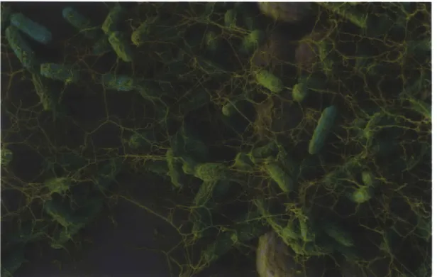
hexagonal features 2.7 pm in height and 3 pm in diameter, separated by 440 nm trenches (designated as "HEX" patterns; Fig. 2.1B). Notably, the spacing of the trenches was more than one standard deviation (SD) below the measured mean cell diameter. We grew wild-type bacteria on smooth and HEX-patterned PDMS coupons submerged in M63+ medium. These static cultures were incubated for 24 h at 37 'C and then prepared for scanning EM imaging (Fig. 2.1 C, D). Surprisingly, our observations indicated that there was more surface coverage by the E. coli cells on HEX than on flat surfaces. Furthermore, we noted the presence of a dense, fibrous network surrounding the surface-bound cells.
A-
B
Figure 2.1. Bacterial surface adhesion. (A) Schematics of en face (top) and cross-sectional (bottom) views of rod-like bacteria adhering to flat (left) or patterned (middle) substrates and attachment of bacteria possessing surface appendages to a patterned substrate (right), when the length scale of surface topography is on the order of the bacterial diameter. (B) Scanning EM of a HEX PDMS substrate. Scale bar is 2 pm. Inset is orthogonal view at lower magnification. Inset scale bar is 5 pm. (C) Scanning EM of wild-type E. coli grown for 24 h at 370C in M63+ on a HEX-patterned PDMS substrate. Inset is higher magnification. (D) E. coli grown on flat PDMS substrate. Inset is higher magnification. Scale bars in C and D are 10 pm, inset scale bars in C and D are 2 pm.
Matrix components are salient characteristics of most biofilms; indeed, many bacterial species, including E. coli, have been shown to produce several kinds of polysaccharide, protein, and DNA elements in their biofilm matrices. Since the E. coli cell bodies could not access the entire surface of HEX-patterned substrates, it seemed likely that the observed fibers were helping to augment surface attachment to these topographies. To identify the components of the fibrous material, we constructed and obtained mutants with deletions in known biofilm-associated genes, including: wcaF, coding for the polysaccharide colanic acid; bcsA, a cellulose gene; fimA, coding for type I pili; csgA, coding for curlin amyloid fiber subunits; and flhD, the master regulator for flagella synthesis. We grew each of these strains on HEX substrates as described above and imaged with a scanning EM (Fig. 2.2). All of the mutants resembled the wild-type, except for
AflhD, which had a clear lack of associated fibers. FlhD is a transcriptional regulator that
controls expression of many genes including, but not limited to the flagellar apparatus [12]. To determine if flagella or motility specifically were required for better colonization, we constructed deletion mutants for fliC, the flagellin subunit gene and motB, a motor protein that enables flagellar rotation (Fig. 2.2). The AfliC mutant displayed the same phenotype as seen for AflhD, indicating that the observed effect was due to the absence of flagella. Furthermore, the AmotB motility mutant was able to colonize the surface and it generated fibers similar to the wild type. These findings are consistent with the fibrous material being predominantly composed of flagellar filaments.
fl- ,A
Figure 2.2. Phenotypes of biofilm-associated knockouts on patterned PDMS. Wild-type (ZK2686) and mutant derivatives (as labeled) were grown on topographically patterned PDMS substrates for 24 or 48 h at 37 'C in M63+. Scanning EM micrographs depict the morphological properties of each strain. Scale bar is 2 gm.
We examined the time-course of adhesion for wild type and AfliC mutant on HEX-patterned versus flat surfaces to determine if flagella play a role in the increased adhesion of the wild type to HEX surfaces observed in Figure 2. 1B, C. To analyze this, we plotted the ratio of biomass on HEX/flat surfaces at different times over a period of 24 h. For both strains, there was
a reduction in adhesion to the HEX surface versus the flat surface at 2 h, the earliest time analyzed (Fig. 2.3A). At later time points, however, the wild-type cells accumulated more on the HEX surfaces than on the flat surfaces. In contrast, the AfliC strain showed more biomass on flat surfaces than on HEX surfaces at all time points, though the ratio approached unity toward 24 h. These data suggest that while adherence of wild-type cells appears to benefit from surface patterning, the cells lacking flagella are unable to exploit the additional surface area provided by the micro-topography.
B
4 MWT I AmotB 2 0 H Flat HEXD
140aa
120 -+E. coli
E
150 - asis-0 C --M63+ only I 120 8 0 4Z9 20 -- i 0 0 2 4 6 0 4 8 "iMM h) o WT md -- WT rmc o AlMOS OFv -- AmosB Mc aA Mt di -*-4-SC rS o control adv -0 control rec 2 1.5 1 0.5 0
Flat HEX HEX wet nonwet
Figure 2.3. Colonization of patterned substrates by wild type and non-motile bacteria and its relationship to surface wetting. (A) Time-course of relative biovolume on patterned (HEX) relative to flat substrates for wild type and AfliC cells at various times. (B) Biovolume of cells adherent to submerged flat or HEX-patterned PDMS coupons after 24 h. Error bars indicate SEM of at least 5 independent experiments (5 z-stacks per experiment). ***, p<0.001 by Student's two-tailed t-test, compared to WT. (C) Optical images of the advancement of the wetting front during culture, at 4 h. The meniscus (dotted line) advances through the patterned
A
1.8 1.6I
1.4 1.2 0.4 0.2 0 -&WT 10W__
Tkm (h) Time (h)F
substrate, exposing channels between surface features, thus increasing available surface area for bacterial attachment. The area to the left of the dotted line is fully wetted, whereas the area to the right of the line contains air pockets. The white arrow indicates the direction of the wetting front progression. Thirty minutes have elapsed between the images on the left and right. Scale bar is 20 pm. (D) Contact angle hysteresis measurements of substrates that have been exposed to growing E. coli cells for increasing incubation periods or M63+ medium only, followed by sonication. Error bars represent SD. ***, p<0.0 01 by Student's two-tailed t-test, comparing WT
to control at 2h.
ttt,
p<0.001 by Student's two-tailed t-test, comparing WT to AfliC or AmotB.See also Figure 3E. (E) Advancing (open symbols) and receding (closed symbols) angles of droplets of distilled water were measured on these substrates after sonication of surfaces to remove adherent bacteria. (F) Biovolume of cells adherent to submerged substrates after 2 h of culture. HEX substrates were either force-wet using ethanol followed by rinsing (HEX-wet), or left in their nonwetting state (HEX-nonwet). Error bars represent SD. * p<O.001 by Student's
two-tailed t-test, comparing HEX-wet to HEX-nonwet.
It had previously been shown that mutants lacking flagella (AfliC) or unable to rotate flagella (AmotB) produce less robust biofilms than wild-type strains on flat surfaces [8]. To determine if this was also the case when cells are grown on patterned surfaces, we compared biofilm production of the wild type to that of the AmotB and AfliC strains at 24h. These strains were each grown on submerged flat and HEX-patterned PDMS coupons, as above, for 24 h and adherent cells were fixed and quantified by acquiring confocal z-stacks of hydrated cells. Consistent with previous findings [8], the AmotB and AfliC strains each had significantly less biomass than the wild type regardless of surface topography (Fig. 2.3B). This indicates that motility, not just the presence of flagella per se is required for optimal biofilm formation. Interestingly, while still less adhesive than wild type, the AmotB mutant accumulated more biomass than AfliC on both flat and patterned surfaces suggesting that the presence of flagella, albeit paralyzed ones, may play a role in adhesion (Fig. 2.3B).
While biofilm formation at 24 h was more robust in the wild type, both the flagella mutant and the wild type showed a dramatic preference for adherence to the flat substrate at the earliest (2 h) time point (see Fig. 2.3A). Why was the patterned substrate successful at preventing adhesion at early time points, but not later? To better understand this phenomenon,
we examined the substrates microscopically during the adhesion process. We noted that the HEX surface remained non-wetting, harboring trapped air bubbles within the trenches until approximately 4 h, when the medium began to displace the entrapped air bubbles (Fig. 2.3C). This property, where the liquid phase rests atop a composite interface of air and solid, is termed the Cassie-Baxter wetting state, and is characteristic of superhydrophobic micro or nano-textured surfaces such as the lotus leaf [13, 14]. We observed that this effect was lost over time in the presence of bacteria, resulting in complete wetting of the substrates (termed the Wenzel wetting state).
The difference in wetting properties of sterile medium versus medium with bacteria could be due to a change in surface tension of the medium or due to a change in surface energy of the substrate. Using the pendant drop method [15], we measured the surface tension of the M63+ medium to initially be 70.1 0.6 mN/m at 20*C. After 16 h of conditioning with E. coli, the medium was extracted by centrifugation and filtration and its surface tension was measured to be 69.1 0.5 mN/m at 200C. Although measurements were not carried out at culture temperatures, the predicted decrease in surface tension by increasing to 37*C would be unlikely to cause wetting. Furthermore, conditioned medium did not cause wetting of fresh HEX substrates incubated at 370C.
To determine if bacteria produce a substance that functions to precondition the surface and allow for increased wetting, we measured contact angle hysteresis (CAH) of water on dry HEX substrates after they had been placed in the presence of growing E. coli for 0 - 8 h (Fig. 2.3D). CAH is the difference between advancing and receding contact angles, and can be due to changes in surface energy or changes in surface topography. This value changes dramatically during Cassie-Baxter to Wenzel wetting transitions, so serves as a sensitive indicator of wetting
state [16]. All bacteria were removed by sonication before measurements, so as to avoid measuring properties of the bacteria themselves. CAH was significantly increased (p<0.001) by 2 h of culture for all strains compared to control and continued to increase over the period measured. The medium-only controls did not wet during this period, resulting in the maintenance of a relatively low CAH. This difference in surface wetting properties indicates that the bacterial modification of the substrate surface energy (rather than modification of the liquid medium) is the dominant contributor to wetting properties. By 5 h, we observed a large increase in CAH for the wild type, but this was still significantly higher than CAH of the two mutant strains (p<0.001). A commensurate increase did not happen until 6 or 8 h in the AmotB and AfliC strains, indicating that the surface wetting brought about by bacteria-surface interactions is aided by the presence of motile flagella. Examining advancing and receding contact angles individually (Fig. 2.3E), we observed that all samples maintained a relatively constant advancing contact angle over time with a slight downward trend, likely due to surface conditioning by bacteria. As the surface transitioned from the Cassie-Baxter to the Wenzel wetting state, there was an increase in drop pinning, which was measured as a decrease in the receding contact angle [16]. It is this receding angle that changed more drastically and differentiated the behavior of the wild-type from that of the mutant strains.
On flat substrates, the lack of microstructure prevents the possibility of a Cassie-Baxter wetting state, thus the surface is entirely available to the cells from the outset. On the structured surfaces, wetting did not significantly occur until after 2 h. During this initial period, only the structure tips (not the trenches) are available to cells, as they cannot penetrate the air-water interface. Only upon wetting does the complete surface become available. We tested this by force-wetting the HEX substrates prior to inoculation with wild-type cells. At 2 h, we measured
adherent biovolume, noting a significant increase in attachment to pre-wet surfaces compared with the untreated HEX substrates (Fig. 2.3F).
We reasoned that if the superior attachment by wild-type cells to HEX substrates is due to the access provided by their flagella, then varying feature size would only affect overall adhesion and biomass insofar as it changes overall surface area. In contrast, the AfliC cells would experience a reduction in attachment whenever portions of the substrate remained inaccessible to the cell body. We compared attachment of wild-type cells and AfliC mutants to substrates as we varied the feature diameters and spacings, maintaining a constant pitch (Fig. 2.4A). Indeed, the AfliC cells increased attachment as the feature spacing became larger, eventually surpassing their adhesion to flat substrates. For wild-type cells, the increased spacing had the opposite effect. Whereas the wild type cells had over four times the biomass of AfliC cells on HEX-patterned substrates (0.44 pm spacing), this difference was less than 2-fold on substrates with 1.70 pm spacing (Fig. 2.4B).
A
Aflic.3.
B -5 4- -- wr/Afic -4 [=WT E ~M fic ;3 -300
0
0 0.5 1 1.5 2 00 0 Spacing (pm)Figure 2.4. Differential response of wild-type and AfliC cells to changes in surface feature spacing. (A) Schematic of underlying surface topography, illustrating increasing spacing with constant pitch (left column) and the scanning EM and confocal images of wild type and AfliC cells (middle and right columns) grown for 24 h on corresponding PDMS substrates and then fixed. Samples were imaged in the hydrated state using confocal microscopy and then
dehydrated and imaged using scanning EM. Scanning EM images are shown, with thickness maps derived from confocal z-stacks shown in the corresponding inset (color mapping is for clarity and has arbitrary scale). Scale bar is 5 pm. (B) Biovolume was quantified for each topographical pattern and normalized to projected surface area. Biovolumes are shown for wild type and AfliC mutants (plotted as bars), as well as their ratios (black squares connected by
lines). Error bars indicate SEM of >26 data points.
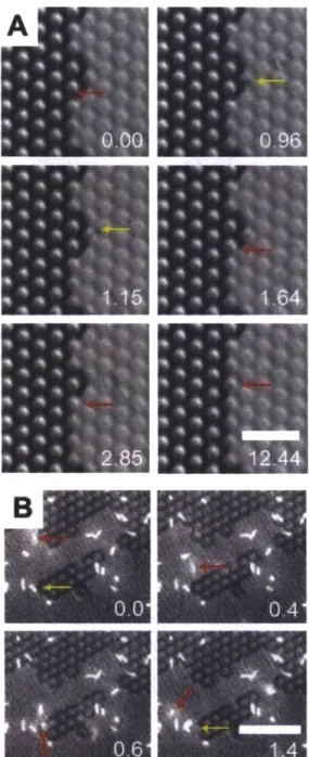
It appears that the benefit of having flagella is greater during adhesion to substrates with trenches smaller than the cell body than during adhesion to flat substrates or substrates with larger feature spacings. This phenomenon is unlikely to be due solely to the motility provided by flagella, since surface access should have been similar for all substrates tested. To further investigate the role of flagella in adhesion of wild-type cells to topographical substrates, we examined their dynamics in live cells during the adhesion process by fluorescently staining their flagellar filaments [17]. Wild-type E. coli cells were placed in contact with HEX substrates and allowed to adhere. During the adhesion process, we observed attachment behavior. We noted that some cells were adhered by their flagella and exhibited tethering behavior (Fig. 2.5A). Additionally, some flagella inserted between surface features and attached within the sub-micron trenches, which was consistent with scanning EM findings, where flagella were observed to adhere between features (Fig. 2.5A-C). After 4 h of incubation, we observed alignment of some flagella with the underlying substrate. There was a tendency of flagella to orient along the planes of symmetry of the substrate (Fig. 2.5D), which implies that the filaments were
Figure 2.5. Flagellar appendages 'reach' and 'grasp' to improve surface adhesion on patterned surfaces. (A) Selected frames from a video of Alexa 594-stained cells taken at 15 frames per second. Times are given (in seconds) in each panel. In frame one, note that the upper cell (red arrow) is fully adherent (it remains stationary throughout the frames). Notably, its middle flagellum is nestled between the surface features slightly out of focus, since it is below
the imaging plane. The other two flagella are resting atop the surface features in the focal plane. The lower bacterium is in the early steps of adhesion, tethered by one flagellum (yellow arrow), also nestled between the surface features. Its remaining free flagella continue to rotate rapidly until 2.45 s, at which time another (short) flagellum makes surface contact (yellow arrow). The cell body continues to slowly reorient as it makes more intimate contact (via other flagella and/or pili) and settles in its final position at 13.14 s. Scale bar is 5 pm. Axes of symmetry of the substrate are indicated by arrows in the bottom-right image. (B) Selected frames from a video of Alexa 594-stained cells taken at 15 frames per second. Times are given (in seconds) in each panel. Note that the cell body of E. coli is attached and tethered via aligned flagellar filaments along the crystallographic axes of the substrate. The flagellum on the left of the cell continues to move between frames, yet is confined to move within the trench between the surface features. Scale bar is 5 pim. Axes of symmetry of the substrate are indicated by arrows in the bottom-right image. (C) Scanning EM images of the same field of wild-type E. coli grown on HEX PDMS posts. Images were acquired with secondary electron (left) and inlens detectors (right). The difference in shadowing between the two images highlights the depth of penetration of the flagellar filaments into the channels between the surface features. Note specifically the region indicated by the red arrow. Scale bar is 2 pm. (D) Image of Alexa 594-stained cells after initial adhesion to HEX substrate. Scale bar is 10 pm. Inset shows angular histogram of filament orientations of adherent E. coli. The histogram illustrates preferential alignment of filaments along the two of the three planes of symmetry of the hexagonal surface pattern. Axes of symmetry of the substrate are indicated with red arrows.
DISCUSSION AND CONCLUSION
We herein set out to characterize the bacterial adhesive response to substrates with regular surface topography. Specifically, we were interested in the role of surface appendages in this response. We tested the hypothesis that surface feature length-scale could, on its own, reduce bacterial attachment by reducing available surface area. Indeed, we observed that sub-micron trenches between features were able to reduce attachment of mutants without flagella. However, the geometric simplification of bacteria as rigid rods becomes invalid when applied to wild-type bacteria possessing surface appendages. Wild-type E. coli achieved better adhesion to surfaces with trenches than to flat surfaces. Because their surface appendages could access the trenches, the wild-type cells actually experienced an increase in surface area on HEX substrates compared to flat, while the non-flagellated cells experienced the predicted decrease (Figure 2.6). These
results indicate that bacterial adhesion to patterned surfaces is far more nuanced than anticipated by simplistic geometric models.
50 -40 -- W.. -f 30 ~20 . 10 0 0 0.5 1 1.5 2 2.5 3 3.5 Inter-post Spacing (rm)
Figure 2.6. Accessible substrate surface area. Simulation of available surface area for wild type and AfliC strains in a unit cell of HEX substrates with post spacing varying as indicated, based on average diameter and standard deviation. Note that wild-type cells can access inter-post regions using flagella, but AfliC cells cannot.
During the early adhesion process, we observed that all strains adhered more to flat substrates than to HEX substrates. Upon microscopic inspection, we could observe a wetting front progressing across the sample at 4-6 h into incubation, consistent with an initial Cassie-Baxter wetting state. Similarly, substrates removed from culture at 2 h appeared to be non-wetting and the medium was observed to easily cascade off the substrates. At later time points, the samples remained wet upon removal. These observations were consistent with CAH measurements taken over 8 h of culture, showing a steady increase in hysteresis over time. Given this finding, it appears that for short durations the meniscus forming over each trench prevents bacterial adhesion and reduces surface availability to only the tips of the bumpy surface projections. At that time structured surfaces inhibit bacterial attachment. AfliC and AmotB mutants were delayed in surface wetting of HEX substrates compared to the wild type, as measured by CAH. We conclude that motile flagella increase the probability of generating