leads to active N-phenyl-4,5-dihydrooxazol-2-amines alkylating
-tubulin Glu198 and prohibitin Asp40
Anna Trzeciakiewicza,b, Sébastien Fortina,b,c, Emmanuel Moreaua,b, René C.-Gaudreaultc,
Jacques Lacroixc, Christophe Chambond, Yves Communale, Jean-Michel Chezala,b,
Elisabeth Miot-Noiraulta,b, Bernadette Bouchona,b, Françoise Degoula,b,*
a U990 INSERM, Rue Montalembert, BP 184, 63000 Clermont-Ferrand, France
b Clermont Université, Université d’Auvergne, Imagerie moléculaire et thérapie vectorisée,
BP 10448, 63000 Clermont-Ferrand, France
c CR-CHUQ, Hôpital Saint-François d’Assise, Université Laval, Unité des Biotechnologies
et de Bioingénierie, Québec, Canada G1L 3L5
d INRA Clermont-Ferrand/Theix, Plate-forme Protéomique, 63122
Saint-Genès-Champanelle, France
e Centre Jean Perrin, Rue Montalembert, 63011 Clermont-Ferrand, France
* Corresponding author at: U990 INSERM, Rue Montalembert, BP 184, 63000 Clermont-Ferrand, France. Tel.: +33 0473150817; fax: +33 0473150801.
E-mail addresses: trzeciakiewicz@gmail.com (A. Trzeciakiewicz),
sebastien.fortin.1@ulaval.ca (S. Fortin), Emmanuel.Moreau@u-clermont1.fr (E. Moreau),
rene.c-gaudreault@crsfa.ulaval.ca (R. C.-Gaudreault), Jacques.Lacroix@crsfa.ulaval.ca (J.
Lacroix), Christophe.chambon@clermont.inra.fr (C. Chambon),
Yves.COMMUNAL@cjp.fr (Y. Communal), j-michel.chezal@u-clermont1.fr (J.-M. Chezal), elisabeth.noirault@inserm.fr (E. Miot-Noirault), bernadette.bouchon@inserm.fr
Abstract
The cyclization of anticancer drugs into active intermediates has been reported mainly for DNA alkylating molecules including nitrosoureas. We previously defined the original cytotoxic mechanism of anticancerous N-phenyl-N’-(2-chloroethyl)ureas (CEUs) that involves their reactivity towards cellular proteins and not against DNA; two CEU subsets have been shown to alkylate -tubulin and prohibitin leading to inhibition of cell proliferation by G2/M or G1/S cell cycle arrest. In this study, we demonstrated that cyclic derivatives of CEUs, N-phenyl-4,5-dihydrooxazol-2-amines (Oxas) are two- to threefold more active than CEUs and share the same cytotoxic properties in B16F0 melanoma cells. Moreover, the CEU original covalent binding by an ester linkage on -tubulin Glu198 and prohibitin Asp40 was maintained with Oxas. Surprisingly, we observed that Oxas were spontaneously formed from CEUs in the cell culture medium and were also detected within the cells. Our results suggest that the intramolecular cyclization of CEUs leads to active Oxas that should then be considered as the key intermediates for protein alkylation. These results will be useful for the design of new prodrugs for cancer chemotherapy.
Keywords: N-Phenyl-N’-(2-chloroethyl)ureas; N-Phenyl-4,5-dihydrooxazol-2-amines; -Tubulin; Prohibitin; Alkylation; Cyclization
1 Introduction
A major challenge for improving cancer chemotherapy is to design and synthesize new drugs that can selectively target cancer cells and bypass cancer chemoresistance systems. In this context, we havedeveloped protein alkylating agents referred to as N-phenyl-N’-(2-chloroethyl)ureas (CEUs) [1,2]. Two CEU subsets have been shown to selectively alkylate proteins through a unique ester linkage involving Glu198 on -tubulin and Asp40 on prohibitin [3,4]. Both proteins are strongly involved in the regulation of cell growth and cell cycle progression [5,6]. Tubulins are the classical targets for antimitotic drugs such as colchicine, vinblastine and paclitaxel [7] blocking the G2/M transition of the cell cycle. Prohibitin, a chaperon protein involved in gene expression, regulates cell cycle and apoptosis, and has been considered as a tumor suppressor [5,8]. However, no consensus for prohibitin function has been established since prohibitin is also required for primary and cancer cell proliferation [9,10]. The decreased G1/S transition in CEU treated cells, associated with prohibitin alkylation is not fully understood and could be linked to either a mitochondrial dysfunction [11] or a nuclear repression of E2F1-mediated transcription [12]. -Tubulin alkylating CEUs induce a G2/M arrest and have obvious antimitotic effects on different types of tumors both in vitro and in vivo [13-17]. Hence, the molecular events underlying CEU antiproliferative activity rely on the functionality of the targeted protein [3].
The requirement of the chlorine atom in the CEU -tubulin alkylation mechanism was clearly established using conventional structure-activity relationship (SAR) studies showing that N-phenyl-N’-ethylureas were devoid of cytotoxicity and unable to alkylate -tubulin [1]. Recently, we unexpectedly found that N-phenyl-4,5-dihydrooxazol-2-amines (Oxas) were more active than their CEU counterparts and still alkylating -tubulin as demonstrated by western blot analysis [18]. It has been reported previously that the presence of oxazolinic heterocycles improves the efficiency of molecules developed for pancreatic cancer treatment [19] as well as of drugs inhibiting angiogenesis [20].
Here, we have determined the cytotoxicity mechanisms of three different Oxas and characterize their covalent binding to target proteins in B16F0 melanoma cells at the molecular level. Our results clearly showed that Oxas and their parent CEUs have the same -tubulin and prohibitin amino acid targets. We further observed, for the first time, that the cyclization of CEUs into Oxas occurs spontaneously in the culture medium and that these Oxa forms were present within the cells. These data suggest that Oxas could be the reactive intermediates of CEUs.
2. Materials and methods
2.1. Chemicals
N-(4-Iodophenyl)-N’-(2-chloroethyl)urea (ICEU), N-(4-iodophenyl)-4,5-dihydrooxazol-2-amine (IOxa), N-(4-tertbutylphenyl)-N’-(2-chloroethyl)urea (tBCEU), N-(4-tertbutylphenyl)-4,5-dihydrooxazol-2-amine (tBOxa), N-(4-cyclohexylphenyl)-N’-(2-chloroethyl)urea (cHCEU), and N-(4-cyclohexylphenyl)-4,5-dihydrooxazol-2-amine (cHOxa) were synthesized as previously described [21,22]. All compounds were dissolved in DMSO at a concentration of 40 mM and stored at -20 °C. The experiments were all performed with a final concentration of 0.5% DMSO in culture medium.
2.2. Cell culture
B16F0 murine melanoma cell line was purchased from the American Type Culture Collection (Manassas, VA, USA). Cells were cultured in DMEM (Invitrogen, Cergy-Pontoise, France) supplemented with 10% fetal bovine serum (Biowest, Paris, France), 1 mM non-essential amino acids (Invitrogen), 1 mM sodium pyruvate (Invitrogen), and 4 g/mL gentamicin (Invitrogen). The cells were maintained at 37 °C in a moisture-saturated atmosphere containing 5% CO2.
2.3. Antiproliferative activity
Cell cytotoxicity was assessed using the resazurin assay as previously described [23]. To determine growth inhibitory concentrations, which is the concentration of the drug required
to inhibit 50% of the tumor cell growth (GI50), 3.5 x 10³ B16F0 cells were seeded into
96-well microtiter plates (NunclonTM, Nunc, Roskilde, Denmark) and maintained at 37 °C in a
moisture saturated-atmosphere containing 5% CO2 for 24 h and then incubated with
escalating drug concentrations (1-50 M) for 48 h. Fluorescence (excitation/emission: 530/590 nm) was measured with a microtiter plate fluorescence reader (Fluoroskan Ascent FL, Thermo Labsystems, Helsinki, Finland).
2.4. Cell cycle progression analysis - flow cytometry
Cell cycle progression was analyzed by flow cytometry with propidium iodide staining as previously described [3].
2.5. Determination of -tubulin and prohibitin alkylation by western blot analysis -Tubulin alkylation was determined by immunoblotting. Briefly, B16F0 cells were harvested from 60 mm Petri dishes (6 x 10⁵ cells/dish), proteins were extracted in RIPA
buffer with EDTA-free protease inhibitor cocktail (Roche Diagnostics, Meylan, France). Protein concentration was determined by Bradford assay using a CooAssay Protein Determination Kit (Interchim, Montluçon, France). For -tubulin alkylation, 50 g of total protein was separated on 10% SDS-polyacrylamide gels. For prohibitin alkylation, 100 g of urea extracted proteins (see Section 2.7) were submitted to IEF on 7-cm IPG strips pH 3-10 (BioRad, Hercules, CA, USA) at 8000 V/h, followed by a 12% SDS-polyacrylamide gel electrophoresis. Gels were transferred onto nitrocellulose membranes (Immobilon NC; Millipore, St-Quentin-en-Yvelines, France). The membranes were blocked in 5% non-fat dry milk in TBS-T (0.1% Tween 20) buffer and the blots were then incubated with a monoclonal anti--tubulin antibody (Anti TUB2.1; Sigma, St-Quentin Fallavier, France) or a monoclonal anti-prohibitin antibody (Abcam, Paris, France), then probed with anti-mouse horseradish peroxidase (HRP) conjugated secondary antibody (Dako, Glostrup, Denmark). The blot signals were detected by chemiluminescence (ECL, Amersham GE Healthcare, Buckinghamshire, UK).
B16F0 cells were seeded in 8-well culture glass slides (BD Biosciences, Erembodegem, Belgium) at a density of 1 x 10⁵ cells/mL for 24 h. Cells were then treated with 0.5- to
1-fold GI50 corresponding to 10 M ICEU, 3 M IOxa, 8 M tBCEU, 8 M tBOxa, 10 M
cHCEU or 5 M cHOxa in vehicle (0.5% DMSO) for 24 h. They were washed twice with PBS and fixed in methanol for 10 min at -20 °C. Non-specific sites were blocked using 5% goat serum for 30 min. Cells were incubated in 1/1000 diluted mouse monoclonal anti--tubulin IgG (Sigma-Aldrich, St. Louis, MO, USA) for 2 h at room temperature before incubating with anti-mouse IgG labelled with Alexa Fluor 488 (Invitrogen) for 1 h. Slides were washed in PBS and then incubated in 10 g/mL Hoechst 33342 for 5 min to visualize the nuclei. Microtubules and nuclei were observed with a fluorescent microscope (Olympus, Center Valley, PA, USA) using FITC and DAPI filter sets, respectively.
2.7. Two-dimensional electrophoresis (2DE)
For the analysis of -tubulin or prohibitin alkylation, two-dimensional electrophoresis was conducted as described by Bouchon et al. [4].
2.8. Mass spectrometry analyses of protein digests
MALDI-TOF MS analyses were performed according to the published procedure on trypsin digests [4] on a Voyager-DE Pro mass spectrometer (ABSciex, Les Ulis, France). MALDI/PSD analysis was performed to characterize CEU binding on prohibitin [3].
Nano ESI-MS/MS was used to localize and characterize -tubulin alkylation. Analyses were performed essentially as described previously [4] by direct infusion of the peptide digest. The MS/MS data from the non-alkylated and alkylated peptides were interpreted manually, assuming the modified masses. Fragments are assigned according to the nomenclature of Roepstorff and Fohlman [24].
2.9. Stability and cellular uptake of drugs - HPLC
Stability and cellular uptake in B16F0 cells was determined by HPLC. Cells were seeded in 100 mm Petri dishes (2 x 10⁶ cells/dish) and cultured for 24 h. They were then exposed to
5- to 10-fold GI50 concentration of drugs, i.e. 100 M (1 mol) ICEU, 30 M (0.3 mol)
IOxa, 80 M (0.8 mol) tBCEU, 80 M (0.8 mol) tBOxa, 100 M(1 mol) cHCEU, 25 M (0.25 mol) cHOxa in 0.5% DMSO in Opti-MEM medium supplemented with CaCl2
for 24 h (T24). Culture medium aliquots were collected in the presence or absence of cells, extracted with 1 volume 0.2% trifluoroacetic acid (TFA) in acetonitrile (ACN) and centrifuged at 13,000 x g for 5 min. For cell lysates, cells were rinsed three times using PBS, scraped, centrifuged at 800 x g for 10 min and the pellets were stored at -80 °C. Free-intracellular drugs were then extracted in 0.1% TFA in ACN /H2O (v/v) and this solution
was further frozen, thawed and sonicated for 15 min. This cycle was repeated twice. After centrifugation at 13,000 x g for 5 min, cellular extracts and previously prepared culture media were analyzed on a HP 1100 series Agilent HPLC (Agilent, Les Ulis, France) equipped with diode array detector using a Kromasil 5 particles C18 column (Interchim, Montluçon, France) at a flow rate of 0.4 mL/min. The gradient used was: 0-3 min: 0.1% TFA/40% ACN in H2O; 3-13 min: linear gradient to 0.1% TFA/100% ACN; and 13-23
min: 0.1% TFA/100% ACN. The column was reequilibrated after each run for 10 min. Drug concentration was assessed at 255 nm and calculated by the peak area compared to standard calibration curve. Each UV peak obtained in HPLC was monitored at least once by ESI-MS/MS to confirm the structure of the product.
3. Results
3.1. Antiproliferative and alkylating properties of CEUs and Oxas in B16F0 melanoma cells
Both CEUs and Oxas exhibited antiproliferative activity in the micro molar range (3-17 M) with GI50 being lower for Oxas when compared to the respective CEUs (Table 1).
Similarly to their CEU counterparts, IOxa and tBOxa induced a cell cycle arrest at the G2/M transition while cHOxa leads to G1/S arrest with 90% of cells accumulated in G0/G1 phase (Supplementary Fig. 1). On western blots, both ICEU/IOxa and tBCEU/tBOxa induced modification of -tubulin migration, which was split into two bands suggesting protein alkylation (Table 1). Incubation of cells with cHCEU/cHOxa changed the pI of
prohibitin, indicating its alkylation (Table 1). -Tubulin alkylating agents such as ICEU, IOxa, tBCEU and tBOxa altered microtubule structure as compared to control conditions. Immunofluorescent staining showed disorganization of the cytoskeleton architecture and the presence of punctuated tubulin aggregates that were not observed in controls (Table 1). Table 1 Anti-proliferative and alkylating properties of CEUs and Oxas in B16F0 melanoma cells.
GI50: drug concentration inhibiting 50% of cell growth; -tubulin alkylation: determined by western blot in
B16F0 cells treated or not (DMSO) for 24h at 5-10 x GI50 drug concentration; microtubule structure:
visualized by immunofluorescence in B16F0 cells treated or not (DMSO) for 24 h at one GI50 drug
concentration; prohibitin alkylation: determined by 2D western blot in B16F0 cells treated or not for 24h at a 5-10 x GI50 drug concentration. Arrows indicate the native form of the protein, asterisks indicate the alkylated
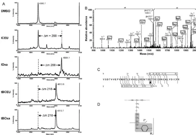
Fig. 1. -Tubulin alkylation by CEUs and their corresponding Oxas. (A) Comparative analyses by MALDI-TOF-MS of tubulin high mass peptides obtained in tubulin tryptic digest after DMSO treatment of cells or after ICEU, IOxa, tBCEU or tBOxa treatment. The low peak intensity observed in the case of ICEU was a partial dealkylation before analysis due to alkaline conditions during trypsin digestion. (B) ESI-MS/MS spectrum of the alkylated peptide obtained by IOxa. (C) Fragmentation pattern along the peptidic chain with localization of the alkylated amino acid on E24 of this peptide. (D) Fragmentation pattern in the bound structure confirming the opening of the oxazolinic heterocycle and its resulting linear structure. Z stands for the bound oxazoline derivative, whose structure is developed in scheme D with a grey background. Z* corresponds to a fragmentation along the bound structure, as indicated in the scheme.8, loss of one water molecule.
3.2. Oxas alkylate -tubulin and prohibitin on the same amino acids as CEUs
The presence of two -tubulin bands after SDS-PAGE of proteins extracted from B16F0 cells treated with IOxa and tBOxa (Table 1) suggested that the drugs bound covalently to
the protein. To characterize the site of interaction, MS analyses were performed on -tubulin treated respectively with ICEU, IOxa, tBCEU, tBOxa (Fig. 1A). Alkylated -tubulin spots isolated from 2DE gels were digested by trypsin and analyzed using MALDI-TOF-MS as previously described [4]. The native -tubulin peptide [Val175-Arg213] on which alkylation had been shown to occur had an apparent molecular weight of 4595.7 (Fig. 1A), while the peptide alkylated by ICEU presented a m of 288, as already observed [4]. The same m/z shift was observed on tubulin extracted from cells treated with IOxa (Fig. 1A). To localize the alkylation site, a nano ESI-MS/MS analysis was performed on this modified peptide (Fig. 1B). The fragmentation pattern allowed localizing the urea derivative on Glu24 of the analyzed peptide corresponding to -tubulin Glu198 (Fig. 1C), as previously shown for ICEU itself [4]. An intense fragmentation (m/z 1546.4, Fig. 1B) occurred at the peptide-like bond of the urea chain bound to Glu198 thus demonstrating the linearity of the added structure (Fig. 1D).
The same approach was used for tBCEU and tBOxa. For both drugs a m/z shift corresponding to the addition of the tBurea derivative was observed (m = 218) indicating alkylation on the same [175-213] -tubulin peptide (Fig. 1A).
Prohibitin alkylation was determined using prohibitin spots isolated from 2DE gels (Fig. 2A). When cHCEU is bound to its target, prohibitin exhibits a modified pI, as compared to the native protein [3]. The m/z 720 corresponding to prohibitin peptide [Ala36-Arg41] observed in the native protein of control cells was shifted in cHCEU- and cHOxa-treated cells to m/z 964 (Fig. 2B). The two modified peptides were further characterized by MALDI-PSD analysis (Fig. 2C). Their fragmentation patterns were identical in cHCEU and cHOxa treated cells, and revealed a modification on Asp5 of this peptide, corresponding to Asp40 of the protein.
Fig. 2. Prohibitin alkylation by cHCEU and its corresponding cHOxa. (A) Prohibitin spots localized on 2D gels. (B) MALDI-TOF-MS analysis of these spots. (C) Fragmentation spectra obtained from the two peptides having a m/z 964. (D) Fragmentation of peptide [AVIFDR], D being alkylated by a cyclo-hexyl-phenyl urea. The bound developed cyclohexyl structure is shown with a grey background to localize M* and M** fragments along the urea chain. ‘‘b’’ and ‘‘y’’ fragments were obtained from N and C-terminal fragmentation, respectively.
3.3. CEU and Oxa stability in culture medium and uptake in B16F0 cells
ICEU/IOxa, tBCEU/tBOxa and cHCEU/cHOxa were separated by HPLC analysis and exhibited retention times of 12.9/4.3 min, 14.2/6.3 min and 16.2/9.8 min, respectively (Fig. 3A). CEUs were spontaneously transformed into Oxas, which remained stable for 24 h in culture media in presence (T24+) or in absence (T24-) of cells (Fig. 3B, Table 2). All the labelled peaks were recovered and analyzed by MS to confirm their identity. The HPLC protocol allowed the recovery of 70-98% of the parent molecules incubated for 24 h in the
cell culture medium without cells (Table 2). Using the same HPLC conditions in the presence of cells, the percentage of detected molecules in the medium was lower suggesting an uptake of 7.4-36.5% of CEUs and/or Oxas by B16F0 cells (Table 2). However, the levels of the intracellular free drug were low (<10%) suggesting that the major part of CEUs and Oxas was bound to proteins or biomolecules [25]. Drugs embedded in the cellular membranes and/or covalently bound to proteins were not detected by this procedure optimized to recover the free compounds. Another possibility was the metabolism of these drugs as our analyses were focused only on CEU and Oxa detection. We can also observe that the percentage of spontaneous CEU cyclization into Oxa was dependent on the molecular structure of the drug (6%, 26% and 49% for cHOxa, IOxa and tBOxa formation, respectively; T24-) (Table 2).
Table 2 Drug recovery in culture medium and in B16F0 cells.
nd: not detected; -: not applicable.
Concentrations of each compound were determined by HPLC and expressed as the percentages of the initial drug input. The culture medium was collected after 24 h of incubation in the absence (T24-) or presence of cells (T24+). Drug recovery in cell pellets was calculated from drug recovery in other compartments and reported to the maximal amount of drugs recovered without cells (T24-). The values represent the mean of at least 2 independent experiments.
Fig. 3. Quantitative HPLC analysis of ICEU, tBCEU or cHCEU in B16F0 cells. (A) Analysis of standard compounds. (B) Analysis of cell culture medium after 24 h of ICEU, tBCEU or cHCEU exposure in the absence (T24-) or presence (T24+) of B16F0 cells (C) Analysis of cell lysates obtained from B16F0 cells cultured for 24 h in serum-free Opti-MEM medium (T24+c) in the presence of 100 M ICEU, 80 M tBCEU or 100 M cHCEU. White arrows indicate CEUs, black arrows Oxas.
4. Discussion
The study of the alkylating properties of CEU represents an interesting field of research since xenobiotics able to bind covalently to cellular proteins are relatively uncommon, especially when generating ester linkages with acidic amino acids such as glutamic and aspartic acids [3]. Here, we have demonstrated that Oxa derivatives are covalently linked to the same proteins and the same amino acids as their parent CEUs notably Glu198 for -tubulin and Asp40 for prohibitin (Figs. 1 and 2). CEUs and Oxas exhibited the same
antiproliferative properties (cell cycle arrest, microtubule disruption specifically for antimitotics) (Table 1, Supplementary Fig. 1), however, Oxas were more active than their CEU counterparts. Altogether, these data suggest that both CEUs and Oxas exert their cytocidal activity through similar mechanisms and that the pharmacological effects of CEUs could be linked to their intramolecular cyclization into Oxas.
Due to the absence of differences in the molecular masses of -tubulin or prohibitin adducts obtained either with CEUs or their corresponding Oxas, we could not determine which molecule was involved in the alkylation reaction. However, fragmentation of the modified peptide allowed to show that bound Oxas had a linear structure (Figs. 1 and 2). Moreover, in culture media CEUs were spontaneously transformed into Oxas. The level of these spontaneously formed Oxas in the medium in presence or in absence of cells was similar, unlike the concentration of CEUs that was decreased in the presence of cells suggesting that CEUs can go through the membranes more easily than Oxas, even if they display quite similar log P value (data form ALOGPS; http://www.vcclab.org/lab/alogps/, data not shown). However, the Oxa level in the culture medium for IOxa-, tBOxa- and cHOxa-treated cells was reduced compared to that without cells (26%, 10% and 33%, respectively) showing that Oxas can penetrate into the cells. The low presence of free Oxas in cells and their higher antiproliferative index (GI50) strongly suggest that Oxas are significantly more
reactive with biomolecules than CEUs.
The chlorine atom being necessary to maintain CEU -tubulin capacity to alkylate proteins [1], we can speculate that cyclization of CEUs into Oxas required this atom to confer electro-attractor property. This cyclization step could involve the formation of a carbocation and/or an aziridinic intermediate (Fig. 4) as recent studies demonstrated that N-acyl-2,2-dimethylaziridines could evolve into oxazolinic forms [26]. These two potential intermediates characterized by very short half-lifes, are difficult to observe in a biological context but it has been established that the aziridinium structures were clearly involved in DNA alkylation by N-nitrosoureas and nitrogen mustards [27]. Interactions between -tubulin and CEUs were studied using CoMFA and CoMSIA models [28]. From these studies, it was deduced that the ethylene chloride group of CEU was essential for the
alkylation of Glu198 and that the stabilization of a complex involving the colchicine-binding site and the CEU preceded the completion of the Glu198 alkylation [29]. This intermediate complex should involve the stabilizing contribution of Cys239 among other amino acids [29]. We are unable to conclude if antimitotic CEUs are transformed into oxazolines in the colchicine-binding site of -tubulin, when the urea group interacting with Cys239 [29] stabilizes the molecules, or react spontaneously with acidic amino acids in a nucleophilic substitution reaction. Recent SAR studies modifying the urea moiety of CEUs indicated that only 2-chloroacetamides and 2-chloroacetylureas retained the cytotoxic properties without -tubulin alkylation [30]. In these two series of molecules, the CEU cyclization into Oxa could not occur reinforcing the hypothesis that cyclization might be necessary for -tubulin alkylation.
Fig. 4. Scheme of the putative mechanisms leading to CEU cyclization into Oxas and reactivity of the protagonists with proteins. CEUs, Oxas and their intermediates could react with acidic part of Glu/Asp residues of target proteins.
New CEUs will be synthesized to elucidate this interesting point concerning the alkylation process of Glu and Asp residues and to enhance our knowledge about the specific interactions occurring in the colchicine-binding site. From a biological point of view, previous in vivo data did not allow detecting oxazolinic forms or dechlorinated CEUs in urines of mice treated with tBCEU [31], while some unidentified metabolites were found in blood, tumors and colon of mice treated with ICEU [32]. In vivo investigations will be pursued to identify these metabolites and to determine precisely the active forms of CEUs or Oxas.
In conclusion, one difficulty of this study was the absence of a mass difference between the CEU or Oxa protein adducts revealed by mass spectrometry analyses, another one was
based on the fact that N-phenyl-N’(2-chloroethyl)ureas cyclized spontaneously in cell culture medium. However, we demonstrated that Oxas had similar mechanisms of cell cytotoxicity and were present in the cells treated with CEU derivatives. We therefore suggest that Oxas constitute key structures for the alkylation of cellular proteins. This study confers new insights for drug conception through the cyclization of chlorinated forms into more active oxazolinic ones.
Acknowledgements
This work was supported by the Canadian Institutes of Health Research [Grant # MOP-79334 and MOP-89707], INSERM, and the Université d’Auvergne. A. Trzeciakiewicz is a recipient of a postdoctoral fellowship from the Université d’Auvergne. S. Fortin is a recipient of a studentship from the Canadian Institutes of Health Research [CGD-83623] and FRONTENAC program from le Fond Québécois de la Recherche sur la Nature et les Technologies (FQRNT). We are grateful to the Centre d’Imagerie Cellulaire Santé (CICS) of Clermont-Ferrand for help in fluorescence microscopy.
We gratefully thank Dr Manfred Theisen for reading and correcting of this manuscript.
Appendix A. Supplementary data
Supplementary data associated with this article can be found, in the online version, at doi:10.1016/j.bcp.2011.02.014.
References
[1] Legault J, Gaulin JF, Mounetou E, Bolduc S, Lacroix J, Poyet P, et al. Microtubule disruption induced in vivo by alkylation of beta-tubulin by 1-aryl-3-(2-chloroethyl)ureas, a novel class of soft alkylating agents. Cancer Res 2000;60:985-92.
[2] Mounetou E, Legault J, Lacroix J, C-Gaudreault R. A new generation of N-aryl-N’-(1-alkyl-2-chloroethyl)ureas as microtubule disrupters: synthesis, antiproliferative activity, and beta-tubulin alkylation kinetics. J Med Chem 2003;46:5055-63.
[3] Bouchon B, Papon J, Communal Y, Madelmont JC, Degoul F. Alkylation of prohibitin by cyclohexylphenyl-chloroethyl urea on an aspartyl residue is associated with cell cycle G(1) arrest in B16 cells. Br J Pharmacol 2007;152:449-55.
[4] Bouchon B, Chambon C, Mounetou E, Papon J, Miot-Noirault E, C-Gaudreault R, et al. Alkylation of beta-tubulin on Glu 198 by a microtubule disrupter. Mol Pharmacol 2005;68:1415-22.
[5] Mishra S, Murphy LC, Murphy LJ. The prohibitins: emerging roles in diverse functions. J Cell Mol Med 2006;10:353-63.
[6] Hammond JW, Cai D, Verhey KJ. Tubulin modifications and their cellular functions. Curr Opin Cell Biol 2008;20:71-6.
[7] Zhou J, Giannakakou P. Targeting microtubules for cancer chemotherapy. Curr Med Chem Anticancer Agents 2005;5:65-71.
[8] Mishra S, Murphy LC, Nyomba BL, Murphy LJ. Prohibitin: a potential target for new therapeutics. Trends Mol Med 2005;11:192-7.
[9] Sievers C, Billig G, Gottschalk K, Rudel T. Prohibitins are required for cancer cell proliferation and adhesion. PloS One 2010;5:e12735.
[10] Schleicher M, Shepherd BR, Suarez Y, Fernandez-Hernando C, Yu J, Pan Y, et al. Prohibitin-1 maintains the angiogenic capacity of endothelial cells by regulating mitochondrial function and senescence. J Cell Biol 2008;180:101-12.
[11] Gregory-Bass RC, Olatinwo M, Xu W, Matthews R, Stiles JK, Thomas K, et al. Prohibitin silencing reverses stabilization of mitochondrial integrity and chemoresistance in ovarian cancer cells by increasing their sensitivity to apoptosis. Int J Cancer 2008;122:1923-30.
[12] Joshi B, Ko D, Ordonez-Ercan D, Chellappan SP. A putative coiled-coil domain of prohibitin is sufficient to repress E2F1-mediated transcription and induce apoptosis. Biochem Biophys Res Commun 2003;312:459-66.
[13] Petitclerc E, Deschesnes RG, Cote MF, Marquis C, Janvier R, Lacroix J, et al. Antiangiogenic and antitumoral activity of phenyl-3-(2-chloroethyl)ureas: a class of soft alkylating agents disrupting microtubules that are unaffected by cell adhesion-mediated drug resistance. Cancer Res 2004;64:4654-63.
[14] Moreau E, Fortin S, Desjardins M, Rousseau JL, Petitclerc E, C-Gaudreault R. Optimized N-phenyl-N’-(2-chloroethyl)ureas as potential antineoplastic agents: synthesis and growth inhibition activity. Bioorg Med Chem 2005;13:6703-12.
[15] Borel M, Degoul F, Communal Y, Mounetou E, Bouchon B, C-Gaudreault R, et al. N-(4-Iodophenyl)-N’-(2-chloroethyl)urea as a microtubule disrupter: in vitro and in vivo profiling of antitumoral activity on CT-26 murine colon carcinoma cell line cultured and grafted to mice. Br J Cancer 2007;96:1684-91.
[16] Fortin S, Moreau E, Patenaude A, Desjardins M, Lacroix J, Rousseau JL, et al. N-Phenyl-N’-(2-chloroethyl)ureas (CEU) as potential antineoplastic agents. Part 2. Role of omega-hydroxyl group in the covalent binding to beta-tubulin. Bioorg Med Chem 2007;15:1430-8.
[17] Moreau E, Fortin S, Lacroix J, Patenaude A, Rousseau JL, C-Gaudreault R. N-Phenyl-N’-(2-chloroethyl)ureas (CEUs) as potential antineoplastic agents. Part 3. Role of carbonyl groups in the covalent binding to the colchicine-binding site. Bioorg Med Chem 2008;16:1206-17.
[18] Fortin JS, Cote MF, Lacroix J, Desjardins M, Petitclerc E, C-Gaudreault R. Selective alkylation of beta(II)-tubulin and thioredoxin-1 by structurally related subsets of aryl chloroethylureas leading to either anti-microtubules or redox modulating agents. Bioorg Med Chem 2008;16:7277-90.
[19] Shaw AY, Henderson MC, Flynn G, Samulitis B, Han H, Stratton SP, et al. Characterization of novel diaryl oxazole-based compounds as potential agents to treat pancreatic cancer. J Pharmacol Exp Ther 2009;331:636-47.
[20] Harris PA, Cheung M, Hunter 3rd RN, Brown ML, Veal JM, Nolte RT, et al. Discovery and evaluation of 2-anilino-5-aryloxazoles as a novel class of VEGFR2 kinase inhibitors. J Med Chem 2005;48:1610-9.
[21] Fortin JS, Lacroix J, Desjardins M, Patenaude A, Petitclerc E, C-Gaudreault R. Alkylation potency and protein specificity of aromatic urea derivatives and bioisosteres as potential irreversible antagonists of the colchicine-binding site. Bioorg Med Chem 2007;15:4456-69.
[22] Mounetou E, Legault J, Lacroix J, C.-Gaudreault R. Antimitotic antitumor agents: synthesis, structure-activity relationships, and biological characterization of N-aryl-N’-(2-chloroethyl)ureas as new selective alkylating agents. J Med Chem 2001;44:694-702.
[23] Debiton E, Madelmont JC, Legault J, Barthomeuf C. Sanguinarine-induced apoptosis is associated with an early and severe cellular glutathione depletion. Cancer Chemother Pharmacol 2003;51:474-82.
[24] Roepstorff P, Fohlman J. Proposal for a common nomenclature for sequence ions in mass spectra of peptides. Biomed Mass Spectrom 1984;11:601.
[25] Saint-Laurent A, Boudreau N, Lariviere D, Legault J, Gaudreault RC, Auger M. Membrane interactions of a new class of anticancer agents derived from arylchloroethylurea: a FTIR spectroscopic study. Chem Phys Lipids 2001;111:163-75. [26] Besbes N, Jellali H, Pale P, Srasra E, Lofti Effrit M. Transformations de Nacylaziridines catalysees par des supports a base de silice et d’alumine: une elucidation mecanistique. C R Chim 2010;13:358-64.
[27] Gnewuch CT, Sosnovsky G. A critical appraisal of the evolution of N-nitrosoureas as anticancer drugs. Chem Rev 1997;97:829-1014.
[28] Fortin S, Labrie P, Moreau E, Wei L, Kotra LP, C-Gaudreault R. A comparative molecular field and comparative molecular similarity indices analyses (CoMFA and CoMSIA) of N-phenyl-N’-(2-chloroethyl)ureas targeting the colchicinebinding site as anticancer agents. Bioorg Med Chem 2008;16:1914-26.
[29] Fortin S, Wei L, Moreau E, Labrie P, Petitclerc E, Kotra LP, et al. Mechanism of action of N-phenyl-N’-(2-chloroethyl)ureas in the colchicine-binding site at the interface between alpha- and beta-tubulin. Bioorg Med Chem 2009;17:3690-7.
[30] Fortin S, Moreau E, Lacroix J, Cote MF, Petitclerc E, C-Gaudreault R. Synthesis, antiproliferative activity evaluation and structure-activity relationships of novel aromatic urea and amide analogues of N-phenyl-N’-(2-chloroethyl)ureas. Eur J Med Chem 2010;45:2928-37.
[31] Maurizis JC, Rapp M, Azim EM, Gaudreault RC, Veyre A, Madelmont JC. Disposition and metabolism of a novel antineoplastic agent, 4-tert-butyl-[3-(2-chloroethyl)ureido]benzene, in mice. Drug Metab Dispos 1998;26: 146-51.
[32] Mounetou E, Miot-Noirault E, Gaudreault RC, Madelmont JC. N-4-Iodophenyl-N’-2-chloroethylurea, a novel potential anticancer agent with colon-specific accumulation: radioiodination and comparative in vivo biodistribution profiles. Invest New Drugs 2009;28:124-31.