HAL Id: hal-01486582
https://hal.archives-ouvertes.fr/hal-01486582
Submitted on 10 Mar 2017HAL is a multi-disciplinary open access archive for the deposit and dissemination of sci-entific research documents, whether they are pub-lished or not. The documents may come from teaching and research institutions in France or abroad, or from public or private research centers.
L’archive ouverte pluridisciplinaire HAL, est destinée au dépôt et à la diffusion de documents scientifiques de niveau recherche, publiés ou non, émanant des établissements d’enseignement et de recherche français ou étrangers, des laboratoires publics ou privés.
How Eye Dominance Strength Modulates the Influence
of a Distractor on Saccade Accuracy
Jérôme Tagu, Karine Doré-Mazars, Christelle Lemoine-Lardennois, Dorine
Vergilino-Perez
To cite this version:
Jérôme Tagu, Karine Doré-Mazars, Christelle Lemoine-Lardennois, Dorine Vergilino-Perez. How Eye Dominance Strength Modulates the Influence of a Distractor on Saccade Accuracy. Investigative Ophthalmology & Visual Science, Association for Research in Vision and Ophthalmology, 2016, 57 (2), pp.534-543. �10.1167/iovs.15-18428�. �hal-01486582�
1
How eye dominance strength modulates the influence of a distractor on 2
saccade accuracy 3
4 5
Jérôme Tagu1, Karine Doré-Mazars1, Christelle Lemoine-Lardennois1 & Dorine
6
Vergilino-Perez1,2
7 8
1
Laboratoire Vision Action Cognition, EA n°7326, Institut de Psychologie, IUPDP,
9
INC, Université Paris Descartes, Sorbonne Paris Cité
10
2
Institut Universitaire de France
11 12 13
Correspondence should be addressed to:
14
Jérôme Tagu, jerome.tagu@parisdescartes.fr
15
Laboratoire Vision Action Cognition, EA n°7326, Institut de Psychologie, Université
16
Paris Descartes
17
71 av. Edouard Vaillant, 92774 Boulogne-Billancourt-Cedex, France
18 19 20 21 22 23 Manuscript information: 24 25 Title: 86 characters. 26
Abstract: In English: 217 words. In French: 248 words.
27
Revised text: 3765 words.
Abstract 29
Purpose. Neuroimaging studies have shown that the dominant eye is preferentially
30
linked to the ipsilateral primary visual cortex. However, its role in perception is still
31
misunderstood. Here we examine the influence of eye dominance and eye
32
dominance strength on saccadic parameters, contrasting stimulations presented in
33
the two hemifields.
34
Methods. Participants with contrasted eye dominance (left or right) and eye
35
dominance strength (strong or weak) were asked to make a saccade toward a target
36
displayed at 5° or 7° left or right of a fixation cross. In some trials a distractor at 3° of
37
eccentricity was also displayed either in the same hemifield as the target (to induce a
38
global effect on saccade amplitude) or in the opposite hemifield (to induce a remote
39
distractor effect on saccade latency).
40
Results. Eye dominance did influence saccade amplitude as participants with strong
41
eye dominance showed more accurate saccades toward the target (weaker global
42
effect) in the hemifield contralateral to the dominant eye than in the ipsilateral one.
43
Such asymmetry was not found in participants with weak eye dominance or when a
44
remote distractor was used.
45
Conclusions. Here we show that eye dominance strength influences saccade target
46
selection. We discuss several arguments supporting the view that such advantage
47
may be linked to the relationship between the dominant eye and the ipsilateral
48
hemisphere.
49
Keywords: Eye dominance, Asymmetry, Saccadic eye movements, Distractor, Global
50
effect
Résumé 52
Introduction : La neuroimagerie suggère qu’il existe une relation privilégiée entre l’œil
53
dominant et le cortex visuel primaire ipsilatéral. Cependant, le rôle de la dominance
54
oculaire dans notre perception et notre action reste mal connu. La présente étude
55
vise ainsi à étudier l’influence de la dominance oculaire et de sa force sur les
56
paramètres saccadiques en manipulant l’hémichamp visuel dans lequel apparaissent
57
les stimuli.
58
Méthode : 92 participants étaient répartis en quatre groupes selon leur dominance
59
oculaire (gauche ou droite) et sa force (forte ou faible). Leur tâche consistait à
60
effectuer une saccade vers une cible présentée à 5° ou 7° d’excentricité, à gauche
61
ou à droite d’une croix de fixation. Dans certains essais, un distracteur présenté à 3°
62
accompagnait la cible, dans le même hémichamp (afin d’induire un effet global) ou
63
dans l’hémichamp opposé (afin d’induire un remote distractor effect).
64
Résultats : Chez les participants ayant une forte dominance oculaire, les saccades
65
étaient plus précises (moins d’effet global) lorsqu’elles étaient dirigées vers une cible
66
présentée dans l’hémichamp controlatéral à l’œil dominant que dans l’hémichamp
67
ipsilatéral. Cet effet n’était en revanche pas présent chez les participants ayant une
68
faible dominance oculaire. Par ailleurs, aucune différence entre hémichamps n’a été
69
trouvée sur la latence des saccades lors de la présentation d’un distracteur éloigné.
70
Conclusion : Cette étude montre que la force de la dominance oculaire module la
71
précision de la sélection saccadique. Nous suggérons que cette modulation soit due
72
à la relation privilégiée entre œil dominant et hémisphère ipsilatéral.
73
Mots-clés : Dominance oculaire, Asymétrie, Saccades oculaires, Distracteur, Effet
74
global
1. Introduction 76
The sighting dominant eye (DE) is the one chosen when performing a
77
monocular task. It is classically determined based on the “hole-in-the-card test”1,
78
which provides a binary measure: left or right DE, according to the eye chosen by the
79
participant for sighting through the hole in a piece of cardboard. However, it has
80
recently been suggested that eye dominance could be assessed more precisely with
81
binocular recordings2. Participants are categorized according to eye dominance
82
strength (i.e., strong or weak eye dominance) based on the analysis of the peak
83
velocity of saccades directed toward an isolated target. Indeed, participants exhibiting
84
higher peak velocities toward the hemifield ipsilateral to the DE whatever the eye
85
being measured are considered as having strong eye dominance, while participants
86
exhibiting higher peak velocities toward the left hemifield with the left eye and toward
87
the right hemifield with the right eye (i.e., standard naso-temporal asymmetry3) are
88
considered as having weak eye dominance2.
89
DE has also been studied with neuroimaging data, showing that it activates a
90
greater part of the primary visual cortex (V1) than the non-dominant eye4. Other
91
evidence5,6 suggests that the V1 ipsilateral to DE is larger5 and more activated6 than
92
the V1 contralateral to DE, suggesting a privileged relationship between DE and
93
ipsilateral V1. Due to the crossing of the optical pathways, the V1 ipsilateral to DE
94
initially processes information presented to the hemifield contralateral to the DE.
95
Recently, it has been examined whether such a relationship could lead to differences
96
in the visuomotor processing of information from the hemifield ipsilateral or
97
contralateral to DE7. Using the Poffenberger paradigm (manual response to a target
98
presented either in the left or in the right hemifield, using either the right or the left
99
hand8), participants exhibited faster reaction times when the target was presented in
the hemifield contralateral to the DE than in the ipsilateral one7. The authors suggest
101
that this advantage of the hemifield contralateral to the DE over the ipsilateral one is
102
linked to the relationship between DE and ipsilateral V1. Indeed, this relationship
103
would lead to a better perceptual processing in the hemifield contralateral to the DE
104
than in the ipsilateral one. Interestingly, in a subsequent study, the authors found this
105
advantage of the hemifield contralateral to the DE only in participants with strong eye
106
dominance9 according to the peak velocity criterion2. The participants with weak eye
107
dominance exhibited the standard Poffenberger effect (i.e., faster reaction times
108
when both the stimulation and the hand are on the same side8), suggesting that the
109
relationship between DE and ipsilateral V1 and the induced perceptual advantage of
110
the hemifield contralateral to the DE occur only when participants have strong eye
111
dominance.
112
The aim of the present study is to further examine the relationship between DE
113
and ipsilateral V1 and its role in perception and action mechanisms. To do so, we
114
assessed the respective influence of eye dominance (left or right) and of eye
115
dominance strength (strong or weak) on a saccadic task. Participants were instructed
116
to make a saccade toward a lateralized target with a distractor presented
117
simultaneously in the same or in the opposite hemifield. It is now well established that
118
a distractor being presented close to the target position modifies saccade amplitude
119
by deviating the saccade to an intermediate position between the two stimuli (global
120
effect, GE) whereas a distractor remote from the target position increases saccade
121
latency (remote distractor effect, RDE)10-12. We therefore hypothesize that a
122
modulation of both effects, depending on the hemifield in which the distractor is
123
displayed, will reflect the influence of eye dominance and of eye dominance strength
124
on saccadic parameters. Indeed, in participants with strong eye dominance the
perceptual advantage of the hemifield contralateral to the DE should result in a
126
greater impact of the distractor presented in this hemifield compared to the ipsilateral
127
one on saccade amplitude and latency. Conversely, we expect no differences
128
between the two hemifields in participants with weak eye dominance, as found in
129
previous studies based on manual reaction times9. Finally, another manipulation
130
involved varying distractor luminance. It was made either as bright as the target or
131
brighter than the target. Indeed, this manipulation is known to provide greater GE
132
when the distractor is brighter than the target13. Greater perceptual weight given to
133
the distractor should differentially modulate the effects of eye dominance and of eye
134 dominance strength. 135 2. Methods 136 2.1. Subjects 137
Ninety-two right-handed participants were divided into four groups according to
138
their eye dominance (left or right) and eye dominance strength (weak or strong) as
139
defined by the analysis of saccade peak velocity2. This classification was made a
140
posteriori after recording eye movement data (See Figures 1 and 2). Hand
141
preference was determined by using the Edinburgh Handedness Inventory14 and eye
142
dominance by using the hole-in-the-card test1 repeated three times.
143
Insert Figures 1 and 2 Here
144
All of the participants had reported normal or corrected to normal vision.
145
Twenty-two had a strong right DE (4M 18 F; mean age: 22.6 years old, SD: 6.41;
146
mean laterality score: 79%, SD: 22.9%). Thirty-five had a weak right DE (7 M 28F;
147
mean age: 21.3 years old, SD: 2.32; mean laterality score: 81%, SD: 16.2%). Ten
had a strong left DE (1 M 9 F; mean age: 21.9 years old, SD: 3.11; mean laterality
149
score: 77%, SD: 18.8%) and twenty-five had a weak left DE (7M 18 F; mean age:
150
23.8 years old, SD: 6.7; mean laterality score: 83%, SD: 20.9%).
151
They gave their informed consent after an explanation of the procedure. The
152
study adhered to the principles of the Declaration of Helsinki and the procedure was
153
approved by the ethics committee of Paris Descartes University (Comité d’Evaluation
154
Ethique en Recherche Biomédicale, IRB number 20130500001072).
155
2.2. Stimuli 156
The initial central fixation was a 0.5° x 0.5° white cross. The saccade target
157
and the distractor were both a 0.5° x 0.5° white circle. All were displayed on a
158
medium gray background with a luminance of 4.5 cd/m². The fixation cross and the
159
saccade target had a luminance of 27 cd/m² and the distractor luminance was either
160
27 cd/m² or 54 cd/m².
161
2.3. Instruments and Eye Movement Recording 162
Stimuli were presented on an Iiyama HM240DT monitor with a refresh rate of
163
170 Hz and a resolution of 800×600 pixels. The experimental sessions took place in
164
a dimly lit room. Subjects were seated 57 cm away from the screen and their heads
165
kept stable with a chin and forehead rest. Movements of the two eyes were recorded
166
with an Eyelink 1000® (SR Research, Ontario, Canada) sampled at 500 Hz and
167
0.25°.
168
Each session began with a 9-point calibration filling the screen. Before each
169
trial, a small circle was presented at the center of the screen in order to compare the
170
actual eye position with the previous calibration. The participants had to fixate the
circle and press a button on a pad. Trial began when comparison was successfully
172
detected (see procedure). Online saccade detection corresponded to
above-173
threshold velocity (30°/s) and acceleration (8000°/s²).
174
2.4. Procedure and design 175
Each participant ran four blocks of 165 trials for a total of 660 trials. The
176
saccade target was always presented in the left hemifield in two blocks and in the
177
right hemifield in the other two blocks. Thus, the uncertainty of target location was
178
reduced by the hemifield blocked design in order to minimize the possible
179
contribution to the distractor effectof decisional and strategy-based processes11. The
180
order of the blocks was counterbalanced across subjects by alternating target side.
181
Each trial of each session began with the presentation of a central fixation
182
cross randomly displayed for 500, 600, 700, 800 or 900 milliseconds. During this
183
delay, the eye position was checked and if the distance between eye position and the
184
center of the cross was greater than 0.75°, the trial was cancelled and repeated later
185
in the session. The initial fixation cross disappeared simultaneously with the target
186
appearance. In the no-distractor control conditions, the target was presented in
187
isolation 3°, 5° or 7° to the left or to the right of the fixation cross on the horizontal
188
axis. In the target-distractor conditions, the target was presented at an eccentricity of
189
5° or 7° to the left or to the right of the fixation cross with a distractor presented at 3°
190
of eccentricity in the same hemifield (testing the GE) or in the opposite one (testing
191
the RDE). Participants were instructed to make a saccade either toward the isolated
192
stimulus in case of no-distractor control condition or to the most eccentric stimulus in
193
case of target-distractor condition.
The whole experiment included 6 no-distractor control conditions (3
195
Eccentricities * 2 Hemifields) and 16 target-distractor conditions testing the GE or the
196
Remote distractor effect (2 target eccentricities * 2 target hemifields * 2 distractor
197
sides * 2 distractor luminances). Each condition included 30 trials. In each block, the
198
target hemifield was held constant whereas all other conditions were intermixed.
199
2. 5. Data Analysis 200
Saccade latency and amplitude were measured. In the no-distractor control
201
conditions, peak velocity of the rightward and leftward saccades was also measured
202
in order to classify participants according to eye dominance strength2. In the
target-203
distractor conditions, we also derived two standard additional measures to examine
204
the effect of the distractor on saccadic behavior. The RDE corresponds to the
205
average saccade latency difference between a given experimental condition and its
206
corresponding control condition when the target is displayed at the same eccentricity
207
with no distractor. The global effect percentage15,16 (GEP) was used to examine the
208
deviation of the saccade endpoint induced by the distractor. The GEP was calculated
209
using the following formula: GEP = 100 * ((A3+5or7° - A3°)/( A5or7° - A3°)), where A3° is
210
the average saccade amplitude to targets presented in isolation at 3°, A5or7° is the
211
average saccade amplitude to targets presented in isolation at 5° or 7°, and A3+5or7° is
212
the average amplitude of saccades evoked by target-distractor pairs. A GEP of 0%
213
means that the saccade landed on the distractor position (maximal GE) and a GEP of
214
100% that the saccade landed on the target position (no GE). In other words, the
215
higher the GEP, the lower the GE. All analyses were run using data from both eyes
216
separately. As our hypotheses do not involve any differences between saccadic
217
parameters of the left and right eyes, and as we indeed do not report such
218
differences, we here present only the data from the right eye.
3. Results 220
3.1. Preliminary analyses 221
Trials with blinks (less than 0.01%) and with latency, amplitude or peak velocity
222
outliers diverging from individual distributions (0.06%) were discarded from further
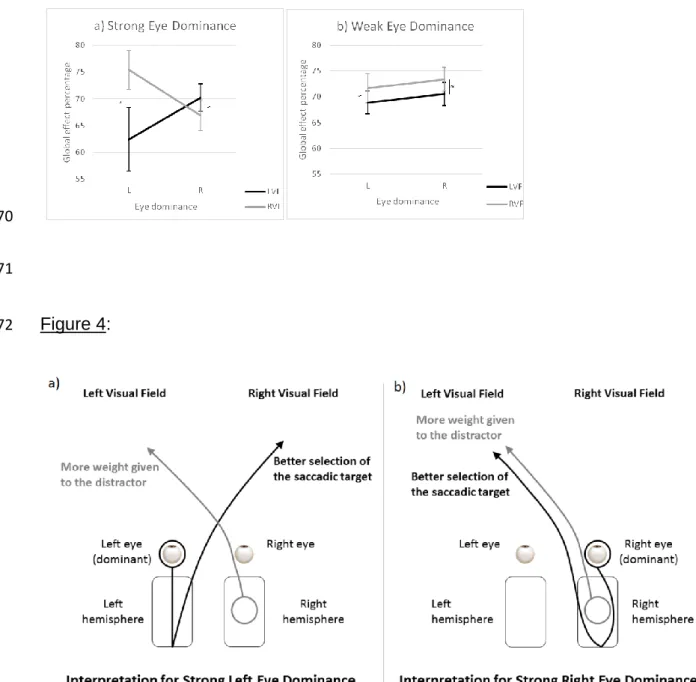
223
analyses. A preliminary analysis showed there was an effect of the block rank on
224
saccade latency (F(3,315) = 8.86, p<.001). Latency was longer on the first block than
225
on the other three blocks. Therefore, in order to keep the number of saccades
226
executed to the left and to the right hemifields constant, the first and the fourth blocks
227
were removed from following analyses.
228
Before analyzing the data on the derived measures to examine the effect of
229
eye dominance and of eye dominance strength on the GEP and the RDE, we
230
conducted a twofold preliminary analysis. We checked that the distractor presented in
231
the hemifield opposite the target increased saccade latency. We also checked that
232
the distractor presented in the same hemifield as the target deviated saccade
233
amplitude. Average saccade latency and amplitude obtained for each condition are
234
presented respectively in Table 1 and 2.
Left Visual Field T3° T5° T5° D3° T5° D-3° T7° T7° D3° T7° D-3° Luminance Distractor D=T D>T D=T D>T D=T D>T D=T D>T Right DE Strong 188 (±28) 181 (±22) 184 (±24) 184 (±26) 201 (±30) 199 (±28) 180 (±29) 185 (±31) 184 (±26) 201 (±31) 204 (±29) Weak 184 (±23) 173 (±18) 179 (±21) 177 (±19) 193 (±20) 193 (±20) 175 (±21) 183 (±20) 182 (±20) 190 (±24) 194 (±27) Left DE Strong 184 (±25) 177 (±17) 179 (±17) 179 (±19) 195 (±21) 200 (±22) 181 (±18) 182 (±19) 183 (±14) 196 (±26) 198 (±26) Weak 186 (±19) 180 (±22) 181 (±20) 182 (±19) 193 (±20) 194 (±18) 175 (±15) 184 (±19) 182 (±19) 194 (±18) 191 (±22)
Right Visual Field
T3° T5° T5° D3° T5° D-3° T7° T7° D3° T7° D-3° Luminance Distractor D=T D>T D=T D>T D=T D>T D=T D>T Right DE Strong 195 (±31) 188 (±22) 188 (±23) 185 (±24) 205 (±28) 203 (±26) 181 (±21) 187 (±23) 188 (±22) 200 (±25) 204 (±28) Weak 189 (±25) 181 (±24) 183 (±24) 183 (±24) 197 (±27) 198 (±28) 179 (±25) 184 (±22) 187 (±24) 197 (±26) 199 (±29) Left DE Strong 186 (±16) 177 (±14) 183 (±16) 180 (±16) 196 (±17) 197 (±17) 174 (±17) 182 (±22) 185 (±24) 195 (±18) 197 (±23) Weak 188 (±22) 180 (±22) 184 (±22) 185 (±20) 199 (±23) 198 (±23) 177 (±20) 186 (±18) 185 (±19) 194 (±19) 201 (±25)
Table 1 : Average latencies (and standard deviations) in milliseconds. Participants were categorized into four groups according to their eye dominance (Right DE; Left DE) 236
and eye dominance strength (Strong; Weak). Their task was to make a saccade toward a target presented either in the Left or in the Right visual field. In no-distractor 237
control conditions, the target (T) could be presented at an eccentricity of 3 (T3°), 5 (T5°) or 7° (T7°) from the initial fixation cross. In target-distractor conditions, the target 238
was presented at an eccentricity of 5 or 7° from the initial fixation cross with a distractor (D) presented either in the same visual field at an eccentricity of 3° (T5° D3° and 239
T7° D3°) or in the opposite visual field at an eccentricity of 3° (T5° D-3° and T7° D-3°). When presented, the distractor could have the same luminance as the target (D=T) or 240
could be brighter than the target (D>T). 241
242 243
244
Left Visual Field
T3° T5° T5° D3° T5° D-3° T7° T7° D3° T7° D-3° Luminance Distractor D=T D>T D=T D>T D=T D>T D=T D>T Right DE Strong 2.9 (±0.2) 4.7 (±0.3) 4.0 (±0.4) 4.1 (±0.3) 4.8 (±0.2) 4.8 (±0.2) 6.6 (±0.3) 5.8 (±0.5) 5.7 (±0.6) 6.7 (±0.3) 6.7 (±0.3) Weak 2.9 (±0.2) 4.6 (±0.3) 4.1 (±0.3) 4.0 (±0.3) 4.7 (±0.3) 4.7 (±0.3) 6.4 (±0.4) 5.6 (±0.6) 5.6 (±0.6) 6.6 (±0.4) 6.5 (±0.4) Left DE Strong 2.8 (±0.2) 4.6 (±0.3) 3.8 (±0.6) 3.9 (±0.5) 4.7 (±0.3) 4.7 (±0.3) 6.5 (±0.4) 5.4 (±1.0) 5.4 (±0.9) 6.6 (±0.3) 6.6 (±0.3) Weak 3.0 (±0.1) 4.8 (±0.2) 4.1 (±0.3) 4.1 (±0.3) 4.8 (±0.3) 4.8 (±0.3) 6.6 (±0.3) 5.7 (±0.6) 5.6 (±0.7) 6.6 (±0.4) 6.6 (±0.4)
Right Visual Field
T3° T5° T5° D3° T5° D-3° T7° T7° D3° T7° D-3° Luminance Distractor D=T D>T D=T D>T D=T D>T D=T D>T Right DE Strong 3.0 (±0.2) 4.8 (±0.2) 4.1 (±0.4) 4.1 (±0.4) 4.9 (±0.2) 4.9 (±0.3) 6.6 (±0.4) 5.7 (±0.6) 5.6 (±0.6) 6.8 (±0.3) 6.8 (±0.3) Weak 3.0 (±0.2) 4.8 (±0.3) 4.2 (±0.3) 4.2 (±0.4) 4.9 (±0.2) 4.9 (±0.3) 6.7 (±0.4) 5.9 (±0.7) 5.8 (±0.8) 6.8 (±0.3) 6.8 (±0.3) Left DE Strong 3.0 (±0.2) 4.8 (±0.3) 4.2 (±0.4) 4.2 (±0.4) 4.9 (±0.4) 4.9 (±0.3) 6.7 (±0.3) 6.0 (±0.7) 6.1 (±0.6) 6.8 (±0.4) 6.8 (±0.4) Weak 3.1 (±0.2) 4.9 (±0.2) 4.3 (±0.3) 4.2 (±0.3) 5.0 (±0.2) 5.0 (±0.2) 6.7 (±0.3) 6.0 (±0.6) 5.8 (±0.7) 6.9 (±0.2) 6.9 (±0.3)
Table 2: Average amplitudes (and standard deviations) in degrees. Participants were categorized into four groups according to their eye dominance (Right DE; Left DE) and 245
eye dominance strength (Strong; Weak). Their task was to make a saccade toward a target presented either in the Left or in the Right visual field. In no-distractor control 246
conditions, the target (T) could be presented at an eccentricity of 3 (T3°), 5 (T5°) or 7° (T7°) from the initial fixation cross. In target-distractor conditions, the target was 247
presented at an eccentricity of 5 or 7° from the initial fixation cross with a distractor (D) presented either in the same visual field at an eccentricity of 3° (T5° D3° and T7° 248
D3°) or in the opposite visual field at an eccentricity of 3° (T5° D-3° and T7° D-3°). When presented, the distractor could have the same luminance as the target (D=T) or 249
could be brighter than the target (D>T). 250
ANOVAs were run on the average saccade latency (Table 1) and on the
251
average saccade amplitude (Table 2) with eye dominance and eye dominance
252
strength as between-subject factors and distractor condition (No distractor, Distractor
253
as bright as the target, Brighter distractor), saccade target eccentricity (5° or 7°) and
254
target presentation hemifield (Left or Right) as within-subject factors. Note that
255
regarding the average saccade latency, the no-distractor control conditions were
256
compared to the opposite-hemifield distractor conditions. Regarding the average
257
saccade amplitude, the no-distractor control conditions were compared to the
same-258
hemifield distractor conditions.
259
A main effect of the distractor was found on saccade latency (F(2,176) = 255.52,
260
p<.001) as well as on saccade amplitude (F(2,176) = 505.67, p<.001). As expected,
261
compared to the no-distractor condition, the presentation of a distractor in the
262
opposite hemifield simultaneously with the target induced an increase of 19 ms of
263
saccade latency (F(1,88) = 341.23, p<.001) whereas the presentation of a distractor
264
within the same hemifield induced a deviation of the saccade of 0.8° closer to the
265
distractor (F(1,88) = 341.23, p<.001). RDE and GE were thus well observed in our
266
experiment.
267
3.2. Main results 268
We then conducted several analyses on the derived measures to examine
269
whether the effects of the distractor could be modulated by eye dominance and eye
270
dominance strength. We expected greater effects of the distractor located in the
271
hemifield contralateral to the DE in the case of strong eye dominance and no
272
difference between the two hemifields in the case of weak eye dominance. Average
saccade latency difference (remote distractor effect) and average global effect percentage are presented respectively in Table 3 274 and 4. 275 276 277 278 279
Table 3: Average remote distractor effect (and standard deviations) in milliseconds. Participants were categorized according to their eye dominance (Right 280
DE; Left DE) and eye dominance strength (Strong; Weak). Their task was to make a saccade toward a target (T) that could appear in the left or in the right 281
visual field, at 5 or 7° of eccentricity. Simultaneously with the target, a distractor (D) was presented at 3° of eccentricity in the visual field opposite the target. 282
This distractor could either have the same luminance as the target (D=T) or be brighter than the target (D>T). 283 284 285 286 287 288 289 290 291
Left Visual Field Right Visual Field
T5° D3° T7° D3° T5° D3° T7° D3° Luminance Distractor D=T D>T D=T D>T D=T D>T D=T D>T Right DE Strong 19 (±17) 18 (±13) 21 (±13) 24 (±12) 17 (±21) 15 (±14) 20 (±14) 24 (±15) Weak 20 (±11) 19 (±11) 15 (±12) 19 (±15) 16 (±15) 17 (±12) 18 (±17) 19 (±16) Left DE Strong 18 (±9) 23 (±11) 15 (±17) 17 (±18) 19 (±14) 21 (±16) 21 (±10) 23 (±13) Weak 13 (±10) 14 (±14) 20 (±14) 16 (±17) 19 (±18) 18 (±12) 17 (±13) 24 (±20)
292 293 294 295 296 297 298
Table 4: Average global effect percentage (and standard deviations). Participants were categorized according to their eye dominance (Right DE; Left DE) and 299
eye dominance strength (Strong; Weak). Their task was to make a saccade toward a target (T) that could appear in the left or in the right visual field, at 5 or 7° 300
of eccentricity. Simultaneously with the target, a distractor (D) was presented at 3° of eccentricity in the same visual field as the target. This distractor could 301
either have the same luminance as the target (D=T) or be brighter than the target (D>T). Remember that the higher the GEP, the lower the distractor effect on 302
saccade amplitude. 303
Left Visual Field Right Visual Field
T5° D3° T7° D3° T5° D3° T7° D3° Luminance Distractor D=T D>T D=T D>T D=T D>T D=T D>T Right DE Strong 63.0 (±12.4) 63.6 (±13.9) 78.0 (±9.9) 76.2 (±11.9) 62.4 (±14.1) 59.9 (±14.1) 74.0 (±13.4) 71.3 (±12.2) Weak 67.2 (±10.2) 63.4 (±9.2) 75.7 (±13.2) 76.0 (±11.9) 69.7 (±11.3) 69.5 (±13.2) 78.4 (±15.9) 76.1 (±17.5) Left DE Strong 55.8 (±19.5) 57.1 (±16.1) 68.7 (±21.8) 68.2 (±17.4) 69.4 (±12.2) 65.9 (±11.8) 81.4 (±12.0) 84.8 (±10.4) Weak 64.5 (±12.3) 62.8 (±11.6) 74.7 (±12.9) 73.6 (±15.8) 68.8 (±11.7) 64.6 (±13.2) 78.5 (±14.4) 74.7 (±15.7)
Two ANOVAs were conducted on the average saccade latency difference
304
(Table 3) and on the GEP (Table 4) with eye dominance (Left or Right) and eye
305
dominance strength (Strong or Weak) as between-subject factors and saccade target
306
eccentricity (5° or 7°), hemifield of target presentation (Left or Right) and distractor
307
luminance (Same or Brighter than the target) as within-subject factors. Concerning
308
the average saccade latency difference (Table 3), we found no main effect or
309
interaction between factors (all p>.10) with the exception of a significant effect of the
310
distractor luminance (F(1,88) = 4.57, p<.001). Saccade latency increased very slightly
311
with a brighter distractor (1.5 ms on average). Data on the GEP (Table 4) indicated a
312
significant effect of target eccentricity (F(1,88) = 131.95, p<.001): GE was higher with
313
shorter target-distractor distance (difference of 11.5%). Distractor luminance also
314
significantly affected GE (F(1,88) = 9.82, p<.005): GE was higher with a brighter
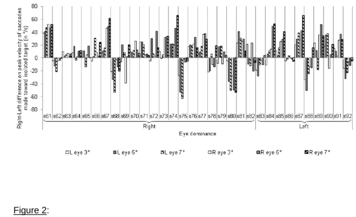
315
distractor with a very slight difference (1.4%). We found no main effect either of eye
316
dominance (F<1) or of eye dominance strength (F(1,88) = 1.05, ns). However, a main
317
effect of the hemifield of presentation was found (F(1,88) = 7.73, p<.01), the deviation
318
of the saccade toward the distractor being greater in the left hemifield (69.1%) than in
319
the right one (71.6%). More interestingly for our purpose, such an effect interacted
320
with eye dominance and eye dominance strength (F(1,88) = 8.86, p<.005). Figure 3
321
presents this interaction between eye dominance (left or right) and hemifield (left or
322
right) in participants with strong (figure 3a) and weak (figure 3b) eye dominance. The
323
effect of the hemifield of presentation did not reach the significance threshold for
324
people with weak eye dominance (F(1,58) = 2.975, p<.10) regardless of their DE (F<1),
325
whereas it was amplified in participants with a strong left DE, the saccade being more
326
deviated toward the distractor presented in the left than in the right hemifield (62.4%
327
vs 75.4%, F(1,9) = 11.92, p<.007). Participants with a strong right DE seemed to show
the reverse effect, with a distractor impact greater in the right hemifield than in the left
329
one, but the difference failed to reach the significance threshold (Figure 3a, F(1,21) =
330
2.92, p<.10). However, it should be noted that an effect of eye dominance strength
331
was found in the right hemifield in participants with a right DE (F(1,55) = 3.87, p<.05)
332
with the distractor effect being greater in participants with strong eye dominance
333
(66.9%) than with weak eye dominance (73.4%).
334
Insert Figure 3 Here
335
4. Discussion 336
4.1. Measuring eye dominance strength: The peak velocity criterion 337
Analyses of saccade peak velocities have been shown useful to estimate eye
338
dominance strength based on binocular recording of eye movements made toward
339
an isolated target2. Accordingly, participants exhibit higher peak velocities toward the
340
hemifield ipsilateral to the DE in case of strong eye dominance and exhibit a
naso-341
temporal asymmetry3 in case of weak eye dominance2. However, note that 2 of the
342
18 participants in the 2012 study exhibited higher peak velocities toward the
343
hemifield contralateral to DE whichever eye they used. In the present study, when we
344
categorized the 92 participants according to eye dominance strength, we noticed that
345
those with strong eye dominance also did not systematically exhibit higher peak
346
velocities toward the hemifield ipsilateral to the DE (see Figure 1). Indeed, 37.5%
347
(12/32) exhibited higher peak velocities toward the hemifield contralateral to the DE.
348
However, the results on the GEP for those 12 participants matched the patterns
349
observed in their eye dominance groups as defined by the hole-in-the-card test, with
350
lower GE (i.e., higher GEP) in the hemifield contralateral to the DE than in the
ipsilateral one. Therefore, these results on GEP as well as the different patterns of
352
peak velocities in the two studies suggest that the criterion for strong eye dominance
353
should finally be to exhibit higher peak velocities toward the same hemifield (left or
354
right) with both eyes, and not only toward the hemifield ipsilateral to the DE.
355
4.2. Distractor Luminance 356
In order to manipulate the perceptual weight of the distractor, the distractor
357
was either as bright as the target or brighter. We did not find a strong modulation of
358
the distractor effect neither for the remote distractor effect nor for the global effect. In
359
remote distractor effect conditions, we observed an only very slight effect of distractor
360
luminance on saccade latency, but no interaction with eye dominance, eye
361
dominance strength or hemifield. A very slight effect of distractor luminance was also
362
found on the GEP. Overall, the manipulation of distractor luminance we used
363
appeared to be not enough important to modify the pattern of results depending on
364
eye dominance and eye dominance strength.
365
4.3. Remote distractor effect 366
A distractor displayed in the hemifield opposite the target hemifield produced a
367
RDE. However, neither eye dominance nor eye dominance strength modulated this
368
effect. Unlike saccade amplitude or saccadic peak velocity2, the presence of
369
asymmetries on saccade latency is unclear in the literature: some studies reported
370
average left-right asymmetries17,18 while others not19,20. However, these studies never
371
took into account eye dominance or even manual laterality. Very few studies have
372
looked the effect of eye dominance but again without consistent results2,21,22. The
373
present study tested these asymmetries on a large sample of participants, and failed
374
to find any left-right asymmetries on saccade latency. The fact that RDE did not differ
between the two hemifields in participants with strong eye dominance suggests that
376
eye dominance does not influence saccade latency, at least in the conditions we
377
tested.
378
4.4. Global effect 379
When the distractor was in the same hemifield as the target, our results show
380
that the distractor had more impact on saccade amplitude (GE) when presented in
381
the hemifield ipsilateral to the DE than in the contralateral one. This was true only in
382
participants with strong eye dominance. However, this contrasts with our assumption
383
based on findings involving the presentation of a unique stimulus2,7,9. Interestingly,
384
the presentation of two stimuli, one of which is the saccade target as used in the
385
present study, specifies the perceptual processing advantage of the hemifield
386
contralateral to the DE, which would finally not occur in the overall hemifield, but
387
would be restricted to the saccade target location. Therefore, in a saccadic task we
388
suggest that the relationship between DE and ipsilateral V1 would lead to a more
389
accurate selection of the saccadic target in this hemifield (i.e., smaller effect of the
390
distractor on saccade amplitude) than in the ipsilateral one.
391
Note that the accurate selection of the saccadic target in the hemifield
392
controlateral to DE for participants with strong eye dominance is hypothesized in light
393
of the relationship between DE and ipsilateral V1, but V1 is only the starting point of
394
the sensori-motor transformation. The signals are then transmitted to the parietal eye
395
fields in the posterior parietal cortex and to the frontal eye fields, close to the
396
precentral sulcus23,24. So, it remains open whether the relationship between DE and
397
ipsilateral V1 will then lead to left-right asymmetries in parietal eye fields and frontal
398
eye fields activations. Future neuroimaging studies could help to clarify this point,
contrasting participants with left and right dominant eye, strong and weak eye
400
dominance.
401
However, our results showed a clear difference between the two hemifields in
402
participants with a strong left DE, but the difference was slighter and did not reach
403
the significance threshold in participants with a strong right DE. To explain this
404
difference between those two groups we propose that two phenomena are involved:
405
on the one hand, in participants with strong eye dominance, the relationship between
406
DE and ipsilateral V1 would induce a more accurate selection of the saccadic target
407
in the hemifield contralateral to the DE than in the ipsilateral one. On the other hand,
408
there would be an attentional bias toward the left hemifield giving more weight to the
409
distractor due to the specialization of the right hemisphere for visuo-spatial
410
attention25-28. Note that this attentional bias is hypothesized for all the participants,
411
and may explain that the distractor deviated saccade amplitude more when
412
presented in the left than in the right hemifield in participants with weak eye
413
dominance (see Figure 3b).
414
Insert Figure 4 Here
415
Figure 4 separately summarizes those two phenomena in participants with a
416
strong left and right DE. In participants with a strong left DE, each phenomenon
417
occurs separately in one hemifield and does not counteract the other one, leading to
418
a great GEP difference between the two hemifields. Conversely, those two
419
phenomena occur in the same hemifield in participants with a strong right DE.
420
Moreover, the attentional bias that gives more weight to the distractor counteracts the
421
accurate selection of the saccadic target. Accordingly, a smaller GEP difference
422
between the two hemifields was found in this population.
5. Conclusion 424
Researchers can now precisely measure participants’ handedness based on
425
questionnaires assessing a percentage of handedness. However, eye dominance is
426
still evaluated based on binary measures. Much research has been carried out to
427
develop a more graduated measure of eye dominance2,9,29-35. We here show different
428
visuomotor influences of eye dominance according to eye dominance strength.
429
Moreover, the use of two stimuli helped to specify the link between DE and ipsilateral
430
V1 (previous studies used a simple target stimulus2,7,9). Indeed, the better processing
431
that it involves in the hemifield contralateral to the DE seems not to operate in the
432
whole hemifield, but seems restricted to the saccade target location. These findings
433
point out the importance of taking into account participants’ eye dominance and eye
434
dominance strength in further visual or visuomotor studies.
435
6. References 436
1. Miles WR. Ocular dominance in human adults. The journal of general psychology.
437
1930;3:412-430.
438
2. Vergilino-Perez D, Fayel A, Lemoine C, Senot P, Vergne J, Doré-Mazars K. Are
439
There Any Left-Right Asymmetries in Saccade Parameters? Examination of
440
Latency, Gain, and Peak Velocity. Investigative ophthalmology & visual science.
441
2012;53:3340-3348.
442
3. Robinson DA. The mechanics of human saccadic eye movement. The Journal of
443
physiology. 1964;174:245-264.
444
4. Rombouts SA, Barkhof F, Sprenger M, Valk J, Scheltens P. The functional basis
445
of ocular dominance: functional MRI (fMRI) findings. Neuroscience Letters.
446
1996;221:1-4.
5. Erdogan AR, Özdikici M, Aydin MD, Aktas Ö, Dane S. Right and left visual cortex
448
areas in healthy subjects with right-and left-eye dominance. International journal
449
of neuroscience. 2002;112:517-523.
450
6. Shima H, et al. Ocular dominance affects magnitude of dipole moment: an MEG
451
study. Neuroreport. 2010;21:817-821.
452
7. Chaumillon R, Blouin J, Guillaume A. Eye dominance influences triggering action:
453
The Poffenberger paradigm revisited. Cortex. 2014;58:86-98.
454
8. Poffenberger AT. Reaction time to retinal stimulation with special reference to the
455
time lost in conduction through nerve centers. Archives of Psychology.
1912;23:1-456
73.
457
9. Chaumillon R, et al. Vers une quantification de la dominance oculaire pour une
458
meilleure prise en charge des pathologies de l’œil. Journal Français
459
d'Ophtalmologie. 2015;38:322-332.
460
10. Walker R, Deubel H, Schneider WX, Findlay JM. Effect of remote distractors on
461
saccade programming: evidence for an extended fixation zone. Journal of
462
Neurophysiology. 1997;78:1108-1119.
463
11. Casteau S, Vitu F. On the effect of remote and proximal distractors on saccadic
464
behavior: A challenge to neural-field models. Journal of vision. 2012;12:1-33.
465
12. Van der Stigchel S, Nijboer TCW. How global is the global effect? The spatial
466
characteristics of saccade averaging. Vision research. 2013;84:6-15.
467
13. Findlay JM. Global visual processing for saccadic eye movements. Vision
468
research. 1982;22:1033-1045.
469
14. Oldfield RC. The assessment and analysis of handedness: the Edinburgh
470
inventory. Neuropsychologia. 1971;9:97-113.
15. Findlay JM, Brogan D, Wenban-Smith MG. The spatial signal for saccadic eye
472
movements emphasizes visual boundaries. Perception & Psychophysics.
473
1993;53:633-641.
474
16. McSorley E, Findlay JM. Saccade target selection in visual search: Accuracy
475
improves when more distractors are present. Journal of Vision. 2003;3:877-892.
476
17. Pirozzolo FJ, Rayner K. Handedness, hemispheric specialization and saccadic
477
eye movement latencies. Neuropsychologia. 1980;18:225-229.
478
18. Hutton JT, Palet J. Lateral saccadic latencies and handedness.
479
Neuropsychologia. 1986;24:449-451.
480
19. De Clerck M, Crevits L, Van Maele G. Saccades: is there a difference between
481
right and left? Neuro-Ophthalmol. 2000;24:327-330.
482
20. Constantinidis TS, Smyrnis N, Evdokimidis I, et al. Effects of direction on saccadic
483
performance in relation to lateral preferences. Exp Brain Res. 2003;150:443-448.
484
21. Kolesnikova OV, Tereshchenko LV, Latanov AV, Shulgovskii VV. Effects of visual
485
environment complexity on saccade performance in humans with different
486
functional asymmetry profiles. Neurosci Behav Physiol. 2010;40:869-876.
487
22. Lazarev IE, Kirenskaya AV. The influence of eye dominance on saccade
488
characteristics and slow presaccadic potentials. Hum Physiol. 2008;34:150-160.
489
23. Pierrot-Deseilligny C, Rivaud S, Gaymard B, Müri R, Vermersch AI. Cortical
490
control of saccades. Annals of neurology. 1995;37:557-567.
491
24. McDowell JE, Dyckman KA, Austin BP, Clementz BA. Neurophysiology and
492
neuroanatomy of reflexive and volitional saccades: evidence from studies of
493
humans. Brain and cognition. 2008;68:255-270.
494
25. Kinsbourne M. The cerebral basis of lateral asymmetries in attention. Acta
495
psychologica. 1970;33:193-201.
26. Bowers D, Heilman KM. Pseudoneglect: effects of hemispace on a tactile line
497
bisection task. Neuropsychologia. 1980;18:491-498.
498
27. Jewell G, McCourt ME. Pseudoneglect: a review and meta-analysis of
499
performance factors in line bisection tasks. Neuropsychologia. 2000;38:93-110.
500
28. Thiebaut de Schotten M, et al. A lateralized brain network for visuospatial
501
attention. Nature neuroscience. 2011;14:1245-1246.
502
29. Purves D, White LE. Monocular preferences in binocular viewing. Proceedings of
503
the National Academy of Sciences. 1994;91:8339-8342.
504
30. Handa T, Shimizu K, Mukuno K, Kawamorita T, Uozato H. Effects of ocular
505
dominance on binocular summation after monocular reading adds. Journal of
506
Cataract & Refractive Surgery. 2005;31:1588-1592.
507
31. Nitta M, Shimizu K, Niida T. The influence of ocular dominance on monovision:
508
the influence of strength of ocular dominance on visual functions. Nippon Ganka
509
Gakkai Zasshi. 2007;111:441-446.
510
32. Johansson J, Pansell T, Ygge J, Seimyr GÖ. Monocular and binocular reading
511
performance in subjects with normal binocular vision. Clinical and Experimental
512
Optometry. 2014;97:341-348.
513
33. Johansson J, Seimyr GÖ, Pansell T. Eye dominance in binocular viewing
514
conditions. Journal of vision. 2015;15:1-17.
515
34. Carey DP. Losing sight of eye dominance. Current Biology. 2001;11:828-830.
516
35. Carey DP, Hutchinson CV. Looking at eye dominance from a different angle: is
517
sighting strength related to hand preference? Cortex. 2013;49:2542-2552.
Figure legends: 519
Figure 1: Average differences of peak velocities of saccades toward isolated targets
520
in left and right visual fields indicating strong eye dominance.
521
Participants were categorized into two groups according to their eye
522
dominance (Left or Right) measured with the hole-in-the-card test. Negative values
523
indicate that saccades toward the left visual field exhibit higher peak velocities than
524
saccades toward the right visual field, and positive values indicate the opposite.
525
Those differences have been calculated for saccades made toward isolated targets
526
presented at 3, 5 or 7° of eccentricity for the right eye (R eye) and the left eye (L
527
eye). All the participants presented in this graph exhibit higher peak velocities toward
528
a same visual field whatever the eye being measured for at least two of the three
529
eccentricities tested. Therefore, they have been categorized as having strong eye
530
dominance.
531
Figure 2: Average differences of peak velocities of saccades toward isolated targets
532
in left and right visual fields indicating weak eye dominance.
533
Participants were categorized into two groups according to their eye
534
dominance (Left or Right) measured with the hole-in-the-card test. Negative values
535
indicate that saccades toward the left visual field exhibit higher peak velocities than
536
saccades toward the right visual field, and positive values indicate the opposite.
537
Those differences have been calculated for saccades made toward isolated targets
538
presented at 3, 5 or 7° of eccentricity for the right eye (R eye) and the left eye (L
539
eye). All the participants presented in this graph exhibit higher peak velocities toward
540
the right visual field with the right eye and toward the left visual field with the left eye
541
(i.e., naso-temporal asymmetry) for at least two of the three eccentricities tested.
542
Therefore, they have been categorized as having weak eye dominance.
Figure 3: Interaction between Eye dominance strength, Eye dominance and
544
Visual field on Global Effect Percentage (GEP).
545
Figure 3a shows the interaction between Eye dominance (L = left DE; R = right
546
DE) and Visual field (LVF = left visual field; RVF = right visual field) in participants
547
with strong eye dominance. Figure 3b shows the same interaction in participants
548
with weak eye dominance. In both graphs, the significant differences are indicated
549
with the symbol * (p<.05) and the differences that failed to reach significance with
550
the symbol ≈ (.05<p<.10). Error bars represent standard errors.
551
Figure 4: Illustration of the two phenomena inferred from our results on the global
552
effect percentage (GEP).
553
Black indicates the relationship between DE and ipsilateral V1, leading to a
554
more accurate saccadic selection in the visual field contralateral to the DE than in
555
the ipsilateral one. This phenomenon occurs in opposite visual fields in
556
participants with a strong left DE (Figure 4a) and with a strong right DE (Figure
557
4b). Gray indicates the second phenomenon, an attentional bias toward the left
558
visual field due to the right hemisphere specialization for visuo-spatial attention,
559
giving more weight to the distractor in this visual field than in the right one.
560
Figure 1: 562 563 564 Figure 2: 565 566 567 568
Figure 3: 569 570 571 Figure 4: 572 573