HAL Id: hal-00469588
https://hal.archives-ouvertes.fr/hal-00469588
Submitted on 31 May 2020
HAL is a multi-disciplinary open access
archive for the deposit and dissemination of
sci-entific research documents, whether they are
pub-lished or not. The documents may come from
teaching and research institutions in France or
abroad, or from public or private research centers.
L’archive ouverte pluridisciplinaire HAL, est
destinée au dépôt et à la diffusion de documents
scientifiques de niveau recherche, publiés ou non,
émanant des établissements d’enseignement et de
recherche français ou étrangers, des laboratoires
publics ou privés.
Copyright
monogalactosyldiacylglycerol synthase MGD1 by
phosphatidic acid and phosphatidylglycerol.
Emmanuelle Dubots, Magali Audry, Yoshiki Yamaryo, Olivier Bastien,
Hiroyuki Ohta, Christelle Breton, Eric Maréchal, Maryse A Block
To cite this version:
Emmanuelle Dubots, Magali Audry, Yoshiki Yamaryo, Olivier Bastien, Hiroyuki Ohta, et al..
Activa-tion of the chloroplast monogalactosyldiacylglycerol synthase MGD1 by phosphatidic acid and
phos-phatidylglycerol.. Journal of Biological Chemistry, American Society for Biochemistry and Molecular
Biology, 2010, 285 (9), pp.6003-11. �10.1074/jbc.M109.071928�. �hal-00469588�
Activation of the Chloroplast Monogalactosyldiacylglycerol
Synthase MGD1 by Phosphatidic Acid
and Phosphatidylglycerol
□SReceived for publication, October 1, 2009, and in revised form, December 2, 2009Published, JBC Papers in Press, December 20, 2009, DOI 10.1074/jbc.M109.071928 Emmanuelle Dubots‡, Magali Audry§, Yoshiki Yamaryo‡, Olivier Bastien‡, Hiroyuki Ohta¶, Christelle Breton§, Eric Mare´chal‡, and Maryse A. Block‡1
From the‡Laboratoire de Physiologie Cellulaire Ve´ge´tale, CNRS/CEA/INRA/Universite´ Joseph Fourier, CEA-Grenoble,
F-38054 Grenoble, France, the§Centre de Recherche sur les Macromole´cules Ve´ge´tales-CNRS, F-38041 Grenoble, France, and the ¶Center for Biological Resources and Informatics and Research Center for the Evolving Earth and Planets, Tokyo Institute of Technology, Yokohama 226-8501, Japan
One of the major characteristics of chloroplast membranes is their enrichment in galactoglycerolipids, monogalactosyldia-cylglycerol (MGDG), and digalactosyldiamonogalactosyldia-cylglycerol (DGDG), whereas phospholipids are poorly represented, mainly as phos-phatidylglycerol (PG). All these lipids are synthesized in the chloroplast envelope, but galactolipid synthesis is also partially dependent on phospholipid synthesis localized in non-plastidial membranes. MGDG synthesis was previously shown essential for chloroplast development. In this report, we analyze the reg-ulation of MGDG synthesis by phosphatidic acid (PA), which is a general precursor in the synthesis of all glycerolipids and is also a signaling molecule in plants. We demonstrate that under phys-iological conditions, MGDG synthesis is not active when the MGDG synthase enzyme is supplied with its substrates only, i.e. diacylglycerol and UDP-gal. In contrast, PA activates the enzyme when supplied. This is shown in leaf homogenates, in the chloroplast envelope, as well as on the recombinant MGDG synthase, MGD1. PG can also activate the enzyme, but compar-ison of PA and PG effects on MGD1 activity indicates that PA and PG proceed through different mechanisms, which are fur-ther differentiated by enzymatic analysis of point-mutated recombinant MGD1s. Activation of MGD1 by PA and PG is pro-posed as an important mechanism coupling phospholipid and galactolipid syntheses in plants.
Chloroplast membrane lipids are mostly composed of non-phosphorous galactoglycerolipids i.e.
monogalactosyldiacyl-glycerol (MGDG)2and digalactosyldiacylglycerol (DGDG) that
represent more than 50 and 30% of thylakoid lipids, respec-tively. Phosphatidylglycerol is the main phospholipid in plas-tids, representing about 10% of thylakoid lipids. Each of those
glycerolipid classes is represented by a range of molecular spe-cies differing in the acyl composition at sn-1 and sn-2 positions of the glycerol backbone. Based on the model of cyanobacterial lipids, the prokaryotic type of glycerolipids contains a 16-car-bon fatty acid at the sn-2 position of glycerol. The eukaryotic type contains an 18-carbon fatty acid at the sn-2 position. Some plants, such as Arabidopsis or spinach, have both prokaryotic-and eukaryotic-type MGDG, whereas other plants, such as pea or cucumber, have only eukaryotic-type MGDG. DGDG is mostly of eukaryotic type in all plants. Chloroplast PG contains exclusively a prokaryotic-type DAG moiety in contrast to non-plastidial PG. These different chloroplast lipids are assembled in the chloroplast envelope (1). Most enzymes have now been identified, but their functioning and regulation remain largely unknown.
MGDG is synthesized by MGDG synthase (MGD), which transfers galactose from UDP-gal to DAG. In Arabidopsis, there are three MGDG synthases, and among them, MGD1, is neces-sary for synthesis of galactolipids and for development of pho-tosynthetic membranes (2, 3). MGD1 utilizes DAG derived from two main sources, either from a DAG de novo synthesized in plastid envelope by double acylation of glycerol-3 phosphate and PA dephosphorylation (prokaryotic DAG) or from a DAG derived from endoplasmic reticulum phosphatidylcholine (PC) possibly through a phospholipase D enzymatic step (eukaryotic DAG) (1). MGD1 can produce prokaryotic- and eukaryotic-type MGDG with the same efficiency (4). MGD1 localizes at the inner envelope membrane (IEM). The protein anchors to the membrane as a monotopic protein through protein-lipid inter-action to the external monolayer of the IEM (5, 6). Native and recombinant MGD1 are known to be active as homodimers (5). Each MGD1 monomer is likely to be organized in two Ross-mann folds (N- and C-domains) (7). Visualization of surface hydrophobic regions suggested that MGD1 interacts with the membrane surface by its N-domain, whereas the C-domain protrudes above the membrane. Although MGD1 is a mem-brane protein and synthesizes MGDG using a lipid substrate (DAG), its mechanisms of interaction with hosting membrane, lipid substrate, and product remain unclear. Moreover, the enzyme activity might potentially be regulated by anionic lipids: PG, SQDG, or PA (8 –10). Because (i) PA is a general precursor in the glycerolipid synthesis pathway and a signaling molecule □S
The on-line version of this article (available at http://www.jbc.org) contains supplemental Fig. S1.
1To whom correspondence should be addressed: CEA-Grenoble, iRTSV/ LPCV, 17 Rue des Martyrs, F-38054, Grenoble, France. Tel.: 33-0-438-78-49-85; Fax: 33-0-438-78-50-91; E-mail: mblock@cea.fr.
2The abbreviations used are: MGDG, monogalactosyldiacylglycerol; MGD, MGDG synthase; CHAPS, 3-[(3-cholamidopropyl)dimethylammonio]-1-propanesulfonic acid; MOPS, 4-morpholine3-[(3-cholamidopropyl)dimethylammonio]-1-propanesulfonic acid; PA, phosphatidic acid; DGDG, digalactosyldiacylglycerol; PG, phosphatidyl-glycerol; DAG, diacylphosphatidyl-glycerol; AEBSF, 4-2-aminoethyl benzenesulfonyl fluo-ride hydrochlofluo-ride; PLD, phospholipase D; DTT, dithiothreitol; Tricine,
N-[2-hydroxy-1,1-bis(hydroxymethyl)ethyl]glycine.
THE JOURNAL OF BIOLOGICAL CHEMISTRY VOL. 285, NO. 9, pp. 6003–6011, February 26, 2010 © 2010 by The American Society for Biochemistry and Molecular Biology, Inc. Printed in the U.S.A.
at INRA Institut National de la Recherche Agronomique on May 13, 2019
http://www.jbc.org/
in plants, and (ii) MGD1 is essential for chloroplast develop-ment, regulation by PA potentially represents a way of control-ling chloroplast biogenesis during plant development. Using both native and recombinant MGD1 protein, we therefore ana-lyzed in this report the enzymatic characteristics of regulation of MGD1 by PA. We also compared the regulation effects of PA and PG on the enzyme.
EXPERIMENTAL PROCEDURES Chemicals
Lipids were purchased as follows, DAG (Sigma D0138), PA (Sigma P9511), 18:1/18:1-PA (Sigma P2767), 16:0/18:1-PA (Avanti Polar lipids 840857c), 16:0/16:0-PA (Sigma P4013), PG (Sigma P0514), and PC (Sigma P3017). 18:1/16:0-PA was puri-fied by two-dimensional TLC after hydrolysis of 18:1/16:0-PC (Sigma P4142) by PLD activity (Sigma L4384) according to the manufacturer’s protocol. Identification of the fatty acid in the
sn-2position was determined by gas chromatography after
hy-drolysis with the Rhizopus arrhizus lipase (Roche, 50 units䡠l⫺1
in 50 mMTris-HCl, pH 7.2, 0.5% Triton X-100) for 1 h at room
temperature. AEBSF, 4-2-aminoethyl benzenesulfonyl fluoride hydrochloride, was purchased from Fluka 76307.
Plant Materials and Growth Conditions
Arabidopsis plants were grown on Murashige and Skoog (MS, Duchefa) agar containing 0.5% sucrose for 15 days
between 22 to 26 °C under a daily 16-h light cycle at 100mol of
photons m⫺2s⫺1. The pld2 mutant (SALK_094369 T-DNA
line of At3g05630 (11)), ecotype Columbia-0, corresponds to
the pld2 line analyzed by (12). The homozygous line was
ini-tially selected from the SALK seed stock and further from a back cross of this line with the parental wild type Columbia line by
kanamycin selection and PCR genotyping using P1 (5
⬘-CGC-ACCACGAGAAAATAGTG-3⬘) and P2
(5⬘-CACTCTGCT-TCCCAATCTGC-3⬘) for the wild-type gene, and P1 and LB
(5⬘-GGCAATCAGCTGTTGCCCGTCTCACTGGTG-3⬘) for the
tagged gene. We verified by RT-PCR with primers 5
⬘-CGACGA-CGGTTTGGGGAG-3⬘ and
5⬘-CTTCCCAATCTGCCACCA-CCAGTC-3⬘ that the mutant line had no expression of the PLD2
gene. Spinach leaves were purchased on the local market.
Lipid Analysis
Lipids were analyzed by TLC generally in CHCl3/acetone/
MeOH/acetic acid/H2O 50:20:10:10:5 (v/v) and quantified by
gas chromatography as described by Ref. 13.
Chloroplast Envelope Isolation from Spinach Leaves
Intact chloroplasts were obtained from spinach (Spinacia
oleracea L.) leaves and purified by isopycnic centrifugation using Percoll gradients (14). Purified envelope membranes from thermolysin-treated chloroplasts were prepared as de-scribed by Ref. 15. Briefly, Percoll-purified intact chloroplasts (final concentration, 1 mg of chlorophyll/ml) were incubated in
the following medium: 0.3Msucrose, 20 mMMOPS-NaOH, pH
7.8, 1 mMCaCl2, for 1 h at 4 °C with 600g䡠ml⫺1of thermolysin
from Bacillus thermoproteolyticus (Sigma). The digestion was
terminated by the addition of 10 mMEGTA. Intact chloroplasts
were then recovered on a second Percoll gradient before lysis in a hypotonic medium and isolation of the envelope fraction on a sucrose gradient.
Preparation of MGD1 Recombinant Proteins
ArabidopsisatMGD1 sequence beginning at A138 and fused
with a His6tag at the C terminus was expressed in Escherichia
coli using the pET expression system (Novagen Inc.) as
described by Ref. 7. Expression was induced by 1 mM
isopropyl-1-thio--D-galactopyranoside for 5 h at 18 °C. The protein was
recovered in buffer containing 50 mMTris-HCl, pH 8.0, 12%
(w/v) glycerol, 1MNaCl, 1 mM-mercaptoethanol, subjected to
chromatography on a Ni2⫹-charged resin (Ni-NTA-agarose,
Qiagen GmbH, Germany), from which it was eluted with
50 –100 mMimidazole. Imidazole was removed from the
pro-tein fraction by gel filtration on BiogelP6DG (Bio-Rad). In this buffer, the recombinant protein was soluble, and its purity was estimated to be more than 95% after SDS-PAGE and Coomassie Blue staining. Recombinant atMGD1 proteins were obtained by site-directed mutagenesis of the protein described above. They were expressed and purified as the wild-type protein.
Cucum-ber csMGD1 beginning at Val-105 and fused to a His6tag at the
C terminus was produced as in Ref. 16. Spinach soMGD1 begin-ning at Leu-99 was expressed and purified as in Ref. 7.
Measurement of MGDG Synthase Activity
Measurement on Leaf Homogenates—Rosette leaves of 2-week-old plants were grounded in liquid nitrogen. The
pow-der was resuspended in 50 mMMOPS-NaOH, pH 7.8, 1 mM
DTT, 6 mMCHAPS, 25 mMKCL, 25 mMKH2PO4, 2% glycerol,
150 mM NaCl, and immediately used for measurement of
MGDG synthase activity. The measurement was monitored at
23 °C with 7 mol% DAG, 350M[14C]UDP-gal (22.9 GBq䡠mol⫺1)
and 1.5 to 4g䡠l⫺1protein. As lipid concentration, the surface
concentration of the relevant lipid relative to all mixed micelle
components ([lipid]/([detergent]⫹⌺[lipids]) expressed in mol%)
was used as a rough representation of the lipid concentration in the vicinity of the enzyme. Labeled galactolipids were analyzed, and radioactivity was measured by liquid scintillation.
Measurement on Chloroplast Envelope—An envelope frac-tion purified from thermolysin-treated spinach chloroplasts
was incubated in 10 mMTricine-NaOH, pH 9, 5 mMEDTA, 1
mMDTT for 15 min at 23 °C. One volume was then added to 1
volume of 100 mMMOPS-NaOH, pH 7.8, 1 mMDTT, 12 mM
CHAPS, 50 mMKCl, 50 mMKH2PO4with 7 mol% DAG, and
750MUDP-[14C]gal (22.9 GBq䡠mol⫺1) for a protein
concen-tration of 0.9 –1.3g䡠l⫺1.
Measurement on Recombinant MGD1—Wild-type or
mu-tant MGD1 proteins were usually incubated in 50 mM
MOPS-NaOH, pH 7.8, 6 mMCHAPS, 150 mMNaCl, 2% glycerol, 1 mM
DTT, 25 mMKCl, 25 mMKH2PO4with 7 mol% DAG and 750
MUDP-[14C]gal (22.9 GBq䡠mol⫺1) for a protein concentration
of 11 ng䡠l⫺1or as indicated. Kinetic data were fitted with the
Hill equation (Equation 1),
V⫽ Vmax⫻关S兴n/共K0.5n⫹关S兴n兲 (Eq. 1) where V is the enzyme velocity, S the PA or PG or UDP-gal
at INRA Institut National de la Recherche Agronomique on May 13, 2019
http://www.jbc.org/
concentration, K0.5the concentration of S where V⫽1⁄2Vmax, and n the Hill coefficient using the nonlinear least squares method (17) implemented in the package from the R software (18).
Protein-Lipid Binding Assays
Lipid-Protein Overlay Assay—1l of lipid solution containing 0.1–10 nmol of PA, DAG, or PG dissolved in chloroform was spot-ted on to nitrocellulose membranes (Schleicher & Schuell) and allowed to dry at room temperature for 1 h. The membrane was blocked in 5% (w/v) fatty acid-free bovine serum albumin in TTBS
(50 mMTris-HCl, pH 7.5, 150 mMNaCl with 0.1%, v/v, Tween
20) for 1 h. The membrane was then incubated overnight at 4 °C with gentle stirring in the same solution containing about
0.5–1.5g䡠l⫺1of MGD1 protein. The membrane was washed
four times for 5 min in TTBS and then incubated for 1 h with anti-His monoclonal antibody 1/5000 (Sigma) or anti-spinach MGD1 (1/500) in TTBS with 3% skimmed milk. The membrane was washed as before, incubated with mouse-horse-radish peroxidase (HRP) or anti-rabbit-HRP conjugate 1/5000 (Jack-son Immunoresearch) in TTBS with 3% skimmed milk, washed four times in TTBS, followed by once in TBS before ECL detection.
Liposome Binding Assay—Dried
lipids were resuspended at 2 g䡠
l⫺1in 50 mMTris-HCl, pH 7, 100
mM NaCl, 1 mM DTT for 1 h at
37 °C. Liposomes were then vigor-ously vortexed for 5 min before
cen-trifugation at 20,000⫻ g for 10 min.
Purified recombinant MGD1 was diluted in the same medium at 0.1
g䡠l⫺1and collected as a
superna-tant after centrifugation at 20,000⫻
gfor 10 min. Protein and liposomes
were incubated together for 30 min at 30 °C. Liposomes were finally harvested as a pellet by
centrifuga-tion at 20,000⫻ g for 10 min, and
protein was analyzed by SDS-PAGE with silver nitrate staining.
RESULTS
Activation of the MGDG Synthase Activity of Leaf Homogenates by PA
In a first evaluation of galactolipid synthesis tuning by specific lipids, MGDG synthase activity was mea-sured in leaf homogenates of 2-week-old Arabidopsis plants. The level of activity was quantified fol-lowing transfer of radioactive galac-tose from UDP-gal onto DAG added to the homogenate. We observed a moderate MGDG synthase activity that was stimulated 5 times by addition of 1.5 mol% of PA (Fig. 1A). Stimulation by PA was not due to con-version of PA into DAG, because in the absence of exogenous DAG, no activity was detected. PLDs are highly active in ground leaves and could generate enough PA to maintain a basal level of MGDG synthase activity in leaf homogenates. We therefore considered inhibiting the presumed formation of PA by phos-pholipases D using AEBSF. AEBSF has been previously reported as an inhibitor of PLD activity in leaf extracts (19, 20),
a property we confirmed in our system (supplemental Fig. S1).
We verified that pure recombinant atMGD1, expected as the most active MGDG synthase in leaf homogenates (see below), was not inhibited by AEBSF (Fig. 1B). We observed that the FIGURE 1. Activation of MGDG synthase by PA in Arabidopsis leaf homogenates. Activity was measured as
described under “Experimental Procedures.” A, effect of PA (1.5 mol%), DAG (7 mol%), and AEBSF (6 mM) on MGDG synthase activity. B, effect of AEBSF (6 mM) on purified at⌬1–137MGD1. 175 ng of purified protein was incubated with 1.5 mol% PA under the same condition as described for leaf homogenates. C, effect of PA (1.5 mol%), DAG (7 mol%), and AEBSF (6 mM) on DGDG synthase activity. D, kinetic analysis of MGDG synthesis: f, control; F, in the presence of AEBSF (6 mM), Œ, with addition of AEBSF after 15 min of incubation (arrow), ⽧, with 1.5 mol% of PA. E, comparison of MGDG synthase activity in wild-type and in pld2. PA (1.5 mol%), DAG (7 mol%) were added as indicated. The inset shows the ratio of the activities measured with addition of DAG in the absence of PA reported to in the presence of PA. F, comparison of DGDG synthase activity in wild-type and in pld2. PA (1.5 mol%), DAG (7 mol%) were added as indicated. Results are average values⫾ S.D. for three independent replicates.
Activation of MGD1 by PA and PG
at INRA Institut National de la Recherche Agronomique on May 13, 2019
http://www.jbc.org/
MGDG synthase activity of leaf homogenates was lower in the presence of AEBSF and that, upon addition of PA, some MGDG synthase activity was restored (Fig. 1A). DGDG synthase activ-ity in leaf homogenates was low and independent of DAG and PA (Fig. 1C). Our conclusion was therefore that PA was an activator of the MGDG synthase of leaf homogenates.
PA in leaf homogenates is likely to be very transient depen-dent on both anabolism and catabolism. This may dynamically regulate MGDG synthase activity. Kinetics analysis of the MGDG synthase activity of leaf homogenates showed that the incorporation of galactose into MGDG was linear with time and stimulated by addition of PA (Fig. 1D). When AEBSF was added at the beginning of the kinetics, no activity was detected in the absence of additional PA. Interestingly, the addition of AEBSF after 15 min of incorporation very rapidly stopped MGDG synthase activity (Fig. 1D). These results were consis-tent with the following: (i) the MGDG synthase activity of leaf homogenates was dependent on the constant presence of PA, (ii) decrease of PA production led to a decrease of the MGDG synthase activity, and (iii) PLDs were able to produce PA nec-essary for MGDG synthase activation.
However, the AEBSF effect was only partially restored by PA addition (Fig. 1A). To confirm that PA issued from PLDs had an impact on the level of MGDG synthase activity in leaf nates, we assayed the MGDG synthase activity of leaf homoge-nates in a mutant deleted of PLD. Twelve PLDs have already been reported in Arabidopsis (21). Most of them are expected to
be active only in the presence of Ca2⫹except the 2 PLD
pro-teins. Because no Ca2⫹was added in our assay and PLD2 had
been previously shown to be involved in the phosphate depri-vation-induced synthesis of galactolipids in roots (12, 22), we
chose to assay a mutant deleted of PLD2. We observed that the
mutant exhibited a little less MGDG synthase activity com-pared with the wild type (Fig. 1E). When PA was added, MGDG synthase activity was more highly increased in the mutant than in the wild type. No activity was detected in the mutant as in the wild type with the addition of PA alone (no addition of DAG). As a result, the ratio of MGDG synthase activity obtained in the
absence to presence of exogenous PA was 2-fold lower in pld2
than in the wild type. The DGDG synthase activity was a little higher in the mutant than in the wild type but still insensitive to addition of DAG or PA (Fig. 1F). These data therefore (i) con-firmed that PA was important for activation of the MGDG
syn-thase, and (ii) indicated that PLD2 was a potent source of PA
for activation of the MGDG synthase in leaf homogenates.
Effect of PA on the MGDG Synthase Activity of Chloroplast Envelope
In leaves, the MGDG synthase enzyme is located in the chlo-roplast envelope (6). To date, the MGDG synthase activity has been classically measured in chloroplast envelope fractions iso-lated from leaves. A sufficient amount of DAG is present in the purified envelope to sustain the activity except when the enve-lope is isolated from thermolysin-treated chloroplasts (15). Indeed thermolysin hydrolyzes the galactolipid-galactolipid galactosyltransferase, which is present on the surface of chlo-roplasts and responsible for DAG formation during isolation of the chloroplast envelope. The supply of exogenous DAG is
therefore required for measurement of MGDG synthase activ-ity in the envelope isolated from thermolysin-treated chloro-plasts. In contrast, PA addition was never reported to be required to detect MGDG synthase activity in the envelope from either thermolysin-treated or non-treated chloroplasts. The PA level in the isolated chloroplast envelope is low and undetectable by conventional techniques. However, our data suggested that a low concentration of PA could be sufficient to activate MGDG synthase. We questioned whether PA was nec-essary for activation of the chloroplast envelope MGDG syn-thase and was present in sufficient amounts in the isolated envelope to allow MGDG synthase activity.
To decrease endogenous PA, a chloroplast envelope fraction was prepared from thermolysin-treated spinach chloroplasts and preincubated to favor envelope-associated PA phosphatase activity. High pH and absence of DAG during pretreatment were expected to allow PA phosphatase activity (23). Then, the envelope fraction was supplemented with DAG and incubated in MGDG synthase activity buffer. We thus observed a barely detectable MGDG synthase activity (Fig. 2A). When exogenous PA was added, the MGDG synthase activity was high in con-trast. Only radiolabeled MGDG was formed indicating the absence of DGDG synthase and galactolipid-galactolipid galac-tosyltransferase activities (Fig. 2B). Several molecular species of PA, either of eukaryotic- or prokaryotic-type, were able to
stim-FIGURE 2. Effect of PA on MGDG synthase measured in isolated spinach
chloroplast envelope. Before the experiment, the envelope fraction was
prepared from thermolysin-treated chloroplasts and incubated in conditions favorable for PA phosphatase activity as described under “Experimental Pro-cedures.” MGDG synthase was then measured as described under “Experi-mental Procedures.” A, kinetic analysis of MGDG synthase activity: control, broken line; with PA 1.5 mol%, solid line. Results are average values⫾ S.D. for three independent measurements. B, analysis of [14C]galactose-labeled lipids
on TLC. C, effect of different lipids on MGDG synthase activity. Activity was measured as described under “Experimental Procedures.” The presence of DAG and additional lipid is indicated below the graph, with for additional lipid fatty acid in sn-1 position on the first line and fatty acid in sn-2 position on the second line. D, effect of 16:0/18:1-PA level on the MGDG synthase activity. Activity was measured as described under “Experimental Procedures.” Results are average values⫾ S.D. for three independent measurements. The curve equation is: V⫽ 3 ⫻ [PA]/(0.5 ⫹ [PA]).
at INRA Institut National de la Recherche Agronomique on May 13, 2019
http://www.jbc.org/
ulate the MGDG synthase activity, although the 16:0/16:0 molecular species was less active than the 16:0/18:1 (Fig. 2C). PG had only a slight effect, whereas PC was not active. The MGDG synthase activity was dependent on the PA concentra-tion and a range of about 0.5 mol% PA (16:0/18:1-PA) was suf-ficient to reach half-maximal activity of the enzyme (Fig. 2D). Our conclusion was that PA had a regulatory effect on the native MGDG synthase present in chloroplast envelope.
Effect of PA on a Recombinant Form of atMGD1
MGDG Synthase Activity—In Arabidopsis, atMGD1 is the main MGDG synthase expressed in leaves and is essential for the development of photosynthetic tissues (2, 3, 6). The full-length mature protein associates with membranes (5). In a
pre-vious work (7), it has been shown that a⌬1–137 truncated form
of atMGD1 is expressed in E. coli as a soluble and active form. Using this form of the enzyme, the effect of PA was analyzed. In the absence of PA, the enzyme alone had no activity (Fig. 3A). In contrast, several acyl species of PA were able to activate the enzyme although less efficiently for the 16:0/16:0 molecular species. PG was partially effective but not PC. A conversion of PA into DAG was excluded because almost no activity was found with PA when exogenous DAG was absent. These results suggested that the MGDG synthase was allosterically activated by PA and that PG could play a similar role as PA.
The MGDG synthase enzymatic velocity versus PA concen-tration showed a similar sigmoid curve with two molecular spe-cies of PA: a eukaryotic-type form, 16:0/18:1, and a prokaryotic-type form, 18:1/16:0 (Fig. 3B). In each case, half-maximal
activation was obtained for⬃0.2 mol% PA. The cooperative
effect of PA on the enzyme could illustrate a relationship with dimerization of the enzyme, because it has been previously shown that the enzyme is active as a dimer (5).
PA binding can change the enzyme behavior regarding sub-strate handling. The enzyme velocity versus UDP-gal concen-tration was compared in the presence of 0.15 and 1.5 mol% of PA (Fig. 3C). With 0.15 mol% of PA, the enzyme velocity versus substrate curve was sigmoid, whereas with 1.5 mol% of PA, the curve was hyperbolic. There was therefore a clear allosteric change of the enzyme for UDP-gal handling depending on PA
concentration. PA predominantly affected the apparent Vmaxof
the enzyme becauseAppVmaxwas increased five times when PA
concentration was changed from 0.15 and 1.5 mol%, whereas
the S0.5for UDP-gal was kept in the same range (80M) in both
conditions. The allosteric activation of the enzyme by PA finally
resulted in an increase of theAppVmaxof the enzyme. These data
therefore indicated that the allosteric activation of the enzyme by PA resulted either from an increase of the apparent affinity of the enzyme for the DAG co-substrate or from an increase of
the true Vmaxof the enzyme.
Lipid Binding Assay of MGD1—To confirm the direct inter-action of MGDG synthase with PA, binding of the protein on PA was tested. By lipid-protein overlay assay, the protein was detected on PA spots (Fig. 4, A and B). The binding started to be visible on 0.5 nmol of PA. Only a weak signal was visible on PG and DAG. A similar binding was observed with other MGD1 protein forms issued from cucumber and spinach (Fig. 4A).
Arabidopsisand cucumber proteins contained a His6tag that
could interact with PA, but, because the spinach MGD1 protein did not contain a tag, the binding was apparently not related to the tag. Liposome binding assays showed that atMGD1 bound to PC liposomes and that the binding was increased when 5 mol% PA was included in the liposomes (Fig. 4C).
Comparative Characterization of MGD1 Activation by PA and PG
To analyze the specific role of PA and PG on the functioning of the enzyme, we compared the enzymatic characteristics of atMGD1 with both activators. In contrast to what was observed
FIGURE 3. Effect of PA on the MGDG synthase activity of at⌬1–137MGD1. MGDG synthase activity was measured as described under “Experimental Procedures.” A, effect of different lipids on the activity. The presence of DAG and additional lipid is indicated below the graph, with for an additional lipid fatty acid in sn-1 position on the first line and fatty acid in sn-2 position on the second line. B, 16:0/18:1-PA and 18:1/16:0-PA concentration dependence of the activity. The measures were done with 5 ng䡠l⫺1of protein. The curve
equation is V⫽ 7.8 ⫻ [PA]1.6/(0.21.6⫹ [PA]1.6) for 16:0/18:1⫺ PA and V ⫽ 7.1 ⫻
[PA]1.9/(0.21.9⫹ [PA]1.9) for 18:1/16:0⫺ PA. C, UDP-gal concentration
depen-dence of the activity for two different concentrations of PA, 0.15 mol% and 1.5 mol%. The curve equation is V⫽ 1.8 ⫻ [UDP-gal]2.7/(782.7⫹ [UDP-gal]2.7) for
0.15 mol% of PA and V⫽ 9.2 ⫻[UDP-gal]1.2/(711.2⫹ [UDP-gal]1.2) for 1.5 mol%
of PA. Results are average values⫾ S.D. for three independent measurements on a single batch of purified protein whereas each part of the figure was done with a separated batch of purified protein.
Activation of MGD1 by PA and PG
at INRA Institut National de la Recherche Agronomique on May 13, 2019
http://www.jbc.org/
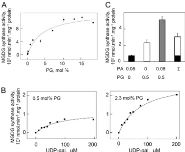
with PA, the enzyme velocity versus PG concentration curve was not sigmoid but hyperbolic (Fig. 5A) indicating a simpler way of interaction of the enzyme with PG than with PA.
Half-maximal activation was obtained with⬃4 mol% PG, suggesting
a lower affinity of the enzyme for PG than for PA. However the maximal activity attained with PG was more elevated than with PA. The apparent affinity of the enzyme for UDP-gal was not modified by different levels of PG but in contrast to what was
observed with PA, at a low concentration of PG, no cooperative effect between UDP-gal concentration and enzymatic activity was observed. This suggested that there was no interference between PG binding and UDP-gal handling by the enzyme (Fig. 5B).
Because the effects of PA and PG look different, we addressed then the question of whether they were synergic. We measured the enzyme activity at concentrations of PA (0.08 mol%) and PG (0.5 mol%) far below those giving the maximal activation of the enzyme. The activity in the presence of both PA and PG was much higher than the sum of the activities obtained separately in the presence of either PA or PG (Fig. 5C). This was clearly in support of distinct binding sites and different roles of PA and PG on the enzyme.
Molecular Discrimination of PA and PG Binding Sites
In a preliminary investigation to discriminate between PA and PG binding sites, we analyzed whether the activations by PA or PG were differentially sensitive to salt and phosphate.
Data show that with 250 mM KCl, activation by PG was
decreased whereas activation by PA was increased (Fig. 6A).
FIGURE 4. Lipid binding assay of MGD1. A, lipid-protein overlay assay of three different MGD1 from Arabidopsis, cucumber, and spinach. at⌬1– 137MGD1 and cs⌬1–104MGD1 harbor a His6tag at their C terminus, whereas
so⌬1–98MGD1 has no His tag. Different lipids were spotted onto nitrocellu-lose membranes as indicated. The experiment was conducted as described under “Experimental Procedures.” B, different amounts of PA or DAG were spotted onto nitrocellulose membranes. C, liposome binding assay with at⌬1–137MGD1. Liposomes were formed of 100% PC or 5% PA and 95% PC. T, total amount of protein; P, liposome pellet; S supernatant.
FIGURE 5. Effect of PG on the MGDG synthase activity of at⌬1–137MGD1. Experiments were conducted as described in the legend to Fig. 3. A, PG con-centration dependence of the activity. The curve equation is V⫽ 13 ⫻ [PG]/ (4⫹ [PG]). B, synergic effect of PG and PA on the activity. The activity was mea-sured with a combination of different mol% concentration of PA and PG as indicated under the graph.⌺ indicates the sum of the activities with only PA or PG. C, UDP-gal concentration dependence of the activity for two different concentrations of PG, 0.5 mol% and 2.3 mol%. The curve equation is V⫽ 1.8 ⫻ [UDP-gal]/(80⫹ [UDP-gal]) for 0.5 mol% of PG and V ⫽ 4.5 ⫻ [UDP-gal]/(80 ⫹ [UDP-gal]) for 2.3 mol% of PG. Results are average values⫾ S.D. for three independent measurements on a single batch of purified protein, whereas each part of the figure was done with a separated batch of purified protein.
FIGURE 6. Molecular discrimination of PA and PG binding on at⌬1–
137MGD1. The activity was measured as described under “Experimental
Pro-cedures” without PA or PG (black bars), with 1.5 mol% of PA (gray bars) or PG (white bars). Results are average values⫾ S.D. for three independent mea-surements. A, effect of salts on MGDG synthase activity. Concentrations of KH2PO4and KCl in the incubation medium are indicated below the graph.
B, effect of point mutations of the protein on activation by PA and PG. SDS-PAGE analysis of the purified point-mutated at⌬1–137MGD1 proteins used for activity measurement is shown above the graph.
at INRA Institut National de la Recherche Agronomique on May 13, 2019
http://www.jbc.org/
This suggested that the activation by PG was specifically depen-dent on electrostatic interaction. Because hydrophobic interac-tions are strengthened by salt, this suggested, on the other hand, that the activation by PA was dependent on hydrophobic
interaction. Incubation with 250 mMKH2PO4confirmed that
activation by PG was inhibited by high salt concentrations. The
inhibition was even stronger than with 250 mMKCl. Activation
by PA was, in contrast, reversed using KH2PO4instead of KCl.
KH2PO4had an inhibitory effect on activation by PA, the
oppo-site to KCl. Because a low concentration of KH2PO4(0.5 mM)
was sufficient to slightly activate the enzyme, we concluded that the phosphate anion directly interacted with the enzyme and that this interaction was competing with PA and PG. It is there-fore likely that phosphate is involved in the binding of both phospholipids to the enzyme. Altogether, main components of PG binding were electrostatic linkage and interaction through the phosphate group, whereas for PA binding they were of hydrophobic linkage and interaction through the phosphate group.
As another way to discriminate between PA and PG binding sites, we analyzed the differential activation by PA and PG of proteins exhibiting point mutations. To our knowledge, no information is available in the literature concerning PG binding sites. Concerning PA binding, there is no consensus sequence, but basic, hydrophobic, and aromatic residues were often reported to be important (24). Some uncharged polar residues such as serine and glutamine might also play a role (25, 26). A first model of plant MGDG synthase has been previously estab-lished using E. coli MURG as a template (7). The model for an MGD monomer comprises 2 Rossman domains (C- and N-do-mains). The binding site for UDP-gal was predicted in the cleft separating the two Rossmann folds involving residues of a con-served UDP-sugar binding pocket in the C-domain. The N-do-main is enriched in basic and hydrophobic residues and was proposed to be involved in lipid binding, either diacylglycerol
binding or membrane anchorage. In this domain, a⬍2-␣2⬎
loop, which has no counterpart in the MURG template, could play a specific role in lipid binding necessary for activity. Based on this information, we decided to test several residues possibly important for substrate binding or lipid binding. We targeted in the N-domain Arg-260, Trp-287, Pro-189, Thr-186, Asp-150, His-155, His-251 and in the C-domain Glu-456. The Arg-260, Trp-287, Pro-189, and Thr-186 residues were selected for a possible interaction with PA. Glu-456 and Asp-150 were selected for a possible interaction with UDP-galactose and His-155 and His-251 with diacylglycerol. The residue replacement was decided on a case by case basis to either change the charge or more generally test the structure of the lateral chain. None of the proteins is active without the addition of either PA or PG (Fig. 6B). Change of the lateral chain of Arg-260 (R260A) did not alter the activity except for a slight improvement of the activation by PG. In contrast, W287A was modified in activa-tion by both PA and PG and, furthermore, activaactiva-tion by PG was
abolished in this mutant. Mutants in the⬍2-␣2⬎ loop, P189A
and T186A, were also modified for both activations. P189A had a notably severe reduction of activation by PG. Mutant E456N was altered for activations by PA and PG. D150N and D150E were almost not affected except for possibly a slight
enhance-ment of activation by PG in D150N. His-155 looked essential because H155R and H155A had no activity at all. H251A was activated by both PA and PG but less efficiently than the wild-type protein. Altogether most of the mutants were affected in both types of activation. Only two mutants, W287A and P189A, showed a specific severe reduction of activation by PG. Because activation by PA was only slightly affected in these mutants, our conclusion was that the catalytic capability of the W287A and P189A proteins was conserved whereas their PG binding capa-bility was modified. R260A and D150N had possibly a light enhancement of activation by PG, whereas their activation by PA was not modified. In conclusion, our data confirm that the N-domain is important for lipid binding and that Trp-287 and Pro-189 play a critical role in the interaction of MGD1 with PG. Finally, drastic reduction of activation by PA was not detected without drastic reduction of activation by PG.
DISCUSSION
In this report, we have shown that MGD1 is allosterically activated by PA and that this activation is mechanistically dif-ferent from the activation by PG. The native enzyme present in leaf homogenates or in the chloroplast envelope as well as the
recombinant Arabidopsis⌬1–137 MGD1 were activated by PA.
The level of PA required for activation was low (k0.5of about 0.2
mol%) and not sufficient to feed MGDG synthesis by transfor-mation of PA into DAG, demonstrating an allosteric regulation of MGD1 by PA.
Modification of PA metabolism by inhibiting its production in leaf homogenates or enhancing its degradation in the chlo-roplast envelope hindered the MGDG synthase activity. The PA steady state concentration in the chloroplast envelope where MGD1 is located is assumed to be very low. It is unde-tectable in the isolated envelope fraction by conventional tech-niques. The PA level in the envelope is possibly close to PA levels in thylakoids, which was reported to be 0.08 mol% mainly prokaryotic PA (27). MGD1 was activated by several molecular species of PA from either a prokaryotic or eukaryotic nature. All PA molecular species were however not similarly efficient, because only partial restoration of PLD inhibition was obtained with exogenous 16:0/18:1-PA in leaf homogenates and also one prokaryotic molecular species of PA, 16:0/16:0-PA, was less efficient than others on MGD1. There are several possible sources of PA in the chloroplast envelope. Prokaryotic forms of PA are produced in the envelope by acylation of glycerol-3-phosphate by GAT (ATS1 gene) and LPAAT (ATS2 gene) (28 – 30). Formed PA, mostly 18:1/16:0-PA and a low level of 16:0/ 16:0-PA, are then precursors in synthetic pathways mainly synthesis of PG and prokaryotic MGDG and also partly of SQDG. Eukaryotic forms of PA are imported from extra-plas-tidial membranes. They likely result from activity of phospho-lipase(s) D and transport to the chloroplast by a system involv-ing the TGD proteins (31). Phospholipase D activity has never been detected in the isolated chloroplast envelope, although proteomics analysis of the envelope reported the presence of
PLD␣1 (32). Our results on pld2 indicated that without the
addition of exogenous PA, the MGDG synthase activity of the mutant leaves was a little reduced compared with the wild type, whereas, after addition of exogenous PA, the activity was much
Activation of MGD1 by PA and PG
at INRA Institut National de la Recherche Agronomique on May 13, 2019
http://www.jbc.org/
higher. These results are consistent with (i) PLD2 being a potent source of PA for activation of MGD1 with (ii) a
counter-balance effect of the PLD2 mutation such as an overexpression
of MGD1. However, considering that PLD2 has been located
in the tonoplast (33), it is not yet clear how PA generated by
PLD2 could reach MGD1 present in the inner envelope
mem-brane. Besides PLD2, some other PLDs could contribute to PA
supply. One other candidate is PLD1, which was shown to be
involved like PLD2 in galactolipid synthesis in roots under
phosphate deprivation. It is possible that overexpression of
PLD2/1 in the initial stage of phosphate deprivation could
enhance MGD1 activity independently of DAG supply in-crease. Several other enzymes determine the fate of PA in the envelope. Plastid PAP hydrolyzes PA into diacylglycerol, which is the substrate for MGDG or SQDG synthesis (23, 34). Alter-natively, a CDP-diacylglycerol synthetase transfers cytidyl from CTP onto PA and sets off the first step for PG synthesis (35). Finally some reports suggest that lipid kinases and acylhydro-lases are active in the chloroplast, which could therefore modify the envelope PA level (36 –38).
Ultimately, the question is the role of MGD1 activation by PA. Because PA is a key signal for many processes in the cell (24, 39), MGD1 activation by PA could relate to a signaling process necessary to couple chloroplast and plant development. In this
sense, with regards to the role of PLD2 under the initial stage
of phosphate deprivation and PLD2 tonoplast localization (12,
22, 33, 40, 41), PA produced by PLD2 could be a switch related
to phosphate sensing. Formation of prokaryotic PA could also be important for MGD1 activity. In support of this, ats2, a mutant devoid of LPAAT, has no formation of prokaryotic PA and is impaired in embryo development (29) although neither formation of prokaryotic galactolipids is essential at this stage (ats1 mutant analysis; Ref. 28, 30), nor envelope-specific PG synthesis (pgp1 mutant analysis; Ref. 35). Another possibility is that MGD1 activation by PA plays a role in the tuning of the different lipid syntheses that occur in the envelope: galactolipid, PG, and SQDG syntheses. For instance, activa-tion of MGD1 by PA could enhance the enzyme funcactiva-tioning when the envelope is initially fed with DAG, while PA is pref-erentially sustaining PG synthesis. Galactolipid synthesis could thus start instantly.
A second point of our results is that activation by PA and PG proceed through different mechanisms. First, we have shown that there is a synergic effect of PA and PG on MGD1 activity. Second, kinetic analysis of enzyme velocity according to activa-tor concentration indicated (i) a more complex and cooperative
way of activation by PA than by PG, (ii) a higherAppVmaxof the
enzyme with PG although the k0.5was much higher than with
PA. Third, the activation by PA and PG was affected differently by salts indicating that the binding of MGD1 with each activa-tor was driven by different types of chemical bond, electrostatic bond for PG activation and hydrophobic bond for PA. Alto-gether, this suggests that PG plays a specific role in the regula-tion of MGD1. PG level in the inner membrane is close to 8
mol%, which is not very far from the k0.5calculated for PG on
the recombinant Arabidopsis⌬1–137 MGD1 (5 mol%).
More-over almost no activity was found in the chloroplast envelope after treatment to deplete PA but activity was enhanced by
addition of PG, indicating that the envelope PG in our assay was not present in sufficient concentration to sustain MGD1 activ-ity. Two residues, Pro-189 and Trp-287, which play a specific role in the activation by PG, are present in the N domain of MGD1. Because the N domain of MGD1 was previously pro-posed to interact with the membrane (7), it is possible that PG is important to anchor MGD1 into the membrane. However, we observed that the recombinant MGD1 could easily associate with PC liposomes. This suggests that PG is not essential for anchoring. MGD1 activation by PG could finally reflect, as suggested above for activation by PA, a reinforced coupling of MGDG synthesis with PG synthesis, considering that MGDG and PG are two essential components of photosyn-thetic membranes.
Acknowledgments—We thank C. Albrieux for technical assistance and Drs G. Curien and F. Rebeille´ for helpful discussions.
REFERENCES
1. Block, M. A., Douce, R., Joyard, J., and Rolland, N. (2007) Photosynth. Res
92,225–244
2. Jarvis, P., Do¨rmann, P., Peto, C. A., Lutes, J., Benning, C., and Chory, J. (2000) Proc. Natl. Acad. Sci. U.S.A. 97, 8175– 8179
3. Kobayashi, K., Kondo, M., Fukuda, H., Nishimura, M., and Ohta, H. (2007) Proc. Natl. Acad. Sci. U.S.A. 104,17216 –17221
4. Mare´chal, E., Block, M. A., Joyard, J., and Douce, R. (1994) J. Biol. Chem.
269,5788 –5798
5. Miege, C., Marechal, E., Shimojima, M., Awai, K., Block, M. A., Ohta, H., Takamiya, K., Douce, R., and Joyard, J. (1999) Eur J Biochem. 265, 990 –1001
6. Awai, K., Marechal, E., Block, M. A., Brun, D., Masuda, T., Shimada, H., Takamiya, K., Ohta, H., and Joyard, J. (2001) Proc. Natl. Acad. Sci. U.S.A.
98,10960 –10965
7. Botte, C., Jeanneau, C., Snajdrova, L., Bastien, O., Imberty, A., Breton, C., and Marechal, E. (2005) J. Biol. Chem. 280, 34691–34701
8. Cove´s, J., Joyard, J., and Douce, R. (1988) Proc. Natl. Acad. Sci. U.S.A. 85, 4966 – 4970
9. Ohta, H., Shimojima, M., Arai, T., Masuda, T., Shioi, Y., and Takamiya, K. (1995) in Plant Lipid Metabolism (Kader, J. C., and Mazliak, P., eds), Klu-wer Academic Publishers, Dordrecht
10. Kelly, A. A., O¨ hman, A., Sedoud, A., and Wieslander, A. (2007) in Current Advances in the Biochemistry and Cell Biology of Plant Lipids(Benning, C., and Olhlrogge, J., eds), Aardvark Global Publishing Company, Salt Lake City
11. Alonso, J. M., Stepanova, A. N., Leisse, T. J., Kim, C. J., Chen, H., Shinn, P., Stevenson, D. K., Zimmerman, J., Barajas, P., Cheuk, R., Gadrinab, C., Heller, C., Jeske, A., Koesema, E., Meyers, C. C., Parker, H., Prednis, L., Ansari, Y., Choy, N., Deen, H., Geralt, M., Hazari, N., Hom, E., Karnes, M., Mulholland, C., Ndubaku, R., Schmidt, I., Guzman, P., Aguilar-Henonin, L., Schmid, M., Weigel, D., Carter, D. E., Marchand, T., Risseeuw, E., Brogden, D., Zeko, A., Crosby, W. L., Berry, C. C., and Ecker, J. R. (2003) Science 301,653– 657
12. Li, M., Qin, C., Welti, R., and Wang, X. (2006) Plant Physiol. 140, 761–770 13. Jouhet, J., Mare´chal, E., Bligny, R., Joyard, J., and Block, M. A. (2003) FEBS
Lett. 544,63– 68
14. Douce, R., and Joyard, J. (1990) Annu. Rev. Cell Biol. 6, 173–216 15. Dorne, A. J., Block, M. A., Joyard, J., and Douce, R. (1982) FEBS Letters 145,
30 –34
16. Yamaryo, Y., Motohashi, K., Takamiya, K., Hisabori, T., and Ohta, H. (2006) FEBS Letters 580, 4086 – 4090
17. Bates, D. M., and Watts, D. G. (1988) Nonlinear Regression Analysis and Its Applications, John Willey and Sons, New York
18. Ihaka, R., and Gentleman, R. (1996) J. Comp. Graph. Stat. 5, 299 –314 19. Andrews, B., Bond, K., Lehman, J. A., Horn, J. M., Dugan, A., and
at INRA Institut National de la Recherche Agronomique on May 13, 2019
http://www.jbc.org/
Cambronero, J. (2000) Biochem. Biophys. Res. Commun. 273, 302–311 20. Andersson, M. X., Kjellberg, J. M., and Sandelius, A. S. (2004) Biochim
Biophys Acta 1684,46 –53
21. Qin, C., and Wang, X. (2002) Plant Physiol. 128, 1057–1068
22. Cruz-Ramírez, A., Oropeza-Aburto, A., Razo-Herna´ndez, F., Ramirez-Cha´vez, E., and Herrera-Estrella, L. (2006) Proc. Natl. Acad. Sci. U.S.A.
103,6765– 6770
23. Malherbe, A., Block, M. A., Joyard, J., and Douce, R. (1992) J. Biol. Chem.
267,23546 –23553
24. Testerink, C., and Munnik, T. (2005) Trends Plant Sci. 10, 368 –375 25. Jones, J. A., Rawles, R., and Hannun, Y. A. (2005) Biochemistry 44,
13235–13245
26. Grange, M., Sette, C., Cuomo, M., Conti, M., Lagarde, M., Prigent, A. F., and Ne´moz, G. (2000) J. Biol. Chem. 275, 33379 –33387
27. Fritz, M., Lokstein, H., Hackenberg, D., Welti, R., Roth, M., Za¨hringer, U., Fulda, M., Hellmeyer, W., Ott, C., Wolter, F. P., and Heinz, E. (2007) J. Biol. Chem. 282,4613– 4625
28. Kunst, L., Browse, J., and Somerville, C. (1988) Proc. Natl. Acad. Sci. U.S.A.
85,4143– 4147
29. Yu, B., Wakao, S., Fan, J., and Benning, C. (2004) Plant Cell Physiol. 45, 503–510
30. Xu, C., Yu, B., Cornish, A. J., Froehlich, J. E., and Benning, C. (2006) Plant Journal 47,296 –309
31. Benning, C. (2009) Annu. Rev. Cell Dev. Biol. 25, 71–91
32. Joyard, J., Ferro, M., Masselon, C., Seigneurin-Berny, D., Salvi, D., Garin, J., and Rolland, N. (2010) Progress in Lipid Research, in press
33. Yamaryo, Y., Dubots, E., Albrieux, C., Baldan, B., and Block, M. A. (2008) FEBS Letters 582,685– 690
34. Nakamura, Y., Tsuchiya, M., and Ohta, H. (2007) J. Biol. Chem. 282, 29013–29021
35. Babiychuk, E., Mu¨ller, F., Eubel, H., Braun, H. P., Frentzen, M., and Kush-nir, S. (2003) Plant Journal 33, 899 –909
36. Siegenthaler, P. A., Mu¨ller, M. O., and Bovet, L. (1997) FEBS Lett. 416, 57– 60
37. Mu¨ller, M. O., Meylan-Bettex, M., Eckstein, F., Martinoia, E., Siegenthaler, P. A., and Bovet, L. (2000) J. Biol. Chem. 275, 19475–19481
38. Seo, Y. S., Kim, E. Y., Kim, J. H., and Kim, W. T. (2009) FEBS Letters 583, 2301–2307
39. Wang, X., Devaiah, S. P., Zhang, W., and Welti, R. (2006) Prog Lipid Res.
45,250 –278
40. Misson, J., Raghothama, K. G., Jain, A., Jouhet, J., Block, M. A., Bligny, R., Ortet, P., Creff, A., Somerville, S., Rolland, N., Doumas, P., Nacry, P., Herrerra-Estrella, L., Nussaume, L., and Thibaud, M. C. (2005) Proc. Natl. Acad. Sci. U.S.A. 102,11934 –11939
41. Li, G., and Xue, H. W. (2007) Plant Cell 19, 281–295
Activation of MGD1 by PA and PG
at INRA Institut National de la Recherche Agronomique on May 13, 2019
http://www.jbc.org/
Christelle Breton, Eric Maréchal and Maryse A. Block
Emmanuelle Dubots, Magali Audry, Yoshiki Yamaryo, Olivier Bastien, Hiroyuki Ohta,
Phosphatidic Acid and Phosphatidylglycerol
doi: 10.1074/jbc.M109.071928 originally published online December 20, 2009 2010, 285:6003-6011.
J. Biol. Chem.
10.1074/jbc.M109.071928 Access the most updated version of this article at doi:
Alerts:
When a correction for this article is posted •
When this article is cited •
to choose from all of JBC's e-mail alerts Click here
Supplemental material:
http://www.jbc.org/content/suppl/2009/12/20/M109.071928.DC1 http://www.jbc.org/content/285/9/6003.full.html#ref-list-1This article cites 37 references, 18 of which can be accessed free at
at INRA Institut National de la Recherche Agronomique on May 13, 2019
http://www.jbc.org/
![[PDF] Cours de base pour debuter et avancer avec CSS | Cours informatique](data:image/gif;base64,R0lGODlhAQABAIAAAP///wAAACH5BAEAAAAALAAAAAABAAEAAAICRAEAOw==)