HAL Id: hal-01802401
https://hal.archives-ouvertes.fr/hal-01802401
Submitted on 12 Nov 2019
HAL is a multi-disciplinary open access
archive for the deposit and dissemination of
sci-entific research documents, whether they are
pub-lished or not. The documents may come from
teaching and research institutions in France or
abroad, or from public or private research centers.
L’archive ouverte pluridisciplinaire HAL, est
destinée au dépôt et à la diffusion de documents
scientifiques de niveau recherche, publiés ou non,
émanant des établissements d’enseignement et de
recherche français ou étrangers, des laboratoires
publics ou privés.
Performances of the CC2 method versus multireference
methods
Nadia Ben Amor, Sophie Hoyau, Daniel Maynau, Valerie Brenner
To cite this version:
Nadia Ben Amor, Sophie Hoyau, Daniel Maynau, Valerie Brenner. Low-lying excited states of model
proteins: Performances of the CC2 method versus multireference methods. Journal of Chemical
Physics, American Institute of Physics, 2018, 148 (18), pp.184105. �10.1063/1.5025942�. �hal-01802401�
Low-lying excited states of model proteins: Performances of the CC2 method versus
multireference methods
Nadia Ben Amor, Sophie Hoyau, Daniel Maynau, and Valérie Brenner
Citation: The Journal of Chemical Physics 148, 184105 (2018); doi: 10.1063/1.5025942 View online: https://doi.org/10.1063/1.5025942
View Table of Contents: http://aip.scitation.org/toc/jcp/148/18 Published by the American Institute of Physics
Low-lying excited states of model proteins: Performances of the CC2
method versus multireference methods
Nadia Ben Amor,1,2,a)Sophie Hoyau,2Daniel Maynau,1,2and Val´erie Brenner3
1CNRS, UPS, LCPQ (Laboratoire de Chimie et Physique Quantiques), IRSAMC, 118, Rte de Narbonne, F-31062 Toulouse Cedex, France
2UPS, LCPQ (Laboratoire de Chimie et Physique Quantiques), IRSAMC, Universit´e de Toulouse, 118, Rte de Narbonne, F-31062 Toulouse Cedex, France
3Laboratoire Interactions, Dynamiques et Lasers, LIDYL, CEA, CNRS, Universit´e Paris-Saclay, 91191 Gif-sur-Yvette Cedex, France
(Received 14 February 2018; accepted 10 April 2018; published online 9 May 2018)
A benchmark set of relevant geometries of a model protein, the N-acetylphenylalanylamide, is pre-sented to assess the validity of the approximate second-order coupled cluster (CC2) method in studying low-lying excited states of such bio-relevant systems. The studies comprise investigations of basis-set dependence as well as comparison with two multireference methods, the multistate complete active space 2nd order perturbation theory (MS-CASPT2) and the multireference difference dedi-cated configuration interaction (DDCI) methods. First of all, the applicability and the accuracy of the quasi-linear multireference difference dedicated configuration interaction method have been demon-strated on bio-relevant systems by comparison with the results obtained by the standard MS-CASPT2. Second, both the nature and excitation energy of the first low-lying excited state obtained at the CC2 level are very close to the Davidson corrected CAS+DDCI ones, the mean absolute deviation on the excitation energy being equal to 0.1 eV with a maximum of less than 0.2 eV. Finally, for the following low-lying excited states, if the nature is always well reproduced at the CC2 level, the differences on excitation energies become more important and can depend on the geometry. Published by AIP
Publishing.https://doi.org/10.1063/1.5025942
I. INTRODUCTION
Many complex molecular systems absorbing light in the near UV spectral range, including those of paramount bio-logical importance like DNA bases or proteins, are endowed with mechanisms of excited-state deactivation following UV absorption.1These processes are of major importance for the photochemical stability of these species since they provide a rapid and efficient way of dissipating the electronic energy in excess into vibration, thus avoiding photochemical processes to take place and then structural damages which can affect the biological function.2,3They are controlled by the energy and
the nature of the electronic excited states of the chromophores, by their couplings and the resulting electron dynamics: UV light absorption populates excited states, which dissipate the electronic energy, either through a relatively slow radiative deactivation process, i.e., photon emission, or, more often and more efficiently, by a radiationless transition, e.g., internal con-version or intersystem crossing. The latter, the nonadiabatic (NA) transfers, often involve ultrafast energy transfers through regions of the potential energy surfaces (PES) corresponding to avoided or surface crossings, of conical nature or not.4–9
In order to investigate conformer-selective dynamics of biologically relevant molecular systems and, in particular, the building block of proteins such as capped peptides, we have developed an original innovative computational strategy. Our
a)Author to whom correspondence should be addressed: benamor@
irsamc.ups-tlse.fr
main goal is to document the basic physical phenomena con-trolling the lifetime of excited states, highlighting the link between electronic dynamics and structure.10,11
The multi-step multi-level computational strategy allows us to both characterize the low-lying excited states of bio-relevant systems and to model efficiently their PES using, first, nonadiabatic dynamic simulations based on time-dependent density functional theory (NA-TDDFT) to provide hints about the critical motions that drive the deactivation. Two better levels of theory are then used to refine these simulations: (i) the standard approximate coupled cluster singles and dou-bles method (CC2)12–16 and (ii) a multireference
configura-tion interacconfigura-tion (MRCI) method.17–19The NA-TDDFT sim-ulations and refinement of the energy profiles at the CC2 level were previously applied on small capped peptides, the N-acetylphenylalanylamide (NAPA) and its N-methylated derivative (NAPMA). It highlighted, for the first time in such systems, the quenching properties of the primary amide group (through its nπ∗COexcited state) along with the effect of vibra-tional energy that facilitated access to the conical intersection (CI) area.10,11 This paper is fully in line with these studies and focuses on the third step of the computational strategy that has, to our knowledge, never been addressed: assessment for such systems of the accuracy of the CC212–16method by
comparison with a MRCI method.17–19
The challenge of such calculations is multiple. First, one of our objectives is not only to assess the accuracy of the CC2 method on the equilibrium geometry of the initially excited state accessible from the Frank-Condon region (generally a
singlet ππ∗ excited state on the aromatic ring) but also to assess them on the equilibrium geometries of all close-low-lying excited singlet states of these systems as well as on relevant geometries of the energy profile of the deactivation mechanisms such as the conical intersections (CI) between excited states. Second, these systems present specificities that are decisive in the choice of methods, i.e., the level of the-ory: (i) their size (medium-size systems where the smallest one, a capped peptide with one residue, already contains at least thirty atoms), (ii) their lack of symmetry, (iii) their great flexibility due to the non-covalent interactions that govern their structure, and (iv) their multiple close-low-lying excited states featuring very different nature [the locally excited state on the peptide bonds (LEpep) or on the aromatic ring (LEπ) and even the charge transfer (CT) state]. Finally, our ulti-mate goal is to apply this computational strategy to capped peptides which contain more than one residue but this lat-ter point will be investigated and discussed in a future paper. Even if the validity of the CC2 method for equilibrium struc-tures and energy profile of both ground and excited states of small peptides has been already established,20–22 there is no
benchmark of the CC2 method for application to the different close-low-lying excited states of such systems. Two of the most recent and extensive benchmarks of the CC2 method which use as reference data experimental values previously compiled from high-resolution gas-phase experiments,23,24 and refer-ences therein, concern the adiabatic transitions of the lowest excited state of a set of different medium-size molecules con-taining either aromatic organic molecules including different conformers (66 molecules: Mean Absolute Error (MAE) of 0.08 eV and Mean Signed Error (MSE) of 0.04 eV)23or cov-ering both organic (polyenes, carbonyl compounds, aromatic hydrocarbons, and heterocycle aromatic compounds) and inor-ganic (main-group and transition metal compounds) systems (79 molecules: MAE of 0.19 eV and MSE of 0.11 eV).24
Con-cerning the theoretical benchmarks, only a few studies using reference values obtained at a very high level of theory, such as MRCI, focus on medium-size molecular systems such as the retinal-chromophore model25or 9H-adenine.26Moreover, these studies deal with energy profiles of mechanisms involv-ing PES of only two states, i.e., the ground state and one excited state.
In this work, we have gone further in investigating the accuracy of the CC2 method for applications to low-lying excited states of capped peptides by performing calculations with the MR-Difference Dedicated Configuration Interaction (DDCI) method.27,28The performances of the CC2 method for the description of both the ground and excited states of a con-former of NAPA, i.e., NAPA B, are first evaluated at the ground state equilibrium geometry. Second, relevant geometries along the energy profile of deactivation mechanisms such as equi-librium geometries of the low-lying excited states or conical intersection (CI) geometry are considered. In this benchmark, the multireference method used as reference is the DDCI method.27,28 This method was previously used for magnetic systems, organic molecules, and carbon nanotubes.17–19,29–32 We report here on the conditions of the quasi-linear version of this multireference method to bio-relevant systems. The appro-priate parameters used in this quasi-linear CI for the capped
peptides are defined and validated by comparison with calcula-tions performed with a more standard multireference method, the multistate complete active space 2nd order perturbation theory (MS-CASPT2).33
II. COMPUTATIONAL DETAILS A. Geometries
Let us define some notations used in the following: pept1 or (1) refers to the first peptide bond in interaction with the phenyl ring (NH–π interaction), while the other peptide bond is denoted (2) or pept2 (see Fig.1). In order to evaluate the performances of the CC2 method, different geometries of the NAPA B conformer (see Fig.1and Appendix S1 in the supple-mentary material) have been investigated with both CC2 and MR methods. First, calculations were performed at the B97-D2/TZVPP (noted DFT-D hereafter) equilibrium geometry of the ground state.34Second, three CC2/cc-pVDZ pertinent
geometries of the energy profile of the deactivation mechanism identified recently and involving the second peptide bond (MII in Refs.10and11) were chosen: (i) the equilibrium geome-try of the first ππ∗ excited state (Mππ∗), (ii) the equilibrium
geometry of the nπ∗CO excited state localized on the second peptide bond [Mnπ∗CO(2)], and (iii) the CI geometry
connect-ing the equilibrium geometries of these two excited states (CIππ∗/nπ∗CO(2)). The last considered geometry was the
par-tially optimized geometry [NH(1) bond length constrained to 1.216 Å] of the CT excited state (M0CT) recently identified in a deactivation mechanism (mechanism I in Refs.10and11). In the following, NH(1) corresponds to the NH bond of the first peptide bond pointing to the phenyl ring.
B. CC2 calculations
The CC212–15calculations were carried out with the TUR-BOMOLE package.35,36All the calculations were performed by using the resolution-of-identity (RI) approximation for the electron repulsion integrals used in the correlation treat-ment and the description of the excitation processes. cc-pVXZ (X = D, T, and Q) Dunning’s correlation consistent basis sets37were employed in connection with optimized auxiliary
basis sets for the RI approximation.38Few calculations were
also performed with the addition of diffuse functions. Frozen core for the 1s electrons was employed, and all calculations were carried out in the C1point-group symmetry. Twenty sin-glet states were considered and D1 and D2 diagnostics and %E1|E1 biorthogonal norm were calculated in order to eval-uate the capability of the CC2 method to properly describe the excited states of such systems.15,39,40Indeed, the D1 and D2 diagnostics, computed from the single and double substitution amplitudes in the CC2 wave function, have been found to be reliable indicators when static or dynamic correlation effects are not adequately treated at the CC2 level: their magnitudes are correlated with the performance of the CC2 method. In addition, the biorthogonal normE1|E1 gives a measure of the weight of the single excitation contributions to an excited state. Indeed, in order to be well described at the CC2 level, an excited state must be dominated by single excitations out of the ground state wave function.
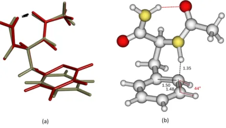
FIG. 1. (a) DFT-D optimized geometry of the ground state of the conformer B of NAPA exhibiting a C7H-bond (red dashes, dNH(2)–O= 2.02 Å) and an NH–π
interaction (black dashes, dNH(1)–Cπ= 2.57 Å); (b) CC2/cc-pVDZ geometries of the energy profile of the MIIdeactivation mechanism of NAPA B: (b1) Mππ∗, the optimized geometry of the ππ∗excited state (red dashes, d
NH(2)–O= 1.95 Å and black dashes, dNH(1)–Cπ= 2.35 Å), (b2) CIππ∗/nπ∗CO(2), the geometry of the CI (red dashes, dNH(2)–O= 2.00 Å and black dashes, dNH(1)–Cπ= 2.42 Å), and (b3) Mnπ∗CO(2), the optimized geometry of the excited state nπ∗COlocalized on the second peptide bond (red dashes, dNH(2)–O= 2.28 Å and black dashes, dNH(1)–Cπ= 2.66 Å); (c) M0CT, the CC2/cc-pVDZ partially optimized geometry with the
NH(1) bond length constrained to 1.216 Å of the intramolecular CT excited state nπ∗
cycle(red dashes, dNH(2)–O= 1.87 Å and black dashes, dNH(1)–Cπ= 1.35 Å).
All the coordinates corresponding to these geometries are reported in thesupplementary material.
C. MR calculations
The complete active space self-consistent field (CASSCF)41/MS-CASPT233 calculations were carried out with the 7.8 MOLCAS package.42The linear scaling MRCI calculations17–19require the use of local orbitals. The CASSCF orbitals were then localized with the DOLO code43,44and the linear scaling MRCI calculations were performed with the EXSCI program.44The Davidson correction was introduced to correct the size-consistency error inherent in the MRCI methods. These multireference calculations were performed with different basis sets. First, the Atomic Natural Orbitals (ANO-L)45basis sets were used with the following
contrac-tion scheme: for C, N, and O, a (14s9p4d3f) set contracted to [3s2p1d] and for H a (8s4p3d) set contracted to [2s1p]. Dunning’s correlation consistent basis sets cc-pVDZ and cc-pVTZ37 were also chosen for comparison with the CC2 results. Finally, as for the CC2 calculations, few computations were performed with the addition of diffuse functions.
A well balanced active space has to be defined to provide a good description of all considered singlet states. These states may correspond to local excitations (LE): (i) centered on the phenyl group (LEπ) and (ii) centered on each peptide bond [n → π∗COwhere n is a N or O lone pair (pure-p lone pair), LEpep, and πCO→ π∗CO] or to excitations corresponding to electronic charge transfer (CT) from the backbone to the phenyl group. Therefore, 18 electrons in 14 orbitals were included in the active space, corresponding to all the π and π∗ orbitals of the phenyl and carbonyl groups and to lone
pairs on the nitrogen and oxygen atoms. However, for both nπ∗COand CT excited state geometries, this active space had to be enlarged in order to include one additional orbital: (i) an n orbital (pept2) for the equilibrium nπ∗CO excited state geometry, i.e., the second lone pair (sigma lone pair) on the sec-ond peptide bsec-ond oxygen atom; (ii) a σNH(1) (pept1) orbital pointing toward the phenyl group for the partially optimized CT excited state geometry, with the NH(1) bond length con-strained to 1.216 Å. In these two cases, 20 electrons in 15 orbitals were included in the active space.
MS-CASPT2 calculations were performed on CASSCF reference wave functions. Average molecular orbitals (MO) are generally a good compromise to obtain the whole set of states involved in the electronic spectrum. State-average orbitals on 14 and 20 singlet states were then optimized in order to obtain the charge transfer states (see discussion in Sec.III). In the MS-CASPT2 method, the use of a level shift allows us to avoid weak intruder states by the addition of a shift parameter to the zeroth-order Hamiltonian. Several tri-als were performed, with various level shift values: 0.0, 0.1, 0.2, 0.3, 0.4, and 0.5 a.u. (Figs. S2-1 and S2-2 of the supple-mentary material). The final choice was made on the basis of the deviation of the reference weight of the zeroth-order wave functions of the excited states compared to the ground state one, as well as on the basis of the stability of the excitation energies. A maximal deviation of 3% for the reference weight was obtained with a level shift of 0.5 a.u. and this value was then used for all the CASPT2 calculations. Another modifi-cation of the zeroth-order Hamiltonian, called the Ionization
Potential Electronic Affinity shift, was introduced in order to reduce the systematic error on the relative energies compar-ing closed shell and open shell states. The 0.25 default value was used for the IPEA shift. The core electrons were frozen. Finally, the Cholesky decomposition technique46–48was used,
with a 10 8a.u. threshold.
The dimension of MRCI calculations is too large to be worth considering. Indeed, mono- and di-excitations on the large (18,14) or (20,15) active spaces would give rise to several billions of determinants. Therefore, different complementary computational strategies were introduced: (i) a reduction of the active space and two different selections of the CI space, (ii) the use of the MR-Difference Dedicated Configuration Interaction (DDCI) method27,28,30instead of MR-Single and Double CI (MR-SDCI), and (iii) the linear scaling MRCI method17–19 using localized orbitals.43 These three points are developed below.
1. Reduction of the active space
The large 18 electrons in 14 orbital active space were reduced, guided by the description of the different states, in the three different following active spaces: (i) πcycleand π∗cycle orbitals for the local excitations centered on the phenyl group (LEπ), corresponding to 6 electrons in 6 orbitals; (ii) the nitro-gen and oxynitro-gen pure-p lone pairs (n), πCOand π∗COorbitals for the local excitations on each peptide bond (LEpep and πCO → π∗CO) leading to 12 electrons in 8 orbitals; (iii) for the charge transfer states (CT), the nitrogen lone pair point-ing toward the phenyl group (nN(1)) and the lone pair (n) of each oxygen atom as well as the anti-bonding π∗cycleorbitals of the phenyl group were included in the last 6 electrons in 6 orbital active space. However, one active orbital was added to properly describe the CT states of the M0
CT and Mnπ∗CO(2)
excited states geometries. In the case of M0
CTgeometry, the σNH(1)orbital of the first peptide bond was added as the amino hydrogen is pointing towards the phenyl group, leading to a CAS(8,7). For the Mnπ∗CO(2) excited state geometry, the
π∗CO (pept2) orbital was added to describe the CT states as a small MR character with a non-negligible weight on the
nπ∗CO (pept2) excitation was found. A CAS(6,7) was then considered.
2. DDCI method
This method was used to reduce the number of determi-nants by neglecting those involving only external orbitals, i.e., two inactive occupied and two virtual orbitals. As the most time consuming part of an SDCI calculation consists in the process-ing of the doubly excited determinants, the computational cost is considerably reduced by neglecting these two holes-two par-ticles determinants. Including only the correlation energy that contributes to the energy difference in a variational CI allows us to obtain accurate vertical and adiabatic excitations, singlet-triplet gaps, and exchange magnetic coupling constants.27,28,49
Beyond the reduction of the determinant basis, the DDCI method also reduces the inherent size-consistency error of the CI method. However, the Davidson correction was applied in order to get the most reliable excitation energies. The corrected excitation energies are noted CAS+DDCI+Q thereafter.
3. Linear scaling MRCI method using localized orbitals
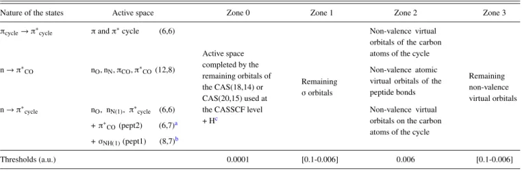
Dealing with local orbitals offers the possibility to neglect long range interactions. Furthermore, it allows dividing the molecular system into regions of unequal importance. Indeed, in the most important region (where the phenomena occur), it is necessary to take into account interactions at a high level of accuracy. The interaction cutoff should then be set at a very small value, while in the rest of the system it can be taken rather large. This cutoff is obtained by means of a threshold on the exchange integral values between orbitals involved in each determinant or integral. Accurate results can then be obtained at a lower cost. Indeed, when thresholds are used on both deter-minants and integrals, the MRCI calculation becomes quasi-linear. The threshold used to eliminate the integrals was taken to be 0.0001 a.u. in all the study. Concerning the selection of the CI space determinants, the definition of the various zones (see TableI) was based on the nature of the localized orbitals: σ, lone pairs, or π. Zone 0 contained the targeted active space and the remaining orbitals of the CAS(18,14) or CAS(20,15) used at TABLE I. Partition of the molecular orbitals in different zones. The corresponding thresholds applied on the exchange integrals in each zone to select the DDCI determinant space are given in a.u.
Nature of the states Active space Zone 0 Zone 1 Zone 2 Zone 3
πcycle→ π∗cycle πand π∗cycle (6,6)
Active space completed by the remaining orbitals of the CAS(18,14) or CAS(20,15) used at the CASSCF level + Hc
Remaining σorbitals
Non-valence virtual orbitals of the carbon atoms of the cycle
Remaining non-valence virtual orbitals
n → π∗CO nO, nN, πCO, π∗CO(12,8) Non-valence atomic
virtual orbitals of the peptide bonds n → π∗ cycle nO, nN(1), π∗cycle (6,6) + π∗ CO(pept2) (6,7)a + σNH(1)(pept1) (8,7)b Non-valence virtual orbitals on the carbon atoms of the cycle
Thresholds (a.u.) 0.0001 [0.1-0.006] 0.006 [0.1-0.006]
aFor the nπ∗
COexcited state geometry. bFor the CT excited state geometry.
the CASSCF level. All orbitals corresponding to the hydrogen atom of the amino group involved in the hydrogen bond (pept2) as well as the one pointing toward the cycle (pept1) were also included in this zone. The remaining σ orbitals were in zone 1. Two additional zones were defined by the non-valence vir-tual orbitals, those of the peptide bonds or those of the cycle (according to the considered states) for zone 2, and the corre-sponding remaining ones for zone 3. The thresholds were dif-ferent among the various zones: 0.0001 a.u. was used for zone 0 while for the non-valence virtual orbitals (zone 2), a threshold of 0.006 a.u. was applied. The remaining non-valence orbitals (zone 3) as well as the σ zone (zone 1) were allowed to be more approximately described depending on the nature of the considered states. Several trials (0.1-0.006 a.u.) were done as presented in Sec. III, in order to get stable and accurate results on the excitation energies. When the exchange integral involved orbitals of different zones, a mean value between the two thresholds was applied. A more detailed presentation of the method can be found in previous studies.17–19
III. RESULTS
Whatever the geometry and the basis set, there are mainly three types of excited states among the twenty first singlet excited states of NAPA B as illustrated in Fig. 2: locally (πcycle, πcycle∗) excited states centered on the phenyl ring (noted hereafter ππ∗), locally (n, π∗CO) excited states centered on a peptide bond (noted hereafter nπ∗CO), and intramolecular CT (n, π∗
cycle) excited states involving an electronic charge trans-fer from the lone pairs of the backbone (one or two peptide bonds) to the phenyl ring (noted hereafter nπ∗
cycle).
A. Ground state geometry
1. CC2 calculations
The relative CC2 excitation energies of the four first low-lying ππ∗, nπ∗CO, and nπ∗cycleexcited states of NAPA B in its DFT-D ground state geometry [Fig. 1(a) and Table S1-1 of the supplementary material] and the contributions of the canonical occupied-unoccupied HF (πcycle, n–π∗cycle, and π∗CO) orbitals to the total wave function change are reported in TableIIfor a series of Dunning’s basis sets. The NAPA B con-former presents a folded form exhibiting both a C7hydrogen bond (2.02 Å) between one hydrogen of the amino group and the oxygen atom of the 1st peptide bond and a NH(1)–Cπ
interaction [dNH(1)–Cπ = 2.57 Å; see Fig. 1(a)]. Whatever the basis set, the D1/D2 values for the ground state were in the range 0.08/0.17 while the D2 value for the excited states was in the range 0.16-0.31 (0.19-0.27 for the first six excited states) with a biorthogonal norm %E1|E1 ≥ 85% (%E1|E1 ≥ 87% for the first six excited states). The ini-tially recommended values for D1/D2for ground state minima were 0.04(0.05)/0.17(0.18) in the case of MP2(CCSD),33,34 but K¨ohn and H¨attig extended these D1/D2 limit values up to 0.15/0.25 in particular from the evaluation of excited states of a set of small-size molecules computed with CC2. Fur-thermore, they proposed a new complementary diagnostics, i.e., the biorthogonal norms %E1|E1 which should be larger than 85%. More recently, for NA-ADC(2)/aug-cc-pVDZ sim-ulations of the 9H-adenine, Barbatti et al. found a D1(MP2) value in the range 0.04-0.06 for the ground state and up to val-ues of 0.09 for the CI area while the D2value [ADC(2)] of the first excited state was between 0.25 and 0.35, the upper region of this domain corresponding to the CI area.26Consequently, although our values remain close to the upper limit, they con-firm the reliability of the CC2 calculations on these systems.
Whatever the basis set, the energy ordering as well as the main character of the first seven excited states remain unchanged: the first and fourth states correspond to ππ∗states on the aromatic ring of the phenylalanine; the second, the third, and the seventh states correspond to nπ∗
COstates localized on the peptide bonds of the backbone. The second state is local-ized on the second peptide bond whereas the third is locallocal-ized on the first peptide bond. The fifth and sixth states corre-spond to intramolecular charge transfer nπ∗
cycle states from the n lone pair orbitals of the backbone to the π∗ aromatic ring orbitals of the phenylalanine. Moreover, from the dou-ble zeta to the quadruple zeta basis set, excitation energies do not vary very strongly; they decrease smoothly with a max-imal decrease of around 0.2 eV for the second ππ∗ excited state, the first nπ∗cycle, and the third nπ∗COstates and a mean decrease of 0.17 eV. This leads to a small variation (<0.09 eV and an absolute value of mean deviation of 0.06 eV) of their relative excitation energy with respect to the energy of first excited states. Finally, as this system presents CT states, the effect of diffuse function addition was also tested. In order to avoid redundancy problems that arise if diffuse functions are supplied on all atoms,50,51we added to the cc-pVXZ (X = D
and T) diffuse functions taken from the aug-cc-pVXZ (X = D and T) basis set for each oxygen and nitrogen atom and for
FIG. 2. Contours of the difference between the CC2 density of the different low-lying excited states (ππ∗(±0.0015
a.u.), nπ∗
CO(±0.03 a.u.), and nπ∗cycle
(±0.003 a.u.)) and that of the S0state
of NAPA B calculated at the CC2/cc-pVDZ optimized geometry of the first ππ∗excited state, the Mππ∗geometry. A density increase (decrease) is indicated in blue (red). Notation: bb for backbone.
TABLE II. CC2 excitation energy (eV) of the first four low-lying ππ∗, nπ∗CO, and nπ∗cycleexcited states of the NAPA B conformer at its DFT-D optimized
ground state geometry [Fig.1(a)and Table S1-1 of thesupplementary material]. The reference energy is the energy of the ground state and several basis sets have been used (cc-pVXZ, X = D, T, and Q). Values in brackets are the contributions of the canonical occupied (πcycle, n)-unoccupied (π∗cycle, π∗CO) HF orbitals to
the total wave function change. Only contributions superior or equal to 10% are indicated and only contributions superior or equal to 2% are taken into account in the summations for each type of contributions (LEπfor πcycle→ π∗cycleexcitation, LEpepfor n → π∗COexcitation, CT for n → π∗cycleexcitation, and CTπ-pep
for πcycle→ π∗CO). The exponent numbers in brackets give the order of stability of each excited state among the twenty computed states.
∆E (eV) CC2 cc-pVDZ CC2 cc-pVTZ CC2 cc-pVQZ πcycle→ π∗cycle (1)5.27 [0.97] (1)5.17 [0.97] (1)5.14 [0.94] (4)6.52 [0.76] + CT [0.15] (4)6.35 [0.83] (4)6.30 [0.87] (8)7.32 [0.79] (8)7.08 [0.75] (9)7.01 [0.44] + LE pep[0.16] + CTπ-pep[0.10] (9)7.35 [0.78] (9)7.13 [0.73] (10)7.02 [0.37] + LE pep[0.21] n → π∗ CO (2)5.68 [0.81]a (2)5.55 [0.61]a (2)5.50 [0.66]a (3)5.81 [0.79]b (3)5.74 [0.69]b (3)5.73 [0.69]b (7)7.00 [0.36] + CT [0.34] (7)6.86 [0.50] + CT [0.26] (7)6.79 [0.44] + LE π[0.32] (13)7.85 [0.34] + CT [0.28] (10)7.25 [0.57] (8)6.91 [0.39] + LE π[0.20] + CTπ-pep[0.18]
n → π∗cycle (5)6.70 [0.54]c+ LEπ[0.19] (5)6.55 [0.50]c+ LEpep[0.13] (5)6.50 [0.50]c+ LEpep[0.12] (6)6.86 [0.74] + LE
π[0.14] (6)6.73 [0.73] + LEπ[0.13] (6)6.68 [0.65] + LEπ[0.11] (11)7.71 [0.76] + LE
pep[0.10] (13)7.57 [0.49] + LEpep[0.28] (15)7.48 [0.58] + LEpep[0.12] (12)7.80 [0.42] + LE
pep[0.36] (15)7.68 [0.64] (16)7.59 [0.38] + LEpep[0.14] + CTπ-pep[0.11] aLone pair localized on the second peptide bond.
bLone pair localized on the first peptide bond.
cLone pair localized on the backbone, i.e., on the two peptide bonds.
only one in two carbon atoms of the aromatic ring. The results reported in Table S3 of thesupplementary materialhighlight a relatively weak effect of these diffuse functions on the exci-tation energies of the low-lying first excited states. Indeed, the diffuse function addition leads to a decrease of around 0.10 eV (respectively, 0.05 eV) for both ππ∗and nπ
CO∗states and of around 0.20 eV (respectively 0.25 eV) for the CT states using the cc-pVDZ (respectively cc-pVTZ) basis set.
In view of these results, all the following calculations are performed within the cc-pVDZ basis set.
2. Multireference calculations
a. MS-CASPT2. First, as it is reported in TableIII, two CASSCF/MS-CASPT2 calculations with the ANO-L Double Zeta plus Polarization (DZP) basis sets, differing by the num-ber of roots, were performed. Indeed, in order to obtain the TABLE III. MS-CASPT2 excitation energy (eV) of the NAPA B conformer at its DFT-D optimized ground state
geometry. The ground state energy is taken as the reference in each method and the basis set used is the ANO-L DZP. 14 and 20 roots have been considered (respectively, noted 14R and 20R). Values in brackets are the sum of the weights of the corresponding determinants in the total wave function. The thresholds considered and the definition of the excitations are those adopted for CC2 calculations and are defined in the caption of TableII, except LEπCOπ∗COfor the πCO→ π
∗
COexcitation. The MR states are reported by gray shading values, the values
in bold correspond to the most important weight of the corresponding determinant in the total wave function of the MR states.
MS-CASPT2 MS-CASPT2
∆E (eV) 14R/ANO-L DZP 20R/ANO-L DZP
πcycle→ π∗cycle 5.06 [0.73] 5.14 [0.67] n → π∗CO 5.80 [0.63]a+ LEπCOπ∗CO[0.13] 5.78 [0.55]a+ CT [0.17] n → π∗ CO 6.00 [0.74]b 6.09 [0.67]b πcycle→ π∗cycle 6.38 [0.87] 6.45 [0.80] MR 6.65 eV CT [0.20] + LEπ [0.19] + LEπCOπ∗CO[0.19] + LEpep[0.17] 6.70 CT [0.14] + LEπ [0.12] + LEπCOπ∗CO[0.20]+ LEpep[0.13] MR 6.88 eV LEπ[0.34]+ CT [0.22] + LEpep[0.10] 6.85 LEπ[0.35]+ LEπCOπ∗CO[0.20] MR 6.99 LEπCOπ∗CO [0.29] + LEπ [0.19] + πCO→ π∗cycle[0.15] πcycle→ π∗cycle 7.03 [0.54] + CT [0.24] 7.10 [0.75] πcycle→ π∗cycle 7.08 [0.49] + CT [0.30] n → π∗ cycle 7.24 [0.54] 7.14 [0.59] + LEπ[0.11] n → π∗cycle 7.32 [0.47] + LEpep[0.20]
aLone pair localized on the second peptide bond. bLone pair localized on the first peptide bond.
second CT state, 14 roots were not sufficient and a 20-root calculation had to be performed to access this latter state.
At the CASSCF level, the four lowest excited states were similar using both root sets except the first nπ∗
COlocalized on the second peptide bond which was strongly modified. Indeed, it was found at 6.11 eV in the calculation with 14 roots (14R) and at 5.66 eV in the 20-root calculation (20R) (Table S4-1 of thesupplementary material). In the MS-CASPT2/14R calcu-lation, only one CT state was found (7.24 eV). On the contrary, in the MS-CASPT2/20R calculation, the two CT states were present among the 20 states and they followed one another in position 9 and 10 (7.14 and 7.32 eV). This illustrated the fact that for such specific states, CT states relatively high in energy, not only the choice of the active space but also the number of roots is crucial for MS-CASPT2 calculations. On the other hand, states which had a strong multireference char-acter (MR states) and which were lower in energy than the first CT appeared in the two calculations: two for the 14R calcula-tion, at 6.65 and 6.88 eV, and three for the 20R calculacalcula-tion, at 6.70, 6.85, and 6.99 eV. Concerning the lowest excited states, the two first ππ∗and the two first nπ∗
CO, the ordering of sta-bility as well as the main character remained unchanged and the excitation energies were very close (<0.1 eV) whatever the number of roots. For both 14R/20R calculations, the following excited states were 1 eV higher and corresponded mainly to di-excited states.
The basis set effects were also tested in order to quantify their order of magnitude. MS-CASPT2/20R calculations using both ANO and cc-pVXZ (X = D and T) basis sets are presented in TableIV. Indeed, these latter were used for further compar-ison with the CC2 results (for which ANO auxiliary basis sets are not available) while ANO basis sets, well adapted to the localization step, were used to compare to CAS+DDCI+Q cal-culations. The results for the ANO-L DZP and cc-pVDZ basis sets were very similar: for the seven low no-MR excited states,
the maximal deviation was 0.11 eV and the mean absolute deviation was 0.05 eV. From the cc-pVDZ to the cc-pVTZ, the nature and the energy ordering of the seven low no-MR excited states remained unchanged. The corresponding exci-tation energies decreased slightly, the maximal deviation being equal to 0.23 eV with a mean value of 0.14 eV. For these two basis sets, two MR states lower in energy than the first CT excited state were also found. Finally, the addition of diffuse functions was tested for the cc-pVDZ basis set (the results are presented in Table S3 of the supplementary material). As for the CC2 method (cf. Sec.III A 1), the results high-lighted a relatively weak effect on the excitation energies: a 0.05 eV average variation for the ππ∗and the nπCO∗states and a 0.12 eV for the CT states were observed.
Finally, the MS-CASPT2 calculations are then carried out in the following considering 20 roots within the ANO-L DZP basis set.
b. CAS+DDCI. First, the different thresholds implied in the CAS+DDCI calculations were adapted to these types of systems. The molecule was divided into four zones and dif-ferent thresholds were applied according to the considered zone. The threshold used was always 0.0001 a.u. for zone 0. Similarly, for the non-valence virtual orbitals of the consid-ered states (zone 2), the threshold was set to 0.006 a.u. These thresholds were low enough to give reliable results. Concern-ing zones 1 and 3, several thresholds were tested. The goal was to determine the highest thresholds giving accurate results for the lowest computational cost. The results are presented in TableV.
Zone 1. When decreasing the threshold used in σ zone from 0.1 to 0.01 a.u., the improvement was quite negligible on the n → π∗
COstates (0.04 eV) whereas it was more important for the πcycle→ π∗cycle(at the most 0.26 eV) and huge for the n → π∗
cycleones (0.74 eV). Finally, the threshold of 0.01 a.u.
TABLE IV. MS-CASPT2/ANO-L DZP and cc-pVXZ (X = D and T) excitation energy (eV) of NAPA B conformer at its DFT-D optimized ground state geometry. The reference energy is that of the ground state in each method and 20 roots have been considered in all calculations. For the definition of the values in bracket, see captions of TablesIIandIII. The MR states are reported by gray shading values, the values in bold correspond to the most important weight of the corresponding determinant in the total wave function of the MR states.
MS-CASPT2 MS-CASPT2 MS-CASPT2
∆E (eV) ANO-L DZP cc-pVDZ cc-pVTZ
πcycle→ π∗cycle 5.14 [0.67] 5.17 [0.73] 5.12 [0.74] n → π∗CO 5.78 [0.55] + CT [0.17] 5.85 [0.51] + LEπCOπ∗CO[0.20] 5.77 [0.57] + LEπCOπ∗CO[0.18] n → π∗ CO 6.09 [0.67] 6.01 [0.51] + LEπCOπ∗CO[0.19] 5.98 [0.52] + LEπCOπ∗CO[0.19] πcycle→ π∗cycle 6.45 [0.80] 6.54 [0.86] 6.36 [0.87] MR 6.70 LEπCOπ∗CO[0.20]+ CT [0.14] + LEpep[0.13] + LEπ [0.12] 6.80 CT [0.26] + LEpep[0.21] + LEπ [0.14] 6.62 LEpep [0.27] + LEπCOπ∗CO [0.17] + LEπ[0.15] + CT [0.10]
MR 6.85 LEπ[0.35]+ LEπCOπ∗CO[0.20] 7.05 LEπ [0.26] + CT [0.21] 6.82 LEπ[0.34]+ LEπCOπ∗CO[0.17]
+ CT [0.17] MR 6.99 LEπCOπ∗CO [0.29] + LEπ [0.19] + πCO→ π∗cycle[0.15] πcycle→ π∗cycle 7.10 [0.75] 7.11 [0.63] + CT [0.10] 6.88 [0.75] n → π∗cycle 7.14 [0.59] + LEπ[0.11] 7.25 [0.40] + LEπ[0.39] 7.08 [0.42] + LEπ[0.30] n → π∗
cycle 7.32 [0.47] + LEpep[0.20] 7.35 [0.45] + LEπ[0.18] 7.14 [0.52] + LEπCOπ∗CO[0.12]
MR 7.28 LEπCOπ∗CO[0.32]+ CT [0.28]
+ LEpep[0.10]
TABLE V. Effect of the thresholds used in the selected DDCI/ANO-L DZP on the excitation energy (in eV) of the different states and corresponding number of determinants (in millions). Starting orbitals are those of the CASSCF(18,14)/14R calculation. Active spaces are defined in TableI.
Thresholds (a.u.)
Zone 1: remaining σ orbitals 0.1 0.01 0.006 0.1 0.1 0.01 0.006
Zone 3: remaining non valence virtual orbitals 0.1 0.1 0.1 0.01 0.006 0.01 0.006 πcycle→ π∗cycle 5.07 5.03 5.03 5.05 5.05 5.04 5.03
7.02 6.89 6.88 6.97 6.96 6.84 6.82
8.20 7.97 7.96 8.13 8.12 7.87 7.85
8.25 7.99 7.98 8.19 8.18 7.91 7.87
8.29 8.19 8.19 8.22 8.22 8.18 8.17
No. of determinants (millions) 21 71 76 44 56 127 161
n → π∗
CO 6.12 6.08 6.06 6.05 6.04 6.01 5.99
6.30 6.26 6.24 6.22 6.20 6.15 6.12
No. of determinants (millions) 65 214 234 192 226 584 739
n → π∗
cycle 7.82 7.08 7.05 7.78 7.76 7.14 7.10
8.39 7.72 7.71 8.42 8.40 7.84 7.82
No. of determinants (millions) 26 92 97 70 85 205 255
gave accurate results for all the excited states since the lowest threshold value of 0.006 a.u. did not change the excitation energies by more than 0.03 eV.
Zone 3. As expected, the threshold of 0.1 a.u. for zone 3, i.e., the remaining virtual non-valence orbitals which do not involve the atoms of the local excitations, gave reason-able results (≤0.1 eV) compared to the threshold of 0.01 a.u for the three types of states. Concerning the excitation energy of the charge transfer states, these orbitals contributed in an equal amount to the differential energies and the excitation energies were stable whatever the threshold used. Decreasing the threshold from 0.01 to 0.006 a.u. improved the results only by 0.01 eV. To conclude, the threshold of 0.01 a.u. for zone 3 gave accurate excitation energies for all the states.
An additional trial was performed to validate these thresh-olds by decreasing the two threshthresh-olds to 0.006 a.u. However, this time consuming calculation gave very similar results to the one with thresholds of 0.01 a.u. Indeed, the largest differ-ence was only of 0.04 eV. Consequently, in the following, the thresholds were set to 0.01 for zones 1 and 3.
Once the quality of the results has been validated, one can also take stock of the performance of the quasi-linear MRCI program. The number of determinants of the non-selected MR-DDCI calculations varied from 2 to 9 × 10⁹, depending on the active space size. Using thresholds of 0.0001 a.u. for zone 0, 0.006 a.u. for zone 2, and 0.01 a.u. for zones 1 and 3 allowed a significant reduction of the CI size. Indeed, the largest CI space (n → π∗COstates) contained 584 × 106of determinants, i.e., 6% of the initial one. The corresponding computational time was 12 h per state and per iteration, and the whole cal-culation took 2 weeks for three roots on a bi-(4c) Intel Xeon E5-2637 v3 machine, using one processor and 30 GB of mem-ory. For comparison, the 0.006 a.u. thresholds calculation took approximately twice as long.
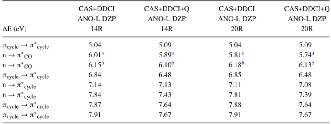
Once the thresholds have been adapted, the effects of the number of roots (14R vs 20R) as well as those of the David-son correction (noted +Q) were tested (TableVI). The effect of the number of roots on most of the excitation energies was small (at most 0.03/0.05 eV for no Q/+Q calculations). As the additional roots implied excitations from and to orbitals of the TABLE VI. CAS+DDCI excitation energy (eV) of the NAPA B conformer at its DFT-D optimized ground state
geometry. The reference energy is that of the ground state in each method and the basis set used is the ANO-L DZP. 14 and 20 roots have been considered and a Davidson correction (+Q) is applied or not.
CAS+DDCI CAS+DDCI+Q CAS+DDCI CAS+DDCI+Q
ANO-L DZP ANO-L DZP ANO-L DZP ANO-L DZP
∆E (eV) 14R 14R 20R 20R πcycle→ π∗cycle 5.04 5.09 5.04 5.09 n → π∗CO 6.01a 5.89a 5.81a 5.74a n → π∗ CO 6.15b 6.10b 6.18b 6.13b πcycle→ π∗cycle 6.84 6.48 6.85 6.48 n → π∗ cycle 7.14 7.13 7.11 7.08 n → π∗cycle 7.84 7.43 7.81 7.39 πcycle→ π∗cycle 7.87 7.64 7.88 7.64 πcycle→ π∗cycle 7.91 7.67 7.91 7.67 aLone pair localized on the second peptide bond.
same nature, the average orbitals were not changed signifi-cantly. The only exception concerned the first nπ∗COlocalized on the second peptide bond (deviation of 0.20/ 0.15 eV for no Q/+Q calculations) which was directly related to the change introduced by the CASSCF calculation (Table S4 of the supplementary material). On the other hand, the David-son correction affected almost all excitation energies with a maximum effect, a decrease of around 0.40 eV, for the sec-ond ππ∗ and CT states. Not only the CAS+DDCI+Q results were in very good agreement with MS-CASPT2 ones, the nature and the energy ordering of the two first ππ∗, nπ∗CO, and nπ∗cycleexcited states were the same but also the maxi-mal deviation was weak, i.e., 0.10/0.07 eV for 14R/20R with a mean absolute value of 0.09/0.05 eV. Originally developed to calculate singlet-triplet gaps in biradicalar systems,52 DDCI allows us to evaluate accurate vertical energy differences,28 from a common set of MOs for all states. The CAS+DDCI+Q method led here to discrepancies within the expected error margins of the MS-CASPT2 method, i.e., ±0.2 eV for the exci-tation energies of the two first ππ∗, nπ∗
CO, and nπ∗cycleexcited states. On the other hand, the MR MS-CASPT2 states lower in energy than the first CT states could not be reproduced by the CAS+DDCI+Q method because of the restricted active space which does not allow the mixing between states of different nature.
Eventually, the CAS+DDCI calculations are carried out in the following considering 20 roots within the ANO-L DZP basis set and taking into account the Davidson correction (CAS+DDCI+Q).
3. CC2 versus MR method
Comparing to the MR method results (MS-CASPT2/20R and CAS+DDCI+Q starting orbitals coming from a 20R CASSCF calculation), it can be observed that the CC2/cc-pVDZ level was in good agreement for the four low-lying excited states while the discrepancy became larger for the states around and beyond 7 eV. The results are presented in Tables II, IV, and VI for comparison. Indeed, the first CT state nπ∗
cycle was found to be 0.44/0.38 eV lower with the CC2 method compared to the MS-CASPT2/CAS+DDCI+Q whereas the two first low-lying ππ∗ states were found to be
0.14-0.07/0.18-0.04 eV higher and the two first nπ∗CO were found to be 0.10-0.28/0.06-0.32 eV lower. The difference between the CC2 excitation energies and the MRCI ones was then not larger than 0.3 eV for the lowest ππ∗and nπ∗
COstates and was of the same order of magnitude than the expected error of the MS-CASPT2 and CAS+DDCI+Q methods. On the con-trary, a larger value was obtained for the excitation energies around and beyond 7 eV (around 0.40–0.60 eV for the CT states). As for CAS+DDCI+Q calculations, there were obvi-ously no MR states in CC2 calculations due to the formalism of the method.
In view of these results, all calculations in the follow-ing are performed with these characteristics: MS-CASPT2/ ANO-L DZP (20R), CAS+DDCI+Q/ANO-L DZP (20R), and CC2/cc-pVDZ.
B. Energetic profile of the deactivation mechanisms of the conformer NAPA B involving the second peptide bond
The relative CC2, MS-CASPT2, and CAS+DDCI+Q excitation energies of the two first ππ∗, nπ∗CO, and nπ∗cycle low-lying excited states of NAPA B are reported in TablesVII–IXfor the three pertinent geometries of the energy profile of the MII deactivation mechanism of the NAPA B conformer,10,11i.e., (i) Mππ∗, the CC2/cc-pVDZ equilibrium
geometry of the ππ∗ [Fig. 1(b1) and Table S1-2 of the
supplementary material], (ii) CIππ∗/nπ∗CO(2), the
CC2/cc-pVDZ CI geometry connecting the equilibrium geometries of the two ππ∗and nπ∗COexcited states [Fig.1(b2)and Table S1-3
of the supplementary material], and (iii) Mnπ∗CO(2), the
CC2/cc-pVDZ equilibrium geometry of the nπ∗
CO excited state localized on the second peptide bond [Fig. 1(b3) and
Table S1-4 of thesupplementary material].
Whatever the geometry, the D1/D2values obtained in the CC2 calculation for the ground state were in the range 0.08-0.13/0.20-0.27 while the D2value for the excited states was in the range 0.17-0.33 with a biorthogonal norm %E1|E1 ≥ 85% (%E1|E1 ≥ 87% for the first six excited states). As previ-ously mentioned, these values confirm the reliability of the CC2 calculations even if they remain close to the upper limit of acceptable values (see Sec.III A 1). As it has been already
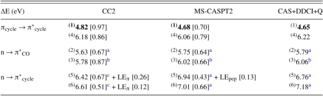
TABLE VII. CC2, MS-CASPT2, and CAS+DDCI+Q excitation energy (eV) of the two first excited states of each nature of the NAPA B conformer. The geometry used is the Mππ∗CC2/cc-pVDZ optimized geometry [see Fig.1(b1)and Table S1-2 of thesupplementary material]. The energy values in bold correspond to the lowest excited state in each method. The exponent numbers in brackets give the order of stability of each excited state among the twenty ones. For the definition of the values in bracket, see captions of TablesIIandIII.
∆E (eV) CC2 MS-CASPT2 CAS+DDCI+Q
πcycle→ π∗cycle (1)4.82[0.97] (1)4.68[0.70] (1)4.65 (4)6.18 [0.86] (4)6.06 [0.79] (4)6.22 n → π∗CO (2)5.63 [0.67]a (2)5.75 [0.64]a (2)5.79a (3)5.78 [0.87]b (3)6.02 [0.66]b (3)6.06b n → π∗cycle (5)6.42 [0.67]c+ LEπ[0.26] (5)6.94 [0.43]a+ LEpep[0.13] (5)6.76a (6)6.61 [0.51]c+ LE π[0.12] (6)7.01 [0.66]a (6)7.18a
aLone pair n localized on the second peptide bond. bLone pair n localized on the first peptide bond.
TABLE VIII. CC2, MS-CASPT2, and CAS+DDCI+Q excitation energy (eV) of the two first excited states of each nature of the NAPA B conformer. The geometry used is the CIππ∗/nπ∗COCC2/cc-pVDZ geometry [see Fig.1(b2) and Table S1-3 of thesupplementary material]. The energy values in bold correspond to the lowest excited state in each method. The exponent numbers in brackets give the order of stability of each excited state among the twenty ones. For the definition of the values in bracket, see captions of TablesIIandIII.
∆E (eV) CC2 MS-CASPT2 CAS+DDCI+Q
πcycle→ π∗cycle (2)4.93[0.52] + LEpep[0.20] (1)4.86[0.59] (1)4.74
(4)6.25 [0.82] (4)6.23 [0.54] + LE pep[0.19] (4)6.27 n → π∗ CO (1)4.91[0.41]a+ LEπ[0.36] (2)5.05[0.63]a+ CT [0.14] (2)5.00a (3)5.77 [0.82]b (3)6.21 [0.49]b+ LE π[0.20] (3)6.12b n → π∗ cycle (5)6.47 [0.59]c+ LEπ[0.20] (6)7.08 [0.63]a (5)6.77a (6)6.62 [0.47]c+ LE π[0.17] + LEpep[0.13] (7)7.20 [0.60]a (6)7.17a πCO→ π∗CO (5)6.36 [0.42] + LEpep[0.21]
aLone pair n localized on the second peptide bond. bLone pair n localized on the first peptide bond.
cLone pair n localized on the backbone, i.e., on the two peptide bonds.
observed in such systems,22 a ππ∗ excitation causes a sig-nificant change in the electron density distribution around the phenyl ring leading to a significant shortening of the NH(1)–Cπ distance in Mππ∗(0.22 Å) whereas parameters such as the
cova-lent bonds, the valence angles, or torsional angles that do not involve the orientation of the backbone relative to the phenyl ring are only weakly changed upon ππ∗excitation (Table S5-1 of thesupplementary material). Indeed, upon excitation, the πsystem tends to extend farther from the ring C6 axis, with a density increase above the C atoms and on the ring edge and a decrease above the CC bonds as illustrated in Fig.2. In Mnπ∗CO(2), the local excitation from the nitrogen and oxygen
lone pairs of the backbone to the CO antibonding π∗orbital of the second peptide bond (see the density transfer illustrated in Fig.2) leads to a characteristic deplanarization of the peptide bond with a pyramidalization of both the carbon and nitrogen atoms: the ω2angle is strongly reduced (∼80◦) and both Σ θNi
and Σ θCi differ from 360◦ by at least 20◦ (see Tables S5-1
and S5-2 and Fig. S5-3 of thesupplementary material). This is accompanied by an elongation of 0.15 Å of the CO bond as well as an elongation of both the NH(2)–O the NH(1)–Cπ dis-tances (+0.26 Å and +0.09 Å). Along the energy profile, the geometry of CIππ∗/nπ∗CO(2) is intermediate between the two
previous geometries but much closer to the Mππ∗ than to the
Mnπ∗CO(2) geometry (see Fig.3): all the characteristic
param-eters, angles and distances, are modified by up to 30% of their total variation along the energy profile (Tables S5-1 and S5-2 and Fig. S5-3 of thesupplementary material).
For Mππ∗and Mnπ∗CO(2) equilibrium geometries, both the
nature and the energetic of the first excited state obtained at the CC2 level are in good agreement with those obtained at the MR level, the energy difference being less than 0.2 eV (see TablesVIIandIX). In the case of the CIππ∗/nπ∗CO(2)
geom-etry, the geometry of the conical intersection identified with
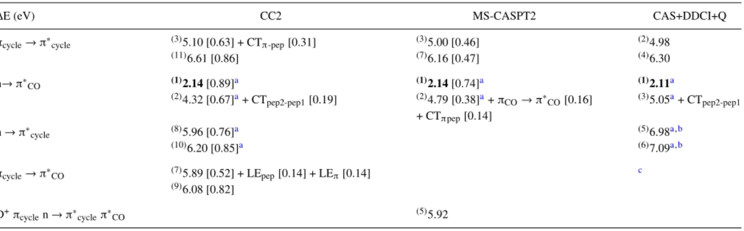
TABLE IX. CC2, MS-CASPT2, and CAS+DDCI+Q excitation energy (eV) of the two first excited states of each nature of the NAPA B conformer. The geometry used is the Mnπ∗CO(2) CC2/cc-pVDZ optimized geometry [see Fig.1(b3)and Table S1-4 of thesupplementary material]. The energy values in bold correspond to the lowest excited state in each method. The exponent numbers in brackets give the order of stability of each excited state among the twenty ones: (i) in the CC2 calculation, the fourth, fifth, and sixth states correspond to nπ∗COat 5.23, 5.58, and 5.72 eV, respectively, and (ii) in the MS-CASPT2 calculation, the fourth
and sixth state correspond to nπ∗COat 5.84 and 6.03 eV. For the definition of the values in bracket, see captions of TablesIIandIII. CTpep1-pep2correspond to
the πCO(peptide bond 2) → π∗CO(peptide bond 1). The orbitals have been optimized at the CASSCF(20,15) level and averaged on 20 roots; these orbitals are
used in the MS-CASPT2 and the DDCI methods.
∆E (eV) CC2 MS-CASPT2 CAS+DDCI+Q
πcycle→ π∗cycle (3)5.10 [0.63] + CTπ-pep[0.31] (3)5.00 [0.46] (2)4.98
(11)6.61 [0.86] (7)6.16 [0.47] (4)6.30 n→ π∗ CO (1)2.14[0.89]a (1)2.14[0.74]a (1)2.11a (2)4.32 [0.67]a+ CT pep2-pep1[0.19] (2)4.79 [0.38]a+ πCO→ π∗CO[0.16] + CTπpep[0.14] (3)5.05a+ CT pep2-pep1 n → π∗ cycle (8)5.96 [0.76]a (5)6.98a,b (10)6.20 [0.85]a (6)7.09a,b πcycle→ π∗CO (7)5.89 [0.52] + LEpep[0.14] + LEπ[0.14] c (9)6.08 [0.82] D+π cyclen → π∗cycleπ∗CO (5)5.92 aLone pair n localized on the second peptide bond.
bWith CAS (6,7).
FIG. 3. Comparison between the CC2/cc-pVDZ geometries of Mππ∗ (in gray), Mnπ∗CO(2) (in dark green), and CIππ∗/nπ∗CO(2)(in light green). To illustrate the change from the second peptide bond and environment point of view, all the carbon atoms of phenyl rings as well as the carbon atom of the CH2group and of the CH(CO–NH2,NH–COCH3) group are superimposed.
the CC2 method, the first ππ∗, and nπ∗COstates are obviously quasi-degenerate at the CC2 level (∆E of 0.02 eV) whereas at the MR level, the energy gap between these two states is more important (MS-CASPT2/CAS+DDCI+Q : +0.19/+0.26 eV), the more stable one being the ππ∗excited state. There is really no reason that the CC2 conical intersection geometry matches exactly the MR one. Moreover, the determination of this geom-etry at the CC2 level results from an interpolation between the geometries of the two minima and corresponds then to an esti-mation of the localization of the CI at the CC2 level.10 On
the other hand, considering the energy difference between the two excited states for this geometry at the MR level which corresponds to a weak lift of degeneracy (∼ 0.2 eV), it is clear that the MR conical intersection geometry must be close to the CC2 one.
The order of stability as well as the nature of the five other states of Mππ∗ given by the CC2 method (Table VII)
are equivalent to that given by the MR methods except for the nπ∗cyclestates in which the lone pairs are localized on the back-bone in the CC2 calculations and only on the second peptide bond in the MR methods. The difference in excitation energies between CC2 and MR methods are similar to that found for the DFT-D ground state geometry: the ππ∗excited states are found to be around 0.1 eV higher, the nπ∗
COexcited states are found to be around 0.2 eV lower, and the nπ∗
cycleexcited states are found to be around 0.45 eV lower. For the CIππ∗/nπ∗CO(2)
geometry (TableVIII), the order of stability of the following excited states is equivalent for the three methods except that in the MS-CASPT2 calculations, a πCO→ π∗COstate is inserted in the fifth position. As for the Mππ∗ geometry, the CC2 and
MR nature of these states is equivalent except again for the lone pairs in nπ∗cyclestates localized on the backbone in the CC2 calculations and only on the second peptide bond with the MR methods. Finally, the difference in excitation energies between CC2 and MR methods is similar to that found for the DFT-D ground state geometry. For Mnπ∗CO(2) (TableIX), as
no charge transfer state was found at the CASSCF/20R level, a 30-root CASSCF calculation was performed but, once more, no CT state was encountered. At the CAS+DDCI+Q level, the two CT states were obtained starting from 20R MOs, not opti-mized for these states, since these latter were not obtained at the CASSCF level. This can explain the quite large discrep-ancy (about 1 eV) between CC2 and CAS+DDCI+Q excitation energies of these states. The excitation energy of the other states does not present a so large discrepancy but this differ-ence remains more important than those obtained for the other discussed geometries.
C. Equilibrium geometry of the CT excited state of the NAPA B conformer identified in the mechanisms involving the first peptide bond
The relative CC2, MS-CASPT2, and CAS+DDCI+Q excitation energies of the two first low-lying ππ∗, nπ∗CO, and nπ∗cycleexcited states of NAPA B for the partially optimized TABLE X. CC2, MS-CASPT2, and CAS+DDCI+Q excitation energy of the two first excited states of each nature
of the conformer NAPA B. The geometry is the CC2/cc-pVDZ partially optimized geometry with the NH(1) bond length constrained to 1.216 Å of the CT excited state [see Fig.1(c)and Table S1-5 of thesupplementary material]. The energy values in bold correspond to the lowest excited state in each method. The exponent numbers in brackets give the order of stability of each excited state among the twenty ones: (i) in the CC2 calculation, the fifth, sixth, and eighth states correspond to nπ∗
cycleat 5.05, 5.36, and 5.63 eV, respectively, and (ii) in the MS-CASPT2
calculation, the fifth and sixth state correspond to nπ∗
COat 5.52 and 6.63 eV. For the definition of the values
in bracket, see captions of TablesIIandIII. The orbitals have been optimized at the CASSCF(20,15) level and averaged on 20 roots; these orbitals are used in the CASPT2 and the DDCI methods.
∆E (eV) CC2 MS-CASPT2 CAS+DDCI+Q
πcycle→ π∗cycle (2)4.28 [0.89] (2)4.07 [0.65] (2)4.11 (3)4.46 [0.72] + CT [0.22] (3)4.38 [0.66] (3)4.86 n → π∗ cycle (1)3.23[0.90]a (1)3.68[0.69]b (1)3.24b,c (4)4.98 [0.83]a+ LE π[0.10] (4)5.47 [0.58]b (4)5.80b,c n → π∗ CO (7)5.55 [0.59]a+ CT [0.13] (7)5.90 [0.38]a+ LEπ[0.16] (5)5.85b (9)5.69 [0.62]a+ CT π-pep[0.13] (8)5.94 [0.52]a (6)6.01d aLone pair localized on the backbone, i.e., on the two peptide bonds.
bLone pair localized on the first peptide bond. cWith CAS(8,7).
FIG. 4. (a) Comparison between the CC2/cc-pVDZ geometries of Mππ∗(in gray) and M0CT (in red). To illustrate
the change from the phenyl ring and environment point of view, backbones are superimposed. (b) For the sake of clarity, the main geometrical parame-ters around the ortho carbon atom of the phenyl ring are presented. Distances are given in Ångstr¨oms and the dihedral angle in degrees.
geometry, with the NH(1) bond length constrained to 1.216 Å, of the first CT excited state (M0
CT) identified in the MI deac-tivation mechanism10 of the NAPA B conformer [Fig.1(c)
and Table S1-5 of thesupplementary material] are presented (TableX).
As previously discussed, the D1/D2values obtained in the CC2 calculation for the ground state, 0.11/0.23, as well as the D2value obtained for the excited states which is in the range 0.19-0.31 with a biorthogonal norm %E1|E1 ≥ 85%, confirm the reliability of the CC2 calculations even if they remain close to the upper limit of acceptable values (see Sec.III A 1). For M0CTin which the NH(1) bond is elongated by around 0.2 Å compared to that of Mππ∗, the excitation corresponds to an
electronic charge transfer from the π (carbonyl) orbitals and the n lone pairs of the backbone to the π∗orbitals of the phenyl ring as it is illustrated in Fig.2. This leads to a strong short-ening of 1.0 Å of the NH(1)–Cπdistance concomitant with a shortening of 0.08 Å of the NH(2)–O distance (see Table S5-1 of thesupplementary material) as well as a strong distortion of the phenyl ring [Figs.4(a)and4(b)] resulting from the dis-ruption of the π conjugation pattern of the phenyl ring in the neighborhood of the ortho carbon atom [an elongation of the two adjacent CC bonds to ∼1.50 Å, Fig.4(b)].
In this geometry, the first excited state is a charge trans-fer state for both the CC2 and MR methods. If for the CC2 method, the involved lone pairs of the backbone are those of both peptide bonds, only the lone pairs of the first peptide bond are involved for the MR methods. Concerning the excitation energy, the CC2 and CAS+DDCI+Q values are very close (∆E = 0.01 eV) whereas the MS-CASPT2 value is 0.45 eV higher. Concerning the following states, both the order of stability and the nature of the first three states are equivalent for the three methods except for the nπ∗
cyclestates in which the lone pairs are localized on the backbone in the CC2 calculation whereas only the lone pairs of the first peptide bond are involved in the MR calculations.
The treatment of the dynamic electronic correlation leads to a different correction in the two MR methods. In the DDCI method, the two holes-two particles determinants are not taken into account compared to the MR-SDCI method. The analysis of the CASPT2 results shows that these excitations contribute for the half to the dynamical correlation energy,
but their contribution is almost the same for all the ground and excited states and leads to a maximal differential value of 0.02 eV between the ground state and the excited states. How-ever, we cannot directly conclude that the difference between DDCI and MS-CASPT2 methods does not come from the two holes-two particles determinants. The active space used in the DDCI method is indeed reduced compared to CASSCF/MS-CASPT2, even if the weights of the reference wave functions in the CI calculation are large enough (> 0.7) to insure a reliable result. Finally, the mean absolute difference between the exci-tation energies obtained with the two MR methods (0.24 eV) is more than twice the value obtained in the other geometries (0.09 eV). This suggests that the difficulty encountered almost systematically at the CASSCF/MS-CASPT2 level to properly describe the CT states could be the reason of these discrepan-cies at the equilibrium geometry of the lowest charge transfer state.
IV. CONCLUSION
First of all, this work allows us to establish the validity of the quasi-linear CAS+DDCI+Q method to treat singlet excited states of bio-relevant systems. Indeed, once their parame-ters are adapted, this method leads—on a building block of proteins, the NAPA system—to a very good agreement with the results obtained with a more standard MR method, the MS-CASPT2. For a series of different geometries, both the excitation energies and the wave functions of the low-lying sin-glet excited states obtained at the CAS+DDCI+Q/20R/ANO-L-DZP level are equivalent to those obtained at the MS-CASPT2/20R/ANO-L-DZP independent of the nature of the excited state. The only non-negligible discrepancies which appear (∆E > 0.2 eV) concern the excitation energies of CT excited states which either are high in energies (see nπ∗
cycle states in TablesVIIIandIX) or which correspond to a strongly distorted geometry (see Table X). Moreover, these calcula-tions highlight various points to be taken into account in MR methods to properly deal with low-lying excited states of such systems. In the MS-CASPT2 calculations, for the low-lying singlet CT states which can be relatively high in energy, not only the choice of the active space is crucial but also the num-ber of roots. For the CAS+DDCI+Q method, an enlargement
of the active space is required for the excitation energy of some low-lying excited states.
Second, the lowest singlet excited state of different CC2/cc-pVDZ geometries can be well described at the CC2/cc-pVDZ level whatever its nature, i.e., a ππ∗ excited state on the phenyl ring, an nπ∗
COexcited state localized on the peptide bond(s), or even an nπ∗
cyclecharge transfer (CT) excited state involving an electronic charge transfer from the lone pairs of the backbone to the phenyl ring. Compared to CC2, the only difference concerns this latter CT state which is localized on a particular peptide bond at the CAS+DDCI+Q level. Furthermore, the CC2/cc-pVDZ excitation energy of the lowest singlet excited state is close to that obtained at the CAS+DDCI+Q/20R/ANO-L-DZP level and the absolute deviation is inferior to 0.2 eV, a value close to the standard error of the MS-CASPT2 method (± 0.2 eV). For the follow-ing low-lyfollow-ing excited states, the nature of these states is also well described by the CC2 method except, as previously, for the electronic charge transfer in the CT states, backbone vs localized lone pair. For the excitation energies, two differ-ent behaviors are observed. For the DFT-D ground state, the Mππ∗and the CIππ∗/nπ∗COgeometries, the difference between
excitation energies is not greater than 0.3 eV for the lowest states—the ππ∗ and the nπ∗CO excited states—and around 0.4 eV for the higher states—the CT states. For the two other geometries, the Mnπ∗CO (2) and the M0CT, the
dif-ference in excitation energies is more pronounced, around 0.4 eV on average with an absolute maximum deviation of 0.8 eV.
Finally, in view of the good performances of the CC2 method on the NAPA system, a building block of proteins containing one residue, it will be very interesting to evaluate if these performances can be extrapolated to larger systems, i.e., systems containing more than one residue. However, even if this work demonstrates that the CAS+DDCI+Q method is appropriate to accurately describe the close-low-lying singlet excited states of a building block of proteins, the active space size which can be affordable in a CASSCF calculation is reached for the NAPA system. Therefore, it will be necessary to consider other solutions to optimize the orbitals, the first step of the CAS+DDCI+Q calculations. A promising solution that we are investigating on capped peptides containing at least two residues is given by the Generalized Active Space Self Consis-tent Field (GASSCF) method,53,54a method which allows us to expand active spaces beyond the CASSCF limit. This work will be reported in a future paper.
SUPPLEMENTARY MATERIAL
Seesupplementary materialfor Appendix S1—Cartesian coordinates of all the geometries of the NAPA B conformer, Appendix S2—dependence of the excitation energy and ref-erence weight of the zeroth-order wave function on the level shift in CASPT2 calculations, Appendix S3—diffuse orbital effects on the excitation energy in CC2 and MS-CASPT2 cal-culations, Appendix S4—first CASSCF excited states for the ground state geometry as a function of the number of roots, and Appendix S5—characteristic structural parameters of the different NAPA B geometries.
ACKNOWLEDGMENTS
This work received financial support from the Agence Nationale de la Recherche (ANR), Grant No. ANR-14-CE06-0019-01-ESBODYR. This work was granted access to the HPC facility of [TGCC/CINES/IDRIS] under Grant Nos. 2015-t2015087412&t2015087372, 2016-t2016087540, and 2017-A0010807540, awarded by GENCI (Grand Equipement National de Calcul Intensif); to the CCRT High Perfor-mance Computing (HPC) facility at CEA under Grant No. CCRT2015/CCRT2016-p606bren; and to the HPC resources of CALMIP supercomputing center under Allocation No. 2016-[P16009].
1W. Domcke and A. L. Sobolewski,Phys. Chem. Chem. Phys.12, 4897
(2010).
2Y. Chen and M. D. Barkley,Biochemistry37, 9976 (1998). 3P. R. Callis and T. Liu,J. Phys. Chem. B108, 4248 (2004).
4M. A. Robb, M. Olivucci, and F. Bernardi, in Encyclopedia of
Computa-tional Chemistry, edited by P. von Ragu´e Schleyer, N. L. Allinger, T. Clark,
J. Gasteiger, P. A. Kollman, H. F. Schaefer, and P. R. Schreiner (John Wiley & Sons, Ltd., Chichester, UK, 2002).
5G. A. Worth and L. S. Cederbaum, Annu. Rev. Phys. Chem. 55, 127
(2004).
6B. G. Levine and T. J. Mart´ınez,Annu. Rev. Phys. Chem.58, 613 (2007). 7Conical Intersections: Electronic Structure, Dynamics & Spectroscopy,
edited by W. Domcke, D. Yarkony, and H. K¨oppel (World Scientific, River Edge, NJ, 2004).
8W. Domcke and D. R. Yarkony,Annu. Rev. Phys. Chem.63, 325 (2012). 9D. R. Yarkony,Chem. Rev.112, 481 (2012).
10M. Maliˇs, Y. Loquais, E. Gloaguen, H. S. Biswal, F. Piuzzi, B. Tardivel,
V. Brenner, M. Broquier, C. Jouvet, M. Mons, N. Doˇsli´c, and I. Ljubi´c,J. Am. Chem. Soc.134, 20340 (2012).
11M. Maliˇs, Y. Loquais, E. Gloaguen, C. Jouvet, V. Brenner, M. Mons,
I. Ljubi´c, and N. Doˇsli´c,Phys. Chem. Chem. Phys.16, 2285 (2014).
12C. H¨attig and F. Weigend,J. Chem. Phys.113, 5154 (2000). 13C. H¨attig and A. K¨ohn,J. Chem. Phys.117, 6939 (2002). 14C. H¨attig,J. Chem. Phys.118, 7751 (2003).
15A. K¨ohn and C. H¨attig,J. Chem. Phys.119, 5021 (2003).
16O. Christiansen, H. Koch, and P. Jørgensen,Chem. Phys. Lett.243, 409
(1995).
17B. Bories, D. Maynau, and M.-L. Bonnet,J. Comput. Chem.28, 632 (2007). 18N. Ben Amor, F. Bessac, S. Hoyau, and D. Maynau,J. Chem. Phys.135,
014101 (2011).
19C. Chang, C. J. Calzado, N. Ben Amor, J. Sanchez Marin, and D. Maynau,
J. Chem. Phys.137, 104102 (2012).
20D. Shemesh, A. L. Sobolewski, and W. Domcke,J. Am. Chem. Soc.131,
1374 (2009).
21N. Doˇsli´c, G. Kovaˇcevi´c, and I. Ljubi´c,J. Phys. Chem. A111, 8650 (2007). 22W. Y. Sohn, V. Brenner, E. Gloaguen, and M. Mons,Phys. Chem. Chem.
Phys.18, 29969 (2016).
23N. O. C. Winter, N. K. Graf, S. Leutwyler, and C. Hattig,Phys. Chem.
Chem. Phys.15, 6623 (2013).
24C. Fang, B. Oruganti, and B. Durbeej,J. Phys. Chem. A118, 4157 (2014). 25D. Tuna, D. Lefrancois, Ł. Wola´nski, S. Gozem, I. Schapiro, T. Andruni´ow,
A. Dreuw, and M. Olivucci,J. Chem. Theory Comput.11, 5758 (2015).
26F. Plasser, R. Crespo-Otero, M. Pederzoli, J. Pittner, H. Lischka, and
M. Barbatti,J. Chem. Theory Comput.10, 1395 (2014).
27J. Miralles, J.-P. Daudey, and R. Caballol,Chem. Phys. Lett.198, 555
(1992).
28J. Miralles, O. Castell, R. Caballol, and J.-P. Malrieu,Chem. Phys.172, 33
(1993).
29T. Krah, N. Ben Amor, and V. Robert,Phys. Chem. Chem. Phys.16, 9509
(2014).
30J. Zapata-Rivera, R. Caballol, and C. J. Calzado,J. Comput. Chem.32, 1144
(2011).
31C. J. Calzado, N. Ben Amor, and D. Maynau,Chem. - Eur. J.20, 8979
(2014).
32C. J. Calzado and D. Maynau,J. Chem. Phys.135, 194704 (2011). 33J. Finley, P.-Å. Malmqvist, B. O. Roos, and L. Serrano-Andr´es,Chem. Phys.