HAL Id: hal-01718514
https://hal.archives-ouvertes.fr/hal-01718514
Submitted on 8 Jun 2021
HAL is a multi-disciplinary open access
archive for the deposit and dissemination of
sci-entific research documents, whether they are
pub-lished or not. The documents may come from
teaching and research institutions in France or
abroad, or from public or private research centers.
L’archive ouverte pluridisciplinaire HAL, est
destinée au dépôt et à la diffusion de documents
scientifiques de niveau recherche, publiés ou non,
émanant des établissements d’enseignement et de
recherche français ou étrangers, des laboratoires
publics ou privés.
Distributed under a Creative Commons Attribution| 4.0 International License
double-strand breaks to prevent translocations
Sarah Cohen, Nadine Puget, Yea-Lih Lin, Thomas Clouaire, Marion
Aguirrebengoa, Vincent Rocher, Philippe Pasero, Yvan Canitrot, Gaelle
Legube
To cite this version:
Sarah Cohen, Nadine Puget, Yea-Lih Lin, Thomas Clouaire, Marion Aguirrebengoa, et al.. Senataxin
resolves RNA:DNA hybrids forming at DNA double-strand breaks to prevent translocations. Nature
Communications, Nature Publishing Group, 2018, 9 (1), pp.533. �10.1038/s41467-018-02894-w�.
�hal-01718514�
Senataxin resolves RNA:DNA hybrids forming at
DNA double-strand breaks to prevent
translocations
Sarah Cohen
1
, Nadine Puget
1
, Yea-Lih Lin
2
, Thomas Clouaire
1
, Marion Aguirrebengoa
1
, Vincent Rocher
1
,
Philippe Pasero
2
, Yvan Canitrot
1
& Gaëlle Legube
1
Ataxia with oculomotor apraxia 2 (AOA-2) and amyotrophic lateral sclerosis (ALS4) are
neurological disorders caused by mutations in the gene encoding for senataxin (SETX), a
putative RNA:DNA helicase involved in transcription and in the maintenance of genome
integrity. Here, using ChIP followed by high throughput sequencing (ChIP-seq), we report
that senataxin is recruited at DNA double-strand breaks (DSBs) when they occur in
tran-scriptionally active loci. Genome-wide mapping unveiled that RNA:DNA hybrids accumulate
on DSB-
flanking chromatin but display a narrow, DSB-induced, depletion near DNA ends
coinciding with senataxin binding. Although neither required for resection nor for timely
repair of DSBs, senataxin was found to promote Rad51 recruitment, to minimize illegitimate
rejoining of distant DNA ends and to sustain cell viability following DSB production in active
genes. Our data suggest that senataxin functions at DSBs in order to limit translocations and
ensure cell viability, providing new insights on AOA2/ALS4 neuropathies.
DOI: 10.1038/s41467-018-02894-w
OPEN
1LBCMCP, Centre de Biologie Integrative (CBI), CNRS, Université de Toulouse, UT3, 118 Route de Narbonne, 31062 Toulouse, France.2Institut de Génétique
Humaine, CNRS, Université de Montpellier, 34396 Montpellier, France. Correspondence and requests for materials should be addressed to
G.L. (email:[email protected])
123456789
M
utations in the SETX gene are responsible for the rare
neurological disorders ALS4, a dominantly inherited
form of amyotrophic lateral sclerosis and ataxia with
oculomotor apraxia type 2 (AOA2) which are associated with an
early onset neurons degeneration (for review see ref.
1). As a
consequence, patients display strong ataxia as well as oculomotor
troubles (AOA2) or muscle weakness (ALS4), generally occurring
before their 30′s. SETX encodes a helicase, strongly conserved
throughout evolution that has been implicated in a large variety
of biological processes, from transcription termination, to meiosis
completion and maintenance of genomic integrity
1. At a
mole-cular level, studies of the yeast senataxin homolog Sen1p
estab-lished that this helicase displays an unwinding activity toward
RNA:DNA hybrids
2–4. Multiple genomic studies in both yeast
and mammals recently unveiled that RNA:DNA hybrids mostly
form as RNA polymerases progress throughout the genes, by the
re-hybridization of the nascent RNA to the template DNA strand,
leading to triple-stranded structure called R-loops (reviewed in
ref.
5). While R-loops display a strong ability to naturally form at
GC-skewed promoters due to an enhanced thermodynamic
sta-bility of C-rich DNA: G-rich RNA duplexes
6,7, multiple
com-plexes regulate their occurrence throughout the genome,
including RNA splicing/processing factors and specific helicases,
such as senataxin
8,9. R-loops formation and processing play a
crucial role in terminating transcription at least in yeast (reviewed
in ref.
8). In agreement, mutations in sen1 trigger defective
transcription termination and increased transcriptional
read-through, especially on short transcribed units such as rDNA,
tRNA, and small non-coding or coding genes (for review see ref.
10). Such a function of R-loops processing and senataxin in
transcriptional termination was also proposed to be conserved in
higher eukaryotes, in a manner that would also involve dsRNA
processing factors such as Drosha and DGCR8 as well as
com-ponents of the RNA exosome (for review ref.
11). However,
beyond their role in regulating transcription, R-loops also
represent a severe threat to genome integrity, proposed to arise
both due to the susceptibility of the displaced single-stranded
DNA to damaging agents as well as their potential to impede
replication fork progression (reviewed in refs.
12–14). In
agree-ment, increased damage occurrence and genome instability was
observed in cells deficient for R-loops processing factors such as
AQR
15. Additionally in yeast, sen1 mutations are associated with
an increased transcription-associated genome instability
16,17. On
the other hand, few studies also suggested that senataxin may play
a more direct role at damage sites. Senataxin/Sen1p interacts with
repair proteins in yeast and mammals and localizes to the site of
damage during replication
18,19. Moreover, SETX mutant mice
display defective meiosis and Spo11-mediated DSB persistence
20.
Finally, depletion of senataxin/Sen1p triggers sensitivity to some
DNA damaging agents such as H
2O
2and UV
21–23, a feature also
observed in AOA2 patient cell lines
22,24,25. However, senataxin
depleted cells are not radiation sensitive
22, suggesting that it may
not function at sites of DNA double-strand break (DSB), a form
of DNA damage largely induced upon irradiation.
Yet, recent studies have suggested that R-loops or/and RNA:
DNA hybrids likely form at DSBs. An assay using a duplex
specific nuclease detected RNA:DNA hybrids upstream the I-SceI
site upon DSB formation
26. Additionally a mutant form of
RNAse H1, devoid of RNA exonuclease activity accumulates at
the site of laser induced damages
27. Finally, in
Schizosacchar-omyces pombe, RNA:DNA hybrids were shown to form during
resection, regulating RPA
filament formation
28. The exact
mechanism that leads to such R-loops or/and RNA:DNA hybrids
formation remains unclear and may either relate to the
tran-scriptional extinction observed at damaged sites or to de novo
RNA PolII loading at DNA ends and subsequent RNA
production at the break point, two features that have been
pre-viously proposed to occur in many organisms (for review see ref.
12).
In order to gain insights into R-loops biology at DSBs, here we
set to assess a potential function of senataxin at sites of DNA
DSBs. Using ChIP-seq and DRIP-seq, we uncovered that
sena-taxin is recruited specifically at DSBs induced in transcriptionally
active genes, which exhibit RNA:DNA hybrids accumulation
following DSB induction. Senataxin distribution around DSBs
coincided with a local decrease in R-loops and senataxin
deple-tion triggered increased DSB-induced RNA:DNA hybrids
for-mation, suggesting that senataxin processes DNA damage
induced RNA:DNA hybrids. We found that senataxin is not
required to sustain resection, nor rapid repair at these DSBs. Yet,
it promotes Rad51 foci formation, counteracts translocations and
sustains viability following DSB production in active genes, hence
identifying a crucial and unanticipated role for senataxin in DSB
repair.
Results
Senataxin is recruited at DSBs produced in active genes. To
assess senataxin recruitment at sites of DSBs, we used the DSB
inducible via AsiSI (DIvA) cell line, which allows to induce clean
DSBs throughout the genome
29,30. In this cell line, 4
hydro-xytamoxifen (4OHT) treatment induces the relocalisation of a
stably expressed restriction enzyme (AsiSI) that in turn triggers
the production of multiple DSBs at annotated positions across the
genome, in a homogeneous manner in the cell population hence
allowing the use of chromatin immunoprecipitation (ChIP)
30. To
obtain a quantitative and genome-wide assessment of senataxin
binding and distribution at DSBs, we performed ChIP followed
by high throughput sequencing (ChIP-seq) against senataxin
before and after DSB induction in DIvA cells (respectively
−4OHT and +4OHT). We observed an accumulation of senataxin
in a 1–2 kb window surrounding AsiSI sites following 4OHT
treatment (see examples in Fig.
1
a). In vivo, AsiSI does not
produce DSB at each of its 1211 annotated recognition sites, likely
due to both DNA methylation and chromatin compaction
30. Our
recent studies allowed us to characterize AsiSI sites cleavage
efficiency, using BLESS (direct in situ breaks labeling, enrichment
on streptavidin and next generation sequencing) throughout the
genome and to identify a set of 80 DSBs robustly induced
fol-lowing 4OHT treatment
31(Clouaire, T. et al., manuscript
sub-mitted). Senataxin binding was significantly enriched following
4OHT on the AsiSI sites population that exhibits cleavage
com-pared to the uncut AsiSI recognition sites (Fig.
1
b). Heatmaps
revealed that senataxin recruitment did not necessarily correlate
with the cleavage efficiency, indicating that genomic and/or
epi-genomic features influence senataxin binding at DSBs (Fig.
1
c).
Importantly, while AsiSI-induced DSBs mostly lie within
promoters or gene bodies of active genes, some do reside in
intergenic regions or in genes exhibiting no or very low level of
RNA PolII (refs.
29,31and see examples later). We previously
reported that preexisting transcriptional activity strongly
influ-ences both DSB repair and signaling events
29–31. Hence, we
further tested whether DSB-induced senataxin recruitment may
vary depending on the transcriptional activity of the broken locus.
For this, we performed ChIP-seq mapping of the elongating form
of RNA polymerase II (RNA PolII-S2P), of the total RNA PolII,
as well as RNA-seq, prior DSB induction. DSBs were further
sorted according to their transcriptional status prior to damage
induction. Senataxin recruitment following DSB induction
correlated with total RNA PolII enrichment levels preceding
damage (Fig.
1
d) as well as with elongating RNA PolII and RNA
levels (Supplementary Fig.
1
A, B). Moreover, inspection of
89 420 000 89 520 000 DSB 30880000 30920000 30960000 31000000 DSB Uncut Cut 1.5 2.0 2.5
Normalized SETX ChIP-seq
read count on +/– 1 kb
c
+/–5 kb +/–5 kb
80 DSBs
cleavage efficiency
−4OHT +4OHT −4OHT +4OHT P=7.4e–19 P=1.7e–35
chr20 chr1
4OHT –
ChIP-seq SETX read count
–/+ 500 bp 0.3 0.2 0.1 0 0.4
Total RNA PolII (-4OHT) High Medium high Medium low
e
0 2 0 0 1 0 1 0.3 42050000 42100000 chr20 RBMXL1 ASXL1Normalized read count
Transcribed loci Untranscribed loci
SETX +4OHT SETX –4OHT BLESS 0 2 0 1 0 1
Normalized read count
0 2 0 1 0 1
Normalized read count
0 1
0 1 Normalized read count
30944000 DSB
0 0.3
−4OHT +4OHT
SETX ChIP-seq count
30950000 0 89456000 DSB 89462000 0 1 1 Normalized read count
0 1 0 2 0 0 1 0 1 0.3 DSB 105200000 105250000 chr13
Normalized read count
0 1 0 2 0 0 1 0 1 0.3 DSB 89400000 89450000 chr1
Normalized read count
0 1 0 2 0 0 1 0 1 0.3 DSB 37300000 37350000 chr20
Normalized read count
0 1 BLESS RNA-seq SETX+4OHT RNA PolII-S2P SETX-4OHT DSB BLESS RNA-seq SETX+4OHT RNA PolII-S2P SETX–4OHT SRSF6 RBMXL1 SLC32A1 BLESS RNA-seq SETX+4OHT RNA PolII-S2P SETX-4OHT BLESS RNA-seq SETX+4OHT RNA PolII-S2P SETX–4OHT • • • •• • • •• Low SETX +4OHT SETX – 4OHT BLESS + – + – + – +
a
b
d
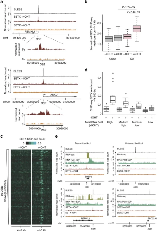
Fig. 1 Senataxin is recruited at DSB induced in active loci. a Genome browser screenshots representing senataxin ChIP-Seq reads count before AsiSI
activation (−4OHT) and after damage induction (+4OHT) at two individual AsiSI sites. The BLESS signal (indicative of cleavage efficiency (Clouaire, T.
et al., manuscript submitted)) is also shown. Close up profiles are also shown below each screenshot. b Box plots representing senataxin ChIP-seq count
before (−4OHT) and after (+4OHT) DSB induction at sites displaying AsiSI-induced cleavage (“cut”, 80 AsiSI sites) or not (“uncut”, 1139 AsiSI sites).
Center line: median; box limits: 1st and 3rd quartiles; whiskers: maximum and minimum without outliers.P values are indicated (Wilcoxon Mann–Whitney
test).c Heatmaps representing senataxin ChIP-seq count over a 10 kb window centered on the DSB before (−4OHT) and after (+4OHT) DSB induction.
DSBs are sorted according to decreasing cleavage efficiency (based on BLESS data set (Clouaire, T. et al., manuscript submitted)). d Box plots representing
senataxin ChIP-seq count before (−4OHT) and after (+4OHT) DSB induction at AsiSI “cut” sites sorted according to total RNA Polymerase II occupancy on
a 10 kb window surrounding AsiSI sites (20 DSBs in each category). Center line: median; box limits: 1st and 3rd quartiles; whiskers: maximum and minimum
without outliers. Points: outliers.e Genome browser screenshots representing senataxin ChIP-Seq reads count before AsiSI activation (−4OHT) and after
damage induction (+4OHT) at four individual AsiSI sites, exhibiting either high (left) or low (right) transcriptional activity (indicated by RNA PolII S2P
ChIP-seq mapping and RNA-seq−4OHT). The BLESS signal (Clouaire, T. et al., manuscript submitted) indicates that all sites display equivalent cleavage
individual sites indicated that senataxin did not accumulate at
broken intergenic or silent regions, although they were robustly
cleaved (see BLESS signal) and showing high level of XRCC4
recruitment (assessed by ChIP-seq
29) (Fig.
1
e, Supplementary
Fig.
1
C). We previously demonstrated that transcriptionally
active genes exhibit preferential binding of Rad51 and
homo-logous recombination (HR) repair
29. In agreement, senataxin
displayed a significantly enhanced recruitment at DSB enriched in
Rad51 compared to DSBs exhibiting low levels of Rad51
(Supplementary Fig.
1
D). Altogether these data indicate that
senataxin is recruited to damage sites with a strong preference for
DSBs induced in transcriptionally active loci, preferentially
repaired by HR.
Senataxin removes DSB-induced RNA:DNA hybrids in active
loci. A well described function of senataxin is its ability to unwind
RNA:DNA hybrids, hence regulating R-loops stability on the
genome. Given senataxin recruitment at DSBs, we further
investigated R-loops distribution at AsiSI-induced DSBs. For this,
we performed DRIP-seq using the S9.6 antibody that displays a
strong specificity for RNA:DNA hybrids
32,33before and after DSB
induction. As expected
6,7, DRIP-seq signal was enriched on active
genes, peaking at TSS and TTS, validating our sequencing results
(Supplementary Fig
2
A, B). Notably, we could observe robust
changes in R-loops distribution following damage, with an
increase of RNA:DNA hybrids around the break site (Fig.
2
a,
Supplementary Fig.
2
C). When taken collectively, following DSB
induction, RNA:DNA hybrids were mildly but significantly
enriched (7% increase) on a 10 kb window surrounding cut sites
compared to uncut sites (Fig.
2
b). Interestingly, while senataxin
accumulation was strongly correlating with the transcriptional
activity of the broken locus (Fig.
1
and Supplementary Fig.
1
), this
was less the case for RNA:DNA hybrids accumulation. Indeed, we
could detect their formation upon damage at few (example
Supplementary Fig.
2
D, E)—but not all (example Supplementary
Fig.
2
F)—untranscribed loci (no detectable signal for RNA-seq or
elongating RNA PolII). While total RNA PolII (hence likely not
in an elongating form) was readily detectable prior DSB induction
at some of these untranscribed loci (Supplementary Fig.
2
D),
others did not display any RNA PolII before damage induction
(Supplementary Fig.
2
E), suggesting that at least in a few
instances RNA:DNA hybrids may also be able to form at
untranscribed loci, devoid of RNA PolII (Discussion), albeit at
lower levels.
Interestingly, at active genes, beyond the accumulation
surrounding the DSBs, we could also detect a decrease in
R-loops across damaged genes bodies and termination sites (Fig.
2
a
for examples, Supplementary Fig.
2
G for averaged profiles).
Moreover, careful examination of our high-resolution data
revealed that despite an increase of RNA:DNA hybrids formation
on ~10 kb window around DSBs, at active genes we could also
observe a sharp 1–2 kb decrease of RNA:DNA hybrids at the
exact sites of senataxin accumulation (Fig.
2
c, d). Depletion of
senataxin using a siRNA (Supplementary Fig.
3
A) triggered an
increase in DSB-dependent RNA:DNA hybrids accumulation
proximally to a DSB (Fig.
2
e). Thus, our high resolution data
reveal that the pattern of R-loops shows complex alterations upon
DSB induction. Altogether our data suggests that RNA:DNA
hybrids form at DSB
flanking chromatin and that, at active genes
senataxin recruitment contribute to their removal in the
immediate vicinity of DSBs.
Senataxin promotes cell survival upon active genes breakage.
To further assess SETX function in DSB repair, we used our
improved version of the DIvA cell line, whereby AsiSI-ER is also
fused to an auxin inducible degron (AID). In this cell line, auxin
addition triggers the degradation of the enzyme and hence repair
of AsiSI-induced DSBs
29. We
first assessed the survival of AID
DIvA cells following break induction and repair, in both control
and senataxin-depleted cells (Supplementary Fig.
3
A). Clonogenic
assays revealed that depletion of senataxin using siRNA does not
trigger cell death in absence of exogenous damage (Fig.
3
a,
−4OHT). DSB induction for 4 h, followed by auxin addition
(+4OHT + auxin) only led to a mild survival defect in control
cells (Fig.
3
a) indicating that these cells recover well from the
induction of DSBs by AsiSI, as previously reported
34. In contrast,
senataxin depleted cells exhibited a strong sensitivity to
AsiSI-induced DSBs (Fig.
3
a, Supplementary Fig.
3
B).
Since AsiSI-induced DSBs exhibit clean DNA ends and
undergo repeated cycles of cleavage, we further tested the effect
of senataxin depletion at DSBs produced by other means.
Etoposide is an inhibitor of Topoisomerase II (TOP II) and
multiples studies have revealed that TOP II exerts a critical
function at active genes in order to unwind supercoils and release
topological constraints that form at transcriptionally active
regions
35. Hence, etoposide-induced DSBs exhibit a biased
distribution throughout the genome, being preferentially located
in active promoters (~30%) and genes bodies (~40%)
36.
Interestingly, senataxin depletion also triggered enhanced
sensi-tivity to etoposide (Supplementary Fig.
3
C). On the other hand,
ionizing radiation (IR) induces DSBs randomly across the
genome, hence being mainly located in intergenic loci that
represent over 95% of higher eukaryotes genomes. Notably, and
in agreement with a previous report
22, senataxin depletion did
not trigger enhanced sensitivity to irradiation (Supplementary
Fig.
3
D). Our data therefore suggest that senataxin exerts an
important function in cell survival specifically following break
induction in active loci.
Senataxin regulates
γH2AX accumulation. To further decipher
the function of senataxin in DSB repair and to understand the
lethality observed following DSB induction in senataxin deficient
cells, we analyzed the effect of senataxin depletion on
γH2AX foci
formation following DSB induction. We found that senataxin
depletion did not abolish foci formation and rather triggered
enhanced
γH2AX signal in 4OHT-treated cells (Fig.
3
b).
Inter-estingly, senataxin depletion also increased
γH2AX signaling
following etoposide treatment (Supplementary Fig.
3
E) but not
following IR (Supplementary Fig.
3
F), suggesting a function of
senataxin in regulating
γH2AX foci formation at DSBs induced in
active genes. To further strengthen these data, we tested whether
a short global transcription extinction preceding break induction
was able to rescue the increased
γH2AX signaling observed in
senataxin-depleted cells. A pretreatment of DIvA cells with
cor-dycepin, a well characterized transcription inhibitor, partially
reduced
γH2AX in damaged DIvA cells following senataxin
depletion (Fig.
3
c). Altogether, our data suggest that senataxin is
involved in regulating
γH2AX establishment at DSBs induced in
transcriptionally active loci.
We next investigated the consequences of senataxin depletion
on DSB repair kinetics. Immunofluorescence performed at
different time points after auxin addition revealed that
γH2AX
foci disappeared with the same kinetics in both control and SETX
siRNA-treated cells (Fig.
4
a, b). To refine this analysis,
experiments were also performed using a high content
micro-scope which allows to sort G1 versus G2 cells based on their DNA
content
31. Similarly we could not detect any delay of
γH2AX foci
clearance following auxin addition in SETX-depleted G1 and G2
cells (Supplementary Fig.
4
A). Additionally, AID DIvA cells allow
to assay repair kinetics at specific DSBs using a protocol based on
the ligation of a biotinylated oligonucleotide followed by
streptavidin purification and quantitative PCR measurement of
purified DNA
30,37. Once more, senataxin depletion did not
trigger repair delay at two DSBs found to be enriched in senataxin
following 4OHT treatment (Fig.
4
c). In addition, this held also
true in G1- and G2-arrested cells following treatment with
lovastatin and RO-3306 respectively (Supplementary Fig.
4
B).
Altogether, these data indicate that while SETX deficiency
triggers enhanced
γH2AX signaling, it is not associated with
delayed DSB repair, neither in G1 nor in G2.
Senataxin regulates repair pathway choice. Given that senataxin
was found to be recruited at DSBs induced in transcriptionally
01.5
Normalized read count
a
0 100 10 30 940 000 31 020 000 ASXL1 chr20 DSB 89 380 000 89 440 000 chr1b
3.0 3.5 4.0 4.5 Uncut CutNormalized DRIP-seq read count on +/– 5 kb
−4OHT +4OHT −4OHT +4OHT P=0.0047 –4OHT +4OHT 0 1.20 1 0 1.5 0 1.5
Normalized read count
0 100 10 0 1.20 1 0 1.5 1.6 1.2 0.8 0.4 0.1 0.05 –5 kb DSB +5 kb DRIP-seq –4OHT +4OHT SETX ChIP-seq –5 kb DSB +5 kb
d
c
DSB RBMXL1 1 0 10 0 89 454 000 89 464 000 DSB 10 kb DRIP(S9.6)+4OHT SETX +4OHTNormalized read count
P=4.5e–22
e
0.12 0.08 0.04 0 DRIP -qPCR (% input) −4OHT +4OHT CTRL siRNA SETX#2 siRNA DRIP(S9.6)–4OHT DRIP(S9.6)+4OHT SETX +4OHT RNAPolII-S2P–4OHT SETX –4OHT RNA-seq DRIP(S9.6)+4OHT DRIP(S9.6)–4OHT SETX +4OHT SETX –4OHT RNAPolIIS2P –4OHT RNA-seq 0 10 0 10 89 458 000 89 459 000 DSB primers +4OHT –4OHT Normalized DRIP-seq read countn =3
Averaged DRIP-seq count
Averaged ChIP-seq count
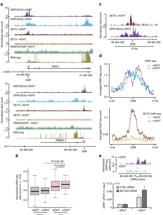
Fig. 2 Senataxin removes DSB-induced RNA:DNA hybrids in active loci. a Genome browser screenshots representing DRIP-seq reads count before
(−4OHT) and after damage induction (+4OHT) at two individual AsiSI sites. Senataxin profiles in both conditions are also shown, together with BLESS
enrichment following DSB induction as well as RNA PolII-S2P and RNA-seq prior to DSB induction.b Box plots representing DRIP-seq reads count before
(−4OHT) and after (+4OHT) DSB induction at sites displaying AsiSI-induced cleavage (“cut”) or not (“uncut”). Center line: median; box limits: 1st and 3rd
quartiles; whiskers: maximum and minimum without outliers.P values are indicated (Wilcoxon–Mann–Whitney test). c Close-up genome browser
screenshot of senataxin and RNA:DNA hybrids at an individual DSB upon DSB induction.d Average DRIP-seq (top) and senataxin ChIP-seq (bottom)
profiles on a ±5 kb window centered on the 80 AsiSI induced DSBs. e DRIP-qPCR in control (CTRL) and senataxin (SETX#2) depleted cells before
(−4OHT) and after (+4OHT) DSB induction in DIvA cells as indicated. The position of the primers used to quantify RNA:DNA hybrids by qPCR are
active loci, which are prone to undergo HR repair
29, we further
investigated the function of senataxin on HR. Senataxin depletion
impaired Rad51 foci formation, while increasing 53BP1
accu-mulation following DSB induction by AsiSI (Fig.
5
a, b). Notably,
the enhanced accumulation of 53BP1 observed in senataxin
depleted cells was partially reversed by pretreating the cells with
transcription inhibitors (cordycepin and 5,6-dichloro-1-β-
D-ribofuranosyl-benzimidazole (DRB)) (Supplementary Fig.
5
A, B).
Next, to further investigate the function of senataxin in repair
pathway choice, we used the reporter constructs previously
developed to quantitatively measure HR
38, single strand
anneal-ing (SSA)
39, and NHEJ
40, using
flow cytometry following I-SceI
transfection. Senataxin depletion triggered a mild decrease of HR
and SSA associated with a similarly mild increase of NHEJ
(Fig.
5
c). Importantly, western blot against I-SceI (myc tagged)
indicated that this was not due to changes in I-SceI expression.
Because generation of ssDNA is the initial step for HR repair,
we further examined the effect of senataxin depletion on DSB end
0 20 40 60 80 100 120 CTRL SETX#2 p=0.03 n =3 p=0.85 p=0.09 CTRL SETX #2 25 γ
H2AX intensity (AU)
p<0.0001 20 15 10 5 +4OHT γH2AX γH2AX γH2AX 0
–4OHT –4OHT+4OHT
–4OHT +4OHT +4OHT –4OHT +4OHT +4OHT +cordycepin CTRL SETX#2 2 4 6 8 10 0 γ
H2AX intensity (AU)
p<0.0001 p=0.0051 CTRL SETX #2 γH2AX γH2AX γH2AX γH2AX γH2AX +cordycepin γH2AX γH2AX +4OHT +cordycepin + 4OHT –4OHT +4OHT –4OHT SETX#2 CTRL
–4OHT +4OHT +4OHT+ IAA
Survival (%) +4OHT+IAA +4OHT –4OHT SETX #2 CTRL
b
c
a
Fig. 3 Senataxin regulates survival andγH2AX upon DSB induction. a Clonogenic assays in AID DIvA cells transfected with control and SETX siRNA, before
and after 4OHT treatment (4 h), followed by auxin (IAA) treatment (4 h) as indicated. Left panel shows a representative experiment. Right panel shows
the average and s.e.m. of three biological replicates.P values are indicated (paired t-test). b γH2AX staining performed in untreated or 4OHT-treated DIvA
cells (4 h), after transfection with control or SETX siRNA as indicated. Scale bar: 10µM. Right panel shows the quantification of the γH2AX nuclear signal
within foci (> 100 nuclei) from a representative experiment. Center line: median; box limits: 1st and 3rd quartiles; whiskers: maximum and minimum
without outliers.P values are indicated (unpaired t-test). c γH2AX staining performed in control or SETX-siRNA transfected DIvA cells as indicated, treated
with 4OHT or pretreated with cordycepin (1 h) previous 4OHT addition (4 h). Scale bar: 10µM. Quantification is shown on the right panel ( > 100 nuclei,
from a representative experiment). Center line: median; box limits: 1st and 3rd quartiles; whiskers: maximum and minimum without outliers.P values are
resection. For this, we used an assay developed previously that
allows to quantitatively measure single stranded DNA (ssDNA)
generated at site specific DSBs
41(Fig.
5
d). Senataxin depletion did
not reduce ssDNA levels at two DSBs induced by AsiSI (Fig.
5
e)
indicating that it is not necessary to promote resection.
Collectively these data indicate that senataxin promotes HR
repair downstream of resection, by promoting Rad51 recruitment
and counteracting 53BP1 accumulation.
SETX depletion enhances translocations. Given the strong
requirement of SETX for survival upon DSB induction in active
genes (Fig.
3
), despite no clear delay in repair kinetics (Fig.
4
), we
next set to assess whether SETX could influence the quality of the
repair reaction, more specifically the frequency of illegitimate
rejoining between distant DSBs, involved in the generation of
translocations. Using high resolution Capture Hi-C, we recently
demonstrated that DSBs can cluster when induced on active
genes and identified the molecular identity of AsiSI-induced DSBs
brought into spatial proximity within nuclear foci
31. Hence, based
on this knowledge we developed an assay to accurately measure
the illegitimate rejoining of closely clustered DSBs. We could
detect rejoining between DSBs induced on the same chromosome
(between MIS12 and TRIM37 as well as in LINC0072 and
LYRM2) (Fig.
6
a), but also between DSBs induced on different
chromosomes (MIS12::LYRM2 and TRIM37::RBMXL1)
(Supple-mentary Fig.
6
A). Importantly, SETX depletion led to a highly
reproducible increase of all four translocations events compared
to control cells (Fig.
6
b, Supplementary Fig.
6
B, C). Notably, this
increase of translocations observed in senataxin depleted cells was
partially rescued by a pretreatment with DRB (Fig.
6
c) or upon
overexpression of RNAseH1, which degrades RNA:DNA hybrids
(Supplementary Fig.
6
C). These data indicate that senataxin plays
a key role in counteracting illegitimate rejoining of DSBs induced
in loci ongoing active transcription.
Discussion
In this study we set to better understand the formation of RNA:
DNA hybrids at DSBs as well as the potential function of RNA:
DNA hybrids removal factors in DSB repair, focusing on
sena-taxin, a well characterized R-loops helicase. We discovered that
senataxin is specifically recruited at DSBs induced in active loci,
where it removes RNA:DNA hybrids forming in cis to broken
loci. Senataxin is further required to regulate
γH2AX signaling, to
promote Rad51 loading and to minimize abnormal rejoining of
distant DNA ends (Fig.
6
d).
Our genome-wide mapping indicates that RNA:DNA hybrids
accumulate in cis to DSBs, as previously proposed
26–28. However,
it seems that proximal DSB-induced RNA:DNA hybrids may
form differently depending on the transcriptional status of the
damaged locus. At active genes, this RNA:DNA hybrids
accu-mulation around DSBs is associated with an otherwise R-loops
decrease across the entire damaged gene body. Several studies
a
–4OHT +4OHT 30 min 60 min 120 min
–4OHT+4OHT 30 60 +4OHT+IAA (min) 120 –4OHT+4OHT 30 60 +4OHT+IAA(min) 120
siRNA CTRL siRNA SETX#2
CTRL SETX#2 0 5 10 15 20 γ
H2AX intensity (AU)
siRNA CTRL siRNA SETX#2 +4OHT+IAA γH2AX γH2AX
b
–4OHT +4OHT 30 60 +4OHT+IAA (min) 120 –4OHT +4OHT 30 60 +4OHT+IAA (min) 120 CTRL SETX#2 Unrepaired DSB (%) DSB-ASXL1 DSB-RBMXL1 0 20 40 60 80 120 100 Unrepaired DSB (%) 0 20 40 60 80 120 100c
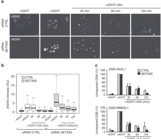
Fig. 4 Senataxin depletion does not delay repair kinetics. aγH2AX staining performed in untreated or 4OHT-treated AID DIvA cells (4 h), followed by
auxin (IAA) addition, after transfection with control or SETX siRNA as indicated. Scale bar: 10µM. b Quantification of the γH2AX nuclear signal ( > 100
nuclei) from a representative experiment, performed in the above condition. Center line: median; box limits: 1st and 3rd quartiles; whiskers: maximum and
minimum without outliers.P values are indicated (unpaired t-test). c Cleavage assay performed in AID-DIvA cells left untreated or treated with 4OHT (4 h)
followed by auxin (IAA) addition (30 min, 60 min, and 120 min), after transfection of control or SETX-directed siRNA. Precipitated DNA was analyzed close to two DSBs, found to recruit SETX after 4OHT. The percentage of sites that remain broken for each DSBs after the indicated time of auxin treatment are
have reported that transcription is downregulated at the damaged
gene, as well as on chromatin proximally
flanking DSBs, although
being globally maintained in the
γH2AX domain at distance from
the break (reviewed in refs.
12,42). The exact mechanism leading
to transcriptional repression in cis to DSBs is not yet clear but
involves the recruitment of chromatin modifying complexes
43–47,
as well as ATM-induced modifications of transcription elongation
factors
45,47yielding to a reduction in the elongating form of RNA
PolII across the gene body
48. Multiple studies have now
estab-lished that R-loops accumulate at sites of paused or slowly
elongating RNA PolII
7,49. Hence, at active genes, DSB-induced
RNA PolII stalling may contribute to the strong RNA:DNA
hybrids and/or R-loops formation in cis to the break.
Notably, although it was not a general feature (Supplementary
Fig.
2
F), we could also identify few seemingly transcriptionally
silent sites that displayed low, but detectable levels of RNA:DNA
hybrids following breakage (Supplementary Fig.
2
D, E), even
when total RNA PolII was not present prior damage
(Supple-mentary Fig.
2
E). Hence de novo RNA PolII recruitment at DNA
ends (a feature recently observed
28,50) may, at least in some
CTRL SETX #2 SETX #2 –4OHT +4OHT CTRL SETX#2
–4OHT +4OHT –4OHT +4OHT
CTRL SETX#2
–4OHT +4OHT –4OHT +4OHT
RAD51 intensity (AU)
0 2 4 6
AsiSI BanI BanI
DSB-ASXL1 740 bp 2000 bp DSB-KDELR3 200 bp 1626 bp 0 1 2 3 4 5 6 7 8 0 1 2 3 4 5 6 7 8 9 p=0.08 CTRL–4OHT CTRL+4OHT SETX–4OHT SETX+4OHT p=0.49 p=0.26 p=0.56 n =4 n =4 DSB-KDELR3 DSB-ASXL1 ssDNA (normalized) siRNA CTRL–4OHT CTRL+4OHT SETX–4OHT SETX+4OHT siRNA CTRL –4OHT +4OHT Rad51 Rad51 Rad51 53BP1 intensity (AU) 0 4 8 12 Rad51 53BP1 53BP1 ssDNA (normalized) ×40 ×40 ×100 ×40 ×40 ×100 P<0.0001 P=0.0028 8 10 0 0.8 0.6 0.4 0.2 1.2 0.8 0.6 0.4 0.2 1.6 1.4 1.2 0.8 0.6 0.4 0.2 1 1.2 HR
GFP-postive cells (AU)
n =3 p=0.12 +IsceI -IsceI SETX CTRL NT NT tub myc 0 1 SSA
GFP-postive cells (AU)
n =4 p=0.06 tub myc 0 1 NHEJ –I–SceI +I–SceI CTRL SETX –I–SceI +I–SceI CTRL SETX –I–SceI +I–SceI CTRL SETX
GFP-postive cells (AU)
n =5 p=0.03 +IsceI –IsceI SETX CTRL NT NT –IsceI +IsceI SETX CTRL NT NT tub myc Rad51 Rad51 53BP1 53BP1 53BP1 53BP1
a
b
2000 bp 740 bp 1626 bp 200 bpe
d
c
instance, produce RNA:DNA hybrids in cis to DSBs. More
dedicated systems that allow to induce DSBs at a larger number of
transcriptionally silent loci (using multiple guides RNAs for Cas9
induced breaks for example) should help to understand the
occurrence of such RNA:DNA hybrids as well as the mechanisms
driving their formation. Additionally, it is important to note here
that our study does not allow to determine whether the
S9.6 signal detected at DSBs represent triple stranded structures
(R-loops) or double-stranded RNA:DNA hybrids. Indeed, in S.
pombe, such hybrids were proposed to form as resection
pro-gresses, by the hybridization of a RNA to the resected single
strand DNA
28. Strand-specific mapping of the RNA engaged in
the hybrids detected at DSB, by DRIPc-seq, should help to
determine whether these resection-dependent RNA:DNA hybrids
are conserved in mammalian cells and whether the increased
S9.6 signal at DSBs observed in this study represent R-loops or
RNA:DNA hybrids.
Regardless of whether they arise from an unscheduled activity
of RNA Polymerase II blocked by the lesion, or by de novo
transcription from DNA-ends, DSB-induced RNA:DNA hybrids
may exert some important function in the DDR. First, these RNA:
DNA hybrids could contribute to setup an adequate chromatin
landscape. Indeed, R-loops at genes have been proposed to
modify chromatin structure. For instance, R-loops trigger H3-S10
phosphorylation linked to chromatin condensation
51,52,]. On
another hand, R-loops also coincide with increased chromatin
accessibility
7and can mediate the recruitment of the TIP60/p400
complex that promotes histone acetylation and nucleosome
remodeling
53. Notably, the Tip60 complex has been repeatedly
found at break site, where it mediates H4 and H2A acetylation
and subsequent recruitment of repair proteins
54–58. Hence one
can hypothesize that R-loops and/or RNA:DNA hybrids will
contribute to set up the proper chromatin landscape required at
DSBs to ensure accurate and timely repair. Second, RNA:DNA
hybrids may contribute in promoting premature transcription
termination of broken genes. Indeed, a large amount of studies
established a function for R-loops, as well as for senataxin, in
terminating transcription including for promoter-associated
bidirectional non-coding RNA (for instance
59,60, reviewed in
refs.
1,11). In this regard it is interesting that dsRNA processing
factors such as Drosha and Dicer contribute to this process
61–63.
Hence, at damaged genes, R-loops and senataxin may promote
premature termination in order to allow either RNA PolII
clearance from the damaged region to favor accessibility of repair
proteins, or efficient recycling of RNA PolII to resume
tran-scription after repair. It is tempting to speculate that the reported
function of Dicer and Drosha in DDR
64may at least in part,
relate to the transcriptional termination at genes experiencing a
DSB.
Interestingly we found that, although senataxin depletion did
not delayed DSB repair, it impaired Rad51 foci formation, and
conversely increased accrual of 53BP1. Decreased HR and
increased NHEJ were also observed following senataxin depletion
using HR and NHEJ reporter constructs although to a milder
extent, which may be due to low R-loops formation on these
substrates. Notably, senataxin depletion did not impede resection,
suggesting that senataxin and/or RNA:DNA hybrids removal are
critical at a step subsequent to ssDNA generation. However,
resection was only monitored up to 1.6 kb from the DSB, so we
cannot exclude a function of senataxin in regulating more long
range resection events. Notably, we also found that senataxin
regulates
γH2AX establishment and counteracts the illegitimate
rejoining of distant DNA ends, suggesting that R-loops removal is
required to minimize translocations and maintain genome
integrity following production of DSB in active genes. The
mechanism by which senataxin impacts on
γH2AX is currently
unknown but may involve the regulation of ATM recruitment or
activity. Alternatively, senataxin and/or R-loops may regulate the
DSB-flanking chromatin structure, modifying its ability to
undergo H2AX phosphorylation. Equally, how senataxin
coun-teracts translocations needs to be investigated. We recently
pro-posed that
γH2AX spreading on entire topologically associated
domains (TAD) likely modifies the properties of the chromatin
fiber which could translate into changes in chromatin mobility
within the nucleus
42,65. Given that DSBs induced in active genes
were found to display enhanced clustering ability
31, we can
speculate that the increased aberrant joining of distant DSBs
observed in senataxin depleted cells arise from an increased DNA
ends mobility triggered by the enhanced
γH2AX establishment.
Importantly, SETX is a gene mutated in two severe
neurolo-gical diseases, AOA2 and ALS4, associated with progressive
neurodegeneration. In this regard it is interesting that DSBs have
been shown to be produced as a consequence of neuronal
activ-ity
66and further genomic studies suggested they likely arise in
active genes
67,68. Given that senataxin exerts its anti-translocation
and survival-promoting functions only for DSBs induced in active
genes, we propose that this
“Transcription Coupled DSB repair”
function of senataxin may contribute to neuron loss in AOA2/
ALS4 patients.
Methods
Cell culture. U20S were retrieved from ATCC and modified with a plasmid
encoding for the restriction enzyme (pBABE-AsiSIER and pAID-AsiSIER)29,30.
U2OS, DIvA (AsiSI-ER-U20S), and AID-DIvA (AID-AsiSI-ER-U20S) cells were
cultured in Dulbecco’s modified Eagle’s medium (DMEM) supplemented with
antibiotics, 10% FCS (InVitrogen) and either 1µg/mL puromycin (DIvA cells) or
800µg/mL G418 (AID-DIvA cells) at 37 °C under a humidified atmosphere with
5% CO2. The cell lines were regularly checked for mycoplasma contamination. For
AsiSI-dependent DSB induction, cells were treated with 300 nM 4OHT (Sigma, H7904) for 4 h. When indicated, 4OHT-treated cells were washed three times in
Fig. 5 Senataxin depletion decreases HR but does not impede resection. a Rad51 staining performed in untreated or 4OHT-treated DIvA cells (4 h), after
transfection with control or SETX siRNA as indicated. Scale bar: 10µM. Right panel shows the quantification of the Rad51 nuclear signal within foci (>100
nuclei) from a representative experiment. Center line: median; box limits: 1st and 3rd quartiles; whiskers: maximum and minimum without outliers.P values
are indicated (unpairedt-test). b 53BP1 staining performed in untreated or 4OHT-treated DIvA cells (4 h), after transfection with control or SETX siRNA as
indicated. Scale bar: 10µM. Right panel shows the quantification of the 53BP1 nuclear signal within foci ( > 100 nuclei) from a representative experiment.
Center line: median; box limits: 1st and 3rd quartiles; whiskers: maximum and minimum without outliers.P values are indicated (unpaired t-test). c HR (top
panel), SSA (middle panel), and NHEJ (bottom panel) usage was measured using cell lines harboring specific reporter constructs38–40in control or
senataxin-deficient (siRNA-transfected) dedicated cells. Myc-I-SceI expression was controlled by western blot in each condition. Mean and s.e.m. of at
least three biological replicates are shown (as indicated).P values are indicated (paired t-test). d The site specific resection assay has been described
earlier. Briefly, DNA purified from damaged or undamaged cells is digested by dedicated restriction enzymes (as indicated) and digestion-resistant DNA
(single stranded DNA) is measured by qPCR, using primers pairs apart from the restriction sites. Here, we optimized this assay at two AsiSI-induced DSBs
that were shown to undergo HR29.e Resection assay at the two DSBs in control or SETX-siRNA transfected cells. Values were normalized against the % of
pre-warmed PBS and further incubated with 500µg/mL auxin (IAA) (Sigma;
I5148) for the indicated time. For transcriptional inhibition, DRB (Sigma, 100μM)
or cordycepin (Sigma, 50µM) was added to the medium 1 h prior to 4OHT (4 h)
and auxin (2 h) treatments. Cells were arrested in G1 using a 48 h treatment with
40μM lovastatin (Mevinolin from LKT Laboratories) and in G2 with a 24 h
treatment with 40μM Ro-3306 (CDK1 inhibitor, Calbiochem). For clonogenic
assays in U20S cells, DSBs were induced either by increasing doses of etoposide (Sigma) for 16 h as indicated or by irradiation with a Cs137 source (Biobeam 8000).
siRNA and plasmid transfection. siRNA transfections were performed with a Cell
Line Nucleofector kit V (Program X-001, Amaxa) according to the manufacturer’s
instructions and cells were assayed 48 h after siRNA nucleofection. The following 2.5 2.0 1.5 1.0 0.5 0 P=0.0006 P=0.0051 MIS12::TRIM37 LINC00271::LYRM2
Translocation frequency (normalized to CTRL)
MIS12::TRIM37 LINC00271::LYRM2 –4OHT +4OHT +IAA H20 100 bp –4OHT +4OHT +IAA H20 100 bp n=5 –4OHT +4OHT+IAA –4OHT +4OHT+IAA siRNA CTRL siRNA SETX 2 1 0.5 1.5 2.5 0
–4OHT +4OHT+IAA –4OHT +4OHT+IAA
–DRB +DRB n=3 MIS12::TRIM37 3 siRNA CTRL siRNA SETX#2 Active gene RNA pol II RNA pol II DSB Damaged gene Elongating polymerase R-loops across gene body
Stalled polymerase
R-loops decreases across gene body
DSB Damaged gene Regulation of γH2AX Rad51 recruitment Inhibition of translocation DSB induction SETX recruitment ATM ? SETX
RNA:DNA hybrids removal
RNA polII clearance? Translocation frequency (normalized to CTRL)
P=0.08
*
R-loops and/or
RNA:DNA hybrids accumulation
a
b
c
RNA pol II RNA pol IId
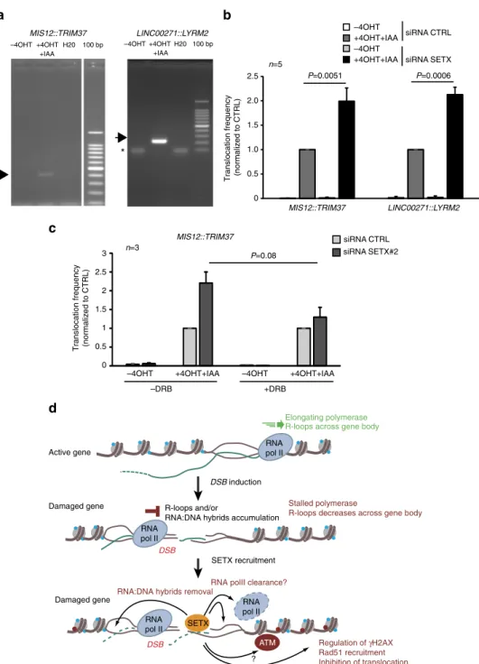
Fig. 6 Senataxin counteracts the formation of translocations. a Rejoining of distant DSBs were detected by PCR, following DSB induction and repair
(+4OHT + IAA 2 h) at breaks recently shown to undergo clustering31. DNA sequencing confirmed the nature of the amplified products. b MIS12::TRIM37
andLINC00271::LYRM2 rejoining frequencies were analyzed before or after 4OHT + IAA treatment, by quantitative PCR in AID DIvA cells transfected with
control or SETX directed siRNA. Mean and s.e.m. offive biological replicates are shown. P values are indicated (one sample t-test). c MIS12::TRIM37
rejoining frequency was analyzed in control or SETX-depleted AID DIvA cells pretreated or not with DRB prior to 4OHT addition as indicated. Mean and s.
e.m. of three biological replicates are shown.P value is indicated (paired t-test). d Model: R-loops form as the RNA Polymerase II progresses across the
gene. The induction of a DSB elicits ATM activity which triggers RNA polymerase II stalling at the vicinity of the DSB, hence decreasing R-loops across the
gene body. On another hand, R-loops and/or RNA:DNA hybrids accumulatein cis to the DSB, due to stalled RNA PolII generating short, abortive, RNAs
which thread back in the DNA duplex, or/and potentially de novo PolII transcription from DNA end. Senataxin is further recruited to remove RNA:DNA
hybrids at the vicinity of the break induced in active loci. Senataxin and/or R-loop removal, regulateγH2AX establishment, promote Rad51 loading and
siRNA against SETX were used: SETX#2 GAGAGAAUUAUUGCGUACU and SETX Smart pool (Dharmacon) containing the following siRNA: #a GCACGU-CAGUCAUGCGUAA, #b UAGCACAGGUUGUUAAUCA, #c AAAGAGUA-CUUCACGAAUU #d GGACAAAGAGUUCGAUAGA. siRNAs efficiency was assessed by mRNAs extraction with a Qiagen RNeasy kit (Qiagen) and reverse transcription with the AMV reverse transcriptase (Promega). cDNAs were
quan-tified by RT-qPCR (primer sequences: SETX_FW
CTTCATCCTCGGA-CATTTGAG and SETX_REV TTAATAATGGCACCACGCTTC) and normalized to RPLP0 cDNA levels (primer sequences: FW GGCGACCTGGAAGTCCAACT and REV CCATCAGCACCACAGCCTTC). For RNAse H1 overexpression, pICE-NLSmCherry and pICE-RNaseHI-NLS-mCherry were transfected 24 h after siRNA transfection using Lipofectamin 2000 (Life Technologies) following manufacturer’s instructions.
Western blot. To assess for SETX depletion, western blot analysis was performed with NuPAGE Bis–Tris 4–12% gels and reagents (Invitrogen) according to the
manufacturer’s instructions. Briefly, cells were lysed in NuPage sample buffer with
reducing agent (Invitrogen) and resolved proteins were transfered onto PVDF membranes (Invitrogen). PVDF membranes were then saturated 1 h in 5% nonfat dry milk with TBS and 0.5% Tween 20 and incubated overnight with the following primary antibodies: SETX (Novus Biologicals, NB100-57542, 1:500) and anti-alpha-tubulin (Sigma-Aldrich, DM1A, 1: 100,000). Horseradish peroxidase-coupled secondary antibodies were from Sigma (anti-mouse, A2554, 1: 10,000; anti-rabbit, A0545, 1: 10,000), and the chemiluminescence Lumilight reagent was from Roche Diagnostic. To analyze I-SceI expression, total cell lysates were pre-pared 24 h after I-SceI plasmid transfection by direct resuspension of cells in Laemmli buffer and sonication. Cell extracts were separated on 10% SDS PAGE and proteins were transferred on nitrocellulose membrane. Primary antibodies were anti-myc (9E10, Roche) and anti-alpha-tubulin (Sigma-Aldrich). Signals were analyzed by autoradiography (for SETX expression) or using a ChemiDoc touch device (BioRad) for I-SceI expression.
ChIP followed by high throughput sequencing and RNA-seq. Cells were crosslinked with formaldehyde (1%) added to the culture medium for 15 min at room temperature. Glycin (0.125 M) was added for 5 min to stop the reaction. Cells were washed twice with cold PBS and harvested by scraping. Pelleted cells were incubated in lysis buffer (Pipes 50 mM pH 8, KCl 85 mM, NP‐40 0.5%), homo-genized with a Dounce homogenizer. Nuclei were harvested by centrifugation and incubated in nuclear lysis buffer: (50 mM Tris pH 8.1, 10 mM EDTA, 1% SDS). Samples were sonicated ten times for 10 s at a power setting of 5 and 50% duty cycle (Branson Sonifier 250), to obtain DNA fragments of about 500–1000 bp. After sonication, samples were diluted ten times in dilution buffer (0.01% SDS, 1.1% Triton X‐100, 1.2 mM EDTA, 16.7 mM Tris pH 8.167 mM NaCl) and
pre-cleared for 2 h with 100μl of protein‐A and protein‐G beads (Sigma), previously
blocked with 500μg of BSA 2 h at 4 °C. Precleared samples were incubated
over-night at 4 °C on a wheel with specific antibodies. For SETX ChIP, 200 µg of
chromatin was immunoprecipitated by using 2µg of anti-SETX (Novus Biologicals,
NB100-57542). For RNA Pol II ChIP, 25µg of chromatin was immunoprecipitated
with 2µg of anti-RNA polymerase II CTD repeat YSPTSPS (phospho S2)
(Chro-motek 3E10), or with 2µg of the anti-total RNA PolII (Bethyl Laboratories
A304-405A). XRCC4 ChIP-seq were published earlier29. Immune complexes were
pre-cipitated with 100μl of blocked protein A/protein G beads for 2 h at 4 C on a
rotating wheel. Beads were washed with dialysis buffer (2 mM EDTA, 50 mM Tris pH 8.1, 0.2% Sarkosyl) once and with wash buffer (100 mM Tris pH 8.8, 500 mM
LiCl, 1% NP‐40, 1% NaDoc) four times. Immunoprecipitated complexes were
re-suspended in 200µl of TE buffer (Tris 10 mM pH8, EDTA 0.5 mM pH8) with 30
µg of RNAse A for 30 min at 37 °C. Crosslink was reversed in the presence of 0.5% SDS at 70 °C overnight with shaking. After a 2 h proteinase K treatment,
immu-noprecipitated and input DNA were purified with phenol/chloroform and
pre-cipitated. Samples were resuspended in 100µl water. For ChIP-Seq, multiple ChIP
experiments were pooled. Immunoprecipitated DNA was subjected to library preparation and single-end sequencing on a NextSeq 500 at EMBL GeneCore (Heidelberg, Germany).
RNA extraction and RNA-seq library preparation. For RNA-seq, DIvA cells (transfected with the control siRNA) were lysed using TRI reagent (SIGMA) and spiked-in with ERCC RNA Spike-In Mix (Thermo Fisher Scientific). Total RNA were recovered by chloroform extraction followed by isopropanol precipitation. Samples were treated with RQ1 RNase-free DNase (Promega) for 1 h at 37 °C and
purified by phenol/chloroform extraction followed by ethanol precipitation.
Ribosomal RNA depletion and RNA-Seq library preparation were performed at EMBL Genomics core facilities (Heidelberg, Germany) using TruSeq Stranded Total RNA (Illumina).
DNA:RNA immunoprecipitation. DRIP assay was carried out according to the
protocol described in ref.7. DIvA cells were treated with 300 nM 4OHT for 4 h,
trypsinized, pelleted at low speed and washed with DPBS (Life technology). Total nucleic acids were extracted with 0.5% SDS /Proteinase K (Thermo Fisher Scien-tific, Waltham, MA) treatment at 37 °C overnight and recovered by
phenol-chloroform extraction and ethanol precipitation. DNA was digested by a restriction enzyme cocktail (20 units each of EcoRI, HindIII, BsrGI, XbaI) (New England Biolabs) in 1× NEBuffer 2, with or without RNase H treatment, overnight at 37 °C. Fragmented DNA was cleaned by phenol-chloroform extraction and ethanol precipitation followed by two washes with 70% ethanol. Air-dried pellets were
resuspended in 10 mM Tris-HCl pH 7.5, 1 mM EDTA (TE). In total, 4µg of digests
was diluted in 450µL of TE, and 10 µL was reserved as input for qPCR. 50 µL of
10× IP buffer was added (final buffer concentration of 10 mM sodium phosphate,
140 mM sodium chloride, 0.05% Triton X-100) and 10µL of S9.6 antibody (1 mg/
ml, kind gift from F. Chedin, UC Davis). Samples were incubated with the antibody
at 4 °C for 2 h on a wheel. 50µL of Protein A/G Agarose (Pierce), previously
washed twice with 700µL of 1× IP buffer for 5 min at room temperature, were
added and samples were incubated for 2 h at 4 °C on a wheel. Each DRIP was then
washed three times with 700µL 1× IP buffer for 10 min at room temperature. After
thefinal wash, beads were resuspended in 250 µL of 1× IP buffer and incubated
with 60 units of Proteinase K for 45 min at 55 °C. Digested DRIP samples were then cleaned with phenol-chloroform extraction and ethanol precipitation.
Air-dried DRIP pellets were resuspended in 45µL of 10 mM Tris-HCl pH 8. DRIP
experiment was assayed by qPCR using primers located at SNRNP (negative control), RPLA13 (positive control) and RBXML1 (AsiSI site):
SNRNP_FW:GCCAAATGAGTGAGGATGGT; SNRNP_REV: TCCTCTCTGCCTGACTCCAT; RPLA13_FW:AATGTGGCATTTCCTTCTCG; RPLA13_REV: CCAATTCGGCCAAGACTCTA. RBMXL1_FW: GATTGGCTATGGGTGTGGAC RBMXL1_REV: CATCCTTGCAAACCAGTCCT
For DRIP-Seq, samples from three DRIP experiments were pooled and sonicated to an average size of 300 bp using a Bioruptor (Diagenode) for 20 cycles of 30 s on, 30 s off, high setting. Immunoprecipitated DNA was subjected to library preparation and single-end sequencing on a NextSeq 500 at EMBL GeneCore (Heidelberg, Germany).
ChIP-seq, seq, and DRIP-seq data set analyses. SETX ChIP-Seq, RNA-seq, and DRIP-Seq samples were sequenced using Illumina NextSeq 500 (single-end, 80-bp reads for SETX ChIP-Seq; paired-(single-end, 75-bp reads for RNA-Seq and single-end, 85 bp reads for DRIP-Seq) at EMBL Genomics core facilities
(Heidel-berg, Germany). The quality of each raw sequencingfile (fastq) was verified with
FastQC (https://www.bioinformatics.babraham.ac.uk/projects/fastqc/). ChIP-Seq
and DRIP-Seqfiles were aligned to the reference human genome (hg19) and
processed using a classical ChIP-seq pipeline: bwa (http://bio-bwa.sourceforge.net/)
for mapping and samtools (http://www.htslib.org/) for duplicate removal (rmdup),
sorting (sort), and indexing (index). RNA-seq was mapped to a custom human genome (hg19 merge with ERCC92 sequences) to avoid mapping ERCC sequences on the human genome and processed as the same way as ChIP-Seq, except for the alignment in paired-end mode with STAR and without remove potential duplicate. Coverage for each aligned ChIP-seq data set (.bam) were computed with the rtracklayer R package and normalized using total read count for each sample.
Coverage data was exported as bigwig (file format) for further processing.
Averaged ChIP-seq, RNA-seq, and DRIP-Seq profiles were generated using the R package ggplot2. For profiles relative to DSB, the x-axis represents genomic position relative to AsiSI site and the y-axis represents the mean coverage at each
bp (Fig.2d). For metagene profiles, the mean coverage was computed in two parts:
first, mean coverage was computed in 200 bp intervals 3 kb upstream TSS and downstream TSS. Second, gene bodies were divided into 100 equally sized bins, so average profiles could be computed as a percent of entire gene length. Profiles were
computed for all genes (Supplementary Fig.2B) or for genes either directly
damaged or located near a DSB (<1 kb) (Supplementary Fig.2G).
To classify DSBs based on transcriptional activity, RNA PolII-S2P ChIP-seq, total RNA PolII ChIP-seq or RNA-seq obtained in DIvA cells prior DSB induction
were computed on a windows of±5 kbp around DSBs. DSBs were ordered based
on their RNA PolII enrichment and discriminated into four categories of 20 DSBs each (low, medium low, medium high, and high).
Box-plots were generated with R-base. The center line represents the median,
box ends represent respectively thefirst and third quartiles, and whiskers represent
the minimum and maximum values without outliers. Outliers were defined as first
quartile−(1.5 × interquartile range) and above third quartile + (1.5 × interquartile
range). Values represent the total normalized read count in a specific genomic
window surrounding AsiSI-induced DSBs or uncut AsiSI genomic sites (“uncut”).
Statistical hypothesis testing was performed using nonparametric paired Mann–Whitney–Wilcoxon (wilcoxon.test() function in R) to tests distribution differences between two populations.
For heatmap representations, average normalized sequencing signal was determined in 500 bp bins centered on each cleaved AsiSI site using custom R/ Bioconductor scripts. The resulting matrix was represented as a heatmap using Java
Treeview (http://www.jtreeview.sourceforge.net). DSBs were ordered based on the
BLESS signal (Fig.1c) or the total RNA PolII enrichment on±5 kb (Supplementary
Fig.1B).
Clonogenic assays. After siRNA transfection, AID-DIvA and U20S cells were seeded at a clonal density in 10 cm diameter dishes. After 48 h, U20S cells were
either exposed at increasing doses of IR or treated with increasing doses of eto-poside, as indicated. AID-DIvA cells were treated with 300 nM 4OHT for 4 h and, when indicated, washed three times in pre-warmed PBS and further incubated with
500µg/mL auxin for another 4 h. After three washes in pre-warmed PBS, complete
medium was added to each AID-DIvA cells dish. After 10 days, U20S cells and AID-DIvA cells were stained with crystal violet (Sigma) and counted. Only colonies containing more than 50 cells were scored.
Immunofluorescence. Cells were plated in glass coverslips, fixed with 4%
paraf-ormaldehyde for 15 min, permeabilized with Triton 0.5%, and blocked with PBS-BSA 3% for 30 min at room temperature. Cells were then incubated with antibody
againstγH2AX (JBW301, 05-636, Millipore, 1:1000) and 53BP1 (Novus Biological
NB-300-104, 1:500) overnight at 4 °C. Cells were washed three times in PBS-BSA 3% and incubated with secondary antibody for 1 h. After three washes (one PBS-BSA 3% and two PBS), nuclei were stained with Hoechst 33342 (Sigma). Staining against Rad51 (Santa cruz sc8349, 1:200) was performed using the following protocol. Cells were plated in glass corverslips, then submitted to a pre-extraction
with ice cold buffer (20 mM HEPES pH 7.5; 20 mM NaCl; 5 mM MgCl2; 1 mM
DTT; 0.5% NP40) for 20 min on ice. Cells werefixed with 4% paraformaldehyde
for 15 min and blocked with PBS-BSA 3% for 30 min at room temperature. Immunofluorescence was carried as previously described. Image acquisition was performed using MetaMorph on a wide-field microscope equiped with a cooled charge-coupled device camera (CoolSNAP HQ2), using a ×40 or ×100 objective. High-throughput microscopy. AID-DIvA and U20S cells were plated in 96-well
plates after transfection. Afterfixation, permeabilization and saturation steps,
γ-H2AX was stained overnight withγH2AX (JBW301, 05-636, Millipore) and the
secondary antibody anti-mouse Alexa 647 (A21235, Molecular Probes). Nuclei
were labeled with Hoechst 33342 (Sigma) at afinal concentration of 1 μg/ml for 5
min.γ-H2AX foci were further analyzed with an Operetta automated high-content
screening microscope (PerkinElmer). For quantitative image analysis, multiple fields per well were acquired with a ×40 objective lens to visualize ~2000 cells per well in triplicate.
γ-H2AX, 53BP1, and Rad51 foci intensity quantification. All quantification was performed using Columbus, the integrated software to the Operetta automated high-content screening microscope (PerkinElmer). DAPI or Hoechst nuclei were selected according to the B method, and appropriate parameters, such as the size
and intensity offluorescent objects, were applied to eliminate false-positive. Then
γ-H2AX, Rad51 and 53BP1 foci were detected with the D method with adjusted parameters to ensure best foci detection: detection sensitivity 0.5–1; splitting
coefficient, 0.5–1; background correction, >0.5–0.9. G1 and G2 nuclei were selected
on the basis of the Hoechst intensity, after visualization of the Hoechst distribution in all cells. Box plots represent the total nuclear signal intensity detected in foci. Repair kinetics at AsiSI sites. Repair kinetics at specific AsiSI-induced DSBs were
measured as described in refs.29,37. Genomic DNA was extracted using the
DNAeasy kit (Qiagen) and in vitro ligation with a biotinylated double-stranded oligonucleotide, ligatable with AsiSI sites, was carried out overnight at 4 °C. T4 ligase was inactivated at 65 °C for 10 min, then ligated DNA was fragmented by EcoRI digestion at 37 °C for 2 h. Digestion was then inactivated at 70 °C for 20 min. Samples were precleared with protein A beads for 2 h at 4 °C on a wheel. Precleared samples were then incubated with streptavidin beads (Sigma) at 4 °C overnight.
Beads were previously saturated with 500μg of BSA 2 h at 4 °C. DNA pulled down
with streptavidin beads was washed once with dialysis buffer (2 mM EDTA, 50 mM
Tris pH 8.1, 0.2% Sarkosyl),five times with wash buffer (100 mM Tris pH 8.8, 500
mM LiCl, 1% NP‐40, 1% NaDoc), and three times in TE buffer (Tris10mM pH8,
EDTA0.5 mM pH8). Beads were resuspended in 100μL of water and digested with
HindIII at 37 °C for 4 h. After phenol/chloroform purification and precipitation,
DNA was resuspended in 100μL water. qPCR was performed using the following
primers: ASXL1-FW CCTAGCTGAGGTCGGTGCTA; ASXL1-REV GAA-GAGTGAGGAGGGGGAGT; RBMXL1-FW GATTGGCTATGGGTGTGGAC; RBMXL1-REV CATCCTTGCAAACCAGTCCT.
Resection assay. Measure of resection was performed as described in ref.41with
the following modifications. DNA was extracted from fresh cells using the DNAeasy kit (Qiagen). In total, 400 ng were digested overnight at 37 °C using the Ban I restriction enzyme (16U per samples) that cuts at ~200 bp and 1626 bp from the DSB-KDELR3 and at 740 bp and 2000 bp for DSB-ASXL1. Digested and undigested samples were also treated with Rnase H (Promega). Ban1 was heat inactivated 20 min at 65 °C. Digested and undigested DNA were analyzed by qPCR using the following primers:
DSB-KDELR3_200 FW: ACCATGAACGTGTTCCGAAT; DSB-KDELR3_200_REV: GAGCTCCGCAAAGTTTCAAG; DSB-KDELR3_1626_FW: CCCTGGTGAGGGGAGAATC; DSB-KDELR3_1626_REV: GCTGTCCGGGCTGTATTCTA; DSB-ASXL1_740 FW: GTCCCCTCCCCCACTATTT; DSB-ASXL1_740_REV: ACGCACCTGGTTTAGATTGG; DSB-ASXL1_2000_FW: GTTCCTGTTATGCGGGTGTT; DSB-ASXL1_2000_REV: TGGACCCCAAATTCCTAAAG.
ssDNA% was calculated with the following equation: ssDNA%= 1/(2(Ct digested
−Ct undigested−1)+ 0.5)*100.
HR, NHEJ, and SSA repair assays. The GCS5 and RG37 cell lines have been
derived from SV40 T-transformed humanfibroblasts (GM639)38,40. The U2OS
SSA has been derived from the osteosarcoma cell line U2OS39. For siRNA
trans-fection, 1 × 10⁵cells were transfected using Interferin (Ozyme, France) with 10 nM
of siRNA according to the manufacturer’s instructions. Plasmid coding for I-SceI
expression was transfected using JetPei (Ozyme, France) according to the manu-facturer’s instructions.Twenty four hours after siRNA transfection, cells were
washed and transfected with the I-SceI coding plasmid (1µg). After 72 h, cells were
collected after trypsin treatment and analyzed byflow cytometry (BD Facscalibur)
to detect and count positive cells in each condition. Percentage of GFP-positive cells was calculated on 25,000 sorted events.
Translocation assay. AID-DIvA cells were treated as indicated and then DNA was extracted from fresh cells using the DNAeasy kit (Qiagen). Illegitimate rejoining frequencies between MIS12 and TRIM37 (chr17_5390209 and chr17_57184285), LINC00217 and LYRM2 (chr6_135819337 and chr6_9034817), MIS12 and LYRM2 (chr17_5390209 and chr6_9034817), or TRIM37 and RBMXL1 (chr17_57184285 and chr1_89433139) were assessed by qPCR using the following primers:
MIS12_Fw: GACTGGCATAAGCGTCTTCG TRIM37_Rev: TCTGAAGTCTGCGCTTTCCA LINC00217_ Fw: GGAAGCCGCCCAGAATAAGA LYRM2_Rev: TCTGAAGTCTGCGCTTTCCA TRIM37_Fw: AATTCGCAAACACCAACCGT RBMXL1_Rev: GCCAATGGAGTTCCCTGAGTC
Results were normalized using two control regions, both far from any AsiSI sites
andγH2AX domain using the following primers:
Ctrl_chr1_82844750_Fw: AGCACATGGGATTTTGCAGG Ctrl_chr1_82844992_Rev: TTCCCTCCTTTGTGTCACCA Ctrl_chr17_9784962_Fw: ACAGTGGGAGACAGAAGAGC Ctrl_chr17_9785135_Rev: CTCCATCATCGCACCCTTTG.
Normalized translocation frequencies were calculated using the DeltaDeltaCt
method from Bio-Rad CFX Manager 3.1 software69.
Data availability. High throughput sequencing data have been deposited to Array Express under accession number E-MTAB-6318. Other data and source codes are available upon request.
Received: 27 July 2017 Accepted: 5 January 2018
References
1. Groh, M., Albulescu, L. O., Cristini, A. & Gromak, N. Senataxin: genome
guardian at the interface of transcription and neurodegeneration. J. Mol. Biol. 429, 3181–3195 (2016).
2. Kim, H. D., Choe, J. & Seo, Y. S. The sen1(+) gene of Schizosaccharomyces
pombe, a homologue of budding yeast SEN1, encodes an RNA and DNA helicase. Biochemistry 38, 14697–14710 (1999).
3. Martin-Tumasz, S. & Brow, D. A. Saccharomyces cerevisiae Sen1 helicase
domain exhibits 5′- to 3′-helicase activity with a preference for translocation
on DNA rather than RNA. J. Biol. Chem. 290, 22880–22889 (2015).
4. Leonaite, B. et al. Sen1 has unique structural features grafted on the
architecture of the Upf1-like helicase family. EMBO J. 36, 1590–1604 (2017).
5. Chedin, F. Nascent connections: R-loops and chromatin patterning. Trends
Genet. 32, 828–838 (2016).
6. Ginno, P. A., Lim, Y. W., Lott, P. L., Korf, I. & Chedin, F. GC skew at the 5′
and 3′ ends of human genes links R-loop formation to epigenetic regulation and transcription termination. Genome Res. 23, 1590–1600 (2013).
7. Sanz, L. A. et al. Prevalent, dynamic, and conserved R-loop structures associate
with specific epigenomic signatures in mammals. Mol. Cell 63, 167–178 (2016).
8. Skourti-Stathaki, K. & Proudfoot, N. J. A double-edged sword: R loops as
threats to genome integrity and powerful regulators of gene expression. Genes Dev. 28, 1384–1396 (2014).
9. Garcia-Muse, T. & Aguilera, A. Transcription-replication conflicts: how they
occur and how they are resolved. Nat. Rev. Mol. Cell Biol. 17, 553–563 (2016). 10. Porrua, O. & Libri, D. Transcription termination and the control of the
transcriptome: why, where and how to stop. Nat. Rev. Mol. Cell Biol. 16, 190–202 (2015).
11. Proudfoot, N. J. Transcriptional termination in mammals: stopping the RNA polymerase II juggernaut. Science 352, aad9926 (2016).