REVIEW ARTICLE
Regulation of telomere addition at DNA
double-strand breaks
Cyril Ribeyre&David Shore
Received: 19 November 2012 / Revised: 28 February 2013 / Accepted: 4 March 2013 / Published online: 17 March 2013 # Springer-Verlag Berlin Heidelberg 2013
Abstract Telomeres constitute the ends of linear eu-karyotic chromosomes. Due to the conventional mode of DNA replication, telomeric DNA erodes at each cell division. To counteract this, a specialized reverse tran-scriptase, telomerase, can elongate chromosome ends to maintain them at a constant average length. Because of their similarity to DNA double-strand breaks (DSBs), telomeres might be expected to induce a DNA damage response, which would lead to repair reactions and the generation of translocations or fusions. Many proteins present at telomeres prevent this by protecting (capping) the chromosome termini. Conversely, a DSB occurring in other regions of the genome, due, for instance, to a stalled replication fork or genotoxic agents, must be repaired by homologous recombination or end-joining to ensure genome stability. Interestingly, telomerase is able to generate a telomere de novo at an accidental DSB, with potentially lethal consequences in haploid cells and, at a minimum, loss of heterozygosity (LOH) in diploid cells. Recent data suggest that telomerase is systematically recruited to DSBs but is prevented from
acting in the absence of a minimal stretch of flanking telomere-repeat sequences. In this review, we will focus on the mechanisms that regulate telomere addition to DSBs. Keywords Telomeres . Telomerase . DNA double-strand breaks . DNA damage checkpoint
Introduction
The ends of linear chromosomes, telomeres, exhibit two distinct features. First, due to the so-called end replication problem (Lingner et al. 1995), telomeric DNA shortens at each cell division. In order to counteract this, a specialized enzymatic complex, the telomerase, has been selected dur-ing evolution. Telomerase elongates the ends of the chro-mosomes through its reverse transcriptase activity, using a short RNA molecule stably associated with the enzyme as a template (Greider and Blackburn 1985, 1987, 1989). Therefore, telomeric DNA is comprised of short tandem repeats. Interestingly, though the sequence of the repeated motif can differ between organisms, all share a high G/C content on the strand that forms the 3′ end acted upon by telomerase. In unicellular organisms like budding yeast, telomerase is constitutively expressed and potentially active during each cell cycle. In contrast, telomerase in humans is expressed in only in a subset of cell types, primarily stem cells, germinal cells and a large fraction of cancer cells. Telomerase reactivation in cancer cells is one of the crucial steps in carcinogenesis, perhaps explaining why telomerase expression is so tightly regulated in long-lived metazoans (for review, see Artandi and DePinho2010).
A second key characteristic of telomeres is their chemical similarity to DNA ends generated by accidental double-strand breaks (DSBs). A DSB represents one of the most harmful types of DNA lesion since it will lead to informa-tion loss if not repaired by either end-joining or
homology-C. Ribeyre
:
D. ShoreDepartment of Molecular Biology, National Center for Competence in Research (NCCR) program“Frontiers in Genetics”, University of Geneva, 30 quai Ernest-Ansermet,
1211 Geneva 4, Switzerland C. Ribeyre
e-mail: cyril.ribeyre@igh.cnrs.fr C. Ribeyre
:
D. Shore (*)Institute of Genetics and Genomics in Geneva, University of Geneva, 30 quai Ernest-Ansermet, 1211 Geneva 4, Switzerland
e-mail: David.Shore@unige.ch C. Ribeyre
Institute of Human Genetics, CNRS, UPR1142, 141 Rue de la Cardonille, 34396 Montpellier Cedex 5, France
based repair mechanisms. However, if a telomere were to be repaired by such mechanisms, this would lead to chromo-some fusions or translocations, explaining why DSB repair must be strongly inhibited at telomeres. Paradoxically, many proteins involved in DSB repair and/or DNA damage check-point activation are present at telomeres. Conversely, telo-mere addition at internal DSBs must be strongly repressed since it would, most of the time, be lethal in haploid cells and would lead to loss of heterozygosity (LOH) in diploid cells. Nevertheless, telomere addition to a DSB has been observed in many organisms (reviewed in Melek and Shippen 1996). Indeed, the telomerase enzyme was first identified in Tetrahymena thermophila (Greider and Blackburn 1985, 1987), which, like many other ciliated protozoa, undergoes developmentally regulated telomere addition (on a massive scale) to gene-sized chromosomal fragments generated in a separate macronucleus. In the majority of eukaryotes that do not undergo this exotic form of chromosome processing, de novo telomere addition may play an important role in the rescue of telomere truncations that might arise when replication forks collapse near telo-meres (Miller et al.2006).
Telomerase core enzyme is composed of a reverse tran-scriptase subunit (hTERT in Homo sapiens; Est2 in Saccharomyces cerevisiae) and a non-coding RNA (hTERC in H. sapiens; TLC1 in S. cerevisiae) used as a template for elongation. In budding yeast, three different protein–protein interactions have been implicated in telomerase recruitment to chromosome ends. First, Cdc13, the large subunit of a telomere-specific RPA-like heterotrimeric complex, appears to recruit telomerase via an interaction with Est1, an essential telomerase co-factor (Bianchi et al. 2004; Evans and Lundblad 1999). Telomerase can also interact, through a 48-nt stem-loop region of telomerase template RNA, TLC1, with the DNA end-binding heterodimer Ku70/80 (Fisher et al.2004; Stellwagen et al.2003). However, direct involve-ment of this interaction in telomerase end recruitinvolve-ment has been recently challenged by the observation that Ku cannot bind simultaneously both TLC1 and DNA (Pfingsten et al. 2012). Finally, telomerase may also be recruited by an inter-action between Rfa2 (one of the subunits of the trimeric, ssDNA-binding RPA complex) and Est1 (Schramke et al. 2004; Luciano et al.2012). The existence of multiple mech-anisms capable of directing telomerase to DNA ends raises the question of how cells prevent telomerase from acting inappropriately at accidental DNA breaks.
In this review, we will focus mainly on regulation of telomere addition to a DSB in budding yeast S. cerevisiae since it is here where the most mechanistic information is available, primarily through studies of de novo telomere ad-dition at induced DSBs engineered to be flanked by telomeric or non-telomeric sequences. For a more detailed discussion of earlier studies addressing de novo telomere addition at DSBs
in yeast, the reader is referred to an excellent review from Kolodner and colleagues (Pennaneach et al.2006).
De novo telomere addition to a DNA double-strand break
Telomere addition to a DSB is a very rare event in S. cerevisiae
Two main pathways can repair a DSB: homology-based mechanisms or non-homologous end-joining, which will religate the two extremities together. Alternatively, a DSB can be“repaired” by telomere addition. The first studies of telomere addition were carried out in budding yeast and exploited a galactose-inducible allele of the endogenous HO endonuclease to efficiently and coordinately produce a single HO break at a defined genomic locus (Diede and Gottschling 1999; Kramer and Haber 1993; Mangahas et al. 2001; Schulz and Zakian 1994). These early studies showed that in ~99 % of cases, a break lacking TG-repeat ends is repaired by RAD52-dependent homologous recom-bination, with telomere addition representing only a small portion of repair events, even in the absence of RAD52 function (Kramer and Haber1993; Mangahas et al.2001). This conclusion was supported by the observation that de novo telomere addition events at an HO-induced DSB are infrequent in haploid cells (Myung and Kolodner 2003). Artificial tethering of Cdc13 or Est1 to a DSB significantly increases repair by telomere healing, suggesting that these factors might be limiting for telomere addition (Bianchi et al.2004). However, recent work has shown that Cdc13 and Est2 are readily detectable at DSBs by chromatin immuno-precipitation (ChIP) (Chung et al. 2010; Oza et al. 2009; Ribaud et al. 2012). This unexpected finding suggests that telomerase is indeed recruited to DSBs but adds a telomere there at a very low frequency. These and other data indicate that a competition between different pathways of repair (homologous recombination, non-homologous end-joining and telomere addition) might ensue following generation of a DSB. One of the main questions driving the field is to understand how telomere addition by the telomerase is regulated under these circumstances.
Preferential telomere addition at or near sequences with homology to telomerase template RNA
Telomere addition to a DSB is not a random event. For example, early experiments showed that even a nearby telomeric sequence of the Tetrahymena type (T2G4repeats)
will strongly promote telomere addition following DSB formation in yeast (Kramer and Haber 1993). Similarly, the majority of telomere addition events observed following
loss of a specific telomere occurred in the vicinity of a microsatellite (CA)17 found ~50 kb internal to the deleted
telomere (Mangahas et al.2001). Importantly, in these cases, the new telomere is not added directly to the TG-repeat sequences, which seem instead to be required for creating a telomerase permissive environment.
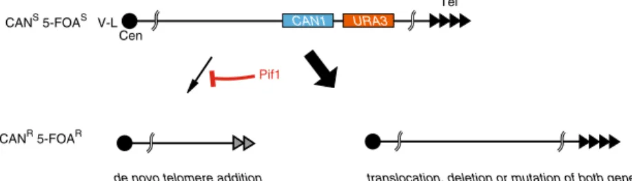
Nevertheless, there is sequence preference at the precise site of telomere addition, which occurs predominantly at short stretches of TG tracts (Chung et al. 2010; Kramer and Haber 1993; Mangahas et al. 2001; Putnam et al. 2004; Schulz and Zakian1994). This has been most exten-sively demonstrated through the use of a “gross chromo-somal rearrangement” (GCR) assay that selects for the simultaneous loss of two counter-selectable marker genes (URA3 and CAN1) placed ~7.5 kb apart from each other at a non-essential region ~20–30 kb from the left end of chro-mosome V (Chen and Kolodner 1999) (see Fig. 1). The frequency of GCR in wild-type cells is extremely low (10
-10
, which is lower than the probability of spontaneous mu-tation of both URA3 and CAN1), but increases dramatically in certain recombination- and replication-defective mutants (Chen and Kolodner 1999). The events that lead to the simultaneous loss of URA3 and CAN1 are mainly deletions, duplications, inversions, translocations or de novo telomere addition. Sequencing of 534 independent telomere addition events that occurred within a 12-kb region of chromosome V-L in wild-type cells revealed a strong preference for telomerase action at GT, TG or GG di-nucleotides (Putnam et al.2004). Significantly, the first nucleotides added were almost always the same and corresponded to a specific part of the TLC1 template (Putnam et al. 2004; Kramer and Haber 1993). Moreover, sequence analysis indicated that telomerase elongation at these sites occurred through suc-cessive annealing/dissociation steps. Altogether, these data indicate that annealing of TLC1 is a crucial step for telomere addition to a DSB. Recently, the Nicolas lab has reported that insertion of two different human minisatellite sequences (CEB25 and CEB1) into the yeast genome strongly in-creases the GCR rate by the promotion of telomere addition events (Piazza et al.2012). For CEB25, telomere additions are due to the presence of Cdc13 binding sites within the
sequence, creating a true telomere seed sequence. However, the ability of CEB1 to generate telomere additions is instead due to the formation of G-quadruplex structures within the minisatellite sequence (Piazza et al. 2012; Ribeyre et al. 2009). These new results indicate that interstitial telomere-like sequences constitute a threat for genome stability through at least two different mechanisms, both leading to inappropriate telomere addition. Interestingly, sequences with homology to TLC1 appear to have been counter-selected in the S. cerevisiae genome, presumably in order to prevent such spontaneous telomere addition events (Mangahas et al.2001).
Mechanisms that inhibit spontaneous telomere addition Although de novo telomere addition is a very rare event in wild-type cells, removal of the nuclear form of the Pif1 helicase (pif1-m2 mutation) strongly increases its frequency (Mangahas et al. 2001; Myung et al. 2001; Schulz and Zakian 1994). Pif1 is a 5′ to 3′ DNA helicase that belongs to a family conserved from yeast to human (reviewed in Bochman et al. 2010). It can unwind a DNA/DNA duplex but acts even more effi-ciently on a DNA/RNA hybrid by first loading onto the DNA strand (Boule and Zakian 2007; Lahaye et al. 1991). It has been proposed that Pif1, through its helicase activity, can disturb the interaction between TLC1 and telomeric single-stranded DNA and, in this way, prevent telomerase action at a normal substrate (Boule et al. 2005; Zhou et al. 2000). Consistent with this, native telomeres are longer when PIF1 function is impaired (Schulz and Zakian 1994).
The action of Pif1 has been extensively studied using the GCR assay described above (Chen and Kolodner 1999), where deletion of PIF1 leads to a ~1,000 increase in GCR frequency (Myung et al.2001). In contrast with deletions of other genes that give rise to a plethora of different rearrangements (e.g. translocations, deletions duplications), the absence of PIF1 leads only to an increase in telomere addition events (see Fig. 1). Moreover, deletion of genes encoding telomerase holoenzyme subunits (EST1, EST2,
CAN1 URA3 Tel Cen V-L CANS 5-FOAS CANR 5-FOAR Pif1
de novo telomere addition translocation, deletion or mutation of both genes Fig. 1 Schematic representation of the “Gross Chromosomal
Rearrangement” assay, showing the arrangement of counter-selectable markers in the starting strain (top), and common outcomes following
selection for canavanine- and 5-FOA-resistant cells in either the ab-sence (left) or the preab-sence (right) of the Pif1 protein
EST3, TLC1) or genes required for normal telomerase action (CDC13, YKU70, YKU80) reduces the GCR rate of PIF1 mutants to wild-type levels. Taken together, these data indi-cate that Pif1 acts specifically to prevent telomerase from
adding telomeres at random to DSBs (Myung et al.2001). In addition, Pif1 might also use its property to disturb G-quadruplex structures to prevent telomere addition (Piazza et al.2012; Ribeyre et al.2009).
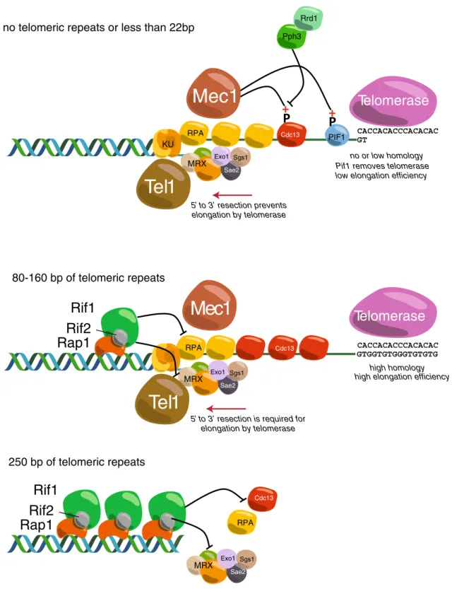
no telomeric repeats or less than 22bp
80-160 bp of telomeric repeats 250 bp of telomeric repeats RPA MRX PIF1 Cdc13
Telomerase
Rif1
Rif2
Rap1
Rif1
Rif2
Rap1
Tel1
Sae2 Exo1 Sgs1 CACCACACCCACACAC GT CACCACACCCACACAC GTGGTGTGGGTGTGTGMec1
MRXTelomerase
Tel1
Sae2 Exo1 Sgs1Mec1
5’ to 3’ resection prevents elongation by telomerasePif1 removes telomerase low elongation efficiency Pph3
Rrd1
high elongation efficiency no or low homology
high homology
5’ to 3’ resection is required for elongation by telomerase KU Cdc13 Cdc13 RPA RPA MRX Sae2 Exo1 Sgs1
Fig. 2 Cartoon depicting some of the molecular events thought to occur at DSBs without flanking TG sequences (top), with 80 to 160 bp of telomeric TG repeats (middle) or with 250 bp of TG-repeat DNA (nearly a full-length native telomere array, bottom). See text for details
In a screen for genes required for telomere addition in a pif1Δ background, Zhang and Durocher identified six new genes (Zhang and Durocher 2010). One of these, RRD1, is required for telomere addition to sites with little or no telomere-like sequences. Rrd1 is a regulator of the Pph3 phosphatase, which is implicated in de-phosphorylation of the serine 306 residue on Cdc13. Significantly, phosphorylation of Cdc13-S306 weakens its physical association with DSBs. Their re-sults suggest that a phosphorylation–de-phosphorylation loop is involved in the regulation of telomere addition to DSBs. It has also been reported recently that Pif1 is phosphorylated by Mec1-Dun1-Rad53 in order to pre-vent telomere addition (Makovets and Blackburn 2009). A mutant that cannot be phosphorylated shows a GCR rate similar to pif1-m2. Notably, this mutant has normal length telomeres, indicating that the role of Pif1 at telomeres can be genetically separated from its role at double-strand breaks. Taken together, these two studies suggest that Mec1 can prevent telomere addition to DSBs through at least two different pathways, one in-volving Cdc13 and the other Pif1 (see Fig. 2, top). DNA resection increases telomere addition
Following DSB formation, 5′ to 3′ exonucleolytic deg-radation (‘resection’) leads to the formation of a 3′ single-stranded DNA. Whilst the MRX (Mre11-Rad50-Xrs2) complex and Sae2 initiate resection, extensive 5′-strand degradation is mediated by at least two different p a t h w a y s ( D n a 2 / S g s 1 o r E x o 1 ) ( M i m i t o u a n d Symington 2008; Zhu et al. 2008). Surprisingly, simul-taneous deletion of SGS1 and EXO1 increases the fre-quency of telomere addition to a DSB (Chung et al. 2010; Lydeard et al. 2010), suggesting that impaired resection at a DSB increases the probability of telomere addition (see Fig. 2, top). However, in strains defective for resection, Pif1 is still able to prevent telomere for-mation, suggesting that resection and Pif1 prevent telo-mere addition by two independent pathways. These data are supported by the fact that Cdc13 binding to a DSB is higher in pif1-m2 or exo1Δ sgs1Δ mutants and even higher in pif1-m2 exo1Δ sgs1Δ triple mutant cells (Chung et al. 2010; Lydeard et al. 2010). Altogether, these results indicate that impaired resection might sta-bilize Cdc13 binding and therefore promote telomere formation. Conversely, when resection is efficient, repair by homologous recombination is more active and pre-vents telomere addition. These findings highlight the possibility of competition between repair pathways at a DSB. In the following section, we will discuss how the balance between repair and telomere addition can be modulated by the presence of telomeric tracts at a DSB.
Telomere addition to a DSB flanked by telomeric repeats Telomere addition is dramatically increased by short telomeric (TG repeat)“seed” sequences
In order to study in detail the factors that control telomere addition at a DSB, researchers have exploited a sys tem i n b ud ding yeast, first described by Gottschling and colleagues (Diede and Gottschling 1999) in which an HO endonuclease-induced DSB is generated immediately adjacent to yeast telomeric (TG repeat) tracts of various lengths (see Fig. 3a). When a DSB is formed in the absence of flanking telomeric sequences and in cells where no homologous sequence is available for recombinational repair, it becomes rap-idly resected and induces a G2/M cell cycle arrest. This leads either to cell death or to adaptation, in which checkpoint arrest is overridden, eventually resulting in loss of the irreparable chromosome (Sandell and Zakian 1993). In contrast, when 80 bp of telomeric repeats is present on the centromere-proximal side of the break, this end is very efficiently elongated by telomerase (as detected by Southern blot analysis; see Fig. 3b), and nearly all cells experiencing the break survive (in the case where sequences on the telomeric side of the HO site are not required for cell viability) (Diede and Gottschling 1999). Efficient telomere formation requires at least 22 bp of TG repeats, which is sufficient for nearly 100 % viability after HO cutting (Hirano and Sugimoto 2007). Constructions with 5, 11 or 17 bp of TG tracts do not show any detectable elongation by Southern blot and only very weak telomere addition frequency using genetic assays (Hirano and Sugimoto 2007; Zhang and Durocher 2010).
Most studies have focused on DSBs flanked on the centromere-proximal side of the HO site by an ~80-bp telomeric tract (TG80) that shows very efficient and rapid telomerase-mediated elongation (Diede and Gottschling 1999; Hirano et al. 2009; Negrini et al. 2007). In contrast, TG160 or TG250 constructs, which are closer to the wild-type telomere tract length, are not elongated and are very efficiently protected against re-section (Hirano et al. 2009; Negrini et al. 2007). Inactivation of Pif1 helicase strongly increases the fre-quency of telomere addition at an HO cut flanked by very short TG seed sequences (5, 11 and 17 bp), but has no effect on telomere addition frequency at TG80 (Zhang and Durocher 2010). The failure of Pif1 to remove telomerase and prevent its action at the longer (TG80) tracts may reflect increased binding of Cdc13 to the resected TG80 ends and thus increased telomerase recruitment (Bianchi and Shore 2007; Sabourin et al. 2007). Telomere addition at a DSB with short telomeric
tracts does not occur when cells are in G1, perhaps because telomerase association is too transient then (Gallardo et al. 2011), but is very efficient when cells are blocked in M phase with nocodazole (Diede and Gottschling 1999). Consistent with this observed cell cycle-regulated action of telomerase, an active Cdk1 is required for elongation (Frank et al. 2006). In addition, primase and DNA polymerasesα and δ are required for telomere elongation at short telomeric tracts (Diede and Gottschling 1999).
Telomerase recruitment and activation are required for elongation of short TG tracts
All of the telomerase components (TLC1, EST1, EST2 and EST3) are required for elongation of a TG80-flanked DSB. Thus, in the absence of TLC1, cell via-bility after HO cutting is almost null (Bianchi et al. 2004; Diede and Gottschling 1999). Consistent with this, Est1 and Est2 are strongly recruited to TG80 ends after cutting (Bianchi et al. 2004; Negrini et al. 2007). In a cdc13-2 mutant, which fails to recruit telomerase (Nugent et al.1996; Bianchi et al. 2004), no elongation is detectable after cutting (Diede and Gottschling1999). Moreover, robust Cdc13 binding is detected at the TG80 ends (Negrini et al. 2007; Hirano and Sugimoto 2007). Telomerase can also be recruited by Ku70/80 (Bianchi
et al. 2004), and consistent with this, in tlc1Δ48 or yku80-135i mutant cells, in which the telomerase–Yku interaction is abrogated, telomere elongation after cut-ting is strongly reduced (Stellwagen et al. 2003). Resection and protein recruitment are similar at telomere-like and non-telomeric DSBs
The MRX (Mre11-Rad50-Xrs2) complex is recruited very early at DSBs and is involved in many aspects of exonucleolytic processing (5′-end resection) and checkpoint activation (Stracker and Petrini 2011). Interestingly, MRX mutants have short telomeres, and the three components of the complex are absolutely required for normal elongation of a TG80 end generated by HO cutting (Diede and Gottschling 2001). Consistent with these findings, Mre11 is recruited to short TG tracts at levels very similar to those observed at a DSB devoid of telomeric tracts (Hirano et al. 2009; Hirano and Sugimoto2007; Negrini et al.2007). This indicates that MRX is required for proper telomere forma-tion at short telomeric tracts. But which of the many roles of MRX are required for telomere elongation?
One of the functions of MRX is to promote recruitment of Tel1 (the yeast ATM-like checkpoint kinase) at both DSBs and telomeres. Tel1 is required for telomere length maintenance (Lustig and Petes 1986) and is recruited to DNA ends through an interaction with the Xrs2 component
a
c
b
HO Gal1 promoter ADE2 LYS2 Tel CEN VII-L HO site Tel CEN ADE2 VII-L 0 1 2 3 4 5 o/n 0 1 2 3 4 5 o/n O/N growth with raffinose 2 hours growth with galactose: HO inductionCells with a small bud are dissected on glucose plate
Cells are checked every 30 min G2/M arrest? YES NO NO ADE2 LYS2 Tel CEN VII-L HO site
Fig. 3 a Schematic representation of the telomere“healing” assay in which galactose-induced expression of the HO endonuclease gene leads to generation of a DSB (top) and, in the case where a telomere “seed” sequence is present, efficient de novo generation of a telomere at the break site, shown by Southern blotting (b, right panel). In cells lacking TG repeats at the break site, the DSB is rapidly degraded (b,
left panel), with rare deletion/joining events detectable following ex-tended growth (left panel, lane marked o/n). c Depiction of a single-cell G2/M arrest assay designed to ask whether DSB flanked on both sides by a short (80 bp) TG-repeat sequence will elicit a cell-cycle arrest following HO cutting
of MRX (Nakada et al.2003). In the absence of Tel1 or in an xrs2-11 mutant unable to recruit Tel1, no elongation is detectable after HO cutting (Frank et al. 2006; Martina et al.2012). Moreover, Tel1, like MRX, is detected at TG80 ends at levels similar to those observed at a DSB (Hirano and Sugimoto 2007). Thus, despite the fact that Tel1 is required for telomere elongation, there is no clear difference in Tel1 recruitment between a DSB with or without telomeric tracts.
One other important role of Mre11 is to initiate the 5′-end resection process at DSBs that normally leads to checkpoint activation and repair by homologous recombination. Mutants of MRE11 defective for resection (Frank et al. 2006) show no elongation at the TG80 ends, indicating that the resection function of MRX is required. In contrast with MRX impairment, inactivation of SAE2 has a weaker, but clearly detectable, effect on TG80 elongation by telomerase (Bonetti et al.2009). Inactivation of SGS1, EXO1 or DNA2 has no detectable effect on telomere elongation, whereas a clear effect is observed in sgs1Δ exo1Δ, dna2-1 exo1Δ and dna2-1 sgs1Δ double mutants, confirming the high redun-dancy of resection pathways. Interestingly, deletion of SGS1 or EXO1 in a sae2Δ mutant completely blocks resection, demonstrating that Sgs1 and Exo1 are responsible for the weak resection that occurs in a sae2Δ mutant. Finally, these data show that resection at TG tracts is essential for elonga-tion by telomerase and is initiated by MRX and Sae2 and then completed by Sgs1, Exo1 and Dna2 (Bonetti et al. 2009). These data are consistent with the fact that a high-level of single-stranded DNA (ssDNA) is present at the TG80 ends (Negrini et al.2007). Consistent with the high-level of resection occurring, binding of the single-stranded specific RPA complex is detected at short telomeric ends, though, interestingly, at lower levels compared to a non-telomeric DSB (Hirano and Sugimoto2007; Negrini et al. 2007). Since Mre11 is required for Cdc13 binding at the TG80 ends (Diede and Gottschling2001), this could explain why resection is required for telomere addition.
Telomerase recruitment at telomeric and non-telomeric DNA ends
Quite surprisingly, considering its strong in vitro bind-ing preference for TG-rich ssDNA substrates, significant Cdc13 accumulation is detected at non-telomeric DSBs, together with the telomerase enzyme itself (Est2) (Oza et al. 2009; Ribaud et al. 2012). Importantly, Est1 and Est2 levels are remarkably similar (as measured by a quantitative qPCR ChIP assay) at non-telomeric DSBs and elongated telomeric DSBs (whether consisting of short TG1-3 or T2AG3 tracts), and their recruitment is
Mre11 dependent (Negrini et al. 2007; Ribaud et al. 2012). This observation further extends the striking
similarity between DSBs and short telomeric ends with respect to both DNA processing (resection) and telome-rase accumulation, and begs that question of why telo-mere addition occurs so infrequently at non-telomeric DSBs whilst ends with short tracts of TG repeats are ‘healed’ by telomerase with remarkable efficiency. One possible explanation for this difference could relate to the relative quantities of RPA and CST binding at the two types of ends: RPA binding is not as high at TG80 ends compared to a DSB, and conversely, CST binding appears to show an opposite bias (consistent with the in vitro binding preference of Cdc13). Surprisingly, inhibi-tion of resecinhibi-tion at DSBs strongly increases the frequen-cy of telomere addition at these ends (Chung et al. 2010), opposite to the effect observed at short TG-tract ends (see Fig. 2, middle). One possible explanation for this apparent paradox is that when TG repeats are present, the equilibrium is in favour of telomere addi-tion instead of repair by homologous recombinaaddi-tion, due to a critical level of Cdc13 binding that depends strong-ly on formation of a sufficientstrong-ly large 3′ overhang. Perhaps when resection is limited at non-TG ends, RPA loading and subsequent steps leading to Rad51 filament formation proceed very slowly, allowing occa-sional activation of the Cdc13–telomerase pathway.
If long telomeric tracts (TG160 or TG250) are pres-ent at the DSB, elongation by telomerase is no longer detected (Hirano et al. 2009; Negrini et al. 2007). Interestingly and in contrast to DSBs and to short telomeric tracts, telomerase is not recruited to TG250 ends (neither Est1 nor Est2 is detected there in signif-icant amounts), suggesting that long telomeric tracts prevent elongation by blocking telomerase recruitment (Negrini et al. 2007). Single-stranded DNA is not detected at long TG tracts either, consistent with the absence or very weak association of Mre11 and Rfa1 at TG160 and TG250 ends (Hirano et al. 2009; Negrini et al. 2007). Interestingly, Cdc13 binding levels are similar at TG80 and TG160 ends, but, in contrast, undetectable at TG250 (Hirano et al. 2009; Negrini et al. 2007). This may indicate that TG160 is not as protected from resection as is TG250. Nevertheless, since Cdc13 is recruited by default to DSBs (Oza et al. 2009; Ribaud et al. 2012), long TG tracts must be able to actively block Cdc13 association. Taken togeth-er, these studies indicate that long telomeric tracts block DSB processing at a very early step in order to prevent elongation by telomerase (see Fig. 2, bottom).
Multiple pathways limit resection at telomeric DSBs As discussed above, DNA resection and ssDNA formation are required for telomere addition at short telomeric tracts,
but are strongly inhibited at long telomeric tracts by at least three different pathways (see Fig.2 for a schematic repre-sentation of the following discussion).
a) Cdc13 prevents extensive ssDNA formation at short telomeric tracts
In addition to its role in telomerase recruitment, Cdc13 is involved in capping since a cdc13-1 mutant displays exten-sive telomeric ssDNA at the restrictive temperature (Garvik et al.1995; Nugent et al.1996). Interestingly, a high amount of ssDNA is detected at TG80, but not at TG250 ends, in a cdc13-1 mutant (Hirano and Sugimoto2007; Negrini et al. 2007), suggesting that Cdc13 may be more important for capping of short TG tracts than long ones. Rfa1, Exo1 and Mec1 proteins are all strongly recruited to TG80 tracts in cdc13-1 mutant cells, consistent with the high level of 5′ end resection detected in this mutant (Hirano and Sugimoto 2007).
b) Ku70 prevents ssDNA formation in G1
The Ku70/80 complex is involved in telomerase recruitment but also in capping. In the absence of Yku70, ssDNA at the TG80 end is increased, but only the G1 phase (Bonetti et al. 2010a; Vodenicharov et al. 2010). Consistent with this, Cdc13 binding is increased in yku70Δ cells, but surprisingly, Mre11 recruitment is not affected (Negrini et al.2007). This is confirmed by the fact that deletion of MRE11 in yku70Δ cells does not reduce resection (Bonetti et al. 2010b) and suggests that Yku70 does not prevent resection by blocking MRX action. Instead, Yku70 is able to block the action of the Exo1 nuclease since ssDNA is no longer observed in yku70Δ exo1Δ double mutants (Bonetti et al. 2010b; Maringele and Lydall2002). At TG250 ends, YKU70 dele-tion has no effect on either Mre11 or Cdc13 recruitment, consistent with the fact that Yku70 recruitment is stronger at TG80 than at TG250 ends (Negrini et al.2007). Finally, it appears that Yku (like Cdc13) is dispensable for capping of long telomeric tracts.
c) Rap1, Rif1 and Rif2 are key regulators of telomere addition at TG tracts
Rap1 binds directly to telomeric duplex DNA repeats, and its C-terminal domain interacts with two telomere-specific proteins, Rif1 and Rif2 (Hirano et al. 2009; Wotton and Shore 1997). As expected, Rap1 is present at TG80 and TG160 ends before and after HO digestion, whereas Rif1 and Rif2 are more readily detectable following generation of a break, for reasons that are still unclear (Hirano et al.2009). In the absence of Rif2, elongation of TG80 ends occurs more rapidly (Diede and Gottschling1999), and TG160 is
now elongated (Hirano et al.2009). In contrast, RIF1 dele-tion has almost no effect on the elongadele-tion by telomerase of either TG80 or TG160 end (Frank et al.2006; Hirano et al. 2009). Simultaneous deletion of both RIF1 and RIF2 pro-vokes a strong synergetic effect on elongation of TG160 ends (Hirano et al.2009), yet surprisingly, it has no effect on the TG250 end (Negrini et al.2007). Consistent with these observations, in a RAP1 mutant lacking the C-terminal (rap1-ΔC), Rif1/Rif2 recruitment domain TG80, but not TG250, ends are more rapidly elongated (Negrini et al. 2007). These data show that Rap1/Rif1/Rif2 are able to negatively regulate the action of telomerase at telomeric DSBs, apparently by preventing telomerase elongation in G1 (Gallardo et al.2011), in a manner inversely related to TG tract length, consistent with the elongated telomere phenotype observed in cells lacking either Rif protein or carrying a rap1-ΔC mutation (Wotton and Shore 1997). Nevertheless, the fact that long TG tracts do not rapidly elongate in rif1 rif2 double mutants suggests that another pathway for telomerase inhibition might be at work at these ends.
What are the mechanisms of action of Rap/Rif complexes at DNA ends? Recent work has shown that in the absence of RIF1 or RIF2, Tel1 and Mre11 are strongly recruited to TG160 ends compared to wild type (Hirano et al. 2009), but quite surprisingly, no increased binding of Mre11 and Cdc13 is detected at TG80 in a rap1-ΔC mutant (Negrini et al. 2007). Artificial tethering of Rif1 and Rif2 to a DSB reduces the level of Tel1 bound, but not that of Mre11 or Xrs2, suggesting that Rif1/2 proteins block the association of Tel1 with end-bound MRX. Interestingly, an N-terminal region of Rif2 interacts in vitro with Xrs2, and since Xrs2 also interacts with Tel1, this raises the possibility of a competition between Rif2 and Tel1 binding that could ex-plain how Rif2 (but not Rif1) can prevent Tel1 binding (Hirano et al.2009). Further support for a Tel1–Rif2 com-petition comes from the recent finding that a hypermorphic allele of TEL1 (TEL1-hy909) behaves like a rif2Δ pheno-copy with respect to resection and telomere addition at a DSB containing a short TG tract, and is unaffected by deletion of RIF2 (Martina et al. 2012). In contrast, Rap1 seems to be able to directly prevent Mre11 binding to DSBs (Hirano et al. 2009). In summary, it appears that Rap1/Rif1/Rif2 cooperate but act through at least partly separable pathways, still poorly understood, to prevent Mre11 and Tel1 binding to telomeric ends.
Recent data show that in the absence of Rif2, ssDNA levels are increased at the TG80 ends. In contrast, Rif1 impairment does not have a strong effect (Bonetti et al. 2010a; Ribeyre and Shore 2012). These data suggest that Rif2 is able to prevent extensive resection at telomeric ends, perhaps by perturbing stable MRX association and therefore resection initiation (Bonetti et al.2010a; Hirano et al.2009).
Interestingly, inactivation of Rap1 and Rif2 enhances the defect of yku70Δ, suggesting that they prevent resection by different pathways (Bonetti et al. 2010a). The absence of Rif1 has a much weaker effect on resection (Bonetti et al. 2010a), which can explain why the effect of rif1Δ on telo-mere elongation immediately following HO cutting is not as severe as that of rif2Δ (Frank et al. 2006). It appears that Rif1 is instead required for preventing resection in situations where capping is already altered, such as in cdc13-1 mutants (Anbalagan et al.2011). Altogether, these data show that Rif1 and Rif2 prevent resection of TG tracts by different pathways. Since DNA resection is required for and would seem to promote elongation by telomerase, this can explain why overelongation by telomerase is observed in these mutants. Interestingly, Rif1 and Rif2 (like Cdc13 and Ku70) are dispensable for capping of TG250 ends, suggesting that Rap1 alone or unknown factors are able to efficiently protect these long ends from resection.
Checkpoint status of telomeric DSBs
a) Short elongating ends do not induce a checkpoint response Until recently, little was known about checkpoint activation at telomeric DSBs. The Mec1 protein, required for phos-phorylation of Rad53, but dispensable for telomere elonga-tion (Frank et al.2006), is recruited to similar level at TG80 and non-telomeric DSBs, but not at TG250 ends or short endogenous telomeres (McGee et al. 2010; Negrini et al. 2007; Ribeyre and Shore2012). When TG80 is present on the centromere-proximal side of the break, Rad53 phosphor-ylation is detectable and is dependent on Mec1 activity (Hirano and Sugimoto2007). Moreover, Tel1 is recruited to short telomeric tracts after cutting but not to long tracts (Hirano and Sugimoto 2007; Ribeyre and Shore 2012). Recent data from our laboratory show that Rad9 is also recruited to short TG80 but not to TG250 ends. In contrast, Rad24, required for loading of the 9-1-1 complex (Rad17-Mec3-Ddc1), is not recruited to either short or long tracts (Ribeyre and Shore 2012). In the view of these data, it seems that short telomeric tracts, but not long ones, might trigger a Mec1/Tel1 DNA damage response.
We recently used an alternative strategy to address the problem. To this end, we designed strains with inducible DSBs flanked on both sides by either short (TG80) or long (TG250) tracts. We then used a previously described single-cell assay to determine if the break induces a G2/M arrest (see Fig.3c) (Michelson et al.2005). Strikingly, little or no cell cycle arrest was observed when the DSB was flanked by either short or long tracts, respectively, despite the fact that short tracts recruit several DNA damage response factors, as mentioned above (Ribeyre and Shore2012). This finding is consistent with earlier results from the Sugimoto group, who
measured phosphorylation of Rad53, a biochemical read-out for checkpoint activation (Hirano and Sugimoto 2007). It appears that telomerase elongation of TG80 ends does not lead to G2/M arrest despite the fact that some checkpoint-related factors are recruited there, perhaps reflecting some inhibitory effect of the telomerase pathway on checkpoint activation.
This somewhat paradoxical behaviour of short de novo telomeres in yeast (abundant accumulation of DNA damage response factors in the absence of checkpoint activation or end fusion) is reminiscent of observations reported recently in which many different mammalian cell lines, usually those possessing relatively low telomerase levels and short telo-meres, were found to display telomeric DNA damage (telo-mere dysfunction-induced foci or TIFs) but no evidence of telomere fusions (Cesare et al. 2009; Kaul et al. 2012). Taken together, these observations suggest that a simple two-state model for telomeres (capped and uncapped) may be an oversimplification and that instead there may exist intermediate states in which a telomere is in an open con-formation, with respect to both the DNA damage response machinery and telomerase, but still not capable of activating a full-blown checkpoint response (Wellinger2010). b) Telomeric ends possess an anticheckpoint activity Remarkably, short, elongating telomeric ends also seem to be able to shut down checkpoint activation occurring from an end devoid of telomeric repeats. Using the same single-cell assay, it has been shown that if TG80 is present on the centromere-proximal side of a DSB, the G2/M arrest initi-ated by HO cutting at this site is abridged (Michelson et al. 2005). The authors of this study postulated that the TG80 end exerts an“anticheckpoint” effect on the non-TG side; in other words, telomeric tracts are able to weaken the check-point created by a nearby, unprotected end. The anticheckpoint function requires a functional checkpoint cascade, but does not depend on DNA repair, DNA resec-tion or telomerase elongaresec-tion, and is not a form of adapta-tion (Michelson et al.2005). Interestingly, the effect is local since a telomeric end cannot shut down the checkpoint created by a HO-induced DSB on another chromosome (Michelson et al.2005). It should be noted that the initial checkpoint status of the elongating telomeric end in these early experiments was unknown.
The anticheckpoint hypothesis was challenged by exper-iments from the Sugimoto laboratory (Hirano and Sugimoto 2007) who presented evidence that the distal (telomere-proximal) fragment produced by HO cleavage in the Weinert laboratory experiments is completely degraded by about 6 h, which could explain why the checkpoint is turned off (absence of the activating signal, ssDNA). Consistent with this explanation, they showed that if the TG-repeat
track is placed on the telomere-proximal side of the break, eliminating the possibility that the unprotected side, consti-tuting essentially all of chromosome VII, could be degraded during the course of the experiment, Rad53 phosphorylation persists, unlike the case where the unprotected fragment is only 10 kb DNA away from the telomere. We have recently re-examined this issue by directly measuring cell cycle progression at the single-cell level following generation of DSBs (Ribeyre and Shore 2012). The transient arrest in these cells is identical regardless of which side of the break contains the TG80 array, demonstrating that the anticheckpoint (the ability of an elongating telomeric end to down-regulate checkpoint arrest caused by a nearby uncapped end) is a real phenomenon. This finding also indicates that bulk gel-based measurements of Rad53 phos-phorylation levels may be a misleading indicator of check-point status.
c) Rif1 and Rif2 block checkpoint activation at telomeric ends by complementary mechanisms
We showed recently that both Rif1 and Rif2 are required to prevent short telomeric (TG80) ends from causing a tran-sient G2/M checkpoint arrest (Ribeyre and Shore 2012). Interestingly, this effect is correlated with the Rif1/2-depen-dent blockage of both Rad9 and Rad24 recruitments at these ends. As pointed out above, loss of Rif2 increases resection of these ends, which could explain why these factors are recruited and trigger a checkpoint response. In contrast, resection is only slightly increased in the absence of Rif1, suggesting that Rif1 might protect ends by directly inhibiting the binding of proteins involved in the DNA damage response. Consistent with this idea, evidence from both the Maringele laboratory and our own suggests that Rif1 is able to directly inhibit the binding of proteins like RPA, Rad24 and Cdc13 (Ribeyre and Shore2012; Xue et al. 2011). In contrast, RIF1 of RIF2 impairment does not lead to checkpoint activation at long (TG250) ends, indicating that Rap1 alone, or in conjunction with other factors, can protect these non-elongating ends. Interestingly, RIF1 (but not RIF2) deletion does increase Cdc13 recruitment to long TG tracts, indicating that these ends are subjected to some resection in the absence of Rif1. However, these ends do not display an increase in RPA binding, nor do they trigger cell cycle arrest (Ribeyre and Shore2012). Taken together, these data suggest that Rif1 and Rif2 prevent checkpoint activa-tion at telomeric DSBs by non-overlapping mechanisms. d) A special case: telomere addition at a DSB flanked by metazoan telomeric repeats
In higher eukaryotes, including humans, telomeric repeats have a slightly different sequence than that found in budding
yeast (T2AG3 versus TG1-3). Modification of the TLC1
template region by a sequence that allows addition of T2AG3 sequences instead of TG1-3 gives rise to perfectly
viable yeast (called“humanized yeast”) that harbour human-like sequences at telomeres (Henning et al. 1998). This suggests that human-like repeats can provide seed se-quences for telomere addition in yeast. In order to determine if T2AG3repeats can indeed permit telomere addition to a
DSB, our laboratory used a HO endonuclease-induced DSB flanked by either 60 or 230 bp of T2AG3repeats (Ribaud et
al. 2012). It appears that 60-bp T2AG3is efficiently
elon-gated by telomerase. In contrast, long T2AG3tracts prevent
telomerase recruitment and elongation yet are nevertheless efficiently capped and do not induce a checkpoint response. In contrast, short T2AG3 is highly resected and recruits
Mec1, Tel1, Mre11 and RPA, in addition to telomerase. This situation is similar to the one observed with short and long TG repeats (Negrini et al. 2007). Surprisingly, the capping function at T2AG3repeats does not require Rap1,
but depends instead on Tbf1, a yeast protein that binds with T2AG3repeats (Brigati et al.1993). Tbf1 is able to prevent
binding of Mre11, Tel1, Mec1 and RPA to these ends in a Rap1/Rif1/Rif2-independent manner, through mechanisms still not understood (Fukunaga et al. 2012; Ribaud et al. 2012). An additional question arising from this experiment is why are short T2AG3-repeat ends elongated by
telome-rase? The fact that these repeats resemble endogenous yeast telomeric repeats, and therefore could be bound by Cdc13 and TLC1, could explain why. In addition, Tbf1 might have a specific function in telomerase activation (Arneric and Lingner 2007). These reports, taken together, suggest that as yet unknown factors might be able to positively regulate telomere addition to a DSB.
Telomere addition in other organisms
Telomere addition is part of normal development in many different organisms (reviewed in Melek and Shippen1996), such as Tetrahymena thermophila (Spangler et al. 1988), Paramecium gender (Baroin et al. 1987; Forney and Blackburn 1988), Ascaris suum (Magnenat et al. 1999; Muller et al.1991) and Plasmodium falciparum (Bottius et al.1998; Cappai et al.1989; Pologe and Ravetch1988).
The first evidence for de novo telomere addition in Drosophila melanogaster emerged in the early 1990s (Biessmann et al. 1990). A recent study using the mega-nuclease I-SceI to induce a DSB in the vicinity of a telomere shows that telomere addition is very efficient in this organ-ism, which uses a retrotransposon-based system of telomere maintenance (Beaucher et al.2012). Inactivation of factors involved in DSB repair by end-joining (LIG4) or homolo-gous recombination (RAD51) increased the frequency of
telomere addition. Surprisingly, deletion of NBS1 (S. cerevisiae XRS2 homolog), required for telomere addition in yeast, also increased the frequency of telomere addition. Although the Drosophila telomere system is unusual in that it does not employ a telomerase enzyme, this result suggests that at least some features underlying the regulation of telomere addition might be different in other organisms compared to S. cerevisiae. This is highlighted by a recent study conducted in the diploid yeast Candida albicans, showing that deletion of RAD52 in this organism leads to a very high level of LOH due to telomere addition (Andaluz et al.2011). Similarly, impairment of homologous recombi-nation increased the frequency of telomere addition in fis-sion yeast (Cullen et al.2007). In contrast, in S. cerevisiae RAD52 deletion does not induce a high rate of spontaneous telomere addition in haploid or diploid cells (Kramer and Haber 1993), presumably due to the action of the Pif1 helicase. Consistent with this explanation, the S. pombe PIF1 homolog PFH1 is not a negative regulator of telomere elongation (Zhou et al.2002).
In humans, subtelomeric rearrangements are associated with mental retardation (Lamb et al. 1989). Among these rearrangements, terminal chromosomal deletions by addi-tion of a telomere have been observed in many cases (Flint et al.1994; Varley et al.2000; Wilkie et al.1990; Wong et al. 1997). In these patients, addition of TTAGGG repeats has been detected and is thought to occur in the germline, where telomerase is active. Interestingly, it seems that in some cases, the telomere is added in a region that has three to four nucleotides of homology with the RNA template of the telomerase. In addition, a GGGGG motif has been found in the vicinity of the breakpoint, again reminiscent of early observations of de novo telomere formation events in yeast (Kramer and Haber1993). Interestingly, it seems that human subtelomeric regions contain hard-to-replicate or secondary structure-prone sequences that could increase the probability of breakage and therefore of chromosome rearrangements in these regions (Hannes et al.2010; Rooms et al. 2007). In addition, minisatellite sequences that increase telomere ad-dition frequency tend to be localized in subtelomeric regions (Piazza et al.2012). Therefore, replication fork stalling and subsequent fork reversal, generating “chicken foot” struc-tures with 3′ ssDNA overhangs, might occur at a high frequency in these regions. Interestingly, these structures have been proposed to be good substrates for telomerase (Dehe et al. 2012), which might explain why telomere addition events are observed in human subtelomeric regions. In order to obtain a deeper understanding of telomere addition in mammalian cells, inducible DSBs generated by the mega-nuclease I-SceI have been studied in human and mouse cell lines. When the DSB is localized in subtelomeric regions, the frequency of telomere addition is quite high in mouse cell lines, but not in a human cancer cell line (Sprung
et al.1999; Kulkarni et al.2010). As expected, telomerase activity is generally required for these telomere addition events (Gao et al. 2008). In contrast, when the DSB is localized in internal sites of the genome (e.g. far away from subtelomeric regions), the frequency of telomere addition is extremely low in both mouse and human cells, with most of the breaks being repaired by end-joining (Honma et al. 2007; Latre et al. 2004; Rebuzzini et al. 2005; Varga and Aplan 2005). This difference between interstitial and subtelomeric DSBs has also been observed in yeast (Ricchetti et al. 2003) and might reflect an inhibition of non-homologous end-joining in the vicinity of telomeres, presumably due to a local effect of the Shelterin components (discussed in Murnane2012). Therefore, the telomere addi-tion might be able to effectively compete with recombina-tion pathways in these regions. Consistent with this, RAD52 deletion in yeast further increases the telomere addition frequency in subtelomeric regions (Ricchetti et al.2003).
Why is the telomere addition frequency so high in mouse cells? Some proteins strongly bound to subtelomeric DNA, like Tbf1 in S. cerevisiae or orphan receptors in human ALT cells (Dejardin and Kingston 2009), might be required for efficient telomere addition. One possibility is that these proteins might be more abundant in mouse due to their longer telomeres. Another possibility could be related to functional differences of the conserved Pif1 helicase (Bochman et al. 2010). Consistent with this notion, the human Pif1 homolog is able to inhibit telomerase activity via its helicase activity (Zhang et al. 2006). In contrast, mouse Pif1 helicase seems to play different roles (Snow et al. 2007), which could explain why the telomere addition frequency is so high in mouse stem cells. Consistent with this, PIF1 impairment in mouse does not lead to increased de novo telomere addition at an I-SceI-induced DSB (Reynolds et al. 2011). Surprisingly, but consistent with the results in Drosophila (Beaucher et al. 2012), the same study showed that an NBS1 hypomorphic allele did not reduce telomere addition frequency, as expected based upon results with XRS2 mutants in yeast (Myung et al. 2001; Reynolds et al.2011).
Summary and conclusions
DNA double-strand breaks pose a serious threat to genome stability and cell survival. Cells thus possess sensitive sur-veillance mechanisms to recognize DSBs and to promote their repair, through either homologous recombination (favoured when a homolog is available), non-homologous end-joining or break-induced replication. At the same time, cells need to hide the“natural” DNA breaks represented by chromosome ends (telomeres) from both checkpoint activa-tion and repair mechanisms. This telomere “capping”
function is accomplished by a set of proteins, referred to as the shelterin complex in mammals, which is assembled on the simple repeated sequences present at telomeres and pre-vents them from joining with each other or with accidental DNA breaks. Mechanisms by which shelterin, or its yeast counterpart, prevents telomeres from provoking checkpoint arrest and recombinational repair are still only partly understood.
The maintenance of a DNA repeat sequence buffer at chromosome ends requires the action of the telomerase enzyme and herein lies a second danger to chromosome stability, namely, the possibility that telomerase will act inappropriately at accidental DSBs, converting them into telomeres. Such events, though rare, have been documented in cells from yeasts to humans, and in this review, we have focused on recent studies in the yeast S. cerevisiae where the genetic and molecular tools available have led to new in-sights. One of the first major insights came from studies that were able to capture randomly occurring, spontaneous “gross chromosomal rearrangement” events in yeast. Although only a small fraction of such events are associated with telomere addition in wild-type cells, deletion of the gene encoding the Pif1 helicase raises their frequency by ~1,000-fold.
Studies in which a DSB is induced at a specific chromo-somal site in all cells of a culture have permitted a much more detailed examination of mechanisms controlling telo-mere addition. Strikingly, DNA ends with as little as 11 bp of TG-repeat sequence show appreciable, though still rela-tively low, levels (<1 %) of telomere addition. This may reflect a strong selective pressure for recognition and repair of telomeres that have become critically short, either through replicative erosion in the absence of telomerase action or through DNA breakage within the telomere-repeat sequences themselves, perhaps provoked by replica-tion fork stalling (Miller et al.2006). Perhaps even more surprising, though, is the recent finding that the telomerase holoenzyme is recruited efficiently to breaks lacking any telomere-like sequences, at least as measured by quantitative ChIP experiments. Furthermore, it has become clear that exonucleolytic processing and ChIP-reported protein cruitment at DSBs flanked by short (80 bp) telomeric re-peats (which are very efficiently converted to full-length telomeres) are both remarkably similar to that observed at non-telomeric ends. It thus appears that regulation of telo-merase action, and not its recruitment, is what determines that only ends with a sufficient number of telomere repeats will actually be acted upon by telomerase. Precisely how this fine regulation is carried out still remains a mystery, though recent studies implicate a phosphorylation loop in-volving the Mec1 (ATR) checkpoint kinase, with both Cdc13 and Pif1 as targets. Additional studies focused on the capping and “anticheckpoint” properties of short
like ends suggest that Rap1 and the telomere-specific Rif1 proteins may also play a critical role, perhaps through controlling end resection and the quantitative bind-ing of the CST versus RPA ssDNA bindbind-ing complexes.
Future studies exploiting the yeast de novo telomere formation system are certain to yield new insights in the coming years into the question of how cells are able to so exquisitely control the decision between recombinational repair and telomere formation at DSBs. Given the fact that many of the factors involved have clear orthologs in humans, these studies may guide future work in more com-plex mammalian systems. Although not specifically addressed here, insights into the regulation of telomerase at DSBs may have important implications for understanding oncogenesis, which is increasingly recognized to be driven by replication stress and its effects on genome stability.
References
Anbalagan S, Bonetti D, Lucchini G, Longhese MP (2011) Rif1 supports the function of the CST complex in yeast telomere capping. PLoS Genet 7(3):e1002024
Andaluz E, Bellido A, Gomez-Raja J, Selmecki A, Bouchonville K, Calderone R, Berman J, Larriba G (2011) Rad52 function pre-vents chromosome loss and truncation in Candida albicans. Mol Microbiol 79(6):1462–1482
Arneric M, Lingner J (2007) Tel1 kinase and subtelomere-bound Tbf1 mediate preferential elongation of short telomeres by telomerase in yeast. EMBO Rep 8(11):1080–1085
Artandi SE, DePinho RA (2010) Telomeres and telomerase in cancer. Carcinogenesis 31(1):9–18
Baroin A, Prat A, Caron F (1987) Telomeric site position heterogeneity in macronuclear DNA of Paramecium primaurelia. Nucleic Acids Res 15(4):1717–1728
Beaucher M, Zheng XF, Amariei F, Rong YS (2012) Multiple path-ways suppress telomere addition to DNA breaks in the Drosophila germline. Genetics 191(2):407–417
Bianchi A, Negrini S, Shore D (2004) Delivery of yeast telomerase to a DNA break depends on the recruitment functions of Cdc13 and Est1. Mol Cell 16(1):139–146
Bianchi A, Shore D (2007) Increased association of telomerase with short telomeres in yeast. Genes Dev 21(14):1726–1730 Biessmann H, Mason JM, Ferry K, d’Hulst M, Valgeirsdottir K,
Traverse KL, Pardue ML (1990) Addition of telomere-associated HeT DNA sequences“heals” broken chromosome ends in Drosophila. Cell 61(4):663–673
Bochman ML, Sabouri N, Zakian VA (2010) Unwinding the functions of the Pif1 family helicases. DNA Repair (Amst) 9(3):237–249 Bonetti D, Clerici M, Anbalagan S, Martina M, Lucchini G, Longhese
MP (2010a) Shelterin-like proteins and Yku inhibit nucleolytic processing of Saccharomyces cerevisiae telomeres. PLoS Genet 6 (5):e1000966
Bonetti D, Clerici M, Manfrini N, Lucchini G, Longhese MP (2010b) The MRX complex plays multiple functions in resection of Yku-and Rif2-protected DNA ends. PLoS One 5(11):e14142 Bonetti D, Martina M, Clerici M, Lucchini G, Longhese MP (2009)
Multiple pathways regulate 3′ overhang generation at S. cerevisiae telomeres. Molecular cell 35(1):70–81
Bottius E, Bakhsis N, Scherf A (1998) Plasmodium falciparum telo-merase: de novo telomere addition to telomeric and nontelomeric sequences and role in chromosome healing. Mol Cell Biol 18 (2):919–925
Boule JB, Vega LR, Zakian VA (2005) The yeast Pif1p helicase removes telomerase from telomeric DNA. Nature 438(7064):57–61 Boule JB, Zakian VA (2007) The yeast Pif1p DNA helicase
preferen-tially unwinds RNA DNA substrates. Nucleic Acids Res 35 (17):5809–5818
Brigati C, Kurtz S, Balderes D, Vidali G, Shore D (1993) An essential yeast gene encoding a TTAGGG repeat-binding protein. Mol Cell Biol 13(2):1306–1314
Cappai R, van Schravendijk MR, Anders RF, Peterson MG, Thomas LM, Cowman AF, Kemp DJ (1989) Expression of the RESA gene in Plasmodium falciparum isolate FCR3 is prevented by a subtelomeric deletion. Mol Cell Biol 9(8):3584–3587
Cesare AJ, Kaul Z, Cohen SB, Napier CE, Pickett HA, Neumann AA, Reddel RR (2009) Spontaneous occurrence of telomeric DNA damage response in the absence of chromosome fusions. Nat Struct Mol Biol 16(12):1244–1251
Chen C, Kolodner RD (1999) Gross chromosomal rearrangements in Saccharomyces cerevisiae replication and recombination defec-tive mutants. Nat Genet 23(1):81–85
Chung WH, Zhu Z, Papusha A, Malkova A, Ira G (2010) Defective resection at DNA double-strand breaks leads to de novo telomere formation and enhances gene targeting. PLoS Genet 6(5): e1000948
Cullen JK, Hussey SP, Walker C, Prudden J, Wee BY, Dave A, Findlay JS, Savory AP, Humphrey TC (2007) Break-induced loss of heterozygosity in fission yeast: dual roles for homologous recom-bination in promoting translocations and preventing de novo telomere addition. Mol Cell Biol 27(21):7745–7757
Dehe PM, Rog O, Ferreira MG, Greenwood J, Cooper JP (2012) Taz1 enforces cell-cycle regulation of telomere synthesis. Mol Cell 46 (6):797–808
Dejardin J, Kingston RE (2009) Purification of proteins associated with specific genomic Loci. Cell 136(1):175–186
Diede SJ, Gottschling DE (1999) Telomerase-mediated telomere addi-tion in vivo requires DNA primase and DNA polymerases alpha and delta. Cell 99(7):723–733
Diede SJ, Gottschling DE (2001) Exonuclease activity is required for sequence addition and Cdc13p loading at a de novo telomere. Curr Biol 11(17):1336–1340
Evans SK, Lundblad V (1999) Est1 and Cdc13 as comediators of telomerase access. Science 286(5437):117–120
Fisher TS, Taggart AKP, Zakian VA (2004) Cell cycle-dependent regulation of yeast telomerase by Ku. Nat Struct Mol Biol 11 (12):1198–1205
Flint J, Craddock CF, Villegas A, Bentley DP, Williams HJ, Galanello R, Cao A, Wood WG, Ayyub H, Higgs DR (1994) Healing of broken human chromosomes by the addition of telomeric repeats. Am J Hum Genet 55(3):505–512
Forney JD, Blackburn EH (1988) Developmentally controlled telomere addition in wild-type and mutant paramecia. Mol Cell Biol 8 (1):251–258
Frank CJ, Hyde M, Greider CW (2006) Regulation of telomere elon-gation by the cyclin-dependent kinase CDK1. Mol Cell 24 (3):423–432
Fukunaga K, Hirano Y, Sugimoto K (2012) Subtelomere-binding pro-tein Tbf1 and telomere-binding propro-tein Rap1 collaborate to inhibit localization of the Mre11 complex to DNA ends in budding yeast. Mol Biol Cell 23(2):347–359
Gallardo F, Laterreur N, Cusanelli E, Ouenzar F, Querido E, Wellinger RJ, Chartrand P (2011) Live cell imaging of telomerase RNA dynamics reveals cell cycle-dependent clustering of telomerase at elongating telomeres. Mol Cell 44(5):819–827
Gao Q, Reynolds GE, Wilcox A, Miller D, Cheung P, Artandi SE, Murnane JP (2008) Telomerase-dependent and -independent chromosome healing in mouse embryonic stem cells. DNA Repair (Amst) 7(8):1233–1249
Garvik B, Carson M, Hartwell L (1995) Single-stranded DNA arising at telomeres in cdc13 mutants may constitute a specific signal for the RAD9 checkpoint. Mol Cell Biol 15(11):6128–6138 Greider CW, Blackburn EH (1985) Identification of a specific telomere
terminal transferase activity in Tetrahymena extracts. Cell 43 (2):405–413
Greider CW, Blackburn EH (1987) The telomere terminal transferase of Tetrahymena is a ribonucleoprotein enzyme with two kinds of primer specificity. Cell 51(6):887–898
Greider CW, Blackburn EH (1989) A telomeric sequence in the RNA of Tetrahymena telomerase required for telomere repeat synthesis. Nature 337(6205):331–337
Hannes F, Van Houdt J, Quarrell OW, Poot M, Hochstenbach R, Fryns JP, Vermeesch JR (2010) Telomere healing following DNA poly-merase arrest-induced breakages is likely the main mechanism generating chromosome 4p terminal deletions. Hum Mutat 31 (12):1343–1351
Henning KA, Moskowitz N, Ashlock MA, Liu PP (1998) Humanizing the yeast telomerase template. Proc Natl Acad Sci U S A 95 (10):5667–5671
Hirano Y, Fukunaga K, Sugimoto K (2009) Rif1 and rif2 inhibit localization of tel1 to DNA ends. Mol Cell 33(3):312–322 Hirano Y, Sugimoto K (2007) Cdc13 telomere capping decreases Mec1
association but does not affect Tel1 association with DNA ends. Mol Biol Cell 18(6):2026–2036
Honma M, Sakuraba M, Koizumi T, Takashima Y, Sakamoto H, Hayashi M (2007) Non-homologous end-joining for repairing I-SceI-induced DNA double strand breaks in human cells. DNA Repair (Amst) 6(6):781–788
Kaul Z, Cesare AJ, Huschtscha LI, Neumann AA, Reddel RR (2012) Five dysfunctional telomeres predict onset of senescence in hu-man cells. EMBO Rep 13(1):52–59
Kramer KM, Haber JE (1993) New telomeres in yeast are initiated with a highly selected subset of TG1-3 repeats. Genes Dev 7 (12A):2345–2356
Kulkarni A, Zschenker O, Reynolds G, Miller D, Murnane JP (2010) Effect of telomere proximity on telomere position effect, chromo-some healing, and sensitivity to DNA double-strand breaks in a human tumor cell line. Mol Cell Biol 30(3):578–589
Lahaye A, Stahl H, Thines-Sempoux D, Foury F (1991) PIF1: a DNA helicase in yeast mitochondria. EMBO J 10(4):997–1007 Lamb J, Wilkie AO, Harris PC, Buckle VJ, Lindenbaum RH, Barton
NJ, Reeders ST, Weatherall DJ, Higgs DR (1989) Detection of breakpoints in submicroscopic chromosomal translocation, illus-trating an important mechanism for genetic disease. Lancet 2 (8667):819–824
Latre L, Genesca A, Martin M, Ribas M, Egozcue J, Blasco MA, Tusell L (2004) Repair of DNA broken ends is similar in embryonic fibro-blasts with and without telomerase. Radiat Res 162(2):136–142 Lingner J, Cooper JP, Cech TR (1995) Telomerase and DNA end
replication: no longer a lagging strand problem? Science 269 (5230):1533–1534
Luciano P, Coulon S, Faure V, Corda Y, Bos J, Brill SJ, Gilson E, Simon MN, Geli V (2012) RPA facilitates telomerase activity at chromosome ends in budding and fission yeasts. EMBO J 31 (8):2034–2046
Lustig AJ, Petes TD (1986) Identification of yeast mutants with altered telomere structure. Proc Natl Acad Sci U S A 83(5):1398–1402 Lydeard JR, Lipkin-Moore Z, Jain S, Eapen VV, Haber JE (2010) Sgs1
and exo1 redundantly inhibit break-induced replication and de novo telomere addition at broken chromosome ends. PLoS Genet 6(5):e1000973
Magnenat L, Tobler H, Muller F (1999) Developmentally regulated telomerase activity is correlated with chromosomal healing during chromatin diminution in Ascaris suum. Mol Cell Biol 19 (5):3457–3465
Makovets S, Blackburn EH (2009) DNA damage signalling prevents deleterious telomere addition at DNA breaks. Nat Cell Biol 11(11):1383–1386
Mangahas JL, Alexander MK, Sandell LL, Zakian VA (2001) Repair of chromosome ends after telomere loss in Saccharomyces. Mol Biol Cell 12(12):4078–4089
Maringele L, Lydall D (2002) EXO1-dependent single-stranded DNA at telomeres activates subsets of DNA damage and spindle check-point pathways in budding yeast yku70Delta mutants. Genes Dev 16(15):1919–1933
Martina M, Clerici M, Baldo V, Bonetti D, Lucchini G, Longhese MP (2012) A balance between Tel1 and Rif2 activities regulates nucleolytic processing and elongation at telomeres. Mol Cell Biol 32(9):1604–1617
McGee JS, Phillips JA, Chan A, Sabourin M, Paeschke K, Zakian VA (2010) Reduced Rif2 and lack of Mec1 target short telomeres for elongation rather than double-strand break repair. Nat Struct Mol Biol 17(12):1438–1445
Melek M, Shippen DE (1996) Chromosome healing: spontaneous and programmed de novo telomere formation by telomerase. BioEssays 18(4):301–308
Michelson RJ, Rosenstein S, Weinert T (2005) A telomeric repeat sequence adjacent to a DNA double-stranded break produces an anticheckpoint. Genes Dev 19(21):2546–2559
Miller KM, Rog O, Cooper JP (2006) Semi-conservative DNA repli-cation through telomeres requires Taz1. Nature 440(7085):824– 828
Mimitou EP, Symington LS (2008) Sae2, Exo1 and Sgs1 collab-orate in DNA double-strand break processing. Nature 455 (7214):770–774
Muller F, Wicky C, Spicher A, Tobler H (1991) New telomere forma-tion after developmentally regulated chromosomal breakage dur-ing the process of chromatin diminution in Ascaris lumbricoides. Cell 67(4):815–822
Murnane JP (2012) Telomere dysfunction and chromosome instability. Mutat Res 730(1–2):28–36
Myung K, Chen C, Kolodner RD (2001) Multiple pathways cooperate in the suppression of genome instability in Saccharomyces cerevisiae. Nature 411(6841):1073–1076
Myung K, Kolodner RD (2003) Induction of genome instability by DNA damage in Saccharomyces cerevisiae. DNA Repair (Amst) 2(3):243–258
Nakada D, Matsumoto K, Sugimoto K (2003) ATM-related Tel1 asso-ciates with double-strand breaks through an Xrs2-dependent mechanism. Genes Dev 17(16):1957–1962
Negrini S, Ribaud V, Bianchi A, Shore D (2007) DNA breaks are masked by multiple Rap1 binding in yeast: implications for telo-mere capping and telomerase regulation. Genes Dev 21(3):292– 302
Nugent CI, Hughes TR, Lue NF, Lundblad V (1996) Cdc13p: a single-strand telomeric DNA-binding protein with a dual role in yeast telomere maintenance. Science 274(5285):249–252
Oza P, Jaspersen SL, Miele A, Dekker J, Peterson CL (2009) Mechanisms that regulate localization of a DNA double-strand break to the nuclear periphery. Genes Dev 23(8):912–927 Pennaneach V, Putnam CD, Kolodner RD (2006) Chromosome healing
by de novo telomere addition in Saccharomyces cerevisiae. Mol Microbiol 59(5):1357–1368
Pfingsten JS, Goodrich KJ, Taabazuing C, Ouenzar F, Chartrand P, Cech TR (2012) Mutually exclusive binding of telomerase RNA and DNA by Ku alters telomerase recruitment model. Cell 148 (5):922–932
Piazza A, Serero A, Boule JB, Legoix-Ne P, Lopes J, Nicolas A (2012) Stimulation of gross chromosomal rearrangements by the human CEB1 and CEB25 minisatellites in Saccharomyces cerevisiae depends on G-quadruplexes or Cdc13. PLoS Genet 8(11): e1003033
Pologe LG, Ravetch JV (1988) Large deletions result from break-age and healing of P. falciparum chromosomes. Cell 55 (5):869–874
Putnam CD, Pennaneach V, Kolodner RD (2004) Chromosome healing through terminal deletions generated by de novo telomere addi-tions in Saccharomyces cerevisiae. Proc Natl Acad Sci U S A 101 (36):13262–13267
Rebuzzini P, Khoriauli L, Azzalin CM, Magnani E, Mondello C, Giulotto E (2005) New mammalian cellular systems to study mutations introduced at the break site by non-homologous end-joining. DNA Repair (Amst) 4(5):546–555
Reynolds GE, Gao Q, Miller D, Snow BE, Harrington LA, Murnane JP (2011) PIF1 disruption or NBS1 hypomorphism does not affect chromosome healing or fusion resulting from double-strand breaks near telomeres in murine embryonic stem cells. DNA Repair (Amst) 10(11):1164–1173
Ribaud V, Ribeyre C, Damay P, Shore D (2012) DNA-end capping by the budding yeast transcription factor and subtelomeric binding protein Tbf1. EMBO J 31(1):138–149
Ribeyre C, Lopes J, Boule JB, Piazza A, Guedin A, Zakian VA, Mergny JL, Nicolas A (2009) The yeast Pif1 helicase prevents genomic instability caused by G-quadruplex-forming CEB1 se-quences in vivo. PLoS Genet 5(5):e1000475
Ribeyre C, Shore D (2012) Anticheckpoint pathways at telomeres in yeast. Nat Struct Mol Biol 19(3):307–313
Ricchetti M, Dujon B, Fairhead C (2003) Distance from the chromosome end determines the efficiency of double strand break repair in subtelomeres of haploid yeast. J Mol Biol 328 (4):847–862
Rooms L, Reyniers E, Kooy RF (2007) Diverse chromosome breakage mechanisms underlie subtelomeric rearrangements, a common cause of mental retardation. Hum Mutat 28 (2):177–182
Sabourin M, Tuzon CT, Zakian VA (2007) Telomerase and Tel1p preferentially associate with short telomeres in S. cerevisiae. Mol Cell 27(4):550–561
Sandell LL, Zakian VA (1993) Loss of a yeast telomere: arrest, recov-ery, and chromosome loss. Cell 75(4):729–739
Schramke V, Luciano P, Brevet V, Guillot S, Corda Y, Longhese MP, Gilson E, Geli V (2004) RPA regulates telomerase action by providing Est1p access to chromosome ends. Nat Genet 36 (1):46–54
Schulz VP, Zakian VA (1994) The Saccharomyces PIF1 DNA helicase inhibits telomere elongation and de novo telomere formation. Cell 76(1):145–155
Snow BE, Mateyak M, Paderova J, Wakeham A, Iorio C, Zakian V, Squire J, Harrington L (2007) Murine Pif1 interacts with telome-rase and is dispensable for telomere function in vivo. Mol Cell Biol 27(3):1017–1026
Spangler EA, Ryan T, Blackburn EH (1988) Developmentally regulat-ed telomere addition in Tetrahymena thermophila. Nucleic Acids Res 16(12):5569–5585
Sprung CN, Reynolds GE, Jasin M, Murnane JP (1999) Chromosome healing in mouse embryonic stem cells. Proc Natl Acad Sci U S A 96(12):6781–6786
Stellwagen AE, Haimberger ZW, Veatch JR, Gottschling DE (2003) Ku interacts with telomerase RNA to promote telomere addition at native and broken chromosome ends. Genes Dev 17(19):2384– 2395
Stracker TH, Petrini JH (2011) The MRE11 complex: starting from the ends. Nat Rev Mol Cell Biol 12(2):90–103
Varga T, Aplan PD (2005) Chromosomal aberrations induced by dou-ble strand DNA breaks. DNA Repair (Amst) 4(9):1038–1046 Varley H, Di S, Scherer SW, Royle NJ (2000) Characterization of
terminal deletions at 7q32 and 22q13.3 healed by de novo telo-mere addition. Am J Hum Genet 67(3):610–622
Vodenicharov MD, Laterreur N, Wellinger RJ (2010) Telomere cap-ping in non-dividing yeast cells requires Yku and Rap1. EMBO J 29(17):3007–3019
Wellinger RJ (2010) When the caps fall off: responses to telomere uncapping in yeast. FEBS Lett 584(17):3734–3740
Wilkie AO, Lamb J, Harris PC, Finney RD, Higgs DR (1990) A truncated human chromosome 16 associated with alpha thalassae-mia is stabilized by addition of telomeric repeat (TTAGGG)n. Nature 346(6287):868–871
Wong AC, Ning Y, Flint J, Clark K, Dumanski JP, Ledbetter DH, McDermid HE (1997) Molecular characterization of a 130-kb terminal microdeletion at 22q in a child with mild mental retardation. Am J Hum Genet 60(1):113–120
Wotton D, Shore D (1997) A novel Rap1p-interacting factor, Rif2p, cooperates with Rif1p to regulate telomere length in Saccharomyces cerevisiae. Genes Dev 11(6):748–760
Xue Y, Rushton MD, Maringele L (2011) A novel checkpoint and RPA inhibitory pathway regulated by Rif1. PLoS Genet 7(12): e1002417
Zhang DH, Zhou B, Huang Y, Xu LX, Zhou JQ (2006) The human Pif1 helicase, a potential Escherichia coli RecD ho-mologue, inhibits telomerase activity. Nucleic Acids Res 34 (5):1393–1404
Zhang W, Durocher D (2010) De novo telomere formation is suppressed by the Mec1-dependent inhibition of Cdc13 accumu-lation at DNA breaks. Genes Dev 24(5):502–515
Zhou J, Monson EK, Teng S, Schulz VP, Zakian VA (2000) Pif1p helicase, a catalytic inhibitor of telomerase in yeast. Science 289 (5480):771–774
Zhou JQ, Qi H, Schulz VP, Mateyak MK, Monson EK, Zakian VA (2002) Schizosaccharomyces pombe pfh1+ encodes an essential 5′ to 3′ DNA helicase that is a member of the PIF1 subfamily of DNA helicases. Mol Biol Cell 13 (6):2180–2191
Zhu Z, Chung WH, Shim EY, Lee SE, Ira G (2008) Sgs1 helicase and two nucleases Dna2 and Exo1 resect DNA double-strand break ends. Cell 134(6):981–994