HAL Id: inserm-00721053
https://www.hal.inserm.fr/inserm-00721053
Submitted on 7 Dec 2012HAL is a multi-disciplinary open access archive for the deposit and dissemination of sci-entific research documents, whether they are pub-lished or not. The documents may come from teaching and research institutions in France or abroad, or from public or private research centers.
L’archive ouverte pluridisciplinaire HAL, est destinée au dépôt et à la diffusion de documents scientifiques de niveau recherche, publiés ou non, émanant des établissements d’enseignement et de recherche français ou étrangers, des laboratoires publics ou privés.
Patched dependence receptor triggers apoptosis through
ubiquitination of caspase-9.
Joanna Fombonne, Pierre-Antoine Bissey, Catherine Guix, Rémy Sadoul,
Chantal Thibert, Patrick Mehlen
To cite this version:
Joanna Fombonne, Pierre-Antoine Bissey, Catherine Guix, Rémy Sadoul, Chantal Thibert, et al.. Patched dependence receptor triggers apoptosis through ubiquitination of caspase-9.. Proceedings of the National Academy of Sciences of the United States of America , National Academy of Sciences, 2012, 109 (26), pp.10510-5. �10.1073/pnas.1200094109�. �inserm-00721053�
Classification: Biological Sciences, Medical Sciences
The Patched dependence receptor triggers apoptosis
through ubiquitination of caspase-9
Joanna Fombonne
1*, Pierre-Antoine Bissey
1*, Chantal Thibert
2, Catherine
Guix
1, Remy Sadoul
2, and Patrick Mehlen
1$1Apoptosis, Cancer and Development Laboratory- Equipe labellisée ʻLa Ligueʼ, LabEx DEVweCAN, Centre de Cancérologie de Lyon, INSERM U1052-CNRS UMR5286, Université de Lyon, Centre Léon Bérard, 69008 Lyon, France;
2
Neurodegenerescence and Plasticity laboratory - Institut des Neurosciences, INSERM U836, Université Joseph Fourier, 38042 Grenoble, France.
$Corresponding author: P. Mehlen; e-mail: patrick.mehlen@lyon.unicancer.fr *These authors contributed equally to this work.
-Manuscript information: 21 pages, 4 figures, 1 Supp Figure, 1 Supp Table.
-Word and character counts: 4759 words, 41965 characters (text: 30805, Fig.1: 3060, Fig.2: 2700, Fig.3: 2700, Fig.4: 2700).
-Abbreviation footnote: Ptc, Patched; Ub, Ubiquitin -Authors declare to have no conflict of interest.
Abstract
Patched (Ptc), the main receptor for Sonic Hedghog, is a tumor suppressor. Ptc has been shown to be a dependence receptor, and as such triggers apoptosis in the absence of its ligand. This apoptosis induction occurs through the recruitment by the Ptc intracellular domain of a caspase-activating complex, which includes the adaptor proteins DRAL and TUCAN, and the apical caspase-9. We show here that this caspase-activating complex also includes the E3 ubiquitin ligase NEDD4. We demonstrate that Ptc-mediated apoptosis and Ptc-induced caspase-9 activation require NEDD4. We show that Ptc, but not Bax, the prototypical inducer of the intrinsic cell death pathway, triggers polyubiquitination of caspase-9. Moreover, a caspase-9 mutant that could not be ubiquitinated failed to mediate Ptc-induced apoptosis. Taken together, these data support the view that the Ptc dependence receptor specifically allows the activation of caspase-9 via its ubiquitination, which occurs via the recruitment by Ptc of NEDD4.
Introduction
Apoptosis is a central mechanism during embryonic development and adult tissue maintenance. Its dysregulation is causally implicated in numerous pathologies such as cancer, neurodegenerative disorders, and autoimmune diseases (1). In higher organisms, apoptosis typically occurs through the activation of caspases, cysteinyl aspartate proteases that are the central executioners of the cell death program (2, 3). As caspases display the property to cleave and activate other caspases in an amplification loop, apoptosis induction is often seen as the result of the initial activation of the so-called apical caspases, -i.e. caspase-8, 10 or caspase-9 (4).
Such apical caspase activation appears to occur in specific and dedicated caspase-activating complexes (5). While the DISC complex allows the recruitment and activation of caspase-8 by death receptors, the apoptosome complex allows the activation of caspase-9 (5-7). Caspase-9 activation typically occurs via the apoptosome, whose composition and mechanism of action have been elucidated (6, 7). However, caspase-9 has also been shown to be activated directly by a specific type of receptor called dependence receptors (8, 9).
Dependence receptors display dual signaling that is dependent on ligand availability: in the presence of their trophic ligands, they transduce various signals, whereas in the absence of their ligands, they are not inactive but rather actively trigger apoptosis(10-12). We recently showed that the dependence receptor Patched triggers apoptosis in the absence of its ligand Sonic Hedgehog through the formation of a caspase-activating complex – i.e., the dependosome- that includes DRAL, TUCAN, and caspase-9 (9). We investigated here the mechanism by which this Ptc-complex triggers caspase-9 activation and we observed the recruitment of Nedd4 in this complex.
Nedd4 plays a major role in protein ubiquitination, which usually requires coordinated action of a Ub-activating enzyme (E1), a Ub-conjugating enzyme (E2), and a Ub ligase (E3) such as Nedd4. Ubiquitination was initially described to play a major role in protein degradation by providing a signal for proteasome-mediated degradation. However, it has been shown recently that ubiquitination is important not
only for protein degradation but also for the regulation of both survival and pro-apoptotic signals (13-15). Considering recent data describing the ubiquitination of caspase-8 as a mechanism for caspase-8 activation (16), we investigated whether caspase-9 activation in the Ptc-complex is associated with caspase-9 ubiquitination. We show here that while caspase-9 is not ubiquitinated upon activation of the intrinsic pathway for apoptosis, caspase-9 is ubiquitinated in the Ptc complex upon SHH withdrawal and we present evidence that caspase-9 ubiquitination in Ptc complex is required for Ptc-mediated apoptosis.
Results and Discussion
Following a two-hybrid screen, using as bait the pro-apoptotic domain of Ptc (Ptc 1165-1392, shown to be required for apoptosis and to interact with caspase-9 (9, 17)), Nedd4, an E3 ubiquitin ligase, was identified as a putative partner of Ptc (Fig.1A). Nedd4 was previously shown to interact with Ptc in Drosophila (18). Nedd4 was further demonstrated to interact with Ptc by co-immunoprecipitation in HEK293T cells (Fig.1B). This interaction appeared specific since, whereas full-length Ptc and Ptc 1-1392 clearly interacted with Nedd4, Ptc deleted of the seventh intracellular domain (Ptc 1-1165) failed to pull down Nedd4 (Fig.1B). The Ptc/Nedd4 interaction was not affected by SHH presence, suggesting that Ptc constitutively interacts with Nedd4 (Fig.1C).
Nedd4 as Ub ligase (E3) plays a major role in protein ubiquitination. Because ubiquitination was initially described to play a major role in protein degradation by providing a signal for proteasome-mediated degradation, the Nedd4 interaction with Ptc might at first appear to be a mechanism to increase proteasome-dependent degradation of Ptc and thus to negatively regulate Ptc-induced apoptosis. However, as shown in Fig.1D, forced expression of Nedd4 in HEK293T failed to reduce the level of Ptc expression, and more importantly, dramatically potentiated Ptc-induced apoptosis as measured by caspase-3 activation. This effect was not related to protein degradation, since treatment with the proteasome inhibitor MG132, while blocking degradation of proteins such as P21, had no effect on the Nedd4-mediated potentiation of Ptc-induced caspase-3 activation (Fig.1E and Suppl. Fig.1A). A similar potentiating effect was observed when cell death per se rather than caspase activation was analysed (Fig.1F). This effect appeared to be specific for Nedd4 since another E3 ubiquitin ligase—mdm2—did not potentiate Ptc-induced apoptosis (Suppl. Fig.1B). Potentiation of Ptc-induced apoptosis was due to the ligase activity of Nedd4, since forced expression of Nedd4 C1286S, a Nedd4 catalytic dead mutant, failed to promote Ptc-induced caspase-3 activation (Fig.1G). We next performed a loss-of-function experiment to determine whether Nedd4 is required for Ptc-mediated apoptosis. As shown in Fig.1H, Ptc-induced apoptosis was completely inhibited when Nedd4 was silenced by a siRNA approach (Suppl. Fig.1C). Thus Nedd4 promotes
Ptc-induced apoptosis via its E3 ligase activity and is required for Ptc pro-apoptotic signaling.
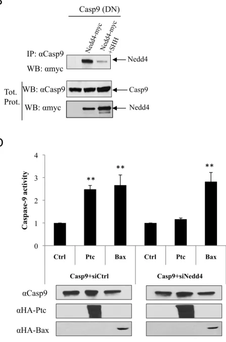
Ptc triggers apoptosis through the recruitment of a caspase-activating complex –the dependosome—that includes DRAL and caspase-9, and leads to direct 9 activation. Nedd4 was shown to be a component of this caspase-activating complex since, in HEK293T co-immunoprecipitation, Nedd4 was pulled down with both DRAL and caspase-9 (Fig.2AB). The presence of SHH, which has been shown to disrupt this caspase-activating complex (9), inhibited the interaction of Nedd4 with caspase-9 (Fig.2B). Moreover, while Ptc triggers an increased caspase-9 activation when Nedd4 was overexpressed, such Ptc-mediated caspase-9 activation was not detected upon forced expression of the Nedd4 catalytic dead mutant (Fig.2C). Conversely Nedd4 silencing by siRNA transfection inhibited caspase-9 activation (Fig.2D), supporting the view that Nedd4 is recruited to the dependosome in the absence of SHH and participates in caspase-9 activation.
We therefore investigated the mechanism by which Nedd4 E3 ligase activity within the dependosome complex promotes caspase-9 activation, and consequently cell death. Considering recent data describing the ubiquitination of caspase-8 as a mechanism for caspase-8 activation (16), we investigated whether caspase-9 activation in the dependosome is associated with caspase-9 ubiquitination. In HEK293T cells forced to express a caspase-9 mutated at its catalytic cysteine (this mutant was used instead of wild-type caspase-9 to avoid caspase amplification), a caspase-9 immunoblot performed after cell extraction with sodium dodecyl sulfate (but not the milder detergent Triton X-100) revealed putative ubiquitination of caspase-9 upon Ptc expression, while Bax, the prototypical inducer of the intrinsic pathway for apoptosis, had no effect (Fig.3A). As expected from the dependence receptor paradigm, the addition of SHH inhibited Ptc-associated caspase-9 ubiquitination. Similarly, enforced expression of Ptc deleted of the pro-apoptotic domain (Ptc 1-1165) was not associated with caspase-9 ubiquitination (Fig.3A). Caspase-9 immunoprecipitation, performed under denaturing conditions to disrupt binding to other proteins, followed by Ub immunoblot, confirmed the ubiquitination of caspase-9 (Fig.3B). Ubiquitination of endogenous caspase-9 was also observed upon Ptc expression after immunoprecipitation of HA-tagged Ub (Fig.3C). To further
analyse whether the caspase-9 recruited to the dependosome is ubiquitinated, we performed Ptc immunoprecipitation and assayed for caspase-9 ubiquitination. As shown in Fig.3D, the Ptc-associated caspase-9 was ubiquitinated. Thus, in the absence of SHH, Ptc triggers ubiquitination of caspase-9 within the dependosome.
Ubiquitination involves covalent attachment of Ub to proteins, and this occurs either through addition of monoubiquitin or polyubiquitin chains linked via internal lysines. While K48-linked polyUb frequently provides a signal for proteasome-mediated degradation, K63-linked chains are more frequently associated with a functional effect on the targeted protein (19). The pattern of ubiquination of Ptc-mediated caspase-9 ubiquitination supports a poly-ubiquitination of caspase-9 (Fig.3ABD), and we therefore investigated the nature of this polyubiquitination. Using specific antibodies recognizing either K48 or K63-linked chain, we observed that Ptc did not trigger any K48-Ub modification of caspase-9, while caspase-9 was clearly covalently linked to K63-Ub (Fig.3E).
To determine whether this 9 K63-ubiquitination could affect caspase-9 activity, we attempted to silence caspase-caspase-9 ubiquitination. Caspase-caspase-9 contains in its P10 fragment 5 putative lysines that represent potential ubiquitination sites (Fig.4A). We therefore mutated these 5 lysines in caspase-9: K394/398/409/410/414R. Enforced expression of Ptc was associated with a strong reduction in caspase-9 ubiquitination when the caspase-9 mutant–i.e., caspase-9 5KR—was used instead of wild-type caspase-9 (Fig.4B). We therefore assessed caspase activation in response to Bax or Ptc expression in the presence of either wild-type caspase-9 or the caspase-9 5KR mutant. A first set of experiments was performed in HEK293T cells. As shown in Fig. 4C, while Ptc triggered caspase-3 activation in wild-type caspase-9 settings, Ptc-induced caspase-3 activation was markedly reduced when caspase-9 5KR was used. The suggested ubiquitination-dependent activation of caspase is specific for Ptc, since Bax triggered caspase activation whether caspase-9 wild-type or 5KR was used (Fig.4D). Since, in the HEK293T cells, endogenous caspase-9 is expressed and may interfere with the ectopically expressed caspase-9, we performed the same experiment in MEF cells deficient for caspase-9 (Suppl. Fig.1D) (20). As shown in Fig.4E, while expression of wild-type caspase-9 allowed Ptc and Bax-induced caspase activation, expression of caspase-9 5KR was associated with
apoptosis induction via Bax but not with Ptc. Thus, Ptc triggers caspase activation via ubiquination of caspase-9. Because caspase-9 cleavage has been shown to play a role in caspase-9 activation and amplification(6), HEK293T cells were forced to express either a non-cleavable form of caspase-9 or a non-cleavable caspase-9 mutated at the 5 lysines that represent potential ubiquitination sites. As shown in Fig. 4F, Ptc triggered caspase-3 activation in uncleavable caspase-9 settings, while it did not when uncleavable caspase-9 5KR was used. Thus, caspase-9 activation mediated by Ptc requires Nedd4 and ubiquitination of caspase-9, but no caspase-9 cleavage.
Together these data support the view that Ptc triggers direct activation of caspase-9 within the dependosome by a mechanism that requires Nedd4 recruitment and caspase-9 ubiquitination. Interestingly, there is a strong analogy with the elegant observation made by Ashkenazi and colleagues, who first described the contribution of cullin3-based polyubiquitination of caspase-8 in caspase-8 activation (16). Interestingly, both type of receptors—the death receptors DR4 and DR5, and the dependence receptor Ptc—recruit at the membrane caspase-activating complexes: the DISC in the former case, and the dependosome in the latter. These then trigger initiator caspase activation, caspase-8 by the death receptors DR4 and DR5, and caspase-9 by the dependence receptor Ptc. It is even more intriguing to note that, while caspase-9 ubiquitination appears to be a prerequisite for caspase-9 activation in the Ptc dependosome, we failed to detect any caspase-9 ubiquitination upon apoptosome activation, and we failed to observe, upon Bax overexpression, any loss of caspase-9 activity using a Ub-dead mutant of caspase-9. Thus, depending on whether an extrinsic or intrinsic pathway is used, two different mechanisms of initiator caspase activation are employed: one requires initiator caspase ubiquitination, be it caspase-8 or caspase-9, and occurs at the membrane; whereas the other is independent of initiator caspase ubiquitination, and occurs in the cytosol. One may wonder what is the role of adding K63 polyubiquitination to promote caspase-9 activation, specifically for the dependosome and not the apoptosome. In the case of caspase-8, ubiquitination was shown to allow caspase-8 translocation from
receptor-associated DISC to Ub-rich foci, although to date there has been no evidence that these Ub-rich foci actively participate in caspase-8 activity. In the case of caspase-9, future biochemical work will need to be performed to define the role of polyubiquitination in caspase-9 catalytic activation.
Experimental Procedures
Site directed mutagenesis and plasmid constructs.
The pRK5-Ptc, pRK5-Ptc-HA, constructs encoding Ptc deleted of its last intracellular domain (pRK5-PtcD7IC) or after caspase cleavage site (D1392) (pRK5-Ptc1-1392) prK5-SHH and pcDNA3-DRAL-3xflagM2 were described in (9). The pcDNA3-Bax-HA, the pcDNA3-Nedd4-myc and the pCMV-Mdm2 were respectively a kind gift from Dr. A. Arrigo Dr. G. Melino and Dr L. Corbo. For direct two-hybrid, the coding sequence of the 4 WW domains of Nedd4 was amplified from pcDNA3-Nedd4-myc by PCR using the primers Nedd4WW-F and Nedd4WW-R (sequences given in Suppl. Table1) and then inserted into pGADT7 plasmid by digestion through EcoRI-XhoI. The pGBKT7-Ptc1165-1392 has been described previously (9). For immunoblot experiments, in order to detect ubiquitination, either pRK5-Ubiquitin-HA or pcDNA3-Ubiquitin-3xflagM2 was used. The pcDNA3-Nedd4C1286S-myc was obtained by directed mutagenesis via Quickchange strategy (Stratagene) on pcDNA3-Nedd4-myc with the primers indicated in Suppl. Table1 (Nedd4C1286S-F and Nedd4C1286S-R). For co-immunoprecipitations, and immunoblots, the pcDNA3-caspase-9 dominant negative (pcDNA3-C9DN) was used for transfection (8). For caspase activity assay active caspase-9 was used (C9) (8). C9DN or pcDNA3-C9 were used as templates to generate the pcDNA3-non cleavable caspase-9 dominant negative (pcDNA3-NCC9-DN) or pcDNA3-non cleavable caspase-9 (pcDNA3-NCC9) constructs by inserting two mutations (D315/330A) via Quickchange strategy (Primers are indicated in Suppl. Table1). The C9DN, pcDNA3-NCC9-DN, pcDNA3-C9 and pcDNA3-NCC9 were then used as templates to generate the mutation of 5 lysines into arginine located at the C-terminus part of the caspase-9 (K394/398/409/410/414R).
Cell cultures, transfection procedures, reagents
Transient transfection of Human Embryonic Kidney 293T cells (HEK293T) was performed with calcium phosphate for co-immunoprecipitation or with Jetprime (Polyplus) for cell death assay and immunoblot according to the manufacturerʼs
instructions. Caspase-9 null Mouse Embryonic Fibroblasts (MEF C9-/-) were a kind gift from Dr. D.Green. MEF cells were cultured in DMEM media supplemented with 10% calf fetal serum and 2µl of β-mercaptoethanol. MEF cells were transfected with Jetprime (Polyplus) for cell death assay. Recombinant SHH-N was from R&D systems and were added at the time of transfection at 600ng/ml. MG132 was used at 0.5 µg/ml for 2h and were purchased from Sigma. For siRNA experiments, cells were transfected with 60 pmols siRNA using Jetprime reagent. Nedd4 and control siRNAs were from Sigma.
Two-hybrid analysis
Matchmaker two-hybrid system III (Clontech) was used according to the manufacturerʼs instructions using AH109 yeast co-transformed with pGBKT7-DNA binding domain GAL4 fused to Ptc 1165-1392 (pGBKT7-Ptc7IC) and the pGADT7-GAL4 transcriptional activation domain AD fused to 4 WW domains of Nedd4 (pGADT7-WWNedd4)(Clontech). Cells were then allowed to grow in the absence of leucine, tryptophane, adenine, and histidine, and in the presence of X-alpha Gal (5-bromo-4-chloro-3-indolyl-D-galactopyranoside (10mg/ml) and 3-amino-1,2,4-triazole (55mM). As a negative control, yeast cells were co-transformed with pGBKT7-Ptc7IC
and empty pGADT7 vector, or with pGADT7-WWNedd4 and empty pGBKT7 vector.
Co-immunoprecipitation and immunoblotting analysis
In cellulo co-immunoprecipitation were carried out on HEK293T cells with various
constructs as described previously (9). For experiments requiring SHH, in addition to transfection with SHH encoding vector, recombinant SHH was added at a final concentration of 600 ng/ml 24 hours before harvesting the cells. HEK293T cells were then lysed in 50 mM HEPES pH 7.6, 150 mM NaCl, 5 mM EDTA and 1% NP-40 in the presence of protease inhibitor cocktail (Roche), and further incubated with Ptc-1 (Santa Cruz Biotechnology, 0.8 μg/ml), FlagM2 (Sigma, 2.4 μg/ml), anti-Myc (Sigma, 2.4 μg/ml) or anti-caspase-9 (Santa Cruz, Biotechnology, 0.8 μg/ml) antibodies and protein-A (Sigma) or protein-G Sepharose (GE Healthcare). Washes were done in 50 mM HEPES pH 7.6, 150 mM NaCl, 5 mM EDTA (4 washes). For western blot and co-immunoprecipitation performed in order to detect ubiquitinated
Caspase-9, HEK293T cells were harvested 48h after transfection. For western blot, cells were lysed in 30 mM Tris pH 7.5, 150 mM NaCl, 10% glycerol and 1% SDS in the presence of protease inhibitor cocktail (Roche). For co-immunoprecipitation, cells were heated in SDS 1% at 95ºC for 5 min and then diluted in 30 mM Tris pH 7.5, 150 mM NaCl, 10% glycerol. Lysates were further incubated with anti-Caspase-9 (Santa Cruz Biotechnology, 0.8 μg/ml) or anti-HA (Sigma, 2.4 μg/ml) antibodies and protein-A Sepharose (Sigma). Washes were performed in 30 mM Tris pH 7.5, 150 mM NaCl, 10% glycerol (4 washes). Immunoblots were performed as previously described (9) using anti-Myc (Sigma, 1/1000), anti-FlagM2 (Sigma, 1/5000), anti-HA (Sigma, 1/10000), anti-Ptc-1 (Santa Cruz Biotechnology, 1/1000), anti-Caspase-9 (Cell Signaling, 1/2000), anti-Caspase-9 (Immunotech, 1/2000), anti-ßactin (Millipore, 1/5000), anti-polyUbiquitin (EnzoLife, 1/2000), anti-Ubiquitin, Lys63-specific (Millipore, 1/2000), anti-Ubiquitin, Lys48-specific (Millipore, 1/2000) and anti-p21 (Dako, 1/1000) antibodies.
Cell death analysis and caspase assays.
Cell death was analyzed 24h after transfection using trypan blue staining procedures. Caspase-3 activity assay was performed 24h after transfection using the caspase-3 fluorometric assay kit (BioVision) as described previously (9). Caspase-9 activity was measured 18h after transfection using the luminescent Caspase-Glo® 9 Assay according to the manufacturerʼs instructions (Promega).
Quantitative Reverse Transcription – Polymerase Chain Reaction.
To assay Nedd4 mRNA expression in HEK293T cells transfected with siRNA against Nedd4, total RNA was extracted from cells with the Nucleospin RNAII kit (Macherey-Nagel, Düren, Germany) and 1μg of RNA was reverse transcribed using the iScript cDNA Synthesis kit (Bio-Rad, Hercules, CA). Real-time Q-RT-PCR was performed on a LightCycler 2.0 apparatus using the LightCycler® TaqMan® Master kit (Roche Applied Science, Basel, Switzerland) according to the manufacterʼs instructions. The ubiquitously expressed ß-actin gene was used as internal control. Primers and probe were given by Universal Probe Library Assay Design Center website (Roche Applied Science). Sequences are shown in Supplementary Table 1.
Statistics.
The statistical significance of differences between groups was evaluated by the Mann – Whitney U test. Mean values for all outcome variables are presented with standard errors of the mean. Data presented are representative of at least four independent experiments. All statistical tests were two-sided, and P values less than 0.05 were considered to be statistically significant.
Acknowledgments:
We wish to thank DE Bredesen for critical reading of the manuscript, AP. Arrigo, D. Green and G. Melino for sharing reagents. This work was supported by institutional grants from the Ligue Contre le Cancer, INCA, ANR, and IP ApoSys.
Correspondence and requests for materials should be addressed to P.M. (e-mail: patrick.mehlen@lyon.unicancer.fr).
References
1. Zornig M, Hueber A, Baum W, & Evan G (2001) Apoptosis regulators and their role in tumorigenesis Biochim Biophys Acta 1551, F1-37.
2. Green D & Kroemer G (1998) The central executioners of apoptosis: caspases or mitochondria? Trends Cell Biol 8, 267-271.
3. Boatright KM & Salvesen GS (2003) Mechanisms of caspase activation Curr
Opin Cell Biol 15, 725-731.
4. Boatright KM, Renatus M, Scott FL, Sperandio S, Shin H, Pedersen IM, Ricci JE, Edris WA, Sutherlin DP, Green DR, et al. (2003) A unified model for apical caspase activation Mol Cell 11, 529-541.
5. Mace PD & Riedl SJ (2010) Molecular cell death platforms and assemblies
Curr Opin Cell Biol 22, 828-836.
6. Pop C, Timmer J, Sperandio S, & Salvesen GS (2006) The apoptosome activates caspase-9 by dimerization Mol Cell 22, 269-275.
7. Riedl SJ & Salvesen GS (2007) The apoptosome: signalling platform of cell death Nat Rev Mol Cell Biol 8, 405-413.
8. Forcet C, Ye X, Granger L, Corset V, Shin H, Bredesen DE, & Mehlen P (2001) The dependence receptor DCC (deleted in colorectal cancer) defines an alternative mechanism for caspase activation Proc Natl Acad Sci U S A 98, 3416-3421.
9. Mille F, Thibert C, Fombonne J, Rama N, Guix C, Hayashi H, Corset V, Reed JC, & Mehlen P (2009) The Patched dependence receptor triggers apoptosis through a DRAL-caspase-9 complex Nat Cell Biol 11, 739-746.
10. Bredesen DE, Mehlen P, & Rabizadeh S (2005) Receptors that mediate cellular dependence Cell Death Differ 12, 1031-1043.
11. Goldschneider D & Mehlen P (2010) Dependence receptors: a new paradigm in cell signaling and cancer therapy Oncogene 29, 1865-1882.
12. Mehlen P & Bredesen DE (2011) Dependence receptors: from basic research to drug development Sci Signal 4, mr2.
13. Wertz IE & Dixit VM (2010) Signaling to NF-kappaB: regulation by ubiquitination Cold Spring Harb Perspect Biol 2, a003350.
14. Sun H, Kapuria V, Peterson LF, Fang D, Bornmann WG, Bartholomeusz G, Talpaz M, & Donato NJ (2011) Bcr-Abl ubiquitination and Usp9x inhibition block kinase signaling and promote CML cell apoptosis Blood 117, 3151-3162. 15. Vucic D, Dixit VM, & Wertz IE (2011) Ubiquitylation in apoptosis: a
post-translational modification at the edge of life and death Nat Rev Mol Cell Biol
12, 439-452.
16. Jin Z, Li Y, Pitti R, Lawrence D, Pham VC, Lill JR, & Ashkenazi A (2009) Cullin3-based polyubiquitination and p62-dependent aggregation of caspase-8 mediate extrinsic apoptosis signaling Cell 137, 721-735.
17. Thibert C, Teillet MA, Lapointe F, Mazelin L, Le Douarin NM, & Mehlen P (2003) Inhibition of neuroepithelial patched-induced apoptosis by sonic hedgehog Science 301, 843-846.
18. Lu X, Liu S, & Kornberg TB (2006) The C-terminal tail of the Hedgehog receptor Patched regulates both localization and turnover Genes Dev 20, 2539-2551.
19. Martinez-Forero I, Rouzaut A, Palazon A, Dubrot J, & Melero I (2009) Lysine 63 polyubiquitination in immunotherapy and in cancer-promoting inflammation
Clin Cancer Res 15, 6751-6757.
20. Marsden VS, O'Connor L, O'Reilly LA, Silke J, Metcalf D, Ekert PG, Huang DC, Cecconi F, Kuida K, Tomaselli KJ, et al. (2002) Apoptosis initiated by Bcl-2-regulated caspase activation independently of the cytochrome c/Apaf-1/caspase-9 apoptosome Nature 419, 634-637.
Figure Legends
Figure 1: Nedd4 is required for Ptc-induced apoptosis.
A. Schematic representation of a two-hybrid screen with the pro-apoptotic domain of
Ptc-1. The Ptc-Nedd4 interaction was confirmed by direct two-hybrid: AH109 yeast transformed either with a Gal4AD plasmid fused with 4 WW domains of Nedd4 and a Gal4BD plasmid fused with the Ptc pro-apoptotic domain (Nedd4+Ptc), with a mock Gal4BD plasmid and a Gal4AD plasmid fused with 4 WW domains of Nedd4 (Nedd4), or with a mock Gal4AD plasmid and a Gal4BD plasmid fused with the Ptc pro-apoptotic domain (Ptc). B, C. Co-immunoprecipitations were performed on HEK293T cells transiently expressing (B) Nedd4-myc alone or with Ptc (Ptc), Ptc-∆7IC (PPtc-∆7IC) or Ptc1-1392 (P1-1392) (C) Ptc-HA alone or with Nedd4-myc in the absence (Nedd4-myc) or in the presence of SHH (Nedd4-myc+SHH). Pull-down with anti-Ptc (B) or anti-myc (C) antibodies was used to immunoprecipitate Ptc and Nedd4, respectively. Nedd4 and Ptc were then revealed by Western blot by using anti-myc and anti-HA antibodies, respectively. Western blot on lysates before pull-down are shown (Tot. prot.). D, E, G, H. Caspase-3 activity assay was performed on HEK293T cells 24h after transfection (D) with empty vector (Ctrl), Ptc encoding vector (Ptc), Nedd4 encoding vector (Nedd4) or Ptc and Nedd4 encoding vectors (Ptc+Nedd4) (E) with Ptc encoding vector (Ptc) or Ptc and Nedd4 encoding vectors (Ptc+Nedd4) in the absence (-) or in the presence (+) of MG132 (G) with empty vector (Ctrl), Ptc encoding vector (Ptc), Nedd4 encoding vector (Nedd4), Ptc and Nedd4 encoding vectors (Ptc+Nedd4), Nedd4C1286S encoding vector (Nedd4 C1286S) or Ptc and Nedd4C1286S encoding vectors (Ptc+Nedd4C1286S) (H) with empty vector (Ctrl) or Ptc encoding vector (Ptc), in the presence of siRNA control (siCtrl) or siRNA Nedd4 (siNedd4). (D, G, H) Anti-HA (αHA-Ptc) and anti-myc (αmyc-Nedd4) immunoblot are shown as a control of specificity and loading. F. Cell death was analyzed by trypan blue exclusion assay in HEK293T cells 24h after transfection of an empty vector (Ctrl), a Ptc encoding vector (Ptc), a Nedd4 encoding vector (Nedd4) or Ptc and Nedd4 encoding vectors (Ptc+Nedd4). For all caspase-3 activity and cell death assays, folds over control of each experiment are represented and error bars are standard errors of the mean. *; p<0.05, **; p<0.01 and ***; p<0.001
Figure 2: Nedd4 is part of the dependosome and regulates caspase-9 activation.
A. DRAL immunoprecipitation (IP:αflag) was performed on HEK293T cells
transfected with either Nedd4 alone or Nedd4 and DRAL together (DRAL-flag). DRAL interaction with Nedd4 was revealed by Western blot using an anti-myc (Nedd4-myc) antibody. Western blot on lysates before pull-down are shown (Tot. prot.). B. Co-immunoprecipitations were performed on HEK293T cells transiently expressing DN-Caspase-9 alone or with Nedd4-myc in the absence (Nedd4-myc) or in the presence of SHH (Nedd4-myc+SHH). Western blot on lysates before pull-down are shown (Tot. prot.). C, D. Caspase-9 activity assay was performed using a proluminogenic 9 substrate 18h after transfection of HEK293T cells with wild-type caspase-9 together (C) with Nedd4 expressing construct or Nedd4C1286S encoding vector or (D) with siRNA control (C9+siCtrl) or siRNA Nedd4 (C9+siNedd4), in the absence (Ctrl) or in the presence of Ptc (Ptc). Anti-HA (αHA-Ptc and αHA-Bax), anti-Caspase-9 (αCanti-Caspase-9) and anti-myc (αmyc-Nedd4) immunoblots are shown as a control of specificity and loading. Folds over control are represented and error bars are standard error of the mean. **; p<0.01
Figure 3: Caspase-9 is polyubiquitinated during Ptc-induced apoptosis.
A. Western blot using caspase-9 antibody was performed on HEK293T cells lysed in
lysis buffer containing 1% SDS, 48h after transient transfection of DN-caspase-9 (DN-C9) together with an empty vector, with Bax encoding vector (Bax-HA), with Ptc encoding vector in the absence (Ptc-HA) or in the presence (Ptc-HA+SHH) of SHH, or with Ptc-∆7IC encoding vector (P∆7IC). ß actin (αß actin), Ptc (αPtc) and Bax (αHA) immunoblot are shown as a control of loading. B, E. Immunoprecipitation of caspase-9 (IP: αCasp9) was performed on SDS-lysed HEK293T cells expressing DN-caspase-9 (DN-C9) and Ubiquitin proteins in the absence or in the presence of Ptc (Ptc). Ubiquitinated Caspase-9 (Ub) was revealed by Western blot using an anti-ubiquitin (B) or anti-K48-anti-ubiquitin and anti-K63-anti-ubiquitin (E) antibodies. Western blots on lysates before pull-down are shown (Tot. prot.). C. Immunoprecipitation of HA-ubiquitinated proteins (IP αHA-Ub) was performed on HEK293T cells lysed in the
presence of SDS and expressing HA-ubiquitin encoding vectors in the absence or in the presence (Ptc) of Ptc. Endogenous caspase-9 was revealed by Western blot using an anti-caspase-9 antibody. Western blot on lysates before pull-down are shown (Tot. prot.). D. Ptc immunoprecipitation (IP α-HA-Ptc) was performed on HEK293T cells expressing DN-caspase-9 (DN-C9) and Ubiquitin proteins in the absence or in the presence of Ptc (Ptc-HA). Ubiquitinated caspase-9 was revealed by Western blot using an anti-caspase-9 antibody. Western blot on lysates before pull-down are shown (Tot. prot.).
Figure 4: Ubiquitination of caspase-9 is required for Ptc-induced caspase activation.
A. Schematic representation of Caspase-9. The position of 5 lysines mutated in
C9-5KR constructs is indicated. B. Anti-caspase-9 immunoblot was performed on HEK293T cells transfected with wild-type caspase-9 (C9) or caspase-9 mutated on 5 lysines (C9-5KR) in the absence (Ctrl) or in the presence of Ptc (Ptc). Anti-HA (αHA-Ptc) immunoblot is shown as a control of loading. C-F. Caspase-3 activity assay was performed in HEK293T cells (C,D,F) or in Mouse embryonic Fibroblast deficient for caspase-9 (MEF C9-/-) (E), 24h after transfection with wild-type caspase-9 (C9), caspase-9 mutated on 5 lysines (C9-5KR) or non cleavable caspase-9 (NCC9) together with either an empty vector (Ctrl), a Ptc encoding vector (Ptc) or a Bax encoding vector (Bax). Anti-HA (αHA-Ptc and αHA-Bax) and anti-caspase-9 (αC9) immunoblots are shown as controls of specificity and loading. Folds over control are represented and error bars are standard error of the mean. **; p<0.01
Supp. Figure 1: Controls for Nedd4 specificity.
A. Immunoblot using p21 antibody on HEK293T cells transfected with an empty
vector (Ctrl) or with Ptc encoding vector (Ptc), Nedd4 encoding vector (Nedd4) or Ptc and Nedd4 encoding vectors (Ptc+Nedd4) in the absence or in the presence of MG132 (0,5 µg/ml). B. Caspase-3 activity assay was performed on HEK293T cells 24 h after transient transfection with an empty vector (Ctrl) or with Ptc encoding vector (Ptc), Mdm2 encoding vector (Mdm2) or Ptc and Mdm2 encoding vectors (P+mdm2). Folds over control are represented and error bars are standard error of
the mean. **; p<0.01. C. Nedd4 mRNA relative expression assessed by Real-time Q-RT-PCR in HEK293T cells transfected with siRNA control (siCtrl) or siRNA Nedd4 (siNedd4) in the absence (Ctrl) or in the presence of Ptc (Ptc). D. Immunoblot performed on wild-type (WT) and Caspase-9 -/- MEF cells (Casp9-/-) using Caspase- 9 and ß-actin antibodies. ß-actin was used as control of loading.
Supp. Table 1. Oligonucleotides used in the manuscript.
The oligonucleotides used for direct mutagenesis and Real-time Q-RT-PCR are shown.
0 1 2 3 4 5 Ctrl Ptc Ctrl Ptc siCtrl siNedd4 C as p as e-3 ac ti vi ty 0 10 20 30 40 50 60 70 80 Ctrl Ptc Nedd4 Ptc + Nedd4 % c el l d eath 0 4 8 12 16 Ctrl Ptc Nedd4 Ptc +Nedd4 C as p as e-3 ac ti vi ty
Figure 1
A
Ptc D1392 Mouse fetal brain cDNA librarypro-apoptotic domain 7IC 1165
F
WB: αmyc WB: αmyc Tot. Prot. Nedd4-myc *** ** *D
αHA-PtcH
αmyc-Nedd4 αHA-PtcG
αHA-Ptc Ptc-HA IP: αmyc WB: αHA WB: αmyc WB: αHA Tot. Prot. ** ** * 0 2 4 6 8 10 12 14 Ctrl Ptc Nedd4 Ptc +Nedd4 Nedd4 C1286S Ptc +Nedd4 C1286S C as p as e-3 ac ti vi ty WB: αPtc IPα Ptc *** ** ** ** 0 2 4 6 8 Ptc Ptc +Nedd4 Ptc Ptc +Nedd4 - MG132 C as p as e-3 ac ti vi ty * *E
αmyc-Nedd4 Nedd4 Ptc Nedd4 PtcΔ7ICC
Ptc Ptc Nedd4 Ptc + Nedd4 Ptc Nedd4B
Figure 2
WB: αflag WB: αmyc IP: αflag WB: αmyc Tot. Prot . Nedd4-mycA
B
D
Nedd4 Nedd4 DRAL IP: αCasp9 WB: αmyc WB: αmyc WB: αCasp9 Tot. Prot. Nedd4 Nedd4 Casp9 Casp9 (DN) ** αCasp9 αHA-Ptc ** 0 1 2 3 4 Ctrl Ptc Bax Ctrl Ptc Bax Casp9+siCtrl Casp9+siNedd4 C as p as e-9 ac ti vi ty ** αHA-BaxC
αCasp9 αHA-Ptc αmyc-Nedd4 ** 0 1 2 3 4 5Nedd4 Ptc+Nedd4 Nedd4 C1286S Ptc+Nedd4 C1286S C as p as e-9 ac ti vi ty
Casp9 (DN) Ub-Casp9
C
A
Casp9 WB: αCasp9 WB: αß actin WB: αHA Bax Ptc WB: αPtc Casp9 (DN) + Ub IgG IP: αCasp9 WB: αUb Ub Tot. Prot. Casp9 WB: αPtc WB: αCasp9 PtcB
Figure 3
D
Ub-HA IP: αHA WB: αCasp9 WB: αCasp9 WB: αPtc Endogenous Casp9 Endogenous Casp9 Ptc Tot. Prot. Casp9(DN)+Ub IP: αHA WB: αCasp9 Tot. prot Casp9 WB: αHA WB: αCasp9 Ptc Casp9 Ub-Casp9 ß actinE
IP: αCasp9 WB: αK48-Ub Casp9(DN)+Ub IP: αCasp9 WB: αK63-Ub Casp9(DN)+Ub Tot. Prot. WB: αHA WB: αCasp9 Casp9 Ptc K63-Ub IgG IgG PtcΔ7IC0 4 8 12 16 Ctrl Bax Ctrl Bax Casp9 Casp9-5KR C as p as e-3 ac ti vi ty 0 1 2 3 4 Ctrl Ptc Bax Ctrl Ptc Bax Casp9 Casp9-5KR C as p as e-3 ac ti vi ty
Figure 4
B
Casp9 WB: αCasp9 WB: αHA Casp9 Casp9-5KR αCasp9 αHA-Ptc 0 2 4 6 Ctl Ptc Ctl Ptc Casp9 Casp9-5KR cas p as e-3 ac ti vi ty ** 0 2 4 6 Ctl Ptc Ctl Ptc NC Casp9 NC Casp9-5KR C as p as e-3 ac ti vi ty ** αCasp9 αHA-PtcC
Ptc ** ** αCasp9 αHA-BaxD
F
HEK293T HEK293T HEK293TA
prodomain D315 D330 p35 p10 p37 AISSLPTPSDIFVSYSTFPGFVSWRDPKSGSWYVETLDDIFEQWAHSEDLQSLLLRVANAVSVKGIYKQMPGCFNFLRKKLFFKTSE
αCasp9 αHA-Ptc αHA-Bax ** ** ** MEF C9-/-0 2 4 6 Ctrl Ptc Mdm2 Ptc+Mdm2 Ca sp ase-‐3 a cGvi ty
Suppl Figure 1
A
MG132 p21 WB: αp21 WB: αß actinB
C
ß acBnD
WB: αCasp9 WB: αß actin MEF ** ** 0 1 2 3 Ctrl Ptc Ctrl Ptc siCtrl siNedd4 N ed d 4 mR N A e xp re ss ion (A .U x 10 3)Nedd4WW-F 5’-CTCCTCTACCTCCAGGGTGGGAAGAGA-‐3’ Nedd4WW-R 5’-CACCCTCGAGCAATCTTGGATCTTCCCAGGTGGTGGT-‐3’ Nedd4C1286S -F 5’-AGCTGCCAAGAGCTCATACCTCATTTAATCGCCTGGACTTGCC -3' Nedd4C1286S-R 5’-GGCAAGTCCAGGCGATTAAATGAGGTATGAGCTCTTGGCAGCT -3' NCC9-D315A -F 5’-CTGGCAGTAACCCCGAGCCAGCTGCCACCCCGTTCCAGGAAGGTTT -3' NCC9-D315A -R 5’-AAACCTTCCTGGAACGGGGTGGCAGCTGGCTCGGGGTTACTGCCAG -3' NCC9-D330A -F 5-GTTTGAGGACCTTCGACCAGCTGGCCGCCATATCTAGTTTGCCCACACCCA -3 NCC9-D330A -R 5-TGGGTGTGGGCAAACTAGATATGGCGGCCAGCTGGTCGAAGGTCCTCAAAC -3 TaqNedd4-F 5’-GGCTCAGAAGATGATAATGCAGA-3' TaqNedd4-R 5’-GCATCTGGTTGGTCCAAAAC-3' Taqβactin-F 5’-ATTGGCAATGAGCGGTTC-3' Taqβactin-R 5’-GGATGCCACAGGACTCCAT-3'
Probe TaqNedd4 5’-GGCTGAGG-3’
Probe Taqβactin 5’-CTTCCAGC-3’