HAL Id: hal-02373675
https://hal.archives-ouvertes.fr/hal-02373675
Submitted on 18 Jun 2020HAL is a multi-disciplinary open access archive for the deposit and dissemination of sci-entific research documents, whether they are pub-lished or not. The documents may come from teaching and research institutions in France or abroad, or from public or private research centers.
L’archive ouverte pluridisciplinaire HAL, est destinée au dépôt et à la diffusion de documents scientifiques de niveau recherche, publiés ou non, émanant des établissements d’enseignement et de recherche français ou étrangers, des laboratoires publics ou privés.
metabolism of the marine diatom Chaetoceros neogracile
Marta Seoane, Carmen González-Fernández, Philippe Soudant, Arnaud
Huvet, Marta Esperanza, Ángeles Cid, Ika Paul-Pont
To cite this version:
Marta Seoane, Carmen González-Fernández, Philippe Soudant, Arnaud Huvet, Marta Esperanza, et al.. Polystyrene microbeads modulate the energy metabolism of the marine diatom Chaetoceros neogracile. Environmental Pollution, Elsevier, 2019, 251, pp.363-371. �10.1016/j.envpol.2019.04.142�. �hal-02373675�
Environmental Pollution
August 2019, Volume 251, Pages 363-371 https://doi.org/10.1016/j.envpol.2019.04.142 https://archimer.ifremer.fr/doc/00492/60415/
Archimer
https://archimer.ifremer.frPolystyrene microbeads modulate the energy metabolism of
the marine diatom Chaetoceros neogracile
Seoane Marta 1, 2, *, González-Fernández Carmen 2, Soudant Philippe 2, Huvet Arnaud 3,
Esperanza Marta 1, Cid Ángeles 1, Paul-Pont Ika 2
1 Laboratorio de Microbiología, Facultad de Ciencias, Universidade da Coruña, Campus da Zapateira
s/n, 15071, A Coruña, Spain
2 Laboratoire des Sciences de l’Environnement Marin (LEMAR), UMR 6539 CNRS/UBO/IRD/IFREMER,
Institut Universitaire Européen de la Mer (IUEM), Technopôle Brest-Iroise, Rue Dumont d’Urville, 29280, Plouzané, France
3 Ifremer, Laboratoire des Sciences de l’Environnement Marin (LEMAR, UMR 6539
CNRS/UBO/IRD/IFREMER), Centre Bretagne, CS 10070, 29280, Plouzané, France * Corresponding author : Marta Seoane, email address : marta.seoane@udc.es
Abstract :
Due to the growing concern about the presence of microplastics (MP) in the environment, the number of studies evaluating the toxicity of these small persistent particles on different marine species has increased in recent years. Few studies have addressed their impact on marine phytoplankton, a subject of great concern since they are primary producers of the aquatic food web. The aim of this study is to unravel the cytotoxicity of 2.5 μg mL−1 unlabelled amino-modified polystyrene beads of different sizes (0.5 and 2 μm) on the marine diatom Chaetoceros neogracile. In addition to traditional growth and photosynthesis endpoints, several physiological and biochemical parameters were monitored every 24 h in C. neogracile cells by flow cytometry during their exponential growth (72 h). Dynamic Light Scattering measurements revealed the strong aggregation and the negative charge of the beads assayed in the culture medium, which seemed to minimize particle interaction with cells and potentially associated impacts. Indeed, MP were not attached to the microalgal cell wall, as evidenced by scanning electron micrographs. Cell growth, morphology, photosynthesis, reactive oxygen species levels and membrane potential remained unaltered. However, exposure to MP significantly decreased the cellular esterase activity and the neutral lipid content. Microalgal oil bodies could serve as an energy source for maintaining a healthy cellular status. Thus, MP-exposed cells modulate their energy metabolism to properly acclimate to the stress conditions.
Graphical abstract
Highlights
► Effects of 0.5 and 2 μm PS-NH2 microplastics (MP) were evaluated on C. neogracile. ► MP showed
negative charge, were aggregated and were not attached to the cell wall. ► Exposure to MP decreased the cellular metabolic activity and neutral lipid content. ► Cells modulate their energy metabolism to properly acclimate to the stress conditions. ► Microalgal oil bodies serve as an energy source for maintaining a healthy status.
M
AN
US
CR
IP
T
AC
CE
PT
ED
1. Introduction 44Plastic wastes constitute a major portion of marine litter (Ruiz-Orejón, 2016). Synthetic 45
polymers have excellent properties for many packaging applications and manufacturing 46
processes that have led to their increasing use throughout the last decades, reaching an 47
annual production of 335 million tons in 2016 (Plastics Europe, 2017). However, the 48
characteristics that make plastics useful materials (low cost, high durability, low density) also 49
make them a menace to the environment (Ryan, 2015). Plastics may persist in the 50
environment for many years and can be easily dispersed by oceanic currents even to remote 51
areas of the world, far away from the source of contamination (Barnes et al., 2009; Peeken et 52
al., 2018; Pham et al., 2014; Ryan et al., 2009). 53
The concern of the scientific community about plastic pollution and its impacts on marine 54
organisms has increased in recent years especially in regards with the high proportion of small 55
persistent particles commonly called microplastics (MP) (Andrady, 2015). MP are defined as 56
plastic particles with a size smaller than 5 mm (NOAA, 2008). Lately, a new category of plastic 57
debris, named nanoplastics (NP), was described as particles <100 nm (Galloway et al., 2017) or 58
<1 μm (Gigault et al., 2018). In this study, we will keep the NP definition as particles <100 nm 59
and therefore, considering MP as particles from 100 nm to 5 mm in size. Both MP and NP may 60
originate from the fragmentation of larger plastic debris (e.g., bags, bottles and fishing nets) in 61
the marine environment caused by a combination of physical and chemical processes such as 62
mechanical abrasion, photochemical and thermo-oxidation, hydrolysis or even biological 63
degradation (Dawson et al., 2018; Gigault et al., 2016; Lambert and Wagner; 2016). In addition, 64
MP could also reach the aquatic environment directly since many products used plastics at the 65
micrometric size (e.g., personal care and household cleaning products or microfibers from 66
synthetic clothing) (Andrady, 2015), while the increased use of NP in diverse industries 67
(cosmetics, drugs, lubricants) may lead to their release in the environment (Hernandez et al., 68
2017; Lusher et al., 2017). MP have been found to be ubiquitously present in all environmental 69
compartments of the aquatic environment, from surface waters and water column to deep-sea 70
sediments (Reviewed in Paul-Pont et al., 2018). In reference to object counts, MP constitute 71
more than 92 % of floating plastics in the oceans (Cole et al., 2011; Eriksen et al., 2014). 72
Regarding reported environmental concentrations, some of the highest have been detected in 73
the southern North Sea reaching 1,700,000 items m-3 (∼ 8.5 mg L-1) for plastic particles > 80 74
µm (Dubaish and Liebezeit, 2013). However, due to the unavailability of methods, there are no 75
field determinations of MP as small as those used in most experimental studies (< 20 µm; 76
Filella, 2015). For nanoplastics, no quantitative data exists in situ and only indirect evidence of 77
their presence has been debated recently (Ter Halle et al., 2017). 78
M
AN
US
CR
IP
T
AC
CE
PT
ED
Exposure laboratory experiments do not reflect the complexity of the marine environment; 79
however, they may contribute to disclose and aware of the effect of plastics debris on marine 80
organisms (Paul-Pont et al., 2018; Phuong et al., 2016). Several studies have investigated how 81
MP can be ingested by aquatic animals leading to negative consequences for them and for the 82
aquatic food chain (Yokota et al., 2017). Nevertheless, there is still a scarcity of knowledge 83
about the impacts of MP on phytoplankton, which constitutes the basis of the aquatic food 84
webs. In particular, diatoms are considered one of the most diverse and ecologically important 85
phytoplanktonic groups and are responsible for approximately 20 % of the overall primary 86
production on Earth, playing a central role in the biogeochemical cycling of important nutrients 87
(Malviya et al., 2016; Rosenwasser et al., 2014). 88
The aim of this study was to assess the potential effects of two different sizes (0.5 μm and 2 89
μm) of unlabelled polystyrene (PS) beads on the marine diatom Chaetoceros neogracile, trying 90
to disclose differential impacts depending on particle size, as smaller particles are expected to 91
be more toxic (Sjollema et a., 2016). To perform the experiments, PS beads with amino (-NH2) 92
surface modifications were used at 2.5 μg mL-1, concentration set to compare with the 72 h 93
EC50 value of 50 nm PS-NH2 NP (data not shown). PS is one of the most used plastics worldwide 94
and it is one of the plastic polymers type most frequently detected as micro-debris in marine 95
environments (Andrady, 2011). PS-NH2 beads have been amply used as model particles in 96
ecotoxicology and have been shown to cause severe damages on different organisms of the 97
aquatic trophic chain (Bergami et al., 2017; Bhattacharya et al., 2010; Canesi et al., 2017; 98
Manfra et al., 2017; Marques-Santos et al., 2018; Pinsino et al., 2017; Tallec et al., 2018). 99
Particle behaviour was measured in Milli-Q water, seawater and in the culture medium using 100
Dynamic Light Scattering (DLS) to assess potential particle aggregation or changes in the 101
particle surface charge. To assess PS-NH2 MP effects on C. neogracile several physiological and 102
biochemical parameters such as cell morphology, autofluorescence, esterase activity, reactive 103
oxygen species (ROS) levels, cytoplasmic membrane potential and neutral lipid content were 104
monitored by flow cytometry (FCM) in addition to traditional growth and photosynthesis 105
endpoints. Furthermore, potential structural damages and disposition or adsorption of 106
unlabelled PS-NH2 beads on microalgal surface were determined using scanning electron 107
microscopy (SEM). 108
2. Materials and methods 109
2.1. Microalgal cultures 110
Chaetoceros neogracile (Bacillariophyceae) was obtained from the Culture Collection of Algae
111
and Protozoa of the Scottish Marine Institute (strain CCAP 1010-3). C. neogracile is a non-112
motile centric marine diatom (width = 4 µm, length = 7 µm) encased in lightly siliceous valves 113
M
AN
US
CR
IP
T
AC
CE
PT
ED
(frustules), covered with an organic coating (Hecky et al., 1973). This species was selected 114
based on its predominance in marine phytoplankton communities (Malviya et al., 2016). Stock 115
microalgal cultures were maintained in filtered (pore size: 0.2 μm) and autoclaved natural 116
seawater (FSW) supplied with 1 mL L-1 of Conway medium (Walne, 1966) and enriched with 117
silica (Na2SiO3 · 9H2O) (1.07 10-4 M). Flasks were kept at 20 °C, with continuous aeration and 118
light at 100 µmol photon m−2s−1. CO
2 was supplied to keep the pH between 7.5 and 7.9. 119
2.2. Particle characterization 120
Unlabelled 0.5 μm and 2 μm PS-NH2 particles were purchased from Micromod (Micromod 121
Partikeltechnologie GmbH). Both particles were characterized by Dynamic Light Scattering 122
(DLS) using Zetasizer Nano Series ZS (Malvern instruments) as conducted by Tallec et al. 123
(2018). Size (Z-average in nm), charge (ζ-potential in mV) and aggregation state (Polydispersity 124
Index, PdI in arbitrary units (a.u.)) were measured in Milli-Q water, FSW and also in filtered (0.2 125
μm) microalgal culture medium. When PdI exceeds 0.2 a.u. particles were considered to be 126
aggregated. Measurements were carried out in triplicate, each containing 13 runs of 10 s for Z-127
average and 20 runs, 3 s delay for ζ-potential following the protocol described in Della Torre et 128
al. (2014). Data were analyzed using Zetasizer Nano Series software, version 6.20. 129
2.3. Microalgal exposure to microplastics 130
Microalgae cells were exposed to both particles in different batch cultures for 72 h, during the 131
exponential growth phase, as recommended in most standardized growth inhibition tests with 132
microalgae (OECD 201, 2011). It has also been shown that during exponential growth phase 133
MP impaired more drastically the major cellular and physiological parameters (Mao et al., 134
2018). Exposures were performed in triplicates in 500 mL glassware balloon flasks filled with 135
300 mL of microalgal culture under the same environmental conditions as stock cultures. 136
Microalgal cells in early exponential growth phase were used as inoculum and initial cell 137
density was adjusted to 4 x 10⁵ cells mL−1. The MP concentration used (2.5 µg mL-1) 138
corresponds to the 72 h EC50 value of 50 nm PS-NH2 NP (data not shown) and was established 139
to compare results with other laboratory studies (Long et al., 2017; Mao et al., 2018; Zhang et 140
al., 2017), but not as high as in most published works, so that we could get closer to worst 141
environmental scenarios (Paul-Pont et al., 2018). This same mass concentration for the two 142
beads tested results in differences in the nominal concentration in number of particles per mL, 143
corresponding to 3.7 x 10⁷ particles mL-1 for the 0.5 μm MP and to 5.7 x 10⁵ particles mL-1 for 144
the 2 μm MP. Before the experiment, MP stock solutions were diluted in Milli-Q water. The 145
volume of the diluted MP solutions added to microalgal cultures to reach the final MP 146
concentration used for the assay (2.5 μg mL-1) never exceeded 0.3 % of the final culture 147
volume. Therefore, in the whole process an approximate dilution of 1/20000 of the 148
M
AN
US
CR
IP
T
AC
CE
PT
ED
commercial MP stock was made. Analyses were performed in fresh samples every 24 h during 149
the 72 h of the test. Additionally, after 48 h of exposure, halfway in the experiment, control 150
and exposed samples were fixed for microscopy observations. 151
2.4. Scanning electron microscopy 152
Samples of control and MP exposed cultures were diluted to 105 cells mL-1 and fixed in 6% 153
glutaraldehyde in 0.1 M sodium cacodylate buffer (1.75% w/v of NaCl, pH 7.2). Suspensions 154
were incubated for 2 hours at 4 °C before being filtered through polycarbonate filters 155
(Nucleopore PC) with a 2 μm pore size. Then, samples were prepared for scanning electron 156
microscopy (SEM) observation following the method previously described by Foulon et al. 157
(2016). Finally, samples were observed with a Hitachi S-3200N microscope. 158
2.5. Flow cytometric analyses 159
Flow cytometric (FCM) analyses were performed using an Easy-Cyte Plus 6HT flow cytometer 160
(Guava Merck Millipore®) equipped with a 488 nm argon excitation laser, detectors of forward 161
(FSC) and side (SSC) light scatter and three fluorescence detectors: green (525 nm ± 15), yellow 162
(583 nm ± 13) and red (680 nm ± 15). All fluorescence measurements were obtained in a 163
logarithmic scale and data were computed as the mean fluorescence value of the cell 164
population in arbitrary units (a.u.). Collected data from the FCM were analyzed with the 165
software InCyte (Millipore). 166
2.5.1. Cellular density and growth rate 167
Direct absolute cell counts were carried out daily by FCM to determine the cellular density of 168
control and treated cultures. Growth rates (µ) were also calculated as described in Seoane et 169
al. (2017a). 170
2.5.2. Morphological parameters and chlorophyll a fluorescence 171
Forward light scatter (FSC) and side light scatter (SSC) values were measured and used as 172
estimates of morphological changes. Natural red autofluorescence, related to chlorophyll a 173
content, was also analysed. 174
2.5.3. Esterase activity 175
Esterase activity in C. neogracile cells was studied using the fluorescein diacetate (FDA) 176
cytometric assay previously described in Seoane et al. (2017b). Cells were incubated with FDA 177
at a final concentration of 6 μM for 10 min at room temperature in the dark. FDA is a non-178
fluorescent, non-polar lipophilic molecule that diffuses across cell membranes. After entering 179
the cell, its acetate residues are cleaved off by non-specific esterases and the polar hydrophilic 180
fluorescent product fluorescein is retained by cells with intact plasma membranes. Since 181
fluorescein is accumulated by viable and active cells, esterase activity is measured by means of 182
the green fluorescent intensity emitted (Prado et al., 2009). 183
M
AN
US
CR
IP
T
AC
CE
PT
ED
2.5.4. Oxidative stress: intracellular levels of reactive oxygen species 184
Reactive oxygen species (ROS) content was measured using 2,7-dichlorofluorescein diacetate 185
(DCFH-DA) as described in González-Fernández et al. (2018). The cells of C. neogracile were 186
incubated with DCFH-DA at a final concentration of 10 μM for 50 min at room temperature in 187
the dark. DCFH-DA is a cell-permeable non-florescent probe that is hydrolysed (de-esterified) 188
intracellularly to form the highly green fluorescent DCF upon oxidation with ROS. Thus, the 189
green fluorescence measured is quantitatively related to the ROS content in cells. 190
2.5.5. Cytoplasmic membrane potential 191
Potential changes in cytoplasmic membrane potential were determined using the bis-(1,3-192
dibutylbarbituric acid) trimethine oxonol (DiBAC4(3)) following the protocol previously 193
described by Seoane et al. (2017a). Cells were incubated with DiBAC4(3) at a final 194
concentration of 2 μM for 10 min at room temperature in the dark. DiBAC4(3) can enter 195
depolarized cells where it binds to intracellular proteins or membranes showing green 196
fluorescent emission (Wolff et al., 2003). 197
2.5.6. Neutral lipid content 198
Neutral lipid content was assessed using the specific lipid droplet stain BODIPY 493/503. This 199
highly lipophilic neutral dye easily goes through cell and organelle membranes and 200
accumulates in intracellular oil-containing organelles, known as lipid bodies, showing green 201
fluorescence. BODIPY has proven to be very effective for lipid measurement in microalgae 202
(Govender et al., 2012; Rumin et al., 2015). Cells of C. neogracile were incubated with BODIPY 203
493/503 at a final concentration of 10 μM for 10 min at room temperature in the dark. 204
2.6. Photosynthetic efficiency 205
The effective quantum yield (QY) of photochemical energy conversion in photosystem II (PSII) 206
was measured by Pulse Amplitude Modulation (PAM) fluorometry using an AquaPen-C AP-207
C 100 fluorometer (Photon Systems Instruments, Czech Republic) equipped with a blue (455 208
nm) LED excitation light. Aliquots of each culture were dark-adapted for 30 minutes before 209
measurements. 210
2.7. Statistical analysis of results 211
Statistical analysis was performed using IBM SPSS Statistics software 21.0. Mean fluorescence 212
values and standard deviation (SD) of the three biological replicates were determined for 213
exposed and control cultures. Data were checked for normal distribution (Shapiro-Wilk test) 214
and homogeneity of variance (Levene test). One-way analysis of variance (ANOVA) was 215
performed to test for differences among treatments at each sampling time. When significant 216
differences were observed, multiple comparisons among treatments were made using the 217
Tukey´s post hoc test. For DLS data, pairwise comparisons were also made to compare particle 218
M
AN
US
CR
IP
T
AC
CE
PT
ED
behaviour (size and charge) between the three media (Supp. Table 1). A p - value < 0.05 was 219
considered statistically significant for all the analyses. 220
3. Results and discussion 221
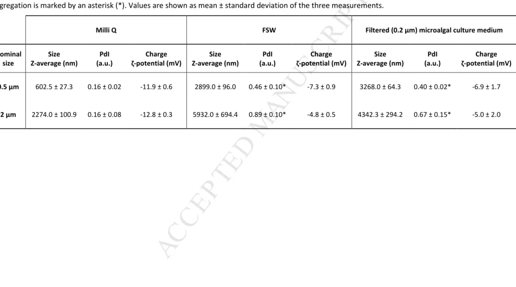
3.1. Particle characterization: actual size, charge and aggregation 222
DLS analysis of the MP suspended in Milli-Q water confirmed the approximate size indicated 223
by the commercial supplier for the two particle sizes used, with Z-averages of 602.5 ± 27.3 and 224
2274.0 ± 100.9 nm, which correspond to the nominal sizes of 0.5 μm and 2 μm, respectively 225
(Table 1). Data also showed an optimal dispersion and stability in Milli-Q water since 226
aggregation was negligible for both particles, as suggested by a PdI < 0.2 (Table 1). However, 227
plastic particles showed a significant increase in Z-average reaching values of 2899.0 ± 96.0 nm 228
and 5932.0 ± 694.4 nm when suspended in FSW and values of 3268.0 ± 64.3 nm and 4342.3 ± 229
294.2 nm in microalgae medium for 0.5 μm and 2 μm PS-NH2 beads, respectively (Table 1; 230
Supp. Table 1). These data indicated an increase in particle size congruent with the observed 231
PdI values (>0.2) that indicated aggregation (Table 1). 232
Regarding the charge, both 0.5 μm and 2 μm PS-NH2 MP showed negative ζ-potential in the 233
three media assayed (Table 1) although a positive charge was expected since the beads used 234
presented amino surface modifications. Therefore, particle characteristics must be assessed 235
prior to performing laboratory exposure to properly interpret particle behaviour and toxicity. 236
Such difference of charge has also been reported by Sun et al. (2018) with 1 μm PS-NH2 beads 237
and by González-Fernández et al. (2018) and Lundqvist et al. (2008) with 100 nm PS-NH2 NP. 238
These discrepancies may derivate from the manufacturing process which may change 239
according to the commercial suppliers. More information and measurements regarding 240
physico-chemical properties of MP is thus a prerequisite for a better interpretation of their 241
behaviour and biological impact in different solutions (González-Fernández et al., 2018). 242
Significant lower absolute values of ζ-potential (closer to zero) were obtained for both particles 243
in FSW and in the culture medium as compared to the values in Milli-Q water (Table 1; Supp. 244
Table 1). These results are in accordance with the results of size and particle aggregation 245
obtained. The ζ-potential is a key indicator of the stability of colloidal dispersions. The 246
magnitude of this value indicates the degree of electrostatic repulsion between particles. 247
Particle solutions with high ζ-potential (negative or positive) are more stable and greatly 248
dispersed, while particles with a ζ-potential between -10 and +10 mV are less stable and tend 249
to aggregate. The high concentration of NaCl and other ions in FSW as well as the presence of 250
proteins or other natural organic matter in the filtered microalgae medium are likely 251
interacting with the particle surface group eliminating the repulsion forces that maintain 252
particles isolated and promoting aggregation (Canesi et al., 2017; Paul-Pont et al., 2018). These 253
M
AN
US
CR
IP
T
AC
CE
PT
ED
surface interactions can explain particle behaviour changes (surface charge, size and 254
aggregation state) observed in FSW and filtered microalgae medium as compared to particles 255
in Milli-Q water (Supp. Table 1). In environmental media, surface charge neutralization and 256
increases in particle size are often observed, due to the formation of a protein coating (called 257
ecocorona; Galloway et al., 2017) on the particle surface (Marques-Santos et al., 2018). 258
Therefore, the medium and related parameters should be always taken into account for 259
particle characterization (Della Torre et al., 2014). 260
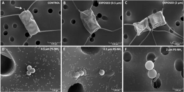
3.2. Scanning electron microscopy observations 261
Micrographs of C. neogracile cells exposed to both MP sizes were presented, evidencing intact 262
frustules, similar to control cells, without MP attached at the surface cell (Fig. 1A, B, C). Most 263
cells showed broken setae, located close to the cell, likely because of the force exerted during 264
the filtration process required for sample preparation for SEM observation. Several studies 265
showed the adsorption of plastic micro and nanoparticles onto the microalgal cell surface 266
(Bhattacharya et al., 2010; Mao et al., 2018) and this adhesion was found to be stronger with 267
positively charged NP than with the negatively charged ones (Bergami et al., 2017; Nolte et al., 268
2017). The observed increase in MP aggregates and the change in MP charge (Table 1) could 269
be responsible for the absence of particle attachment to the cell wall and its associated 270
impacts. However, it cannot be excluded that a previous interaction between MP and 271
microalgae occurred. Although the beads tested showed negative charges, MP could be 272
adsorbed on microalgae by weak chemical bonds and SEM preparation could have detached 273
them from the cell wall. Nevertheless, Long et al. (2017) showed that aggregation between C. 274
neogracile cells and 2 μm uncharged PS MP is rare during exponential growth (about 2%) and
275
mainly occurred during the stationary growth phase (<20%). 276
Micrographs also evidenced free small MP aggregates and bacteria attached to the filter 277
and/or associated to MP aggregates (Fig. 1D, E, F). Bacteria could interact with plastic particles 278
and colonize them, as it has been shown by Foulon et al. (2016) with 5 μm PS MP. Accordingly, 279
bacteria concentration during the experiment was measured by flow cytometry and no 280
significant differences were found among control and exposed cultures (Supp. Table 2). 281
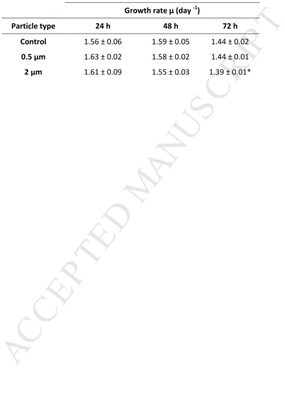
3.3. Little effect of MP exposure on cell growth 282
Only a slight but significant decrease in growth rate was detected in cultures exposed to 2 μm 283
MP for 72 h (ANOVA; F(2,6) = 13.25; p < 0.01) (Table 2). The strong aggregation pattern of the 284
MP in the microalgal medium observed in this study (Table 1) could reduce their bioavailability 285
and explain their limited impact on growth, as previously hypothesized in other studies with PS 286
particles (Bergami et al., 2017; Della Torre et al., 2014; Gambardella et al., 2018). Previous 287
studies that tested different MP concentrations of similar size range, reported no effect on the 288
M
AN
US
CR
IP
T
AC
CE
PT
ED
growth of microalgae. Long et al. (2017) did not observe adverse effects on the growth and 289
chlorophyll fluorescence of C. neogracile cells exposed to 0.04 μg mL-1 of uncharged 2 μm PS 290
MP. Davarpanah and Guilhermino (2015) did not found significant effects on the growth of the 291
marine green microalgae Tetraselmis chuii exposed for 96 h to concentrations ranging from 292
0.046 to 1.472 μg mL-1 of red fluorescent polyethylene microspheres (1-5 μm diameter). 293
Sjollema et al. (2016) analyzed the growth and photosynthetic capacity of the marine green 294
microalgae Dunaliella tertiolecta exposed to three sizes (50 nm, 0.5 and 6 μm) of uncharged PS 295
beads and also observed that these beads had negligible effects on microalgae growth, except 296
for nano-sized particles (50 nm) at high exposure concentrations (250 μg mL-1), suggesting that 297
the effect on microalgal growth increases with decreasing bead size. 298
Deleterious effects of MP on microalgal growth were only detected at very high 299
concentrations, even higher than the concentration used in this study (2.5 μg mL-1). However, 300
influence of particle size was rarely considered and controlled which are making comparisons 301
difficult. Mao et al. (2018) showed that 10, 50 and 100 μg mL-1 of 0.1 and 1 μm PS MP caused a 302
dose-dependent negative effect on the growth and photosynthetic activity of the freshwater 303
green microalgae Chlorella pyrenoidosa during its logarithmic growth phase. The mechanism 304
associated to the toxicity was attributed to the physical damage and oxidative stress. 305
Gambardella et al. (2018) exposed the marine microalgae D. tertiolecta to a wide range of 306
0.1 μm PS MP concentrations (0.001-0.01-0.1-1-10 μg mL-1) for 72 h and also observed a dose-307
dependent growth inhibition. At the highest MP concentration they tested (10 μg mL-1) about 308
40% of growth inhibition was observed. Authors suggested that this growth inhibition was due 309
to the fact that microalgae energy sources were used in detoxification processes, such as the 310
generation of extracellular polysaccharides. It is also noteworthy that most of the published 311
works assessing the effects of MP on phytoplankton were made with green microalgae, and 312
scarce data about the effects of the small MP fraction on other phytoplankton taxonomic 313
groups is available. 314
3.4. MP did not cause significant alterations of cell morphology and photosynthesis-related 315
parameters 316
Potential changes in the structural properties of the diatom C. neogracile exposed to the MP 317
were studied by FCM based on light diffraction. Neither of the two particle sizes significantly 318
altered the morphology of microalgal cells, since no changes were detected in the FSC and SSC 319
signals (Supp. Table 3). Similarly, Long et al. (2017) did not observed FSC and SSC changes in C. 320
neogracile cells exposed to 0.04 μg mL-1 of uncharged 2 μm PS MP for all duration of culture 321
growth. The fact that 0.5 and 2 μm PS-NH2 MP aggregate and become negatively charged may 322
have affected the effectiveness of physical adsorption of these MP onto the cell wall. 323
M
AN
US
CR
IP
T
AC
CE
PT
ED
Environmental stressors could affect the function of photosynthetic systems, thereby affecting 324
the fluorescence emission (Geoffroy et al., 2007; Juneau et al., 2002). Natural red 325
autofluorescence measured by FCM and effective quantum yield (QY) measured by PAM in C. 326
neogracile cells did not show significant changes upon exposure to 2.5 µg mL-1 of 0.5 and 2 μm 327
PS-NH2 MP as compared to control condition (Supp. Table 3). Mao et al. (2018) showed that 328
upon exposure of 0.1 μm and 1 μm PS MP at 10 µg mL-1, adsorption of MP onto the surface of 329
the freshwater microalgae Chlorella pyrenoidosa during its exponential growth phase could 330
provoke shading effects, hindering algal photosynthesis. However, this phenomenon appears 331
to be negligible for the particles tested, as shown in SEM micrographs, probably due to the 332
electrostatic repulsion exerted by its observed negative charge on microalgal cell membrane 333
(Table 1). 334
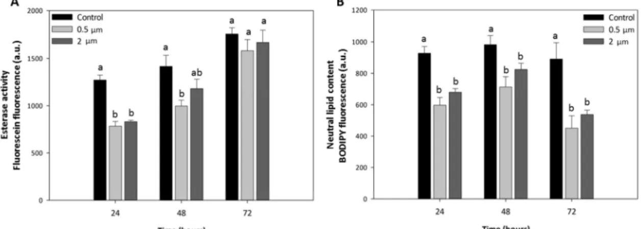
3.5. Decrease in esterase activity as an early response to MP exposure 335
A significant decrease in esterase activity of C. neogracile cells exposed to the MP tested with 336
respect to control cells was observed after 24 h (ANOVA; F(2,6) = 7.07; p < 0.05) and 48 h 337
(ANOVA; F(2,6) = 4.96; p < 0.05) (Fig. 2A). This reduction was more pronounced after 24 h of 338
exposure, followed by a slight recovery at 48 h. After 72 h, esterase activity appeared fully 339
recovered as significant differences were not observed anymore among treatments (ANOVA; 340
F(2,6) = 1.35; p > 0.05) (Fig. 2A). Recovery from the detrimental effects caused by MP on 341
microalgae was also observed by Mao et al. (2018) when cultures started the stationary phase. 342
Representative flow cytometric histograms showing shifts in the green fluorescence intensity 343
related to the esterase activity of C. neogracile cells in control cultures and cultures exposed to 344
both MP are shown in Supp. Fig. 1. 345
The lack of effects on key parameters such as growth, morphology or photosynthesis was 346
explained due to the absence of physical adsorption of MP onto diatom´s cell wall. However, 347
the decrease in esterase activity observed could be attributed to the contact between the MP 348
and the microalgae during the culture. Contact, even temporal, could be detected and 349
considered as a stress by the cells and may be translated into biochemical signals, triggering a 350
response to deal with. It could be described as the “billiard ball effect”. As documented in 351
humans, cells may sense mechanical cues, although the details underlying how cells respond 352
to mechanical forces are not well understood yet (Yusko et al., 2014). The potential release of 353
chemicals from MP could be another explanation to the indirect toxicity detected. Monomers 354
and/or additives incorporated during manufacture could be toxic to microalgae and could 355
interfere with biological processes, explaining the decrease observed in the general metabolic 356
activity of cells (Fig. 2A). Many exposure experiments reported significant toxicity of plastic 357
leachates on aquatic organisms (reviewed in Hermabessiere et al., 2017). Virgin MP, frequently 358
M
AN
US
CR
IP
T
AC
CE
PT
ED
used as model materials in aquatic toxicity laboratory studies, are supposed to be free of 359
additives or residual monomers. However, some studies suggest that commercial virgin plastic 360
pellets may also leach toxic unknown chemicals (Nobre et al., 2015). Martínez-Gómez et al. 361
(2017) reported significant toxicity of virgin and aged PS MP on the fertilization and larval 362
development of sea urchins, and plastic leachates were found to have higher toxicity than the 363
virgin and aged materials themselves. Toxicity of leachates from plastic products obtained 364
after 72 h of leaching was also observed in the copepod Nitocra spinipes (Bejgarn et al., 2015). 365
Moreover, it cannot be excluded the potential repercussions that amino functional groups 366
could have on cells. MP are coated with -NH2 groups by chemical bonds and during laboratory 367
exposure, light or other environmental factors could lead to bond cleavage. 368
As compared to the other parameters analysed, the FDA assay appeared more sensitive to 369
detect physiological changes in cells exposed to these 0.5 and 2 μm (nominal sizes) particles 370
after a short period of time. Esterases involved in the FDA assay turn over on a time frame of 371
several hours (Jochem, 2000). Therefore, this technique seems appropriate to detect changes 372
in metabolic activity on a day-to-day or even shorter basis, which makes it well suited to 373
monitor short-term phytoplankton responses to environmental changes or to diverse 374
pollutants (Esperanza et al., 2015; Franklin et al., 2001; Prado et al., 2009; Seoane et al., 2017a; 375
Seoane et al., 2017b). Our results showed that, with more subtle measurements, we can 376
detect the impact of MP. This brings out the suitability and sensitivity of this assay with the 377
flow cytometry technique to assess the effects of plastic debris on marine phytoplankton. 378
3.6. Unaltered ROS production and membrane potential during MP exposure 379
Microalgae may have higher levels of ROS as a result of changes in environmental conditions 380
or the presence of contaminants. When there is an imbalance between the production of ROS 381
and the cellular antioxidant defence mechanisms, oxidative stress increases leading to several 382
cellular damages. In the present study, ROS production in C. neogracile cells was not 383
significantly affected by the presence of MP in the medium (Supp. Table 4). ROS 384
overproduction has been previously observed by Mao et al. (2018) during the exponential 385
growth phase of the freshwater microalgae Chlorella pyrenoidosa exposed to 0.1 μm and 1 μm 386
PS particles at very high concentrations (10, 50 and 100 μg mL-1). However, it seems that with 387
the 0.5 μm and 2 μm MP we tested (nominal sizes), the concentration used (2.5 μg mL-1) was 388
not high enough to alter the intracellular equilibrium of C. neogracile ROS levels. 389
Regarding cytoplasmic membrane potential, no significant alterations were observed in cells 390
exposed to MP (Supp. Table 4). Interactions between MP and biological membranes are driven 391
by particle size, since small particles are suspected to interact more with biological membranes 392
(Nel et al., 2006; Verma and Stellacci, 2010), and by particle surface properties, notably the net 393
M
AN
US
CR
IP
T
AC
CE
PT
ED
surface charge (Nolte et al., 2017). Taking into consideration the particle size, internalization of 394
MP used (0.5 and 2 μm) was discarded on intact cells. Algal cell walls are semipermeable and 395
the diameter of their pores determines its sieving properties (Navarro et al., 2008). Diatoms´ 396
wall pores are typically between 3 and 50 nm (Sanka et al., 2017). As long as there are no holes 397
in the cell wall or loss of viability, only particles with a size smaller than that of the largest pore 398
are expected to pass through the cell wall. Thus, the algal cell wall pore size is too small to 399
transport MP used through the cell. Gambardella et al. (2018) evaluated MP internalization in 400
the green microalgae D. tertiolecta using 0.1 μm fluorescent PS MP. Although MP caused algal 401
growth inhibition, they also discard MP internalization into cells, since all fluorescence beads 402
were observed as aggregates in the medium, out of the microalgal cell surface. With regard to 403
the charge, it is possibly related to differences in effectiveness of physical adsorption onto the 404
cell wall, since cationic particles interact with membranes more easily than anionic ones 405
(Bhattacharya et al., 2010; Nel et al., 2009). In the present study, the aggregation of MP and 406
the negative charge measured seems to minimize MP effects. MP concentration and time 407
exposure are also important factors for MP impacts. Mao et al. (2018) showed membrane 408
damages such as cell wall thickening and loss of viability in C. pyrenoidosa exposed to PS MP at 409
the higher concentration they tested (100 μg mL-1) in long-term exposure (30 days). However, 410
at the concentration used in our short-term study, we did not observed effects on membrane 411
potential. 412
3.7. Diminished neutral lipid content in MP-exposed cells 413
Exposure to 0.5 and 2 μm PS-NH2 MP resulted in a significant decrease in the cellular neutral 414
lipid content with respect to control cells at all tested times (ANOVA 24 h; F(2,6) = 37.50; 415
p < 0.001) (ANOVA 48 h; F(2,6) = 12.08; p < 0.01) (ANOVA 72 h; F(2,6) = 18.46; p < 0.01) (Fig. 2B). 416
After 72 h, the lipid content of MP-exposed cells was reduced by half with respect to control. 417
Representative flow cytometric histograms showing shifts in the green fluorescence intensity 418
related to the neutral lipid content of C. neogracile cells in control cultures and cultures 419
exposed to both MP are shown in Supp. Fig. 2. 420
Microalgae generally accumulate neutral lipids, mainly triacylglycerol (TAG), in specific 421
organelles called lipid bodies, upon stresses such as nutrient limitation, elevated 422
temperatures, unfavourable light intensities, alkaline pH, high salinity or dehydration 423
(Zienkiewicz et al. 2016). In laboratory cultures, microalgae begin to accumulate lipids in the 424
stationary phase of growth, when shortage of nutrients arrives (Huerlimann et al., 2010; Xu et 425
al., 2008). Previous studies found disturbances in lipid metabolism after exposure to MP in 426
other species such as marine fishes and mussels (Von Moos et al., 2012; Yin et al., 2018). 427
Although exposure to MP does not usually cause mortality in marine organisms, it has been 428
M
AN
US
CR
IP
T
AC
CE
PT
ED
observed that it can affect them by altering their feeding behaviour and reducing their energy 429
reserves, with consequences for growth and reproduction (Galloway et al., 2017). Microalgal 430
oil bodies have a dynamic nature and appear to function as transient reservoirs, as the storage 431
lipids are quick catabolized in response to environmental changes (Maeda et al., 2017). In the 432
present study, oxidative stress was not detected in MP-exposed cells; therefore, the decrease 433
in lipids observed cannot be associated with its oxidation, but to modulation of energy 434
metabolism to properly acclimate to the stress conditions, maintaining a healthy status. As 435
discussed previously, despite not having observed MP attached to the cell surface it cannot be 436
excluded an interaction between MP and microalgae and an indirect toxicity due to i) the 437
contact between MP and cells during the culture and ii) the potential toxic compounds 438
(monomers, oligomers, additives or other chemicals) that could be leached from MP. The 439
“consumption” of lipid reserves could be interpreted as a cell response to overcome the stress 440
provoked by MP exposure and for the maintenance of the normal growth, photosynthesis and 441
even the membrane integrity. Thus, microalgal oil bodies could serve as an energy source for 442
their recovery. This decrease in the lipid content could also have ecological implications for 443
food web trophic functioning by reducing microalgae nutritional quality for primary consumers 444
and onward. 445
4. Conclusions 446
MP aggregated in FSW and in filtered culture medium, producing secondary particles with 447
different properties, which make difficult assessing influence of size on MP toxicity. In 448
addition, their ζ-potential in FSW and in the culture medium outlined low negative values, fact 449
that could have influenced their aggregation state and their interaction with the cell surface. It 450
highlights the necessity to characterize behaviour of plastic particles in assay media before 451
exposure to avoid bias in results interpretation. 452
Direct toxicity on key parameters such as cell growth, morphology, photosynthesis, ROS 453
content and membrane potential was not observed and SEM micrographs showed that MP 454
were not attached to the microalgal cell wall. However, indirect toxicity was detected because 455
we unravelled a significant decrease in the esterase activity and the lipid reserves of MP-456
exposed cells. Results suggest that microalgal oil bodies could serve as an energy source for 457
the maintenance of the normal cellular growth, photosynthesis and membrane integrity to 458
overcome the stress produced by MP exposure. 459
Acknowledgements 460
This work was supported by the ANR CESA (ANR-15-CE34-0006-02, NANOPLASTICS project) 461
and by the Unique Inter-ministerial Fund (FUI) as part of the MICROPLASTIC2 project. 462
M
AN
US
CR
IP
T
AC
CE
PT
ED
M.S. acknowledges a pre-doctoral grant from “Campus do Mar” and all the LEMAR team for 463
their help during the course of this project. 464
Authors would thank Philippe Miner for his help with microalgal cultures at the Ifremer 465
facilities. We are also very grateful to Philippe Eliès from the PIMM core and to Olivier Lozach 466
from the COSM team at the University of Western Brittany for the scanning electron 467
microscopy observations and for providing us the equipment for DLS measurements, 468
respectively. 469
References 470
Andrady, A.L., 2011. Microplastics in the marine environment. Mar. Pollut. Bull. 62, 1596– 471
1605. 472
Andrady, A.L., 2015. Persistence of plastic litter in the oceans. In: Bergmann, M., Gutow, L., 473
Klages, M. (Eds.), Marine Anthropogenic Litter. Springer International Publishing, 474
Cham, pp. 57-72. 475
Barnes, D.K., Galgani, F., Thompson, R.C., Barlaz, M., 2009. Accumulation and fragmentation 476
of plastic debris in global environments. Philos. T. Roy. Soc. B. 364, 1985–1998. 477
Bejgarn, S., MacLeod, M., Bogdal, C., Breitholtz, M., 2015. Toxicity of leachate from 478
weathering plastics: An exploratory screening study with Nitocra spinipes. 479
Chemosphere 132, 114–119. 480
Bergami, E., Pugnalini, S., Vannuccini, M. L., Manfra, L., Faleri, C., Savorelli, F., Dawson, K.A., 481
Corsi, I., 2017. Long-term toxicity of surface-charged polystyrene nanoplastics to 482
marine planktonic species Dunaliella tertiolecta and Artemia franciscana. Aquat. 483
Toxicol. 189, 159–169. 484
Bhattacharya, P., Lin, S., Turner, J. P., Ke, P. C., 2010. Physical adsorption of charged plastic 485
nanoparticles affects algal photosynthesis. J. Phys. Chem. C 114, 16556–16561. 486
Canesi, L., Balbi, T., Fabbri, R., Salis, A., Damonte, G., Volland, M., Blasco, J., 2017. 487
Biomolecular coronas in invertebrate species: implications in the environmental 488
impact of nanoparticles. NanoImpact 8, 89-98. 489
Cole, M., Lindeque, P., Halsband, C., Galloway, T.S., 2011. Microplastics as contaminants in 490
the marine environment: A review. Mar. Pollut. Bull. 62, 2588-2597. 491
Davarpanah, E., Guilhermino, L., 2015. Single and combined effects of microplastics and 492
copper on the population growth of the marine microalgae Tetraselmis chuii. Estuar. 493
Coast. Shelf Sci. 167, 269–275. 494
Dawson, A. L., Kawaguchi, S., King, C. K., Townsend, K. A., King, R., Huston, W. M., Bengtson 495
Nash, S. M., 2018. Turning microplastics into nanoplastics through digestive 496
fragmentation by Antarctic krill. Nat. Commun. 9, 1001. 497
Della Torre, C., Bergami, E., Salvati, A., Faleri, C., Cirino, P., Dawson, K.A., Corsi, I., 2014. 498
Accumulation and embryotoxicity of polystyrene nanoparticles at early stage of 499
development of sea urchin embryos Paracentrotus lividus. Environ. Sci. Technol. 48, 500
12302–12311. 501
Dubaish, F., Liebezeit, G., 2013. Suspended microplastics and black carbon particles in the 502
Jade system, southern North Sea. Water Air Soil Pollut. 224, 1352-1359. 503
Eriksen, M., Lebreton, L.C.M., Carson, H.S., Thiel, M., Moore, C.J., Borerro, J.C., Galgani, F., 504
M
AN
US
CR
IP
T
AC
CE
PT
ED
Ryan, P.G., Reisser, J., 2014. Plastic Pollution in the World’s Oceans: More than 5 505
Trillion Plastic Pieces Weighing over 250,000 Tons Afloat at Sea. PLoS One 9, 1–15. 506
Esperanza, M., Seoane, M., Rioboo, C., Herrero, C., Cid, Á., 2015. Chlamydomonas reinhardtii 507
cells adjust the metabolism to maintain viability in response to atrazine stress. Aquat. 508
Toxicol. 165, 64–72. 509
Filella, M., 2015. Questions of size and numbers in environmental research on microplastics: 510
methodological and conceptual aspects. Environ. Chem. 12, 527–538. 511
Foulon, V., Le Roux, F., Lambert, C., Huvet, A., Soudant, P., Paul-Pont, I., 2016. Colonization of 512
polystyrene microparticles by Vibrio crassostreae: Light and electron microscopic 513
investigation. Environ. Sci. Technol. 50, 10988–10996. 514
Franklin, N.M., Stauber, J.L., Lim, R.P., 2001. Development of flow cytometry-based algal 515
bioassays for assessing toxicity of copper in natural waters. Environ. Toxicol. Chem. 516
20, 160–170. 517
Galloway, T. S., Cole, M., Lewis, C., Atkinson, A., and Allen, J. I., 2017. Interactions of 518
microplastic debris throughout the marine ecosystem. Nat. Ecol. Evol. 1, 116. 519
Gambardella, C., Morgana, S., Bramini, M., Rotini, A., Manfra, L., Migliore, L., Piazza, V., 520
Garaventa, F., Faimali, M., 2018. Ecotoxicological effects of polystyrene microbeads in 521
a battery of marine organisms belonging to different trophic levels. Mar. Environ. Res. 522
141, 313–321. 523
Geoffroy, L., Gilbin, R., Simon, O., Floriani, M., Adam, C., Pradines, C., Cournac, L., Garnier-524
Laplace, J., 2007. Effect of selenate on growth and photosynthesis of Chlamydomonas 525
reinhardtii. Aquat. Toxicol. 83, 149–58.
526
Gigault, J., Halle, A. Ter, Baudrimont, M., Pascal, P. Y., Gauffre, F., Phi, T. L., El Hadri, H., 527
Grassl, B., Reynaud, S., 2018. Current opinion: What is a nanoplastic? Environ. Pollut. 528
235, 1030-1034. 529
Gigault, J., Pedrono, B., Maxit, B., Ter Halle, A., 2016. Marine plastic litter: the unanalyzed 530
nano-fraction. Environ. Sci. Nano 3, 346–350. 531
González-Fernández, C., Tallec, K., Le Goïc, N., Lambert, C., Soudant, P., Huvet, A., Suquet, 532
M., Berchel, M., Paul-Pont, I., 2018. Cellular responses of Pacific oyster (Crassostrea 533
gigas) gametes exposed in vitro to polystyrene nanoparticles. Chemosphere 208,
764-534
772. 535
Govender, T., Ramanna, L., Rawat, I., Bux, F., 2012. BODIPY staining, an alternative to the Nile 536
Red fluorescence method for the evaluation of intracellular lipids in microalgae. 537
Bioresour. Technol. 114, 507–511. 538
Hecky, R.E., Mopper, K., Kilham, P., Degens, E.T., 1973. Amino-acid and sugar composition of 539
diatom cell-walls. Mar. Biol. 19, 323-331. 540
Hermabessiere, L., Dehaut, A., Paul-Pont, I., Lacroix, C., Jezequel, R., Soudant, P., Duflos, G., 541
2017. Occurrence and effects of plastic additives on marine environments and 542
organisms: a review. Chemosphere 182, 781–793. 543
Hernandez, L.M., Yousefi, N., Tufenkji, N., 2017. Are there nanoplastics in your personal care 544
products? Environ. Sci. Technol. Lett. 4, 280-285. 545
Huerlimann, R., Rocky, N., Heimann, K., 2010. Growth, lipid content, productivity and fatty 546
acid composition of tropical microalgae for scale-up production. Biotechnol. Bioeng. 547
107, 245-257. 548
Jochem, F. J., 2000. Probing the physiological state of phytoplankton at the single-cell level. 549
M
AN
US
CR
IP
T
AC
CE
PT
ED
Sci. Mar. 64, 183-195. 550Juneau, P., El Berdey, A., Popovic, R., 2002. PAM fluorometry in the determination of the 551
sensitivity of Chlorella vulgaris, Selenastrum capricornutum and Chlamydomonas 552
reinhardtii to copper. Arch. Environ. Contam. Toxicol. 42, 155–164.
553
Lambert, S., Wagner, M., 2016. Characterisation of nanoplastics during the degradation of 554
polystyrene. Chemosphere 145, 265-268. 555
Long, M., Paul-Pont, I., Hégaret, H., Moriceau, B., Lambert, C., Huvet, A., Soudant, P., 2017. 556
Interactions between polystyrene microplastics and marine phytoplankton lead to 557
species-specific hetero-aggregation. Environ. Pollut. 228, 454–463. 558
Lundqvist, M., Stigler, J., Elia, G., Lynch, I., Cedervall, T., Dawson, K. A., 2008. Nanoparticle 559
size and surface properties determine the protein corona with possible implications 560
for biological impacts. Proc. Natl. Acad. Sci. 105, 14265–14270. 561
Lusher, A. L., Hollman, P. C. H., Mendoza-Hill, J. J., 2017. Microplastics in Fisheries and 562
Aquaculture: Status of Knowledge on Their Occurrence and Implications for Aquatic 563
Organisms and Food Safety. FAO Fisheries and Aquaculture Technical Paper. No. 615, 564
p. 147. 565
Maeda, Y., Nojima, D., Yoshino, T., Tanaka, T., 2017. Structure and properties of oil bodies in 566
diatoms. Phil. Trans. R. Soc. 372, 20160408. 567
Malviya, S., Scalco, E., Audic, S., Vincent, F., Veluchamy, A., Poulain, J., Wincker, P., Iudicone, 568
D., de Vargas, C., Bittner, L., Zingone, A., Bowler, C., 2016. Insights into global diatom 569
distribution and diversity in the world's ocean. Proc. Natl. Acad. Sci. 113, E1516-570
E1525. 571
Mao, Y., Ai, H., Chen, Y., Zhang, Z., Zeng, P., Kang, L., Li, W., Gu, W., He, Q., Li, H., 2018. 572
Phytoplankton response to polystyrene microplastics: Perspective from an entire 573
growth period. Chemosphere 208, 59–68. 574
Marques-Santos, L. F., Grassi, G., Bergami, E., Faleri, C., Balbi, T., Salis, A., Damonte, G., 575
Canesi, L., Corsi, I., 2018. Cationic polystyrene nanoparticle and the sea urchin 576
immune system: biocorona formation, cell toxicity, and multixenobiotic resistance 577
phenotype. Nanotoxicology 0, 1–21. 578
Martínez-Gómez, C., León, V. M., Calles, S., Gomáriz-Olcina, M., Vethaak, A. D., 2017. The 579
adverse effects of virgin microplastics on the fertilization and larval development of 580
sea urchins. Mar. Environ. Res. 130, 69-76. 581
National Oceanic and Atmospheric Administration (NOAA), 2008. Proceedings of the 582
International Research Workshop on the Occurrence, effects, and fate of Microplastic 583
Marine debris. C Arthur, J. Baker, and H. Bamford (eds). NOAA Technical 584
Memorandum NOS-OR&R-30. 585
Navarro, E., Baun, A., Behra, R., Hartmann, N.B., Filser, J., Miao, A.J., Quigg, A., Santschi, P.H., 586
Sigg, L., 2008. Environmental behavior and ecotoxicity of engineered nanoparticles to 587
algae, plants, and fungi. Ecotoxicology 17, 372–386. 588
Nel, A., Xia, T., Mädler, L., Li, N., 2006. Toxic potential of materials at the nano level. Science 589
311, 622-627. 590
Nel, A.E., Mädler, L., Velegol, D., Xia, T., Hoek, E.M.V., Somasundaran, P., Klaessig, F., 591
Castranova, V., Thompson, M., 2009. Understanding biophysicochemical interactions 592
at the nano-bio interface. Nat. Mater. 8, 543-557. 593
Nobre, C. R., Santana, M. F. M., Maluf, A., Cortez, F. S., Cesar, A., Pereira, C. D. S., et al., 2015. 594
M
AN
US
CR
IP
T
AC
CE
PT
ED
Assessment of microplastic toxicity to embryonic development of the sea urchin 595
Lytechinus variegatus (Echinodermata: Echinoidea). Mar Pollut. Bull. 92, 99–104.
596
Nolte, T. M., Hartmann, N. B., Kleijn, J. M., Garnæs, J., van de Meent, D., Jan Hendriks, A., 597
Baun, A., 2017. The toxicity of plastic nanoparticles to green algae as influenced by 598
surface modification, medium hardness and cellular adsorption. Aquat. Toxicol. 183, 599
11–20. 600
OECD 201, 2011. Alga Growth Inhibition Test (201). OECD Guideline for Testing of Chemicals. 601
Organization for Economic Cooperation and Development, Paris, France. 602
Paul-Pont, I., Tallec, K., Gonzalez-Fernandez, C., Lambert, C., Vincent, D., Mazurais, D., 603
Zambonino-Infante, J.L., Brotons, G., Lagarde, F., Fabioux, C., Soudant, P., Huvet, A., 604
2018. Constraints and priorities for conducting experimental exposures of marine 605
organisms to microplastics. Front. Mar. Sci. 5, 252. 606
Peeken, I., Primpke, S., Beyer, B., Gütermann, J., Katlein, C., Krumpen, T., Bergmann, 607
M., Hehemann, L., Gerdts, G., 2018. Arctic sea ice is an important temporal sink and 608
means of transport for microplastic. Nat. Commun. 95, 2041–1723. 609
Pham, C. K., Ramirez-Llodra, E., Alt, C.H.S., Amaro, T., Bergmann, M., Canals, M., Company, 610
J.B., Davies, J., Duineveld, G., Galgani, F., Howell, K.L., Huvenne, V.A., Isidro, E., Jones, 611
D.O., Lastras, G., Morato, T., Gomes-Pereira, J.N., Purser, A., Stewart, H., Tojeira, I., 612
Tubau, X., Van Rooij, D., Tyler, P.A., 2014. Marine litter distribution and density in 613
European seas, from the shelves to deep basins. PLoS ONE 9(4), e95839. 614
Phuong, N. N., Zalouk-Vergnoux, A., Poirier, L., Kamari, A., Châtel, A., Mouneyrac, C., Lagarde, 615
F., 2016. Is there any consistency between the microplastics found in the field and 616
those used in laboratory experiments? Environ. Pollut. 211, 111–123. 617
Plastics Europe, 2017. Plastics – the facts 2017. In: An Analysis of European plastics 618
production, demand and waste data. Technical Report. 619
Prado, R., García, R., Rioboo, C., Herrero, C., Abalde, J., Cid, Á, 2009. Comparison of the 620
sensitivity of different toxicity test endpoints in a microalga exposed to the herbicide 621
paraquat. Environ. Int. 35, 240–247. 622
Rosenwasser, S., van Creveld, G.S., Schatz, D., Malitsky, S., Tzfadia, O., Aharoni, A., Levin, Y., 623
Gabashvili, A., Feldmesser, E., Vardi, A., 2014. Mapping the diatom redox- sensitive 624
proteome provides insight into response to nitrogen stress in the marine 625
environment. Proc. Natl. Acad. Sci. U. S. A. 111, 2740–2745. 626
Ruiz-Orejón, L.F., 2016. Floating plastic debris in the central and western mediterranean sea. 627
Mar. Environ. Res. 120, 136–144. 628
Rumin, J., Bonnefond, H., Saint-Jean, B., Rouxel, C., Sciandra, A., Bernard, O., Cadoret, J.P., 629
Bougaran, G., 2015. The use of fluorescent Nile red and BODIPY for lipid 630
measurement in microalgae. Biotechnol. Biofuels 8, 42. 631
Ryan, 2015. A Brief History of Marine Litter Research. In: Bergmann, M., Gutow, L., Klages, M. 632
(Eds.), Marine Anthropogenic Litter. Springer International Publishing, pp. 1-25. 633
Ryan, P.G., Moore, C.J., van Franeker, J.A., Moloney, C.L., 2009. Monitoring the abundance of 634
plastic debris in the marine environment. Philos. Trans. R. Soc. B 364, 1999–2012. 635
Sanka, I., Suyono, E.A., Alam, P., 2017. The effects of diatom pore-size on the structures and 636
extensibilities of single mucilage molecules. Carbohydr. Res. 448, 35–42. 637
Seoane, M., Esperanza, M., Cid, Á., 2017a. Cytotoxic effects of the proton pump inhibitor 638
omeprazole on the non-target marine microalga Tetraselmis suecica. Aquat. Toxicol. 639
M
AN
US
CR
IP
T
AC
CE
PT
ED
191, 62–72. 640Seoane, M., Esperanza, M., Rioboo, C., Herrero, C., Cid, Á., 2017b. Flow cytometric assay to 641
assess short-term effects of personal care products on the marine microalga 642
Tetraselmis suecica. Chemosphere, 171, 339–347.
643
Sjollema, S.B., Redondo-Hasselerharm, P., Leslie, H. a., Kraak, M.H.S., Vethaak, a. D., 2016. 644
Do plastic particles affect microalgal photosynthesis and growth? Aquat. Toxicol. 170, 645
259–261. 646
Sun, X., Chen, B., Li, Q., Liu, N., Xia, B., Zhu, L., Qu, K., 2018. Toxicities of polystyrene nano- 647
and microplastics toward marine bacterium Halomonas alkaliphila. Sci. Total Environ. 648
642, 1378–1385. 649
Tallec, K., Huvet, A., Di Poi, C., González-Fernández, C., Lambert, C., Petton, B., Le Goïc, N., 650
Berchel, M., Soudant, P., Paul-Pont, I., 2018. Nanoplastics impaired oyster free living 651
stages, gametes and embryos. Environ. Pollut. 242, 1226–1235. 652
Ter Halle, A., Jeanneau, L., Martignac, M., Jardé, E., Pedrono, B., Brach, L., Gigault, J., 2017. 653
Nanoplastic in the North Atlantic Subtropical Gyre. Environ. Sci. Technol. 51, 13689– 654
13697. 655
Verma, A., Stellacci, F., 2010. Effect of surface properties on nanoparticle-cell interactions. 656
Small 6, 12-21. 657
Von Moos, N., Burkhardt-Holm, P., Köhler, A., 2012. Uptake and effects of microplastics on 658
cells and tissue of the blue mussel Mytilus edulis L. after an experimental exposure. 659
Environ. Sci. Technol. 46, 11327–11335. 660
Walne, P. R., 1966. Experiments in the Large-scale Culture of the Larvae of Ostrea edulis L. 661
Fishery In, Her Majesty’s Stationery Office, London. 662
Wolff, C., Fuks, B., Chatelain, P., 2003. Comparative study of membrane potential- sensitive 663
fluorescent probes and their use in ion channel screening assays. J. Biomol. Screen 8, 664
533-543. 665
Xu, Z.B., Yan, X.J., Pei, L.Q., Luo, Q.J., Xu, J.L., 2008. Changes in fatty acids and sterols during 666
batch growth of Pavlova viridis in photobioreactor. J. Appl. Phycol. 20, 237–243. 667
Yin, L., Chen, B., Xia, B., Shi, X., Qu, K., 2018. Polystyrene microplastics alter the behavior, 668
energy reserve and nutritional composition of marine jacopever (Sebastes schlegelii). 669
J. Hazard. Mater. 360, 97–105. 670
Yokota, K., Waterfield, H., Hastings, C., Davidson, E., Kwietniewski, E., Wells, B., 2017. Finding 671
the missing piece of the aquatic plastic pollution puzzle: Interaction between primary 672
producers and microplastics. Limnol. Oceanogr. Lett. 2, 91–104. 673
Yusko, E.C., Asbury, C.L., 2014. Force is a signal that cells cannot ignore. Mol. Biol. Cell 25, 674
3717–3725. 675
Zhang, C., Chen, X., Wang, J., Tan, L., 2017. Toxic effects of microplastic on marine microalgae 676
Skeletonema costatum: interactions between microplastic and algae. Environ. Poll.
677
220, 1282-1288. 678
Zienkiewicz, K., Du, Z. Y., Ma, W., Vollheyde, K., Benning, C., 2016. Stress-induced neutral 679
lipid biosynthesis in microalgae — Molecular, cellular and physiological insights. 680
Biochim. Biophys. Acta 1861, 1269–1281. 681
M
AN
US
CR
IP
T
AC
CE
PT
ED
Table 1. Characterization of PS-NH2 MP in Milli Q water, filtered natural seawater (FSW) and microalgae medium using DLS analysis. Z-average (nm),
polydispersity index - PdI (a.u.) and ζ-potential (mV), referred to a PS-NH2 MP solution concentration of 100 μg mL-1 are reported. PdI > 0.2 showing
aggregation is marked by an asterisk (*). Values are shown as mean ± standard deviation of the three measurements.
Milli Q FSW Filtered (0.2 μm) microalgal culture medium
Nominal size Size Z-average (nm) PdI (a.u.) Charge ζ-potential (mV) Size Z-average (nm) PdI (a.u.) Charge ζ-potential (mV) Size Z-average (nm) PdI (a.u.) Charge ζ-potential (mV) 0.5 μm 602.5 ± 27.3 0.16 ± 0.02 -11.9 ± 0.6 2899.0 ± 96.0 0.46 ± 0.10* -7.3 ± 0.9 3268.0 ± 64.3 0.40 ± 0.02* -6.9 ± 1.7 2 μm 2274.0 ± 100.9 0.16 ± 0.08 -12.8 ± 0.3 5932.0 ± 694.4 0.89 ± 0.10* -4.8 ± 0.5 4342.3 ± 294.2 0.67 ± 0.15* -5.0 ± 2.0
M
AN
US
CR
IP
T
AC
CE
PT
ED
Table 2. Growth rates μ (day−1) of C. neogracile cultures exposed to 0 and 2.5 µg mL-1 of 0.5 μm and 2 μm PS-NH2 MP at each sample time. Data are given as mean values ± standard deviation of three replicates. Significant differences with respect to control at a level of significance of 0.05 (p < 0.05) are represented by an asterisk (*).
Growth rate μ (day -1)
Particle type 24 h 48 h 72 h
Control 1.56 ± 0.06 1.59 ± 0.05 1.44 ± 0.02 0.5 μm 1.63 ± 0.02 1.58 ± 0.02 1.44 ± 0.01 2 μm 1.61 ± 0.09 1.55 ± 0.03 1.39 ± 0.01*
M
AN
US
CR
IP
T
AC
CE
PT
ED
Figure 1. Scanning electron micrographs showing a control cell (A) and cells exposed to 0.5 μm (B) and 2 μm PS-NH2 MP (C). 0.5 μm (D, E) and 2 μm (F) PS-NH2 beads micro-aggregates and bacteria are also shown. Arrow indicates a rimoportula (tubular process through the valve of some diatoms). Scale bars: 5 μm (A, B, C); 1 μm (D, E); 2 μm (F).
M
AN
US
CR
IP
T
AC
CE
PT
ED
Figure 2. Esterase activity (A) and neutral lipid content (B) of C. neogracile cells in control cultures and cultures exposed to 2.5 μg mL-1 of 0.5 and 2 μm PS-NH2 MP for 24, 48 and 72 h. Significant differences between treatments are marked with lowercase letters (p < 0.05).
M
AN
US
CR
IP
T
AC
CE
PT
ED
Highlights1. Effects of 0.5 and 2 μm PS-NH2 microplastics (MP) were evaluated on C. neogracile 2. MP showed negative charge, were aggregated and were not attached to the cell wall 3. Exposure to MP decreased the cellular metabolic activity and neutral lipid content 4. Cells modulate their energy metabolism to properly acclimate to the stress conditions 5. Microalgal oil bodies serve as an energy source for maintaining a healthy status