HAL Id: hal-02140502
https://hal.archives-ouvertes.fr/hal-02140502
Submitted on 26 May 2020
HAL is a multi-disciplinary open access
archive for the deposit and dissemination of
sci-entific research documents, whether they are
pub-lished or not. The documents may come from
teaching and research institutions in France or
abroad, or from public or private research centers.
L’archive ouverte pluridisciplinaire HAL, est
destinée au dépôt et à la diffusion de documents
scientifiques de niveau recherche, publiés ou non,
émanant des établissements d’enseignement et de
recherche français ou étrangers, des laboratoires
publics ou privés.
Copyright
Polyunsaturated fatty acid metabolites: biosynthesis in
Leishmania and role in parasite/host interaction
Lucie Paloque, Teresa Pérez-Berezo, Anne Abot, Jessica Dalloux-Chioccioli,
Sandra Bourgeade-Delmas, Pauline Le Faouder, Julien Pujo, Marie-Ange
Teste, Jean Marie François, Nils Helge Schebb, et al.
To cite this version:
Lucie Paloque, Teresa Pérez-Berezo, Anne Abot, Jessica Dalloux-Chioccioli, Sandra
Bourgeade-Delmas, et al.. Polyunsaturated fatty acid metabolites: biosynthesis in Leishmania and role in
par-asite/host interaction. Journal of Lipid Research, American Society for Biochemistry and Molecular
Biology, 2019, 60 (3), pp.636-647. �10.1194/jlr.M091736�. �hal-02140502�
Copyright © 2019 Paloque et al. Published under exclusive license by The American
also in the Mediterranean. It is the second-most lethal
parasitic infection worldwide, with more than 20,000 annual
deaths (http://www.who.int/leishmaniasis/en). This disease
is supported by the intracellular development of a protozoan
parasite belonging to the Leishmania genus. Metacyclic
promastigotes are transmitted by the Phlebotominae sandfly
during a blood meal on mammals, where they infect
macro-phages prior to differentiating into amastigote forms and
proliferating inside the host cell. Procyclic promastigotes
differentiate into metacyclic promastigotes during
metacy-clogenesis, a process that is triggered by an environmental
pH decrease from neutral to acidic pH inside the sandfly
vector (1, 2). Metacyclogenesis involves important
morpho-logical and biochemical modifications, and approximately
300 genes have been described to be specifically regulated
according to the parasite stage (3–6). Macrophages are the
primary replication site for Leishmania and the main effector
cells to fight it (7). Leishmania infection leads to the
develop-ment of macrophages that overexpress anti-inflammatory
molecules such as interleukin (IL) 10 and transforming
growth factor and the induction of proinflammatory
cyto-kine production such as TNF-, IL-1, and IL-6 (8). M2
macrophages control disease severity and protect the host
Abstract Inside the human host, Leishmania infection starts
with phagocytosis of infective promastigotes by macrophages.
In order to survive, Leishmania has developed several
strate-gies to manipulate macrophage functions. Among these
strat-egies, Leishmania as a source of bioactive lipids has been
poorly explored. Herein, we assessed the biosynthesis of
polyunsaturated fatty acid metabolites by infective and
non-infective stages of Leishmania and further explored the role
of these metabolites in macrophage polarization. The
con-centration of docosahexaenoic acid metabolites, precursors
of proresolving lipid mediators, was increased in the infective
stage of the parasite compared with the noninfective stage,
and cytochrome P450-like proteins were shown to be
impli-cated in the biosynthesis of these metabolites. The treatment
of macrophages with lipids extracted from the infective
forms of the parasite led to M2 macrophage polarization and
blocked the differentiation into the M1 phenotype induced
by IFN-. In conclusion, Leishmania polyunsaturated fatty
acid metabolites, produced by cytochrome P450-like protein
activity, are implicated in parasite/host interactions by
pro-moting the polarization of macrophages into a proresolving
M2 phenotype.—Paloque, L., T. Perez-Berezo, A. Abot, J.
Dalloux-Chioccioli, S. Bourgeade-Delmas, P. Le Faouder, J.
Pujo, M-A.Teste, J-M. François, N. H. Schebb, M. Mainka,
C. Rolland, C. Blanpied, G. Dietrich, J. Bertrand-Michel, C.
Deraison, A. Valentin, and N. Cenac. Polyunsaturated fatty
acid metabolites: biosynthesis in Leishmania and role in
parasite/host interaction. J. Lipid Res. 2019. 60: 636–647.
Supplementary key words cytochrome P450 • infection •
macro-phages • -3 • metabolism • arachidonic acid • eicosanoids
Leishmaniasis is a plague affecting millions of people
around the world, most notably in developing countries but
This work was supported by grants from Région Midi-Pyrénées (N.C.) and Euro-pean Commission Marie Curie Action EPIMACASE Grant IOF-627487 (C.D.). Manuscript received 19 December 2018 and in revised form 8 January 2019. Published, JLR Papers in Press, January 9, 2019DOI https://doi.org/10.1194/jlr.M091736
Polyunsaturated fatty acid metabolites: biosynthesis in
Leishmania and role in parasite/host interaction
Lucie Paloque,
1,*
,†Teresa Perez-Berezo,
1,§Anne Abot,
§Jessica Dalloux-Chioccioli,**
Sandra Bourgeade-Delmas,* Pauline Le Faouder,** Julien Pujo,
§Marie-Ange Teste,
††Jean-Marie François,
††Nils Helge Schebb,
§§Malwina Mainka,
§§Corinne Rolland,
§Catherine Blanpied,
§Gilles Dietrich,
§Justine Bertrand-Michel,** Céline Deraison,
§Alexis Valentin,
2,* and Nicolas Cenac
2,3,§UMR152 Pharma Dev,* Université de Toulouse, IRD, UPS, 31400 Toulouse, France; LCC CNRS,† UPR8241, Université de Toulouse, UPS, INPT, 31400 Toulouse, France; IRSD,§ Université de Toulouse, INSERM, INRA, INP-ENVT, 31024 Toulouse, France; MetaToulLipidomics Facility,** INSERM UMR1048, 31432 Toulouse, France; LISBP,†† Université de Toulouse, CNRS, INRA, INSA, 31400 Toulouse, France; and Faculty of Mathematics and Natural Sciences,§§ University of Wuppertal, 42119 Wuppertal, Germany
Abbreviations: AA, arachidonic acid; Arg1, arginase; BMDM, bone marrow-derived macrophage; CYP, cytochrome P450; DiHDPE, dihy-droxydocosapentaenoic acid; EpDPE, epoxydocosapentaenoic acid; EpETrE, epoxyeicosatrienoic acid; HDoHE, hydroxydocosahexaenoic acid; IL, interleukin; LA, linoleic acid; MeOH, methanol; NDGA, nordi-hydroguaiaretic acid; PDx, protectin Dx; PG, prostaglandin; qPCR, quantitative PCR; RvD, resolvin D; SPM, specialized proresolving mediator; 5-oxo-ETE, 5-oxoeicosatetraenoic acid; 7-MaR1, 7-maresin 1; 17-ODYA, 17-octadecynoic acid.
1 L. Paloque and T. Perez-Berezo contributed equally to this work. 2
A. Valentin and N. Cenac are co-senior authors.
3 To whom correspondence should be addressed.
e-mail: [email protected]
The online version of this article (available at http://www.jlr.org) contains a supplement.
at INRA Institut National de la Recherche Agronomique, on May 6, 2019
www.jlr.org
Downloaded from
.html
from the detrimental effect of an excessive type 1 T helper
cell response, making “symbiotic” survival between the host
and parasite more likely (7, 9, 10). In order to survive in the
macrophage, Leishmania has to prevent or inhibit a variety
of intracellular mechanisms of parasite elimination. Among
the strategies developed by the parasite for manipulating
macrophage functions (7, 9, 10), Leishmania as a source of
bioactive lipids has been poorly explored (11), despite the
fact that very close protozoan pathogens, Trypanosoma sp.,
are known to produce a number of oxylipins (12). Herein,
we explore the biosynthesis of PUFA metabolites in
Leishma-nia infantum during infectivity acquisition and their putative
role in the interaction with macrophages that trigger a
min-imal proinflammatory response in the host for the benefit
of the parasite.
METHODS
Ethics statement
Male C57Bl/6J mice (6–8 weeks; Janvier Labs, Saint-Quentin-Fallavier, France) were used to produce primary cultures of mac-rophages. All procedures were performed in accordance with the Guide for the Care and Use of Laboratory Animals of the Euro-pean Council and were approved by the Animal Care and Ethics Committee of US006/CREFE (CEEA-122).
Leishmania culture conditions
The Leishmania species used in this study was Leishmania infan-tum (MHOM/MA/67/ITMAP-263). Procyclic promastigotes were cultured in the log phase in RPMI 1640 medium (VWR, Fontenay-sous-Bois, France) supplemented with 10% FCS (VWR), 2 mM
l-glutamine (Thermo Fisher Scientific, Illkirch-Graffenstaden,
France), 25 mM HEPES (Thermo Fisher Scientific), 100 U/ml penicillin (Thermo Fisher Scientific), 100 µg/ml streptomycin (Thermo Fisher Scientific), and 50 µg/ml geneticin (Sigma- Aldrich, Lyon, France), pH 7.7, at 24°C.
Cell culture experiments
CHO cells (CCL-61; ATCC, Molsheim, France) were main-tained in Ham’s F12 Nutrient Mixture (Thermo Fisher Scientific) supplemented with 5% FCS and antibiotics (100 U/ml penicillin and 100 µg/ml streptomycin) at 37°C in 5% CO2.
Bone marrows were isolated from murine femurs. Cells were cultured in RPMI 1640 supplemented with 20% FBS (VWR), 100 U/ml penicillin, and 100 µg/ml streptomycin and supplemented with 20 ng/ml mouse macrophage colony-stimulating factor (R&D Systems, Abingdon, UK) for 7 days.
Induction of Leishmania metacyclogenesis
Procyclic promastigotes in the log phase were centrifuged at 900 g for 10 min. Cells were then suspended in RPMI 1640 com-plete medium (pH 5.4) and incubated at 24°C for 24 h to obtain metacyclic promastigotes.
Inhibition of lipid metabolism by different inhibitors
Metacyclic promastigotes (2 × 10⁶ promastigotes/ml in RPMI 1640 complete medium, pH 5.4) were treated with either 100 µM nordihydroguaiaretic acid (NDGA; Alfa Aesar, Karlsruhe, Germany), 100 µM 17-octadecynoic acid (17-ODYA; Cayman Chemicals, Ann Arbor, MI), 10 µM clotrimazole (Sigma-Aldrich), or 10 µM tioconazole (Sigma-Aldrich) for 24 h at 24°C.
Leishmania oxylipin extraction
Parasites were harvested by centrifugation at 900 g for 10 min and then washed three times with PBS (Sigma-Aldrich). Pellets of 1 × 10⁸ parasites in 200 µl PBS were snap-frozen and stored in liquid nitro-gen prior to lipid extraction. Thawed pellets in PBS were transferred to lysing matrix tubes containing 5 µl internal standard mixture [lipoxin A4-d5, leukotriene B4-d4, and 5-HETE-d8 at 400 ng/ml in methanol (MeOH); Cayman Chemicals] and immediately crushed with a FastPrep-24 Instrument (MP Biomedical, Santa Ana, CA). Af-ter two crush cycles (6.5 m/s, 30 s), 10 µl suspension were withdrawn for protein quantification, and 0.3 ml cold MeOH were added. Sam-ples were centrifuged thereafter at 1,016 g for 15 min at 4°C, and the resulting supernatants were submitted to solid-phase extraction of lipids using HRX-50 mg 96-well plates (Macherey-Nagel, Hoerd, France). In brief, plates were conditioned with 2 ml MeOH and 2 ml water-MeOH (90:10; v/v). Samples were loaded at flow rate of about 1 drop per 2 s and, after complete loading, columns were washed with 2 ml water-MeOH (90:10; v/v). The columns were dried there-after under aspiration, and lipids were eluted with 2 ml MeOH. Solvent was evaporated under N2, and samples were resuspended in 10 µl MeOH and stored at 80°C for LC/MS/MS analysis or macro-phage treatments.
LC/MS/MS measurements of proinflammatory and
proresolving lipids
The quantification of 6-keto-prostaglandin (PG) F1, thrombox-ane B2, PGE2, PGA1, 8-iso-PGA2, PGE3, 15d-PGJ2, PGF2, PGD2, lipoxin A4, lipoxin B4, resolvin D (RvD) 1, RvD2, 7-maresin 1 (7-MaR1), leukotriene B4, leukotriene B5, protectin Dx (PDx), 18-hydroxyeicosapentaenoic acid, 5,6-DiHETE, 15-HETE, 12-HETE, 8-HETE, 5-HETE, 13-HODE, 9-HODE, 17-hydroxydocosahexae-noic acid (HDoHE), 14-HDoHE, 14(15)-epoxyeicosatrie17-hydroxydocosahexae-noic acid (EpETrE), 11(12)-EpETrE, 8(9)-EpETrE, 5(6)-EpETrE, and 5-oxoeicosatetraenoic acid (5-oxo-ETE) was performed at different stages of the life cycle of L. infantum and in stimulated macrophages. To simultaneously separate the 32 lipids of interest and the 3 deuter-ated internal standards, LC/MS/MS analysis was performed as previously described (13) on an Agilent LC1290 Infinity ultra-high-performance liquid chromatography system coupled to an Agilent 6460 triple quadrupole mass spectrometer (Agilent Technologies, Les Ulis, France) equipped with electrospray ionization operating in the negative mode. Reverse-phase ultra-high-performance liquid chromatography was performed using a ZORBAX SB-C18 column (inner diameter: 2.1 mm; length: 50 mm; particle size: 1.8 µm; Agi-lent Technologies) with a gradient elution.
LC/MS/MS measurements of epoxy and hydroxyl PUFAs
The detection of oxylipins was adapted from Rund et al. (14) and performed using an Agilent 6460 triple quadrupole mass spectrometer equipped with an Agilent 1290 Infinity HPLC system and ZORBAX Eclipse Plus C18 column (inner diameter: 2.1 mm; length: 150 mm; particle size: 1.8 µm). For HPLC separation, the mobile phase A was 0.1% acetic acid in water (v/v), and the mo-bile phase B was acetonitrile, MeOH, and acetic acid (800/150/1; v/v/v). Oxylipins were separated using an optimized 32.2-min gradient of 0.0–1.0 min, 26% B; 1.0–1.5 min, 35% B; 1.5–8.0 min, 49.5% B; 8.0–16.0 min, 59.8% B; 16.0–25.1 min, 72% B; 25.1–27.6 min, 98% B; 27.6–29.7 min, 98% B; 29.7–29.8 min, 26% B; and 29.8–32.2 min, 26% B. The flow rate was set at 0.35 ml/min, and the column temperature was set at 40°C. Electrospray ionization was used in the negative mode. The MS parameters were as follows: source temperature, 325°C; desolvatation temperature, 350°C; cone gas flow, 10 l/min; desolvatation gas flow, 12 l/min; nebulizer pres-sure, 30 psi; capillary voltage, 3,000 V; and nozzle voltage, 2,000 V. Data were acquired in dynamic multiple reaction monitoring mode with optimized conditions (ion optics and collision energy). Peak
at INRA Institut National de la Recherche Agronomique, on May 6, 2019
www.jlr.org
Downloaded from
detection and integration analysis were done using MassHunter quantitative analysis software (Agilent Technologies).
Leishmania fatty acid extraction
Parasites were harvested by centrifugation at 900 g for 10 min and then washed three times with PBS (Sigma-Aldrich). Pellets of 1 × 10⁸ parasites in 200 µl PBS were snap-frozen and stored in liq-uid nitrogen prior to lipid extraction. Thawed pellets in PBS were transferred to lysing matrix tubes with 1 ml MeOH-EGTA (5 mM). After homogenization, 75% of the sample was extracted accord-ing to Bligh and Dyer (15) in dichloromethane-MeOH-water (2.5:2.5:2.1; v/v/v) in the presence of the internal standard hep-tadecanoate acid (2 g) for the free fatty acid profiling. The lipid extract was then directly methylated in 1 ml 14% boron trifluo-ride MeOH solution (Sigma-Aldrich) and 1 ml heptane at room temperature for 10 min. After the addition of 1 ml water to the crude, methylated free fatty acids were extracted with 3 ml hep-tane, evaporated to dryness, and dissolved in 20 l ethyl acetate. In addition, 25% of the crushed parasite was extracted in the pres-ence of 2 g glyceryl triheptadecanoate to profile the total fatty acids. The lipid extract was hydrolyzed in potassium hydroxide (0.5 M in MeOH) at 50°C for 30 min and transmethylated in 1 ml 14% boron trifluoride MeOH solution and 1 ml heptane at 80°C for 1 h. After the addition of 1 ml water to the crude, methylated total fatty acids were extracted with 3 ml heptane, evaporated to dryness, and dissolved in 20 l ethyl acetate.
GC with flame ionization detection of total and free fatty acids
Methylated free or total fatty acids (1 l) were analyzed by GC/ LC (16) on a Clarus 600 Perkin Elmer system using FAMEWAX RESTEK fused silica capillary columns (inner diameter: 0.32 mm; length: 30 m; film thickness: 0.25 m). The oven temperature was programmed from 110 to 220°C at a rate of 2°C/min, and the carrier gas was hydrogen (0.5 bar). The injector and detector were set at 225 and 245°C, respectively. Peak detection and inte-gration analysis were done using Azur software.
Leishmania RNA extraction
RNA extraction from 2 × 10⁷ procyclic promastigotes and meta-cyclic promastigotes was performed with the TRIzol + RNA extrac-tion kit (Thermo Fisher Scientific) according to the manufacturer’s instructions.
RT-qPCR
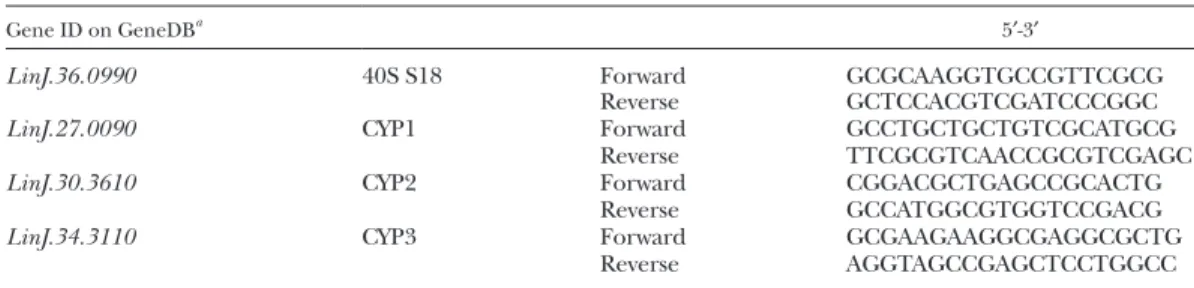
RNA (1 g) was reverse-transcribed using the SuperScript First-Strand Synthesis System (Thermo Fisher Scientific) accord-ing to the manufacturer’s recommendations. Transcriptional lev-els of specific mRNAs were determined by quantitative PCR (qPCR) using the SYBR Green PCR Master Mix and performed on a LightCycler 480 (Roche Diagnostics, Meylan, France). Primers (Eurofins Genomics, Les Ullis, France) used for qPCR are listed in
Table 1 for cytochrome P450 (CYP). Primers (Eurofins Genomics)
used to characterize macrophage phenotype were edited by
Accarias et al. (17). Expression ratios were calculated according to the expression level of the housekeeping gene LinJ.36.0990 coding for 40S ribosomal protein S18 (18) and RLP19 in the para-site and macrophage, respectively.
Leishmania CYP cloning
DNA was extracted from 2 × 10⁷ procyclic promastigotes with the Wizard Genomic DNA Purification Kit (Promega, Charbonnières-les-Bains, France) according to the manufacturer’s instructions for CYP2 and CYP3. L. infantum JPCM5 genome chromosome 27 (CYP1) was purchased from CliniSciences (Nanterre, France) cloned in the pUC57 vector with XbaI/NotI used as the cloning site. Primers (Eurofins Genomics) used for CYP cloning were 5′-CACCTTCTCAGCCTTGGGTTCCA-3′ and 5′-ATGGCAGCGTT-TAGTCGTCTCC-3′ for CYP2 and 5′-CCCGCGTACGGCGGCT-TCTTT-3′ and 5′-ATGGCCCCCACTGTCTCGCCA-3′ for CYP3. CYP-amplified inserts were subcloned in pCI-Neo (Promega) between XbaI and NotI restriction sites downstream to the consti-tutive promoter cytomegalovirus. Plasmid construction was verified by sequence alignment with a reference sequence from GenBank.
Transfection assay in the CHO cell line
Twenty-four hours before transfection, CHO cells were seeded in 24-well plates at 50% confluence in Opti-MEM medium. Trans-fections were carried out using Gene Juice transfection reagent (Merck, Kenilworth, NJ) with 500 ng of empty pCI-neo plasmid (Promega) containing CYP1, CYP2, or CYP3. Following an over-night incubation, culture medium was removed and replaced. Twenty-four hours thereafter cells were placed in HBSS (200 µl) containing vehicle (HBSS), 10 µM arachidonic acid (AA), or 10 µM DHA for 10 min at room temperature. Cells and HBSS were then collected for lipid quantification.
Bone marrow macrophage stimulation
Macrophage progenitors were isolated from the femurs of C57BL6 WT mice. Cells were differentiated and maintained in culture with 20 ng/ml macrophage colony-stimulating factor for 7 days before use. Bone marrow-derived monocytes were stimulated for 24 h with oxylipin extracted from procyclic or metacyclic pro-mastigotes (10 µl in MeOH). For some experiments, monocytes were also incubated with oxylipin extracted from Leishmania or vehicle (MeOH) and costimulated with 20 ng/ml IFN for 24 h. Cells were collected in HBSS and submitted to lipidomic analysis. For transcriptional analysis, cells were lysed in TRIzol, and mRNA was collected according to the manufacturer’s instructions.
Statistical analysis
Data are presented as means ± SEMs. GraphPad Prism version 6.0 (GraphPad, San Diego, CA) was used for statistical analysis. Multiple comparisons within groups were performed by the Kruskal-Wallis test followed by Dunn’s post hoc test. Comparisons among groups were performed by the Mann-Whitney test. Statistical sig-nificance was accepted at P < 0.05.
TABLE 1. Primers for RT-qPCR
Gene ID on GeneDBa 5′-3′
LinJ.36.0990 40S S18 Forward GCGCAAGGTGCCGTTCGCG
Reverse GCTCCACGTCGATCCCGGC
LinJ.27.0090 CYP1 Forward GCCTGCTGCTGTCGCATGCG
Reverse TTCGCGTCAACCGCGTCGAGC
LinJ.30.3610 CYP2 Forward CGGACGCTGAGCCGCACTG
Reverse GCCATGGCGTGGTCCGACG
LinJ.34.3110 CYP3 Forward GCGAAGAAGGCGAGGCGCTG
Reverse AGGTAGCCGAGCTCCTGGCC
a
See http://www.genedb.org.
at INRA Institut National de la Recherche Agronomique, on May 6, 2019
www.jlr.org
Downloaded from
.html
RESULTS
PUFA metabolite production by L. infantum varies during
infectivity acquisition
Free PUFA metabolite production during
metacyclogen-esis was assessed by LC/MS/MS in procyclic promastigotes
(noninfective stage) and metacyclic promastigotes
(infec-tive stage) obtained in vitro by preconditioning procyclic
promastigotes at acidic pH 5.4 for 24 h (19). Of the 32
PUFA metabolites assessed, 10 were detected and
quanti-fied in both the procyclic and metacyclic stages of L.
infan-tum (Fig. 1A). These metabolites were derived from linoleic
Fig. 1. PUFA metabolite production by L. infantum varies during infectivity acquisition. A: Production of PUFA metabolites assessed by LC/
MS/MS on Leishmania lipid extracts from procyclic and metacyclic promastigotes. Data shown are from six experiments conducted in tripli-cate; means ± SEMs are shown (n = 6). *P < 0.05, **P < 0.01, and ***P < 0.001 compared with the procyclic group. B: Percentage of main PUFAs (LA, ALA, DGLA, AA, and DHA) in free and total fatty acids assessed by GC with flame ionization detection on Leishmania lipid ex-tracts from procyclic and metacyclic promastigotes. Data shown are from three experiments conducted in triplicate; means ± SEMs are shown (n = 3). ALA, -linolenic acid; DGLA, dihomo--linolenic acid.
at INRA Institut National de la Recherche Agronomique, on May 6, 2019
www.jlr.org
Downloaded from
acid (LA) (9- and 13-HODE), AA [8(9)-EpETrE, 5-HETE,
8-HETE, 12-HETE, 15-HETE, and 5-oxo-ETE], and DHA
(14- and 17-HDoHE). The infective form (metacyclic
pro-mastigote stage) of the parasite showed an increased
concentration of 8-HETE, 14-HDoHE, and 17-HDoHE
compared with the noninfective form (Fig. 1A). On the
contrary, the quantity of 9-HODE and 8(9)-EpETrE was
re-duced during metacyclogenesis, as shown by the decreased
concentration of these metabolites in the metacyclic
com-pared with procyclic stage of the parasite (Fig. 1A). As the
quantified metabolites were epoxy or hydroxyl PUFAs, we
developed a method to fully quantify specifically these
fam-ilies (supplemental Figs. S1, S2). We confirmed the
in-crease in the concentration of 14-HDoHE, 17-HDoHE, and
8-HETE and the decrease of 9-HODE and 8(9)-EpEtrE in
metacyclic compared with procyclic promastigotes (Table
2, supplemental Figs. S1, S2). In addition, we observed an
increase in the concentration of
9,10-dihydroxyoctadece-noic acid, 9,10-DiHODE, 12-HODE, 11-HETE, 4-HDoHE,
20-HDoHE, and 19(20)-epoxydocosapentaenoic acid
(Ep-DPE) (Table 2, supplemental Figs. S1, S2). The
concentra-tion of their precursors was not different between the two
stages of the parasite (Fig. 1B), suggesting that the increase
in metabolite concentration is not dependent on the
amounts of precursors.
CYP-like proteins are responsible for PUFA metabolite
production in Leishmania
In an attempt to elucidate whether the PUFA
metabo-lism in L. infantum results from nonenzymatic autoxidation
or from an enzymatic activity, metacyclic promastigotes
were treated with different enzyme inhibitors. The
treat-ment of metacyclic promastigotes with either NDGA,
17-ODYA, mammalian CYP -hydroxylase inhibitor (20), or
clotrimazole and tioconazole, fungal CYP-dependent
14-demethylase inhibitors (21, 22), impaired the production
of most of the PUFA metabolites (Fig. 2). NDGA was the
only inhibitor that decreased the concentration of all of
the metabolites tested (Fig. 2). Other inhibitors decreased
differentially the concentration of the different
metabo-lites (Fig. 2). We highlighted five different profiles: 1)
9-HODE, 13-HODE, and 14-HDoHE decreased by NDGA,
17-ODYA, and clotrimazole; 2) 5-HETE, 12-HETE, and
5-oxo-ETE decreased only by NDGA; 3) 8-HETE and
17-HDoHE decreased by NDGA and 17-ODYA; 4) 15-HETE
decreased by all of the inhibitors; and 5) 8(9)-EpETrE
de-creased by NDGA, clotrimazole, and tioconazole.
The Leishmania genome database (GeneDB; Wellcome
Trust Sanger Institute, Hinxton, UK; http://www.genedb.
org) annotates only three putative CYP-like proteins coded
by LinJ.27.0090, LinJ.30.3610, and LinJ.34.3110 genes.
TABLE 2. PUFA metabolite concentration (nmol/1 × 108 parasites) in L. infantum at the procyclic or metacyclic
promastigote stage
Metabolites Procyclic Metacyclic P
9,10-DiHOME 17.68 ± 0.48 21.21 ± 0.69 0.0048 9,10-DiHODE 10.92 ± 0.21 12.01 ± 0.32 0.0298 15,16-DiHODE 5.83 ± 0.16 5.957 ± 0.12 0.8092 9-HODE 396.80 ± 34.93 249.91 ± 34.33 0.0298 10-HODE 14.24 ± 0.92 15.08 ± 0.75 0.4649 12-HODE 17.44 ± 0.81 19.32 ± 0.27 0.0298 13-HODE 962.11 ± 66.13 1,065.00 ± 67.89 0.3743 15-HODE 8.11 ± 0.10 7.41 ± 0.41 0.2198 5-HETE 26.29 ± 5.73 26.69 ± 4.25 1.0000 8-HETE 5.26 ± 0.29 13.68 ± 3.60 0.0048 11-HETE 3.87 ± 0.38 9.70 ± 0.90 0.0048 12-HETE 12.61 ± 3.94 12.15 ± 3.27 1.0000 15-HETE 43.57 ± 7.46 50.98 ± 1.15 0.7163 18-HETE 110.30 ± 7.48 112.01 ± 3.34 0.8092 5(S)-HETrE 0.970 ± 0.09 1.347 ± 0.13 0.1262 8(S)-HETrE 38.12 ± 6.69 44.35 ± 8.47 0.6879 12(S)-HETrE 12.25 ± 0.98 14.17 ± 0.93 0.1474 15(S)-HETrE 31.26 ± 2.36 39.37 ± 1.83 0.0766 8(9)-EpETrE 17.20 ± 2.07 2.91 ± 2.91 0.0247 4-HDoHE 146.10 ± 2.56 218.80 ± 17.02 0.0048 7-HDoHE 32.44 ± 2.44 37.94 ± 1.83 0.1262 8-HDoHE 114.80 ± 16.21 123.70 ± 10.91 0.1269 10-HDoHE 48.33 ± 3.24 47.18 ± 12.96 0.3760 11-HDoHE 63.44 ± 11.77 90.39 ± 8.89 0.1262 13-HDoHE 64.82 ± 3.81 64.05 ± 3.99 1.0000 14-HDoHE 112.10 ± 6.27 177.00 ± 18.81 0.0126 16-HDoHE 59.07 ± 3.22 69.35 ± 3.96 0.1262 17-HDoHE 129.1 ± 13.84 268.5 ± 39.05 0.0022 20-HDoHE 248.91 ± 24.76 327.50 ± 22.75 0.0431 7(8)-EpDPE 38.82 ± 3.271 46.52 ± 2.80 0.0646 10(11)-EpDPE 18.27 ± 1.89 24.83 ± 3.67 0.2403 13(14)-EpDPE 20.62 ± 2.43 19.75 ± 2.29 0.9360 16(17)-EpDPE 15.30 ± 2.08 19.19 ± 1.67 0.0646 19(20)-EpDPE 56.28 ± 5.09 69.91 ± 1.36 0.0301 10,11-DiHDPE 3.31 ± 0.29 3.64 ± 0.23 0.2598 13,14-DiHDPE 3.56 ± 0.30 3.99 ± 0.20 0.4616 16,17-DiHDPE 1.98 ± 0.63 2.22 ± 0.73 0.9348
DiHOME, dihydroxyoctadecenoic acid; HETrE, dihydroxyeicosatrienoic acid.
at INRA Institut National de la Recherche Agronomique, on May 6, 2019
www.jlr.org
Downloaded from
.html
These genes, conserved among species, are described here
as CYP1, CYP2, and CYP3, respectively. Our data confirm
the expression of these three CYP enzymes in Leishmania at
the two different stages (Fig. 3A). In order to further
deci-pher the putative role of CYP1, CYP2, and CYP3 enzymes in
the biosynthesis of PUFA metabolites, CHO cells
express-ing Leishmania recombinant CYP1, CYP2, or CYP3 enzymes
were treated with either AA or DHA as fatty acid metabolite
precursors, and the quantification of epoxy and hydroxyl
PUFA metabolites was performed (Fig. 3B, C). Only
15-HETE (Fig. 3B) and 4-, 14-, 16-, and 17-HDoHE (Fig. 3C)
were significantly increased in CYP1- and CYP3-transfected
cells compared with untransfected cells after AA (Fig. 3B)
or DHA treatment (Fig. 3C). In addition, transfection by
CYP3 increased the concentration of 10,11- and
13,14-dihy-droxyeicosatetraenoic acid (DiHDPE) (Fig. 3C). By
con-trast, the transfection of CHO cells with CYP2 had no effect
on PUFA metabolite synthesis (Fig. 3B, C). Thus, CYP1 and
CYP3 are implicated in the biosynthesis of AA- and
DHA-derived metabolites in Leishmania.
Lipids extracted from Leishmania promote the synthesis
of bioactive lipids in macrophages
To further explore the role of PUFA metabolites in the
parasite/host interaction, lipids extracted from procyclic
and metacyclic promastigotes were assessed for their ability
to induce the synthesis of lipid mediators in mouse bone
marrow-derived macrophages (BMDMs). PUFA
metabo-lites quantified in the parasite alone [9- and 13-HODE; 5-,
8-, 12-, and 15-HETE; 8(9)-EpETrE; and 14- and 17-HDoHE]
were increased in treated macrophages (Fig. 4). Treatment
with the parasite lipid extracts did not modify the
produc-tion of PGs such as PGD
2, 15d-PGJ
2, PGE
2, PGF
2, PGE
3, and
8-iso-PGA
2or thromboxane B2, lipoxin A4, and 14(15)-
EpETrE in BMDMs (supplemental Fig. S2). In contrast, the
synthesis of the specialized proresolving mediators (SPMs)
Fig. 2. Production of PUFA metabolites by metacyclic promastigotes is repressed by inhibitors of lipid metabolism. Production of PUFA
metabolites by metacyclic promastigotes was assessed by LC/MS/MS after 24 h of treatment with 100 µM NDGA, 100 µM 17-ODYA, 10 µM clotrimazole, or 10 µM tioconazole compared with untreated metacyclic promastigotes. Data shown are from three experiments conducted in triplicate; means ± SDs are shown (n = 3). *P < 0.05, **P < 0.01, and ***P < 0.001 significantly different from control group.
at INRA Institut National de la Recherche Agronomique, on May 6, 2019
www.jlr.org
Downloaded from
at INRA Institut National de la Recherche Agronomique, on May 6, 2019
www.jlr.org
Downloaded from
.html
RvD2, PDx, and 7-MaR1 was significantly higher in BMDMs
treated with metacyclic lipid extracts than those treated
with procyclic lipid extracts (Fig. 4B).
Lipids extracted from the metacyclic stage of Leishmania
promote the polarization of macrophages into a
proresolving M2 phenotype
M0 macrophages derived from bone marrow were
treated with lipids extracted from the two stages of the
par-asite. Only metacyclic lipid extracts induced the
transcrip-tional upregulation of the arginase 1 (Arg1) and CD206
genes, two bona fide markers of the alternatively activated
M2 macrophage phenotype (Fig. 5A) (23). At the same
time, no change in the expression level of the main
mark-ers of M1 macrophage phenotypes was observed,
irrespec-tive of parasite stages. When M0 macrophages were treated
with lipids extracted from the two stages of the parasite
concomitantly with INF- to induce M1 differentiation,
only metacyclic lipid extracts decreased several M1 markers
induced by IFN- treatment (Fig. 5B).
DISCUSSION
Metacyclogenesis is known to be crucial for infectivity
ac-quisition by promastigotes. Indeed, metacyclic
promasti-gotes are primarily characterized by their ability to infect
macrophages (6, 7, 24, 25) thanks to a higher resistance to
host complement-mediated lysis, which is necessary to save
time to infect macrophages. This is essentially due to the
structure and size of the surface lipophosphoglycan of
metacyclic promastigotes (25, 26) and the inactivation of
C3b by leishmanolysin (gp63), a protein that is not present
in procyclic promastigotes (4). Our study describes the
ability of metacyclic promastigotes to promote their own
survival by modifying the host macrophage phenotype
through the synthesis of specific PUFA metabolites.
We first described the production by L. infantum of 10
different PUFA metabolites derived from LA (18:2n-6), AA
(20:4n-6), and DHA (22:6n-3). The relative abundance of
these different metabolites is in agreement with the
abun-dance of their precursors (from least to most abundant:
AA, dihomo--linolenic acid, DHA, -linolenic acid, and
LA) (27). Contrary to its mammalian host, Leishmania is
able to produce LA from oleic acid (18:1n-9) via an oleate
desaturase. LA is then desaturated either to -linolenic
acid (18:3n-6) by 6 desaturase activity or to -linolenic
acid (18:3n-3) by the linoleate (15) desaturase. The
-linolenic acid is the major accumulated product in
Leishma-nia (28, 29), which explains the unique DHA-AA ratio
quantified in Leishmania as DHA derived from the -isomer
and AA from the -isomer. Independent of the amount of
AA and DHA, which is similar in metacyclic promastigotes
compared with procyclic promastigotes, our results
high-light the overproduction of different PUFA metabolites in
metacyclic promastigotes. The use of NDGA, mainly known
to inhibit mammalian 5-lipoxygenase, highly repressed the
synthesis of all the PUFA metabolites in L. infantum. Due to
the absence of the lipoxygenase coding gene in the
Leish-mania genome, we further assessed whether the NDGA
in-hibitory effect on PUFA metabolite synthesis was dependent
on its known activity as a nonspecific oxidase/reductase
inhibitor or CYP inhibitor (30). Treatment with different
CYP inhibitors differentially decreased the different PUFA
metabolites. In mammalian cells, CYP -hydroxylases
pro-vide pathways for the production of bioactive metabolites
from AA such as 18-, 19-, and 20-HETE mainly by the CYP4F
family (31–33). Interestingly, 17-ODYA, which decreased
the concentration of several metabolites in Leishmania, is a
suicide inhibitor of CYP4F, indicating possible -hydroxylase
activity present in the parasite (32). In addition, CYP2C8
and CYP2C9, characterized by their epoxygenase activity,
may also produce HETE and HODE by bisallylic
hydroxyl-ation activity (34). These results, in line with previous
stud-ies, suggest that CYP enzymes may participate in the
synthesis of PUFA metabolites in Leishmania. Even though
there are no studies available addressing the functional
in-volvement of CYP in PUFA metabolism in Leishmania, CYP
enzymatic activities were described in trypanosomatids,
in-cluding Leishmania sp. (35). A pivotal role of this enzyme in
Leishmania biology is supported by the antileishmanial
ac-tivity of the azole antifungals, which are known to act as
CYP inhibitors (36–38). The Leishmania genome database
(GeneDB, Wellcome Trust Sanger Institute, Hinxton, UK)
annotates only three putative CYP-like proteins, described
here as CYP1, CYP2, and CYP3. This group of proteins is
annotated as membrane-associated proteins with
monoox-ygenase activity; however, there are limited experimental
data focusing on their mechanisms of action and function
in the Leishmania life cycle. CYP1, classified as CYP5122A1
by the Cytochrome P450 Nomenclature Committee, has
been reported to play a key role in ergosterol metabolism
and to be associated with mechanisms of drug resistance
in L. donovani (39) as well as in cell growth and infectivity
(40). Promastigotes heterozygote for CYP1 gene deficiency
show a lesser capacity to differentiate into metacyclic forms
and to subsequently cause infection (40). CYP2 seems to be
underregulated in L. infantum axenic amastigotes during
amastigote morphogenesis (41). By expressing Leishmania
recombinant CYP1, CYP2, or CYP3 enzymes in CHO cells,
we demonstrated the ability of CYP1 and CYP3 to
synthe-tize PUFA metabolites, mainly hydroxylated DHA and in
particular 4-, 14-, 16-, and 17-HDoHE. The concentration
Fig. 3. CYP-like proteins are responsible for PUFA metabolite production in Leishmania. A: mRNA expression of CYP1, CYP2, and CYP3
genes in procyclic and metacyclic promastigotes determined by RT-qPCR. Data shown are from five experiments conducted in triplicate; means ± SEMs are shown (n = 5). B: Concentration of AA-derived metabolites in CHO cells expressing recombinant CYP1, CYP2, or CYP3 determined after cell media supplementation with AA. Data shown are from six experiments conducted in duplicate; means ± SEMs are shown (n = 6). *P < 0.05 and **P < 0.01 significantly different from the empty group. C: Concentration of DHA-derived metabolites in CHO cells expressing recombinant CYP1, CYP2, or CYP3 determined after cell media supplementation with DHA. Data shown are from six experi-ments conducted in duplicate; means ± SEMs are shown (n = 6). *P < 0.05 significantly different from the empty group.
at INRA Institut National de la Recherche Agronomique, on May 6, 2019
www.jlr.org
Downloaded from
Fig. 4. Lipids extracted from Leishmania promote the synthesis of lipid mediators by BMDMs. A: Production of PUFA metabolites assessed
by LC/MS/MS in BMDMs treated with 0% (M0), 50%, or 100% of lipids extracted from 1 × 10⁸ procyclic and metacyclic promastigotes. Data shown correspond to three experiments conducted in triplicate; means ± SEMs are shown (n = 3). *P < 0.05, **P < 0.01, and ***P < 0.001 significantly different from the M0 group. B: Production of proresolving mediators assessed by LC/MS/MS in BMDMs treated with 0% (M0), 50%, or 100% of lipids extracted from 1 × 10⁸ procyclic and metacyclic promastigotes. Data shown are from three experiments conducted in triplicate; means ± SEMs are shown (n = 3). **P < 0.05 and ***P < 0.01 significantly different from the M0 group and ###P < 0.001 significantly
different from the lipid extracts of the procyclic group.
at INRA Institut National de la Recherche Agronomique, on May 6, 2019
www.jlr.org
Downloaded from
.html
of several PUFA metabolites, increased in metacyclic
pro-mastigotes, was not affected by the transfection of the
dif-ferent CYPs in CHO cells, demonstrating that other
enzymes may be implicated in PUFA metabolite synthesis.
Among them, it is important to note the increase in
19(20)-EpDPE concentration, which plays a critical role in
monocyte lineage recruitment during inflammatory
reso-lution (42).
We also demonstrated that lipids extracted from
metacy-clic promastigotes did not modify the production of PGs by
macrophages but were able to increase the synthesis of the
SPMs RvD2, PDx, and 7-MaR1. This increase in SPMs could
result from the increase of their precursors (supplemental
Fig. S2), 14-HDoHE for maresin and 17-HDoHE for
resol-vin and protectin (43). Our results are in agreement with a
previous study demonstrating a key role of RvD1 in diffuse
cutaneous leishmaniasis and in intracellular L. amazonensis
replication in human macrophages (44). Upon parasitic
challenge, it has been shown that Leishmania actively
ma-nipulated the metabolic pathway to avoid the M1
polariza-tion of macrophages (7). We assessed the involvement of
PUFA metabolites from the parasite in this process. Lipid
extracts from metacyclic promastigotes were also able to
induce transcriptional upregulation of markers of the
al-ternatively activated M2 macrophage phenotype (Fig. 5A).
Our results are in agreement with the mouse model of
adi-pose tissue inflammation, in which DHA and RvD1 intake
induces a shift of the macrophage phenotype with an
in-crease in Arg1 and CD206, concomitantly with a
downregu-lation of TNF- and IL-6 expression (45). Transcriptional
levels of TNF-, IL-6, and the typical transcriptional factor
of the M1 phenotype induced by IFN-, IFN regulatory
Fig. 5. Lipids extracted from metacyclic
promasti-gotes promote the polarization of macrophages into a proresolving M2 phenotype. A: mRNA expression of markers of M1 and M2 polarization quantified in BMDMs after treatment with lipid metabolites from procyclic and metacyclic promastigotes. Data shown are means of fold changes ± SEMs compared with the undifferentiated monocyte (M0) from three inde-pendent experiments conducted in triplicate (n = 3). **P < 0.01 significantly different from the procyclic lipid extract group. B: Gene expression of M1 and M2 markers in BMDMs after treatment with lipid extracts from procyclic and metacyclic promastigotes and/or IFN-. Data shown are means of fold changes ± SEMs compared with the undifferentiated monocyte (M0) from three independent experiments conducted in triplicate (n = 3). *P < 0.05 and **P < 0.01 significantly different from the IFN- group. iNOS, inducible nitric oxide synthase; IRF5, IFN regulatory factor 5; Socs3, suppressor of cytokine signaling 3.
at INRA Institut National de la Recherche Agronomique, on May 6, 2019
www.jlr.org
Downloaded from
factor 5, decreased after coincubation with parasitic lipid
extracts from the infective form of Leishmania. In L.
major-infected mice, TNF- inhibition is associated with the
up-regulation of Arg1 and CD206 without affecting inducible
nitric oxide synthase expression (26). The balance in favor
of Arg1 rather than inducible nitric oxide synthase
re-presses reactive oxygen species production (46) and
in-creases polyamine biosynthesis, which is essential for the
intracellular growth of Leishmania (26, 47). Our data
dem-onstrate that DHA- and AA-derived metabolites from
meta-cyclic promastigotes also induce a minimal proinflammatory
response in the host to favor parasite growth. Therefore,
metabolites derived from DHA and specifically
overrepre-sented in metacyclic promastigotes (14- and 17-HDoHE)
could participate in building a safer environment to
facili-tate infection by affecting the polarization of immune
re-sponse during infection.
The results presented here demonstrate the production
of PUFA-derived metabolites in the Leishmania parasite.
Biosynthesis of these metabolites partially depends on
Leish-mania CYP-like protein (CYP1 and CYP3) activity. Infectivity
acquisition is accompanied by an increased production of
14- and 17-HDoHE, precursors of the proresolving lipid
me-diators maresin, D-series resolvin, and protectin (43). The
treatment of macrophages with lipids extracted from the
infective forms polarizes macrophages into an M2
pheno-type. M2 macrophages produce high levels of proresolving
bioactive lipids (RvD2, 7-Mar1, and PDx) and several
mark-ers of proresolving phenotypes as CD206 or Arg1,
promot-ing the survival and proliferation of Leishmania inside the
host cell.
The authors thank Jean-Luc Parrou for his advice on CYP cloning. The authors also thank Genotoul, Anexplo, US006/INSERM, the University of Toulouse, and the INSERM MetaToulLipidomics Facility-MetaboHUB ANR-11-INBS-010, where lipidomic analysis was performed.
REFERENCES
1. Bates, P. A., and L. Tetley. 1993. Leishmania mexicana: induction of metacyclogenesis by cultivation of promastigotes at acidic pH. Exp.
Parasitol. 76: 412–423.
2. Zilberstein, D., and M. Shapira. 1994. The role of pH and tem-perature in the development of Leishmania parasites. Annu. Rev.
Microbiol. 48: 449–470.
3. Saraiva, E. M., L. H. Pinto-da-Silva, J. L. Wanderley, A. C. Bonomo, M. A. Barcinski, and M. E. Moreira. 2005. Flow cytometric assess-ment of Leishmania spp metacyclic differentiation: validation by morphological features and specific markers. Exp. Parasitol. 110: 39–47.
4. Mojtahedi, Z., J. Clos, and E. Kamali-Sarvestani. 2008. Leishmania major: identification of developmentally regulated proteins in pro-cyclic and metapro-cyclic promastigotes. Exp. Parasitol. 119: 422–429. 5. Alcolea, P. J., A. Alonso, M. J. Gomez, M. Postigo, R. Molina, M.
Jimenez, and V. Larraga. 2014. Stage-specific differential gene ex-pression in Leishmania infantum: from the foregut of Phlebotomus perniciosus to the human phagocyte. BMC Genomics. 15: 849. 6. Alcolea, P. J., A. Alonso, A. Sanchez-Gorostiaga, M. Moreno-Paz,
M. J. Gomez, I. Ramos, V. Parro, and V. Larraga. 2009. Genome-wide analysis reveals increased levels of transcripts related with infectivity in peanut lectin non-agglutinated promastigotes of Leishmania in-fantum. Genomics. 93: 551–564.
7. Olivier, M., and D. J. Gregory. 2008. Interactions between Leishmania and the host macrophage. In Leishmania: After the Genome. P. J. Myler and N. Fasel, editors. Caister Academic Press, Norfolk, UK. 239–262.
8. Lefèvre, L., G. Lugo-Villarino, E. Meunier, A. Valentin, D. Olagnier, H. Authier, C. Duval, C. Dardenne, J. Bernad, J. L. Lemesre, et al. 2013. The C-type lectin receptors dectin-1, MR, and SIGNR3 con-tribute both positively and negatively to the macrophage response to Leishmania infantum. Immunity. 38: 1038–1049.
9. Liu, D., and J. E. Uzonna. 2012. The early interaction of Leishmania with macrophages and dendritic cells and its influence on the host immune response. Front. Cell. Infect. Microbiol. 2: 83.
10. de Menezes, J. P., E. M. Saraiva, and B. da Rocha-Azevedo. 2016. The site of the bite: Leishmania interaction with macrophages, neu-trophils and the extracellular matrix in the dermis. Parasit. Vectors.
9: 264.
11. Kabututu, Z., S. K. Martin, T. Nozaki, S. Kawazu, T. Okada, C. J. Munday, M. Duszenko, M. Lazarus, L. W. Thuita, Y. Urade, et al. 2003. Prostaglandin production from arachidonic acid and evi-dence for a 9,11-endoperoxide prostaglandin H2 reductase in Leishmania. Int. J. Parasitol. 33: 221–228.
12. Noverr, M. C., J. R. Erb-Downward, and G. B. Huffnagle. 2003. Production of eicosanoids and other oxylipins by pathogenic eu-karyotic microbes. Clin. Microbiol. Rev. 16: 517–533.
13. Le Faouder, P., V. Baillif, I. Spreadbury, J. P. Motta, P. Rousset, G. Chene, C. Guigne, F. Terce, S. Vanner, N. Vergnolle, et al. 2013. LC-MS/MS method for rapid and concomitant quantification of pro-inflammatory and pro-resolving polyunsaturated fatty acid metabolites. J. Chromatogr. B Analyt. Technol. Biomed. Life Sci. 932: 123–133.
14. Rund, K. M., A. I. Ostermann, L. Kutzner, J. M. Galano, C. Oger, C. Vigor, S. Wecklein, N. Seiwert, T. Durand, and N. H. Schebb. 2018. Development of an LC-ESI(-)-MS/MS method for the simultaneous quantification of 35 isoprostanes and isofurans derived from the major n3- and n6-PUFAs. Anal. Chim. Acta. 1037: 63–74.
15. Bligh, E. G., and W. J. Dyer. 1959. A rapid method of total lipid extraction and purification. Can. J. Biochem. Physiol. 37: 911–917. 16. Lillington, J. M., D. J. Trafford, and H. L. Makin. 1981. A rapid and
simple method for the esterification of fatty acids and steroid car-boxylic acids prior to gas-liquid chromatography. Clin. Chim. Acta.
111: 91–98.
17. Accarias, S., G. Lugo-Villarino, G. Foucras, O. Neyrolles, S. Boullier, and G. Tabouret. 2015. Pyroptosis of resident macrophages differ-entially orchestrates inflammatory responses to Staphylococcus au-reus in resistant and susceptible mice. Eur. J. Immunol. 45: 794–806. 18. Alcolea, P. J., A. Alonso, M. J. Gomez, A. Sanchez-Gorostiaga, M.
Moreno-Paz, E. Gonzalez-Pastor, A. Torano, V. Parro, and V. Larraga. 2010. Temperature increase prevails over acidification in gene expression modulation of amastigote differentiation in Leishmania infantum. BMC Genomics. 11: 31.
19. da Luz, R. I., M. Vermeersch, J. C. Dujardin, P. Cos, and L. Maes. 2009. In vitro sensitivity testing of Leishmania clinical field isolates: preconditioning of promastigotes enhances infectivity for macro-phage host cells. Antimicrob. Agents Chemother. 53: 5197–5203. 20. Edson, K. Z., and A. E. Rettie. 2013. CYP4 enzymes as potential drug
targets: focus on enzyme multiplicity, inducers and inhibitors, and therapeutic modulation of 20-hydroxyeicosatetraenoic acid (20-HETE) synthase and fatty acid omega-hydroxylase activities.
Curr. Top. Med. Chem. 13: 1429–1440.
21. Zhang, W., Y. Ramamoorthy, T. Kilicarslan, H. Nolte, R. F. Tyndale, and E. M. Sellers. 2002. Inhibition of cytochromes P450 by antifun-gal imidazole derivatives. Drug Metab. Dispos. 30: 314–318.
22. McLean, K. J., K. R. Marshall, A. Richmond, I. S. Hunter, K. Fowler, T. Kieser, S. S. Gurcha, G. S. Besra, and A. W. Munro. 2002. Azole antifungals are potent inhibitors of cytochrome P450 mono- oxygenases and bacterial growth in mycobacteria and streptomycetes.
Microbiology. 148: 2937–2949.
23. Murray, P. J., J. E. Allen, S. K. Biswas, E. A. Fisher, D. W. Gilroy, S. Goerdt, S. Gordon, J. A. Hamilton, L. B. Ivashkiv, T. Lawrence, et al. 2014. Macrophage activation and polarization: nomenclature and experimental guidelines. Immunity. 41: 14–20.
24. Yao, C., Y. Li, J. E. Donelson, and M. E. Wilson. 2010. Proteomic examination of Leishmania chagasi plasma membrane proteins: contrast between avirulent and virulent (metacyclic) parasite forms.
Proteomics Clin. Appl. 4: 4–16.
25. Sacks, D. L. 1989. Metacyclogenesis in Leishmania promastigotes.
Exp. Parasitol. 69: 100–103.
at INRA Institut National de la Recherche Agronomique, on May 6, 2019
www.jlr.org
Downloaded from
.html
26. Schleicher, U., K. Paduch, A. Debus, S. Obermeyer, T. Konig, J. C. Kling, E. Ribechini, D. Dudziak, D. Mougiakakos, P. J. Murray, et al. 2016. TNF-mediated restriction of arginase 1 expression in myeloid cells triggers type 2 NO synthase activity at the site of infection. Cell
Reports. 15: 1062–1075.
27. Bouazizi-Ben Messaoud, H., M. Guichard, P. Lawton, I. Delton, and S. Azzouz-Maache. 2017. Changes in lipid and fatty acid composi-tion during intramacrophagic transformacomposi-tion of Leishmania don-ovani complex promastigotes into amastigotes. Lipids. 52: 433–441. 28. Alloatti, A., and A. D. Uttaro. 2011. Highly specific methyl-end fatty-acid
desaturases of trypanosomatids. Mol. Biochem. Parasitol. 175: 126–132. 29. Uttaro, A. D. 2014. Acquisition and biosynthesis of saturated and
unsaturated fatty acids by trypanosomatids. Mol. Biochem. Parasitol.
196: 61–70.
30. Capdevila, J., L. Gil, M. Orellana, L. J. Marnett, J. I. Mason, P. Yadagiri, and J. R. Falck. 1988. Inhibitors of cytochrome P-450-dependent arachidonic acid metabolism. Arch. Biochem. Biophys.
261: 257–263.
31. Hsu, M. H., U. Savas, K. J. Griffin, and E. F. Johnson. 2007. Human cytochrome p450 family 4 enzymes: function, genetic variation and regulation. Drug Metab. Rev. 39: 515–538.
32. Kalsotra, A., and H. W. Strobel. 2006. Cytochrome P450 4F subfam-ily: at the crossroads of eicosanoid and drug metabolism. Pharmacol.
Ther. 112: 589–611.
33. Hardwick, J. P. 2008. Cytochrome P450 omega hydroxylase (CYP4) function in fatty acid metabolism and metabolic diseases. Biochem.
Pharmacol. 75: 2263–2275.
34. Bylund, J., T. Kunz, K. Valmsen, and E. H. Oliw. 1998. Cytochromes P450 with bisallylic hydroxylation activity on arachidonic and lin-oleic acids studied with human recombinant enzymes and with hu-man and rat liver microsomes. J. Pharmacol. Exp. Ther. 284: 51–60. 35. Berger, B. J., and A. H. Fairlamb. 1993. Cytochrome P450 in
try-panosomatids. Biochem. Pharmacol. 46: 149–157.
36. Croft, S. L., and G. H. Coombs. 2003. Leishmaniasis–current che-motherapy and recent advances in the search for novel drugs.
Trends Parasitol. 19: 502–508.
37. Roberts, C. W., R. McLeod, D. W. Rice, M. Ginger, M. L. Chance, and L. J. Goad. 2003. Fatty acid and sterol metabolism: potential
antimicrobial targets in apicomplexan and trypanosomatid parasitic protozoa. Mol. Biochem. Parasitol. 126: 129–142.
38. Balding, P. R., C. S. Porro, K. J. McLean, M. J. Sutcliffe, J. D. Marechal, A. W. Munro, and S. P. de Visser. 2008. How do azoles in-hibit cytochrome P450 enzymes? A density functional study. J. Phys.
Chem. A. 112: 12911–12918.
39. Pandharkar, T., X. Zhu, R. Mathur, J. Jiang, T. D. Schmittgen, C. Shaha, and K. A. Werbovetz. 2014. Studies on the antileishmanial mechanism of action of the arylimidamide DB766: azole inter-actions and role of CYP5122A1. Antimicrob. Agents Chemother. 58: 4682–4689.
40. Verma, S., A. Mehta, and C. Shaha. 2011. CYP5122A1, a novel cyto-chrome P450 is essential for survival of Leishmania donovani. PLoS
One. 6: e25273.
41. Rosenzweig, D., D. Smith, F. Opperdoes, S. Stern, R. W. Olafson, and D. Zilberstein. 2008. Retooling Leishmania metabolism: from sand fly gut to human macrophage. FASEB J. 22: 590–602.
42. Gilroy, D. W., M. L. Edin, R. P. De Maeyer, J. Bystrom, J. Newson, F. B. Lih, M. Stables, D. C. Zeldin, and D. Bishop-Bailey. 2016. CYP450-derived oxylipins mediate inflammatory resolution. Proc. Natl. Acad.
Sci. USA. 113: E3240–E3249.
43. Chiang, N., and C. N. Serhan. 2017. Structural elucidation and physiologic functions of specialized pro-resolving mediators and their receptors. Mol. Aspects Med. 58: 114–129.
44. Malta-Santos, H., B. B. Andrade, D. L. Zanette, J. M. Costa, P. T. Bozza, C. Bandeira-Melo, A. Barral, J. Franca-Costa, and V. M. Borges. 2017. Resolvin D1 drives establishment of Leishmania ama-zonensis infection. Sci. Rep. 7: 46363.
45. Titos, E., B. Rius, A. Gonzalez-Periz, C. Lopez-Vicario, E. Moran-Salvador, M. Martinez-Clemente, V. Arroyo, and J. Claria. 2011. Resolvin D1 and its precursor docosahexaenoic acid promote reso-lution of adipose tissue inflammation by eliciting macrophage po-larization toward an M2-like phenotype. J. Immunol. 187: 5408–5418. 46. Rath, M., I. Muller, P. Kropf, E. I. Closs, and M. Munder. 2014.
Metabolism via arginase or nitric oxide synthase: two competing arginine pathways in macrophages. Front. Immunol. 5: 532.
47. Colotti, G., and A. Ilari. 2011. Polyamine metabolism in Leishmania: from arginine to trypanothione. Amino Acids. 40: 269–285.
at INRA Institut National de la Recherche Agronomique, on May 6, 2019
www.jlr.org
Downloaded from