HAL Id: hal-03034241
https://hal.archives-ouvertes.fr/hal-03034241
Submitted on 1 Dec 2020
HAL is a multi-disciplinary open access
archive for the deposit and dissemination of sci-entific research documents, whether they are pub-lished or not. The documents may come from teaching and research institutions in France or abroad, or from public or private research centers.
L’archive ouverte pluridisciplinaire HAL, est destinée au dépôt et à la diffusion de documents scientifiques de niveau recherche, publiés ou non, émanant des établissements d’enseignement et de recherche français ou étrangers, des laboratoires publics ou privés.
Tularemia: A Case Series of Patients Diagnosed at the
National Reference Center for Rickettsioses From 2008
to 2017
Anne Darmon-Curti, François Darmon, Sophie Edouard, Aurélie Hennebique,
Thomas Guimard, Guillaume Martin-Blondel, Timothée Klopfenstein,
Jean-Philippe Talarmin, Didier Raoult, Max Maurin, et al.
To cite this version:
Anne Darmon-Curti, François Darmon, Sophie Edouard, Aurélie Hennebique, Thomas Guimard, et al.. Tularemia: A Case Series of Patients Diagnosed at the National Reference Center for Rickettsioses From 2008 to 2017. Open Forum Infectious Diseases, Oxford University Press, 2020, Open Forum Infectious Diseases, 7 (11), pp.1-9. �10.1093/ofid/ofaa440�. �hal-03034241�
Accepted Manuscript
© The Author(s) 2020. Published by Oxford University Press on behalf of Infectious Diseases Society of America.This is an Open Access article distributed under the terms of the Creative Commons Attribution-NonCommercial-NoDerivs licence (http://creativecommons.org/licenses/by-nc-nd/4.0/), which permits non-commercial reproduction and distribution of the work, in any medium, provided the
Tularemia: a case series of patients diagnosed at the National Reference Center for rickettsioses from 2008 to 2017.
Anne Darmon-Curti1, François Darmon2, Sophie Edouard1, Aurélie Hennebique3, 4, Thomas Guimard
5
, Guillaume Martin-Blondel6,7, Timothée Klopfenstein8, Jean-Philippe Talarmin9, Didier Raoult1,10,
Max Maurin3,4, and Pierre-Edouard Fournier10,11
1
Aix Marseille University, IRD, MEPHI, IHU-Méditerranée Infection, Marseille, France.
2
LIGM (UMR 8049), Ecole des Ponts ParisTech, UPE, Marne-la-Vallée, France
3
Centre National de Référence des Francisella, Institut de Biologie et de Pathologie, Centre Hospitalier Universitaire Grenoble Alpes, Grenoble, France.
4
Université Grenoble Alpes, Centre National de la Recherche Scientifique, TIMC-IMAG, Grenoble, France.
5
Infectious Diseases and Emergency Department, Centre Hospitalier de La Roche sur Yon, La Roche sur Yon, France.
6
Department of Infectious and Tropical Diseases Toulouse University Hospital, Toulouse, France.
7
UMR INSERM/CNRS 1043, Centre de Physiopathologie Toulouse-Purpan, Toulouse, France.
8
Department of Infectious Diseases, Besancon University Hospital, 25000 Besançon, France.
9
Infectious Diseases and Internal Medicine, Cornouaille Hospital, Quimper, France
10
Centre National de Référence des Rickettsia, Coxiella et Bartonella, IHU-Méditerranée Infection, Marseille, France.
11
Aix Marseille University, IRD, Service de Santé des Armées, VITROME, IHU-Méditerranée Infection, Marseille, France.
Accepted Manuscript
To whom the correspondence should be addressed:pierre-edouard.fournier@univ-amu.fr anne.darmon@ap-hm.fr
IHU - Méditerranée Infection
19-21 Boulevard Jean Moulin
13005 Marseille
Phone number: +33 4 13 73 24 01
Fax number: +33 4 13 73 24 02
Summary of the article main point:
This case series presents 177 patients with tularemia in France between 2008 and 2017: Glandular and ulcero-glandular forms were the most frequent. Two aortitis, an infectious endocarditis, a myocarditis, an osteoarticular infection and a splenic hematoma were diagnosed.
Accepted Manuscript
AbstractINTRODUCTION: We describe the epidemiological, clinical and prognostic aspects of 177 tularemia cases diagnosed at the National Reference Center for rickettsioses, coxiellosis and bartonelloses between 2008 and 2017.
METHODS: All patients with a microbiological diagnosis of tularemia made in the laboratory were included. Clinical-epidemiological data were collected retrospectively from clinicians in charge of patients using a standardized questionnaire. Diagnostic methods used were indirect
immunofluorescence serology, real-time polymerase chain reaction and universal PCR targeting the 16S rRNA gene.
RESULTS: The series included 54 females and 123 males (sex ratio 2.28, mean age 47.38 years). Eighty-nine (50.2%) were confirmed as having tularemia on the basis of a positive F. tularensis PCR or seroconversion, and 88 (49.8%) were considered as probable due to a single positive serum. The regions of France that were most affected included Pays de la Loire (22% of cases), Nouvelle
Aquitaine (18.6% of cases) and Grand-Est (12.4% of cases). Patients became infected mainly through contact with rodents or game (38 cases, 21.4%), through tick-bites (23 cases, 12.9%) or during outdoor leisure activities (37 cases, 20.9%). Glandular and ulcero-glandular forms were the most frequent (109 cases, 61.5%). Two aortitis, an infectious endocarditis, a myocarditis, an osteoarticular infection and a splenic hematoma were also diagnosed. Tularemia was discovered incidentally in 54.8% of cases. Seventy-eight patients were hospitalized, no deaths were reported.
DISCUSSION: Our data suggest that in an endemic area and/or in certain epidemiological contexts, tularemia should be sought in order to allow an optimized antibiotic therapy and a faster recovery.
Key Words: Tularemia; Francisella tularensis; case series; France; diagnosis
Accepted Manuscript
IntroductionTularemia is a zoonotic disease caused by Francisella tularensis. This facultative intracellular Gram-negative bacillus was isolated for the first time from flying squirrels in Tulare County in the USA by
Mac Coy and Chapin 1. Tularemia is endemic in North America2, Asia3 and Europe4–8.
Francisella tularensis subspecies tularensis (type A), almost restricted to North America, is
responsible for the most severe diseases. The lethality may be as high as 30% in untreated pulmonary
forms9. In contrast, F. tularensis subspecies holarctica (type B) is widely distributed in the Northern
Hemisphere, but also in southern Australia, and associated with a lethality rate < 1%. Clinical signs
vary according to the geographical area, season and thus mode of contamination6,7,10–13. Six clinical
forms are classically recognized: the ulceroglandular (ulcer associated with lymphadenopathy) and glandular (lymphadenopathy) forms, upon skin inoculation after animal contact (most often lagomorphs or small rodents) or by arthropod bite; the oculoglandular (conjunctivitis) form after conjunctival inoculation; the oropharyngeal (sore throat) form upon ingestion of contaminated water
or food14; and two systemic diseases, including the pneumonic (pneumonia) and the typhoidal forms
(mimicking symptoms of typhoid). Frequents complications of lymphadenopathies are suppurations
and even skin fistulas. Other complications such as sepsis or meningitis15 are more rarely reported and
usually occur in patients with comorbidities and/or immunosuppression.
Current knowledge of tularemia comes from data reported in highly endemic areas such as North
America11,16 and Northern Europe10. In contrast, few data exist for Western Europe17–19.
Here, we present the epidemiological, clinical, diagnostic and treatment data of 177 patients
diagnosed with tularemia in the French reference center for rickettsioses, coxiellosis and bartonelloses in Marseille, France, from January 1, 2008 to December 31, 2017.
Accepted Manuscript
MethodsDefinition of tularemia cases:
All patients with a suspected diagnosis of tularemia were considered as confirmed when they
exhibited compatible clinical findings and at least: (1) a positive real-time PCR (RT-PCR) and/or 16S rRNA PCR for F. tularensis; (2) seroconversion; or (3) a four-fold increase in immunofluorescence
serological titers, as previously described 20. Culture was carried out only on PCR-positive samples.
Patients with compatible clinical findings and a single positive serological test were considered probable cases.
Patients and clinical samples:
The present study is a retrospective analysis of epidemiological and clinical data from patients with tularemia. Data had been prospectively collected since 2008 using an anonymized and standardized questionnaire and stored on a secured computer by the French reference center for tularemia.
Patient Consent Statement
The present study was validated by the Ethics Committee of the “Institut hospitalo-universitaire” (IHU) Méditerranée Infection under reference 2017-029.
The consent form to be signed by patients was sent to the clinicians, who obtained the signature before transmitting the data.
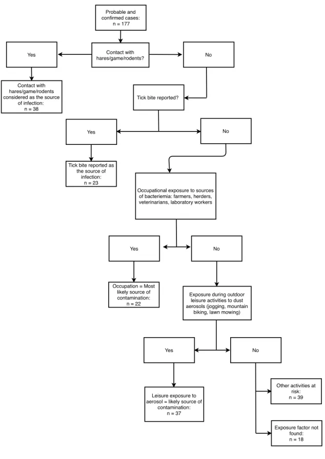
Mode of transmission:
We assigned the most likely source of contamination according to the scheme already used by Maille
et al. 18:
- Any patient infected with F. tularensis who reported direct contact with hares during the month before the onset of symptoms would have been infected by this exposure;
Accepted Manuscript
- A tick bite reported during the month before the onset of symptoms would be the route of contamination for any patient unless the patient also reported direct contact with hares;- An occupational exposure during the month before the onset of symptoms would constitute a circumstance of contamination unless the patient also reported direct contact with hares or a tick bite;
- Recreational activities resulting in exposure to aerosols or dust in the forest during the month before the onset of symptoms would be circumstances of contamination unless the patient reported direct contact with hares, tick bite or occupational exposure.
Laboratory diagnosis
Serology was performed using indirect immunofluorescence. Immunoglobulin G and M titers were measured. For this purpose, a formalin-inactivated antigen prepared in-house from a biovar I strain of
F. tularensis subspecies holarctica was used as previously described 20.
When lymph node biopsy specimens were available, a F. tularensis-specific RT-PCR assay targeting the yqaB gene (Ftul0541F, Ftul0541R and Ftul0541P), was also used for the detection of the
bacterium, as previously described 22. In addition, a patient with hip arthritis was detected positive
using a broad range PCR assay followed by sequencing the 16S RNA gene with the fD1 and rP2
primers was used22.
Outcome
Therapeutic success was defined as the resolution of symptoms (fever, abscess) one month after appropriate antibiotics (fluoroquinolone, tetracycline, or aminoglycoside) had been discontinued.
We considered as therapeutic failure the persistence of symptoms and/or the occurrence of complication(s) despite appropriate antibiotic.
Surgery was performed either as a diagnostic and/or a curative procedure. Diagnostic surgery was performed in patients presenting with enlarged lymph nodes of unknown or uncertain etiology.
Accepted Manuscript
Curative surgery was performed in patients with a complicated form such as abscess, prosthesis infection, aortitis.Statistics
The Fisher’s exact test was used to compare the number of therapeutic failures with doxycycline and fluoroquinolones, and assess the correlation between immunosuppression and the need for surgical treatment as well as the correlation between immunosuppression and the occurrence of therapeutic failure.
An ANOVA test was carried out to investigate statistical correlations between the occurrence of therapeutic failure or the need for surgical treatment and the time between the onset of symptoms and effective antibiotic therapy.
Results:
Confirmed and probable cases (Supplementary Figure1):
A total of 251 patients were included. For 68 patients, the laboratory criteria were not fulfilled. Another six patients that had been infected abroad were excluded. Overall, 89 confirmed and 88 probable cases (total 177 patients) were included in the study.
Epidemiological data:
The male-female sex ratio was 2.28 (54 female and 123 male). The mean age of patients was 47.38 +/- 17.8 years (range 2 to 89 years).
A total of 173 cases were sporadic, and four cases were clustered in two groups: two were household cases, and two were patients infected during an orientation race in a forest. The geographical
distribution of infected patients is depicted in Figure 1.
Exposure factors are detailed in Figure 2. The most common risk factor was a contact with hares, game (including boar, deer and doe) or rodents (21.5%), followed by exposure to aerosols (20.9%). No specific exposure factor was identified in 18 patients.
Accepted Manuscript
Contact with hares, game or rodents occurred more often in the winter season, while tick-bites were mostly reported in Summer (Supplementary Figure 2).Clinical data (Tables 1 and 2):
Significant background:
Thirteen of 142 patients (9.15%) were immunocompromised: eight patients had diabetes (one type I and seven type II). Four patients were on immunosuppressive therapy: one on mycophenolate mofetil, one on methotrexate, and two on anti-TNFα. The thirteenth patient suffered from liver cirrhosis. One patient had a vascular prosthesis.
The average incubation time of tularemia, calculated from 49 patients’ data, was 9.28+/- 9.7 days (range 1 to 43 days). Supplementary Figure 3 represents the distribution of the incubation times, which mostly ranged from one to five days. In our series, 7 patients exhibited unusually long incubation delays, ranging from 16 to 43 days.
Clinical forms:
Sixty-one (34.5%), 48 (27.1%), 32 (18%), 14 (7.9%), nine (5.0%) and four (2.3%) patients presented with an ulceroglandular, glandular, pleuropulmonary, typhoidal, oropharyngeal, or oculoglandular forms, respectively. We identified nine (5.1%) atypical forms which are presented in Supplementary Tables 1 and 2: a combined oculoglandular and ulceroglandular form, a pre-thyroid abscess, an ulceroglandular form associated with aortitis, a pleuropulmonary form associated with aortitis, a pleuropulmonary form associated with myocarditis, an aortic endocarditis, a total hip prosthesis infection, a pleuropulmonary form complicated by a splenic hematoma, and a pleuropulmonary form complicated by pericarditis.
The hip prosthesis infection occurred in a 49-year-old immunocompromised patient with cirrhosis. While drunk and walking outside, this male patient fell and suffered both femoral neck fracture and skin lacerations. He had a total hip replacement. In addition to hepato-renal decompensation of his cirrhosis with hepatic encephalopathy, the immediate aftermath of surgery was complicated by a
Accepted Manuscript
hematoma of the hip that had to be surgically drained. 16S rRNA PCR testing of a hematoma sample revealed the presence of F. tularensis. No culture was performed. The evolution of the illness was favorable with a three-month treatment with doxycycline followed by a one-stage exchange arthroplasty and additional antibiotic therapy with ciprofloxacin and gentamicin for three months.The case of splenic hematoma occurred in a 73-year-old male with a pleuropulmonary form of tularemia. This non-immunocompromised patient had a history of pericarditis, hypothyroidism and coronary stent placement. The diagnosis of tularemia was confirmed by PCR-detection of F.
tularensis DNA in a mediastinal lymph node. A spontaneous splenic hematoma developed during
hospitalization for tularemia as a complication of a moderate splenomegaly. The evolution of the illness was favorable after 21 days of doxycycline treatment.
We also diagnosed tularemia in a patient who developed a pre-thyroid lodge abscess without having any evident history of exposure to F. tularensis. While performing a mediastinoscopy for a
mediastinal lymph node biopsy, the surgeon unexpectedly drained an abscess from the pre-thyroid chamber. The diagnosis of tularemia was made by F. tularensis-specific PCR from the abscess pus. The evolution of the illness was favorable after surgical drainage and doxycycline therapy.
Symptoms
The patients’ symptoms are summarized in Table 2. The main symptoms included fever (85.0%), general impairment, (33.3%), chills (26.4%), myalgia (25.5%), sweats (25.0%) and arthralgia (21.2%).
Location of lymphadenopathies:
The upper limbs were the most frequent location, in 56 patients (31.6%), followed by the chest (46, 30.0%), lower limbs (38, 21.4%), neck (31, 17.5%) and abdomen (2, 1.1%). Twenty-nine patients (16.4%) had enlarged nodes in various anatomical sites.
Accepted Manuscript
Inoculation skin lesions:Twenty-nine patients (17.0%) exhibited an inoculation lesion on the upper limbs, 23 (13.5%) on the lower limbs, five (2.9%) on the trunk, three (1.7%) on the scalp, and one (0.6%) patient had both upper limb and facial ulcers.
Secondary dermatological lesions (all dermatological manifestations following the initial inoculation ulcer):
Nineteen patients had secondary dermatological lesions, including seven with lymphangitis ranging from the primary skin lesion to an adenopathy, five had a rash (location and type not specified), three had nodosum, one had cellulitis, one had erythema multiforme, one had pustules, and one had livedo reticularis.
Microbiological results
Initial presumed diagnosis:
For 67/148 patients (45.2%) for whom a suspected diagnosis was specified, tularemia was the main diagnostic request. In the other cases, the main suspected etiologies were: cat-scratch disease (44, 27.7%), Q fever (19, 12.8%), rickettsiosis (12, 7.9%), Whipple’s disease (3, 2.0%), ehrlichiosis (2, 1.3%) and anaplasmosis (1, 0.7%).
Confirmation of tularemia diagnosis:
Six cases (6.9%) were confirmed by seroconversion and five (5.8%) by a four-fold rise in antibody titers between two serum samples.
Confirmed diagnosis of tularemia was obtained in 78 patients (86%) by PCR testing of lymph node (64), skin (6), lung (3) or aorta biopsies (2), bronchoalveolar lavage (1) and joint (1) fluid.
In addition, F. tularensis was identified by 16S rRNA PCR-sequencing of a bacterial strain that could not be routinely identified.
Accepted Manuscript
Cultures were performed for 59 of the 78 PCR-proven specimens, and 6 (1%) were positive.Blood tests:
Blood test results are summarized in Supplementary Table 3. Twenty-two out of 91 (24.1%) patients had leukocytosis, 1.1% had leukopenia, 31.3% had liver cytolysis, 6.7% had thrombocytopenia, and 4.0% had thrombocytosis. The mean CRP rate was 81.6 +/- 60.5 mg/L (range 4 to 248).
Evolution:
Seventy-eight (53.4%) patients were hospitalized. Sixty (40.5%) developed complications, including lymph node abscesses (39), fistulizations (14), persistent asthenia (2), aortitis (2), and one case each of myocarditis, pericarditis and splenic hematoma.
Antibiotic therapy:
One hundred and seventy-two patients received an antibiotic therapy active against F. tularensis, including 118 who received one antibiotic. The most commonly prescribed antibiotics were doxycycline (106 patients) and an oral fluoroquinolone (42). Two patients were administered a fluoroquinolone and doxycycline. Table 3 and Supplementary Table 4 specify the effectiveness of treatments known to be active against tularemia. The mean duration of antibiotic therapy was 16 (min 7, max 28, SD 4.5) days for doxycycline, and 21 (min 7, max 84, SD 18.3) for fluoroquinolones. There were more therapeutic failures for fluoroquinolones (ciprofloxacin, levofloxacin, ofloxacin and unspecified fluoroquinolones, 14/43 [33%]) than for doxycycline (9/91 [9.9%], p=0.02). The mean duration between the onset of symptoms and the onset of effective antibiotics was 38.6 days (range 5 to 71, SD 16.7) for patients who presented with a therapeutic failure, versus 42.5 days (range 1 to 175, SD 33.4) for patients who recovered (p=0.64).
In contrast, 13 patients recovered without any effective antibiotic therapy, including four who benefited from lymph node excision, six who received an ineffective empirical antibiotic treatment, and three who neither received antibiotics nor underwent surgery. Their characteristics are specified in Supplementary Table 5. None of the thirteen patients was immunocompromised. There were four
Accepted Manuscript
pleuropulmonary forms, three oropharyngeal forms, three glandular forms, two typhoidal forms and one oculoglandular form.Surgery (Supplementary Table 4):
Eighty patients (51.6%) underwent surgery: 31 (20%) had a lymph node puncture, 47 had lymph node removal (30.3%), 1 (0.6%) had an aortic allograft, and 1 (0.6%) had a total hip prosthesis
replacement.
Thirty-four patients (21.9%) underwent surgery before effective antibiotic therapy, seventeen (10.9) after, eight (5.1%) at the same time. For twenty-one patients, the date of surgery was unknown. No data about surgery was available for 22 patients.
For patients who underwent surgery, the mean delay between onset of symptoms and effective antibiotic therapy was longer (52.1 days, range 7 to 175, SD 37.9) than for those who did not have surgery (mean 32.1 days, range 1 to 87 SD 19.5, p=0.006). In immunocompromised patients, 5/13 patients (38.5%) benefited from surgery, compared to 21/127 (16.6%) in immunocompetent patients (p=0.06).
Discussion
We describe 177 cases of tularemia diagnosed in the French reference center for rickettsioses,
coxiellosis and bartonelloses from January 1st, 2008 to December 31st, 2017. During this period, the
French reference center for tularemia in Grenoble recorded 317 cases, and Santé Publique France 611 cases. Among our 177 cases, three were previously described in a published series by the French
reference center for tularemia 19 and 44 cases included in a study published by Santé Publique France,
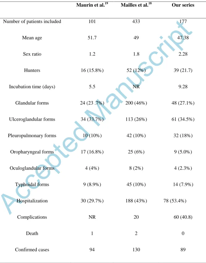
the French national public health agency 18. Table 1 compares these three French series.
The most affected regions were “Nouvelle Aquitaine”, “Pays de la Loire” and “Grand Est” (Figure 1),
as previously described 18,19. The annual distribution of cases is similar to that described in the
literature19: tick-borne contamination occurred more often in Summer, while contamination during
hunting or contact with infected animals occurred more often in Winter. The mean incubation time
Accepted Manuscript
was 9.28 days, which is longer than that usually described (3 to 5 days16). As expected, the most
common forms were ulceroglandular (34.5%) and glandular (27.1%). These forms had a significantly
higher therapeutic failure rate than the other forms (19.2% versus 7.6%, p<10-2). We also diagnosed
rare forms (Supplementary Tables 1 and 4), including one case each of hip prosthesis infection, splenic hematoma and pre-thyroid lodge abscess. To date, few cases of joint prosthetic infections have
been reported, affecting the knees 23,24 or hips 25. We assume that the hip prosthesis infection
complicated a skin wound contamination with F. tularensis. In contrast, we could not find any previous case of spontaneous splenic hematoma complicating a splenomegaly, or pre-thyroid abscess,
in the course of tularemia. However, a case of parapharyngeal abscess was reported in the literature26.
In addition, we diagnosed two cases of F. tularensis aortitis, one of which had previously been
published27, and a case of pericarditis complicating a pleuropulmonary form. Other cases of
pericarditis have been described in the literature28, including one previously diagnosed in our
laboratory in 2007 29. We also diagnosed a case of F. tularensis aortic endocarditis 30. Only four
confirmed cases of F. tularensis endocarditis have been described to date30–32. We also described a
probable case of myocarditis complicating a pleuropulmonary form. Indeed, the diagnosis was made on the basis of a positive serology and histological findings observed in a mediastinal
lymphadenopathy biopsy. To date, only two cases of F. tularensis myocarditis have been described in
the literature33, 34.
Seventy-eight of the 177 patients (53.4%) required hospitalization. The clinical signs exhibited by patients were not specific (Table 2). In our series, the majority of patients had fever (85%), lymphadenopathy (72.2%) and inoculation lesions (36%). During our study, we did not find a statistical relationship between the time to introduce effective treatment and the occurrence of therapeutic failures. The diagnosis was obtained in 99 patients (56%) by serology, and in 78 patients (44%) by PCR.
The antibiotic that was most commonly used in our series was doxycycline (administered 106 times as monotherapy), followed by a fluoroquinolone alone (42 patients). In our series, the observed
Accepted Manuscript
therapeutic failure rates for doxycycline and fluoroquinolones (9.9% and 32.5%, respectively) were high. It was previously demonstrated that the earlier effective antibiotic therapy is administered totularemia patients, the less often therapeutic failure occurs35,36. We observed long delays prior to
antibiotic therapy onset, which may explain such high therapeutic failure rates. However, the
therapeutic failure rate was significantly higher for fluoroquinolones (p = 0.002), which are currently
recommended with doxycycline as first line treatment in the less severe cases37 by the World Health
Organization (WHO)38. In their series, Rojas-Moreno described in their retrospective analysis of 17
patients no case of therapeutic failure under doxycycline for a mean duration of 21 days in addition to
surgical treatment 39. Castrillon et al. and Eliasson et al. both described more therapeutic failures with
doxycycline than with fluoroquinolones (42.8% and 3% therapeutic failures with doxycycline,
respectively, and 4.5% and no therapeutic failure with fluoroquinolones 7,10). In our series, among the
42 patients on fluoroquinolone alone, only 11 patients received ciprofloxacin 500mg 2 times per day (Table 3). We acknowledge the fact that the high failure rate observed for fluoroquinolones may at least in part be explained by the heterogeneity in molecule, dosage and treatment duration used.
Due to its clinical and pathological presentation, tularemia can be confused with cat-scratch disease or
tuberculosis 40,41. In our series, the suspected diagnosis by clinicians was tularemia in only 67 patients
(45.2%), and Bartonella infection in 44 patients (27.7%). Our data suggest that tularemia is probably underdiagnosed in France due to its unspecific clinical presentation and insufficient knowledge of the disease by clinicians. We suggest that tularemia should systematically be considered among the differential diagnoses of enlarged lymph nodes or prolonged fever in patients with outdoor activities.
Conclusion
Tularemia is endemic in France in the Nouvelle Aquitaine, Grand Est and Pays de la Loire regions. This infection is probably underdiagnosed. Although the ulceroglandular form was the most common, we identified rare forms of the disease such as splenic hematoma and osteoarticular infection. In endemic areas and in a consistent epidemiological context, diagnosing this disease allows optimized patient management.
Accepted Manuscript
References1. McCoy Georges W; Chapin Charles W. Further Observations on a Plague-Like Disease of
Rodents with a Preliminary Note on the Causative Agent , Bacterium tularense. J Infect Dis. 1912;10(1):61-72.
2. Petersen JM, Carlson JK, Dietrich G, et al. Multiple Francisella tularensis subspecies and
clades, tularemia outbreak, Utah. Emerg Infect Dis. 2008;14(12):1928-1930.
3. Chitadze N, Kuchuloria T, Clark D V., et al. Water-borne outbreak of oropharyngeal and
glandular tularemia in Georgia: Investigation and follow-up. Infection. 2009;37(6):514-521.
4. Hauri AM, Hofstetter I, Seibold E, et al. Investigating an airborne tularemia outbreak,
Germany. Emerg Infect Dis. 2010;16(2):238-243.
5. Kantardjiev T, Ivanov I, Velinov T, et al. Tularemia outbreak, Bulgaria, 1997-2005. Emerg
Infect Dis. 2006;12(4):678-680.
6. Reintjes R, Dedushaj I, Gjini A, et al. Tularemia outbreak investigation in Kosovo: case
control and environmental studies. Emerg Infect Dis. 2002;8(1):69-73.
7. Pérez-Castrillón JL, Bachiller-Luque P, Martín-Luquero M, Mena-Martín FJ, Herreros V.
Tularemia epidemic in northwestern Spain: clinical description and therapeutic response. Clin
Infect Dis. 2001;33:573-576.
8. Eliasson H, Broman T, Forsman M, Bäck E. Tularemia: Current Epidemiology and Disease
Management. Infect Dis Clin North Am. 2006;20(2):289-311.
9. Ellis J, Oyston PCF, Green M, Titball RW. Tularemia. ClinMicrobiolRev.
2002;15(0893-8512):631-646.
10. Eliasson H, Back E. Tularaemia in an emergent area in Sweden: an analysis of 234 cases in
five years. Scand J Infect Dis. 2007;39(10):880-889.
Accepted Manuscript
11. Evans ME, Gregory DW, Schaffner W, McGee ZA. Tularemia: a 30-year experience with 88
cases. Medicine (Baltimore). 1985;64(4):251-269.
12. Helvaci S, Gedikoǧlu S, Akalin H, Oral HB. Tularemia in Bursa, Turkey: 205 cases in ten
years. Eur J Epidemiol. 2000;16(3):271-276.
13. Feldman KA, Enscore RE, Lathrop SL, et al. An Outbreak of Primary Pneumonic Tularemia
on Martha’s Vineyard. N Engl J Med. 2001;345(22):1601-1606.
14. Maurin M, Gyuranecz M. Tularaemia: clinical aspects in Europe. Lancet Infect Dis.
2016;16(1):113-124.
15. Gangat N. Cerebral abscesses complicating tularemia meningitis. Scand J Infect Dis.
2007;39(3):258-261.
16. Matyas BT, Nieder HS, Telford SR. Pneumonic tularemia on Martha’s Vineyard: Clinical,
epidemiologic, and ecological characteristics. Ann N Y Acad Sci. 2007;1105:351-377.
17. Pilo P, Johansson A, Frey J. Identification of Francisella tularensis cluster in central and
western Europe. Emerg Infect Dis. 2009;15(12):2049-2051.
18. Mailles A, Vaillant V. 10 years of surveillance of human tularaemia in France. Euro Surveill.
2014 Nov 13;19(45):20956
19. Maurin M, Pelloux I, Brion JP, Del Banõ JN, Picard A. Human Tularemia in France,
2006-2010. Clin Infect Dis. 2011;53(10):2006-2006-2010.
20. Institut de veille sanitaire (InVS). Tularémie : définition de cas. Saint-Maurice: InVS. Available from: http://www.invs.sante.fr/Dossiers-
thematiques/Maladies-infectieuses/Zoonoses/Tularemie/ Comment-signaler-et-notifier-cette-maladie
21. Gouriet F, Levy PY, Samson L, Drancourt M, Raoult D. Comparison of the new InoDiag
automated fluorescence multiplexed antigen microarray to the reference technique in the serodiagnosis of atypical bacterial pneumonia. Clin Microbiol Infect. 2008;14(12):1119-1127.
Accepted Manuscript
22. Angelakis E, Roux V, Raoult D, Rolain J. Real-time PCR strategy and detection of bacterial
agents of lymphadenitis. Euopean J Clin Microbiol Infect Dis. 2009;28(11):1363-1368.
23. Cooper C., Van Caeseele P, Canvin J, Nicolle L. Chronic Prosthetic Device Infection with
Francisella tularensis. Clin Infect Dis. 1999;29:1589-1591.
24. Chrdle A, Trnka T, Musil D, Fucentese SF, Bode P, Keller PM. Francisella tularensis
Periprosthetic Joint Infections Diagnosed with Growth in Cultures. Am Soc Microbiol. 2019;57(August):1-7.
25. Rawal H, Patel A, Moran M. Unusual case of prosthetic joint infection caused by Francisella
Tularensis. BMJ Case Rep. 2017:3-5.
26. Koc S, Gürbüzler L, Yaman H, Eyibilen A, Salman N, Ekici A. Tularaemia presenting as
parapharyngeal abscess: Case presentation. J Laryngol Otol. 2012;126(5):535-537.
27. Briere M, Kaladji A, Douane F, et al. Francisella tularensis aortitis. Infection.
2016;44(2):263-265.
28. ADAMS CW. Tularemic pericarditis; report of two cases and review of literature. Dis Chest.
1958;34(6):632-639.
29. Landais C, Levy PY, Habib G, Raoult D. Pericardial effusion as the only manifestation of
infection with Francisella tularensis: A case report. J Med Case Rep. 2008;2:3-5.
30. Gaci R, Alauzet C, Selton-Suty C, et al. Francisella tularensis endocarditis: two case reports
and a literature review. Infect Dis (Auckl). 2017;49(2):128-131.
31. Tancik CA, Dillaha JA. Francisella tularensis Endocarditis Trovafloxacin-Induced Acute
Hepatitis. Clin Infect Dis. 2000;30:399-400.
32. Salit IE, Liles WC, Smith C. Tularemia endocarditis from domestic pet exposure. Am J Med.
2013;126(10):e1.
Accepted Manuscript
33. Franco S, Prieto JM, Balaguer I, Alvarez AP. Infection due to Francisella tularensis,
myocarditits ant dilated myocardiopathy. Enferm Infecc Microbiol Clin. 2010;28(10):752-753.
34. Frischknecht M, Meier A, Mani B, et al. Tularemia : an experience of 13 cases including a rare
myocarditis in a referral center in Eastern Switzerland ( Central Europe ) and a review of the literature. Infection. 2019;0(0):0.
35. Meric M, Willke A, Finke EJ, et al. Evaluation of clinical, laboratory, and therapeutic features
of 145 tularemia cases: The role of quinolones in oropharyngeal tularemia. Apmis. 2008;116(1):66-73.
36. Gozel MG, Engin A, Altuntas EE, et al. Evaluation of clinical and laboratory findings of
pediatric and adult patients with oropharyngeal tularemia in turkey: A combination of surgical drainage and antibiotic therapy increases treatment success. Jpn J Infect Dis. 2014;67(4):295-299.
37. Caspar Y, Hennebique A, Maurin M. Antibiotic susceptibility of Francisella tularensis subsp.
holarctica strains isolated from tularaemia patients in France between 2006 and 2016. J
Antimicrob Chemother. 2017;(January):1-5.
38. World Health Organization WHO. WHO Guidelines on Tularaemia. 2007:115.
39. Rojas-Moreno C, Bhartee H, Vasudevan A, Adiga R, Salzer W. Tetracyclines for Treatment of
Tularemia : A Case Series. Open forum Infect Dis. 2018;Sept:1-3.
40. Turhan V, Berber U, Haholu A, Salihoglu M, Ulcay A. Differential diagnosis of cervical
lymphadenitis mimicking malignancy due to tularemia: our experiences. Indian J Pathol
Microbiol. 2013;56(3):252-257.
41. Yildirim Ş, Turhan V, Karadenizli A, et al. Tuberculosis or tularemia? A molecular study in
cervical lymphadenitis. Int J Infect Dis. 2014;18(1):47-51.
Accepted Manuscript
ACKNOWLEDGMENTS:The authors thank the clinicians who provided the patients' clinical informations. The study
was funded by the Mediterranee Infection foundation and the French nationa research agency
under reference ANR-10-IAHU-03
AUTHOR’S CONTRIBUTION:
Anne Darmon-Curti and Pierre-Edouard Fournier analyzed the data;
Anne Darmon-Curti and Pierre-Edouard Fournier wrote the manuscript;
François Darmon performed the statistical analysis;
Aurélie Hennebique, Max Maurin, Thomas Guimard, Guillaume Martin-Blondel, Timothée
Klopfenstein, and Jean-Philippe Talarmin provided information on many patients;
Sophie Edouard and Didier Raoult critically revised the manuscript.
Potential conflicts of interest.
The authors of this manuscript have no conflicts of interest related to the work presented. All
authors have submitted the ICMJE Form for Disclosure of Potential Conflicts of Interest.
Accepted Manuscript
Table 1: Comparison of the data in our series with 2 other French seriesMaurin et al.19 Mailles et al.18 Our series
Number of patients included 101 433 177
Mean age 51.7 49 47.38
Sex ratio 1.2 1.8 2.28
Hunters 16 (15.8%) 52 (12%) 39 (21.7)
Incubation time (days) 5.5 NR 9.28
Glandular forms 24 (23 .7%) 200 (46%) 48 (27.1%) Ulceroglandular forms 34 (33.7%) 113 (26%) 61 (34.5%) Pleuropulmonary forms 10 (10%) 42 (10%) 32 (18%) Oropharyngeal forms 17 (16.8%) 25 (6%) 9 (5.0%) Oculoglandular forms 4 (4%) 8 (2%) 4 (2.3%) Typhoidal forms 9 (8.9%) 45 (10%) 14 (7.9%) Hospitalization 30 (29.7%) 188 (43%) 78 (53.4%) Complications NR 20 60 (40.8) Death 1 2 0 Confirmed cases 94 130 89
Accepted Manuscript
Strain isolation 16 30 0
PCR 39 75 78
Serologies (seroconversion, titer x4)
39 11 11
Probable cases 7 303 88
Cases in common with our series 3 63
Accepted Manuscript
Table 2: Clinical data of 177 patients in France (2008-2017): Comparison with data from 5previously reported case seriesA
Ref11 Ref8 Ref40 Ref7 Ref19 Our study
Country USA Sweden Turkey Spain France France
Number of patients (M/F) 88 (3.5) 234 (1.05) 205 (0.55) 142 (0.59) 101 (1.37) 177 (2.28)
Study period 1949-1979 2000-2004 1988-1998 1997-1998 2006-2010 2008-2017
Age ranges (means) 2-82 (NI) 1-88 (48.3) 5-75 (NI) 14-82 (52) 5-95 (52) 2-89 (47.4)
Main sources of infection Hares Mosquitos Water Hares Hares Hares
Fever 85 83 66 90.8 67.3 85.0 Headache 45 32 15 9.9 NA 13.4 Myalgia 31 NA 6 25.4 20.8 25.5 Arthralgia 15 NA NA 14.8 16.8 21.2 Cough 38 25 NA 24 5.9 9.2 Sore throat 15 3 58.5 16.9 15.8 7.0
Nausea and/or vomiting 17 23 NA 24 5.9 2.8
Diarrhea 10 9 NA 2.1 3.9 6.4
Inoculation lesion 59 88 NA 61.4 31.7 35.6
Secondary 5.7 29.9 14 17.6 2.9 11.9
Accepted Manuscript
dermatological lesion Lymphadenopathy 86 91.9 85 69.7 69.3 87.8 Pharyngitis 23.8 NA 29 16.2 19.8 4.2 Conjunctivitis NA NA 2 NA 2.9 3.5 Hepatosplenomegaly 16 NA 2 NA 2.9 5.6 Worsening of condition NA NA NA 38.7 3.9 33.8M/F: Male/female sex ratio, NA: no data available
A
: All data are given as percentages unless otherwise specified.
Accepted Manuscript
Table 3: Antibiotic therapy administred and therapeutic failure ratesAntibiotic therapy1
Total number of patients on antibiotic therapy
N (%) TF /* (%)
Doxycycline, 200 mg/d 104 (59) 9 /91 (9.9)
Ciprofloxacin, 500 mg X2/d 21 (12) 7/21 (33)
Unspecified fluoroquinolone and dosage 11(6.2) 4/9 (44)
Ofloxacin, 200 mg X2/d 5 (2.8) 1/5 (20)
Levofloxacin, 500 mg/d 4 (2.3) 2/4 (50)
Doxycycline, 200mg/d + IV amikacin 2 (1.1) 0/2 (0)
Ciprofloxacin, 750mg X2/d + IV gentamicin + doxycycline, 200mg/d 1 (0.56) 0 /1(0)
Unspecified fluoroquinolone + IV gentamicin 1 (0.60) 0 (0)
Levofloxacin, 500mg X2/d + doxycycline, 200mg/d 1 (0.60) 0/1 (0)
Ofloxacin, 400 mg X2/d 1 (0.60) 0/1(0)
IV amikacin 1 (0.60) 0/1 (0)
Accepted Manuscript
IV gentamicin 1 (0.60) 0/1 (0)
No antibiotic active on Francisella tularensis 13 (7.3) 0 /12(0)
No data 11 (6.2) 0 /1 (0)
N = number of patients; TF = therapeutic failure; * = number of patients for whom data is available; mg/d = milligram/day; X2/d = two times per day; IV = intravenous
1
If not specified, antibiotics were administred orally.
Accepted Manuscript
FIGURE TABLE AND GRAPHICSFigure 1: Geographical distribution of tularemia cases