HAL Id: inserm-00802220
https://www.hal.inserm.fr/inserm-00802220
Submitted on 19 Mar 2013
HAL is a multi-disciplinary open access
archive for the deposit and dissemination of
sci-entific research documents, whether they are
pub-lished or not. The documents may come from
teaching and research institutions in France or
abroad, or from public or private research centers.
L’archive ouverte pluridisciplinaire HAL, est
destinée au dépôt et à la diffusion de documents
scientifiques de niveau recherche, publiés ou non,
émanant des établissements d’enseignement et de
recherche français ou étrangers, des laboratoires
publics ou privés.
Saleh Al-Muhsen, Severine Letuve, Alejandro Vazquez-Tello, Mary Angeline
Pureza, Hamdan Al-Jahdali, Ahmed Bahammam, Qutayba Hamid, Rabih
Halwani
To cite this version:
Saleh Muhsen, Severine Letuve, Alejandro Vazquez-Tello, Mary Angeline Pureza, Hamdan
Al-Jahdali, et al.. Th17 cytokines induce pro-fibrotic cytokines release from human eosinophils.
Respi-ratory Research, BioMed Central, 2013, 14 (1), pp.34. �inserm-00802220�
R E S E A R C H
Open Access
Th17 cytokines induce pro-fibrotic cytokines
release from human eosinophils
Saleh Al-Muhsen
1, Severine Letuve
2, Alejandro Vazquez-Tello
1, Mary Angeline Pureza
1, Hamdan Al-Jahdali
1,3,
Ahmed S Bahammam
4, Qutayba Hamid
5and Rabih Halwani
1*Abstract
Background: Subepithelial fibrosis is one of the most critical structural changes affecting bronchial airway function during asthma. Eosinophils have been shown to contribute to the production of pro-fibrotic cytokines, TGF-β and IL-11, however, the mechanism regulating this process is not fully understood.
Objective: In this report, we investigated whether cytokines associated with inflammation during asthma may induce eosinophils to produce pro-fibrotic cytokines.
Methods: Eosinophils were isolated from peripheral blood of 10 asthmatics and 10 normal control subjects. Eosinophils were stimulated with Th1, Th2 and Th17 cytokines and the production of TGF-β and IL-11 was determined using real time PCR and ELISA assays.
Results: The basal expression levels of eosinophil derived TGF-β and IL-11 cytokines were comparable between asthmatic and healthy individuals. Stimulating eosinophils with Th1 and Th2 cytokines did not induce expression of pro-fibrotic cytokines. However, stimulating eosinophils with Th17 cytokines resulted in the enhancement of TGF-β and IL-11 expression in asthmatic but not healthy individuals. This effect of IL-17 on eosinophils was dependent on p38 MAPK activation as inhibiting the phosphorylation of p38 MAPK, but not other kinases, inhibited IL-17 induced pro-fibrotic cytokine release.
Conclusions: Th17 cytokines might contribute to airway fibrosis during asthma by enhancing production of eosinophil derived pro-fibrotic cytokines. Preventing the release of pro-fibrotic cytokines by blocking the effect of Th17 cytokines on eosinophils may prove to be beneficial in controlling fibrosis for disorders with IL-17 driven inflammation such as allergic and autoimmune diseases.
Keywords: Asthma, Eosinophils, Th17 cytokines, Pro-fibrotic cytokines, TGF-β, IL-11 Introduction
Asthma is a chronic inflammatory disorder of the lung that is usually associated with airway tissue remodelling. This term refers to the structural changes affecting lung tissue which normally include epithelial detach-ment, increased airway smooth muscle (ASM) mass, subepithelial fibrosis, mucous gland and goblet cell hyper-plasia, vascular changes, and edema [1-4]. Subepithelial fibrosis is one of the most critical structural changes associated with airway remodeling. In normal subjects, a
loose array of collagen fibrils resides beneath the basal membrane. In asthmatics, however, this layer is replaced by a dense network of extra-cellular matrix (ECM) proteins including collagens [5]. ECM protein depo-sition is known to be regulated by a number of cyto-kines and growth factors including TGF-β [6]. Several reports have shown that the majority of TGF-β1 mRNA positive cells in bronchial biopsies of severe asthmatics were eosinophils [7-9]. Eosinophils were also shown to produce IL-11 mRNA and protein [10]. These reports suggested that eosinophils could play an important role in regulating tissue fibrosis. IL-5 deficient mice experiments [11] and human studies [12] supported this hypothesis. In addition to lowe-ring eosinophil levels, using anti-IL-5 antibodies was
* Correspondence:rhalwani@ksu.edu.sa
1Asthma Research Chair and Prince Naif Center for Immunology Research,
Department of Paediatrics, College of Medicine, King Saud University, Riyadh, Saudi Arabia
Full list of author information is available at the end of the article
© 2013 Al-Muhsen et al.; licensee BioMed Central Ltd. This is an Open Access article distributed under the terms of the Creative Commons Attribution License (http://creativecommons.org/licenses/by/2.0), which permits unrestricted use, distribution, and reproduction in any medium, provided the original work is properly cited.
shown to be associated with reduced expression of ECM proteins particularly tenascin, lumican, and procollagen III [12].
Since its recent discovery, IL-17 has been described to be involved in various aspects of asthma pathogenesis. Elevated IL-17A levels were shown to correlate with in-creased airway hyper-responsiveness (AHR) in asthmatics [13]. In fact, IL-17 was shown to modulate airway struc-tural cells leading to tissue remodeling. Over-expression of IL-17 F resulted in goblet cell hyperplasia and mucin gene expression [14]. In addition, using an in vitro cell migration assay, Change et al. have recently shown that Th17-associated cytokines IL-17A, IL-17 F, and IL-22 promote migration of human ASMCs. These effects were shown to be mediated by selective activation of receptors on ASMCs, with IL-17A and IL-17 F acting through p38 MAPK activation while IL-22 acting through a distinct nuclear factor kB (NF-kB)–dependent signaling pathway [15]. These studies indicated for a role of IL-17 in airway remodeling and hence in regulating asthma pathogenesis.
Eosinophils have receptors for a number of mediators that are associated with asthma including Th1, Th2, and Th17 cytokines [16-18]. The expression of IL-17 cyto-kines was also associated with subepithelial fibrosis [19-21]. In fact, Th17 cytokines were shown to trigger the expression of pro-fibrotic cytokines in bronchial fibroblasts [22]. We, hence, hypothesized that IL-17 cytokines may induce eosinophils to produce pro-fibrotic cytokines. In this paper, we stimulated eosinophils, isolated from normal and asthmatic subjects, with Th17 cytokines as well as a group of Th1 and Th2 cytokines known to be associated with asthma. Eosinophil production of TGF-β and IL-11 pro-fibrotic cytokines was then investigated.
Materials and methods
Study subjects
Ten subjects with severe asthma (6 males and 4 females, mean age 33.3 ± 2.6) who met the criteria defined by ATS on refractory asthma [23] were recruited. To be classified as severe asthmatics, patients must have had high-dose inhaled corticosteroid: Budesonide 160 μg/twice a day (or equivalent) or daily anti-leukotriene for >50% of the last year, and at least 1 other add-on therapy on daily basis for the previous 12 months. They were also required to have two of the following criteria: daily short-acting β-agonist, persistent FEV1 <60% and FEV1/FVC <75% predicted, 1 urgent visit or at least 3 steroid bursts in the previous year, prompt deterioration with <25% steroid dose reduction, or previous near-fatal asthma within the last 3 years. Subject characteristics are summarized in Table 1. Exclusion criteria included smoking history or any other pulmonary diseases or co-existing medical conditions such as cardiac and renal diseases and uncontrolled hypertension. Ten normal control subjects (6 males and 4 females, mean age: 38.2 ± 3.4) were also recruited. All normal control subjects were non-smokers with normal lung function, no history or symptoms of allergy and respiratory diseases, and were not taking any medications for the preceding four weeks. The Ethics Committee of the King Khalid University Hospital in Riyadh reviewed and approved the study, and all subjects recruited signed written informed consent for the drawing of peripheral venous blood for the isolation of eosinophils.
Isolation and culture of eosinophils
Peripheral venous blood were drawn from patients with severe asthma (120-180 ml) (60 ml every 3 months) and from normal control subjects (180-240 ml) (60 ml
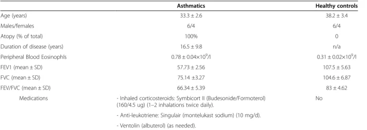
Table 1 Demography and spirometry data of the recruited subjects
Asthmatics Healthy controls
Age (years) 33.3 ± 2.6 38.2 ± 3.4
Males/females 6/4 6/4
Atopy (% of total) 100% 0
Duration of disease (years) 16.5 ± 9.8 n/a
Peripheral Blood Eosinophls 0.78 ± 0.04×10⁹/l 0.31 ± 0.02×10⁹/l
FEV1 (mean ± SD) 57.73 ± 2.56 107.5 ± 5.63
FVC (mean ± SD) 75.14 ±3.27 104.6 ± 6.87
FEV/FVC (mean ± SD) 66.34 ± 5.39 83 ± 4.62
Medications - Inhaled corticosteroids: Symbicort II (Budesonide/Formoterol) (160/4.5 ug) (1–2 inhalations twice daily).
No
- Anti-leukotriene: Singulair (montelukast sodium) (10 mg/d). - Ventolin (albuterol) (as needed).
Except for the medications indicated above, the patients were not receiving any other immunosupressive drugs. FEV1forced expiratory volume; FVC forced
every 3 months). Eosinophils were isolated by negative selection using MACS Isolation Kit (Miltenyi Biotec, Auburn, CA, USA) as previously described [24]. Neutrophils, monocytes and T cells were labeled with anti-CD16, anti-CD14 and anti-CD3 Abs respectively bound to immunemagnetic beads and separated with MACS LD Separation column. Eosinophil purity was consistently >98% as evaluated by Hema3 (Fisher) staining and the viability of freshly isolated eosino-phils was >99% as evaluated by Trypan blue dye exclusion. Isolated eosinophils were then cultured in RPMI + 10% FCS in the presence of 30 pg/ml IL-5 cytokine required for eosinophil survival in vitro [25,26]. Eosinophil viability ranged between 85 and 92% following stimulation and culture.
ELISA assay
Eosinophils (2×10⁶cells/ml cultured in 24 well plate) were stimulated with Th1 (IL-2, IFN-γ) (50 ng/ml), Th2 (IL-4, IL-5, IL-9, IL-13) (50 ng/ml), and Th17 (IL-17A, IL-17 F, IL-23) (10, 25, 50, or 100 ng/ml) cytokines (R&D Systems, Minneapolis, Minn., USA) for 24 hrs and supernatants were collected. In some experiments, eosinophils were treated with p38 mitogen-activated protein kinase (MAPK) inhibitors (SB 203580; 5 μM, Invivogen San Diego, CA, USA) or PI3K inhibitor (PI103; 5 μM, Cayman Chemical, Ann Arbor, Mich., USA) 2 hours prior to stimu-lation with IL-17. Levels of secreted TGF-β and IL-11 in supernatants were determined using ELISA assay (R&D Systems, Minneapolis, Minn., USA) according to the manufacturer instructions.
RNA extraction and real-time RT-PCR
Eosinophils were stimulated with cytokines (Th1 (50 ng/ml), Th2 (50 ng/ml) or Th17 (50 ng/ml)) for 4 hours prior to cell harvest. In some experiments, eosinophils were treated with p38 MAPK inhibitors (SB 203580; 5 μM), or PI3K inhibitor (PI103; 5 μM) 2 hours prior to stimulation with IL-17. Cells were then harvested, total RNA extracted (levels of RNA extracted from 2×10⁶ eosinophils were 18.4 ± 4.7 μg for asthmatic eosinophils and 16.3 ± 3.9 μg for healthy controls) (RNeasy Mini kit, Qiagen, CA, USA) and modulations of the level of expression of TGF-β and IL-11 mRNA were determined using quantitative RT-PCR (Applied Biosystems, 7900 Fast RT-PCR system). Specific primers for TGF-β and IL-11 were as follows: TGF-β: Forward: 5-CTGGACACCCTAA CCGTGAT-3, Reverse: 5-CTAGGCCGTGCTGCTGCT-3; IL-11: Forward: 5-GTGGCCAGATACAGCTGTCGC-3, Reverse: 5- GGTAGGACAGTAGGTCCGCTC-3. Relative expressions of TGF-β and IL-11 genes normalized with GAPDH were determined by the delta-delta Ct method [27].
Assessment of p38 MAPK phosphorylation by western analysis
2×10⁶eosinophil cells were starved using medium with 0.1% FBS for 18 hours. Cells were stimulated with 50 ng/mL IL-17A and IL-17 F for 0, 10, and 20 minutes and total proteins were extracted using lysis buffer (1% Triton X-100 containing protease and phosphatase inhibi-tor cocktails (Roche, Mannheim, Germany). Protein lysates (10 μg) were then resolved on 10% acrylamide SDS-PAGE gel and blots were probed with antibodies to phosphorylated p38 MAPK (Millipore) and total p38 MAPK (Millipore). Membranes were analyzed with an Odyssey IR scanner using Odyssey imaging software 3.0 (LI-COR Biosciences, Inc).
Statistical analysis
Data are presented as mean ± SD. Expression of
pro-fibrotic cytokines was evaluated using ANOVA
followed by Bonferroni-Dunn post hoc test. Non-parametric Mann–Whitney U test (Systat, version 7.0, SPSS, Chicago, IL) was used to evaluate significance in differential phosphorylation of MAPK. Values of p < 0.05 were considered statistically significant.
Results
Th1 and Th2 cytokines do not induce expression of eosinophil derived pro-fibrotic cytokines
The patho-physiological characteristics of lung tissue inflammation during severe asthma differ significantly from those of the milder disease. While the airway tissues of mild asthmatics usually present preferential Th2 cytokine profile [28], those from severe asthmatics show a Th17 lymphocyte infiltration and elevated cytokine levels, particularly Th1 cytokines (IFN-γ, IL-2), IL-17 and TGF-β [29-31]. Many T-helper cytokines were shown to play a significant role in regulating TGF-β expression and function in different types of cells [32-34]. However, their direct role in regulating eosinophil ability to produce pro-fibrotic cytokines was not studied. To investi-gate that, we first determined the basal expression levels of pro-fibrotic cytokines within peripheral blood eosinophils of 10 asthmatic and non-asthmatic individuals using real time RT-PCR. The levels of expression of TGF-β and IL-11 mRNA in eosinophils isolated from asthmatic individuals (ct values: TGF-β: 27.53 ± 0.21 IL-11: 28.80 ± 1.2) were comparable to those isolated from healthy controls (ct values: TGF-β: 27.70 ± 0.29 IL-11: 29.56 ± 0.86) (Figure 1A). Eosinophil supernatant IL-11 and TGF-β cytokines levels were also determined in the two groups using ELISA assay (Figure 1B). Similarly, no change in the secreted levels of these pro-fibrotic cytokines was detected between the two groups. We then investigated whether Th1 and Th2 cytokines play a role in regulating eosino-phils pro-fibrotic cytokines production. To do that, we
A
B
0 0.1 0.2 0.3 0.4 0.5 0.6 0.7 IL -1 1 /GA P D H Asthmatic Healthy 0 10 20 30 40 50 60 70 TGF-1 ( p g /m l) 0 10 20 30 40 50 60 70 IL-11 ( pg/ m l )C
D
Medium Th-2 GM-CSF Th-1 50ng/ml 0 20 40 60 80 100 120 140 160 IL -11 m R N A (% b a sa l) 0 20 40 60 80 100 120 140 160 180 TG F -β 1 m R N A (% b a sa l)stimulated 2×10⁶ eosinophil cells isolated from 10 asth-matic as well as healthy individuals with Th1 (IL-2, IL-12, and IFN-γ), and Th2 (IL-4, IL-5, IL-9, and IL-13) cytokines as well as GM-CSF for 4 hrs. Total RNA was then extracted from stimulated eosinophils and the level of IL-11 and TGF-β was determined using real time RT-PCR. As shown in Figure 1C-D, stimulating asthmatic eosinophils with Th1 or Th2 cytokines did not affect TGF-β (ct values range: 27.63 ± 0.21 to 27.58 ± 0.79) (Figure 1C) or IL-11 (ct values range: 28.67 ± 0.84 to 28.76 ± 0.18) (Figure 1D) m-RNA levels. Similar results were obtained at higher concentrations of Th1 and Th2 cytokines (100 ng/ml) as well as for eosinophils isolated from healthy controls (data not shown). These results indicated that neither Th1 nor Th2 cytokines play a significant role in regulating expression of eosinophil derived pro-fibrotic cytokines.
Th17 cytokines enhance the expression of eosinophil derived pro-fibrotic cytokines in asthmatic individuals
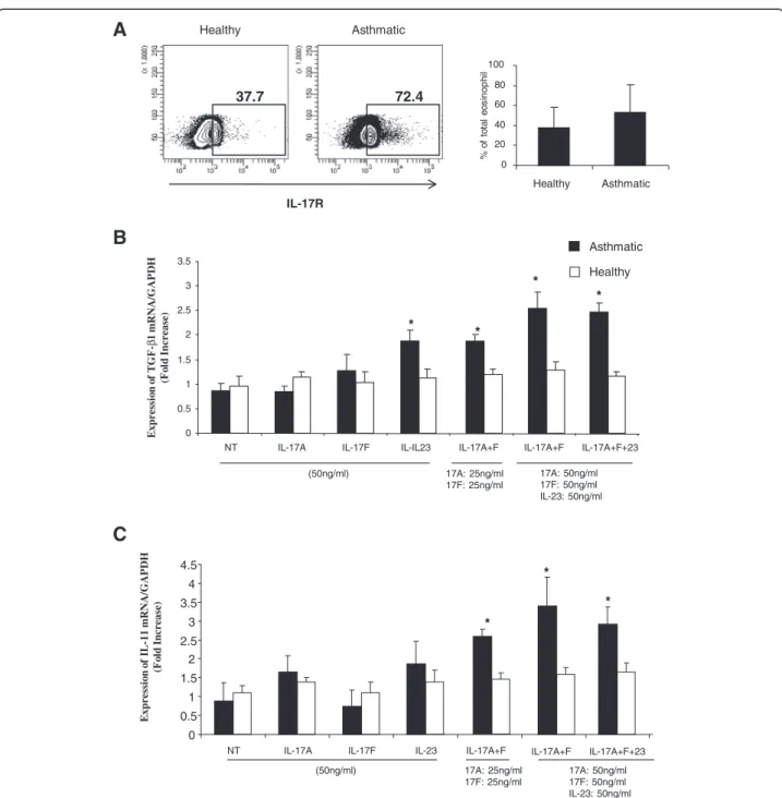
IL-17A enhanced the production of IL-6 and IL-11 in bronchial fibroblasts [22] while IL-17 F was shown to induce the expression of TGF-β in human umbilical vein endothelial cells (HUVECs) [35]. IL-17A and IL-17 F were recently shown to be over expressed in bronchial lung tissue of asthmatic patients compared to healthy controls [29] and their level of expression was associated with the severity of the diseases. Interestingly, using FACS and western analysis, eosinophils were also shown to express receptors for Th17 cytokines [16]. We, therefore, hypothesised that Th17 cytokines may induce eosinophils to produce pro-fibrotic cytokines. To investi-gate that, we first determined the expression levels of IL-17R on eosinophils isolated from both groups. As in-dicated in Figure 2A, eosinophils from both healthy and asthmatic subjects express IL-17R. Although asthmatic eosinophils express higher levels of IL-17R, this increase did not reach significance. We next stimulated 2×10⁶ eosinophils, isolated from 10 severe asthmatic patients and 10 healthy controls, with IL-17A, IL17F, as well as IL-23, another Th17 cytokine for 4 hrs. Total RNA was then extracted and eosinophil expression of TGF-β and IL-11 mRNA was measured using real-time PCR. As shown in Figure 2B, contrary to stimulating eosinophils with IL-17A and IL-17 F alone, stimulation with a com-bination of IL-17A + F (ct value: 24.39 ± 0.17, P = 0.031;
n = 10), or IL-23 (ct value: 24.42 ± 0.21, P = 0.025; n = 10) alone, induced a significant increase in the expression of eosinophil derived TGF-β. Further increase in TGF-β ex-pression was observed when stimulating with double the amount of the combined cytokines IL-17A + F (ct value: 24.06 ± 0.4, P = 0.012; n = 10) and or IL-17A + F + IL-23 (ct values: 23.98 ± 0.16, P = 0.018; n = 10). Inter-estingly, this increase in TGF-β production was only ob-served within eosinophils isolated from asthmatic patients. Stimulation of eosinophils isolated from non-asthmatic in-dividuals with Th17 cytokines had no effect on TGF-β production (ct values: IL-17A + F: 25.29 ± 0.15; IL-23: 25.22 ± 0.18; IL-17A + F (50 ng): 25.36 ± 0.14; IL-17A + F + 23 (50 ng): 25.78 ± 0.11, p = NS) (Figure 2B). Similarly, a combination of IL-17A and IL-17 F at different concentra-tions or IL-17A + F + IL-23 induced a significant increase in IL-11 mRNA expression within eosinophils isolated from asthmatics (ct values: IL-17A + F (25 ng): 24.61 ± 0.37, p = 0.029; 17A + F (50 ng): 23.71 ± 0.70, p = 0.009; IL-17A + F + 23 (50 ng): 23.80 ± 0.37, p = 0.014, n = 10)
but not healthy subjects (ct values: IL-17A + F
(25 ng): 25.18 ± 0.10; IL-17A + F (50 ng): 25.10 ± 0.11; IL-17A + F + 23 (50 ng): 25.00 ± 0.13, p = NS) (Figure 2C). To determine effective concentration inducing eosinophils release of TGF-β and IL-11 cytokines, a dose response ef-fect of Th17 cytokines was performed. Eosinophils were treated with increasing concentration of Th17 cytokines and levels of TGF-β and IL-11 in their supernatant were determined using ELISA assay (Figure 3A). Al-though low concentrations of Th17 cytokines in-duced pro-fibrotic cytokine secretion, a significant enhancement of TGF-β and IL-11 release was only attained at 50 ng/ml and above. At this concentration, the level of eosinophil derived TGF-β was significantly increa-sed following treatment with a combination of IL-17A + F (P = 0.032; n = 10), 23 (P = 0.049; n = 10) alone, or IL-17A + F + IL-23 (P = 0.043; n = 10) (Figure 3A). Similarly, IL-11 secreted levels were significantly upregulated
following stimulation with a combina- tion of
IL-17A + F (P = 0.035; n = 10), IL-23 (P = 0.048; n = 10)
alone, or IL-17A + F + IL-23 (P = 0.043; n = 10)
(Figure 3B). This data suggest that, in an asthmatic vironment, an additive effect of Th17 cytokines en-hance the production of eosinophils derived pro-fibrotic cytokines.
(See figure on previous page.)
Figure 1 Basal expression of pro-fibrotic cytokines by human eosinophils isolated from asthmatic and controls subjects. Eosinophils were isolated from 10 asthmatic and 10 controls subjects and total RNA was extracted from 2×10⁶million cells and quantified using real-time
PCR. A: Level of expression of TGF-β1 and IL-11 mRNA in eosinophils of asthmatic versus control subjects (n = 10). B: Levels of TGF-β1 and IL-11 cytokines within the supernatant of un-stimulated eosinophils (n = 10) as determined by ELISA assay. (C-D) Effect of Th1 and Th2 cytokines on asthmatic eosinophil TGF-β1 and IL-11 transcripts levels. Level of expression of TGF-β1 (C) and IL-11 (D) mRNA as quantified by real-time PCR following 4 hours exposure to mediators. Data is presented as percentage of basal expression (n = 10).
IL-17 cytokine enhance eosinophil derived TGF-β and IL-11 production through P38 MAP kinase activation
P38 mitogen-activated protein kinase (MAPK), being at a critical junction of the IL-17 signaling pathways, has been shown by various reports to be a key regulator element for the activity of IL-17 cytokines [15,16,36]. To
study the mechanism behind Th17 cytokines enhance-ment of eosinophil derived TGF-β production, eosino-phils were isolated from peripheral blood of 10 asthmatic patients as described above. 2×10⁶cells were treated, or not, with p38 MAPK or PI3K inhibitors (SB2035802 and PI103, respectively), or diluent control
A
B
Asthmatic Ex p re ss io n o f TG F -β 1 m RNA/ G AP DH (F o ld Increase) Ex p re ss io n o f IL -1 1 m RNA/ G AP DH (F o ld In crease) 0 0.5 1 1.5 2 2.5 3 3.5 4 4.5NT IL-17A IL-17F IL-23 IL-17A+F IL-17A+F+23
* * * IL-17A+F 17A: 25ng/ml 17F: 25ng/ml (50ng/ml) 17A: 50ng/ml 17F: 50ng/ml IL-23: 50ng/ml 17A: 50ng/ml 17F: 50ng/ml IL-23: 50ng/ml 0 0.5 1 1.5 2 2.5 3 3.5
NT IL-17A IL-17F IL-IL23 IL-17A+F IL-17A+F+23
Healthy * * * * IL-17A+F 17A: 25ng/ml 17F: 25ng/ml (50ng/ml)
C
0 20 40 60 80 100 Healthy Asthmatic Asthmatic % o f to ta l e o s in op hi l IL-17R Healthy 37.7 72.4Figure 2 IL-17 and IL-23 enhance eosinophil expression of pro-fibrotic cytokines. (A) Surface expression of IL-17R on eosinophils (1×10⁶cells) isolated from healthy and asthmatics was determined by flow cytometry. Blots are representative data for eosinophils isolated from one healthy control and one asthmatic patient. The graph shows arithmetic mean ± SD of IL-17R positive eosinophils as percentage of total eosinophils (n = 5). 2×10⁶ peripheral blood eosinophils isolated from 10 asthmatic and 10 controls subjects were stimulated with IL-17A, F, and IL-23 (50 ng/ml or 25 ng/ml) alone or in combination for 4 hrs. Total RNA was extracted and mRNA levels of TGF-β and IL-11 were then quantified using real-time PCR. mRNA expression levels of TGF-β (B) and IL-11 (C) were normalized with GAPDH for asthmatic versus healthy individuals.
(DMSO) 2 hours prior to stimulation with IL-17. As shown in Figure 4, inhibiting phosphorylation of p38 MAPK significantly decreased the level of TGF-β (P = 0.011 (IL-17A + F); P = 0.015 (IL-17A + F + 23); n = 10) and IL-11 (P = 0.021 (IL-17A + F); P = 0.026 (IL-17A + F + 23); n = 10) secreted into eosinophil supernatants 24 hrs following Th17 cytokine stimulation (Figure 4A, B). This blocking effect was only specific to p38 MAPK as diluent control or inhibitor of another kinase (PI3K) did not affect the supernatant levels of TGF-β and IL-11 (P = NS; n = 10). This data indicated that p38 MAPK activation is critical for IL-17 induced eosinophil derived pro-fibrotic cytokine production. To confirm p38 MAPK phosphory-lation following treatment with IL-17 cytokines, 2×10⁶ eosinophil cell were treated with IL-17A + F (50 ng/ml each) for 0, 10 and 20 minutes and the level of p38 MAPK
phosphorylation was then determined using western analysis. As shown in Figure 4C, stimulating eosi-nophils with a combination of IL-17A and IL-17 F resulted in phosphorylation of p38 MAPK which seems to peak at 10 minutes (3.5 fold increase, p = 0.039). Inhibiting p38 MAPK, PI3K, or ERK1/2, however, did not interfere with the ability of IL-23 to stimulate eosinophil to produce pro-fibrotic cytokines. This indicated that IL-23 may use other mechanisms to stimulate pro-fibrotic cytokine release that need to be further investigated.
Discussion
Eosinophils constitute a major source of TGF-β in asth-matic lung tissue [7-9]. Reduction of lung eosinophilia by anti–IL-5 therapy in humans [12] or genetic knock down in mice [11] significantly reduced airway fibrosis
IL
-11 (p
g/
m
l) Asthmatic Healthy
Cytokine conc. Cytokine conc.
B
* 0 50 100 150 200NS IL-17A IL-17F IL-23 IL-17A+F IL-17A+F+23
Asthmatic Healthy IL -11 (pg/ m l) * * (50ng/ml) TG F -β 1 (p g/ m l) Asthmatic Cytokine conc. 0 50 100 150 200 250 300 350 400
NS 17A 17F IL-23 IL-17A+F IL-17A+F+23
Asthmatic Healthy TG F -β 1 (p g/ m l) * * *
A
(50ng/ml) 0 50 100 150 200 250 NS 10ng 25ng 50ng 100ng 0 50 100 150 200 250 NS 10ng 25ng 50ng 100ng IL-17A IL-17F IL-23 IL-17A+F IL-17A+F+23 0 50 100 150 200 250 300 350 400 NS 10ng 25ng 50ng 100ng Healthy Cytokine conc. 0 50 100 150 200 250 300 350 400 NS 10ng 25ng 50ng 100ngFigure 3 Th17 cytokines enhance eosinophil production and release of pro-fibrotic cytokines. Levels of TGF-β (A) and IL-11 (B) in the supernatant of stimulated eosinophils (1×10⁶cells/0.5 ml) were determined 24 hrs following Th17 cytokine stimulation (0-100 ng/ml) using ELISA assay. Results are expressed as the arithmetic mean ± SD from 5 independent experiments. * = p < 0.05.
and pulmonary TGF-β1 levels. Here, we show, for the first time, that Th17 cytokines enhance eosino-phil derived TGF-β and IL-11 production. This effect of Th17 cytokines was prominent on eosinophils isolated from asthmatics but not healthy subjects. Our results clearly demonstrate that eosinophils con-stitute an additional site of action for Th17 cytokines in asthma supporting a role for IL-17 in regulating fibrosis and airway remodeling.
Although Th2 (IL-4, IL-5, and IL-13) cytokines has earlier been reported to regulate the expression of TGF-β1 by eosinophils [37,38], other studies had shown no effect of these cytokines on TGF-β expression [39]. Our results support the latest reports as we did not see any increase in
TGF-β or IL-11 mRNA or protein expression following stimulation with Th2 cytokines. Similarly, Th1 cyto-kines had no effect on eosinophil derived TGF-β expression. In fact, IFN-γ was previously shown to inhibit TGF-β production in human airway epithelial cells which is in consistence with our findings [38].
The enhancement of eosinophil derived pro-fibrotic cytokine release upon IL-17 cytokines stimulation was only significant in eosinophils isolated from asthmatic individuals. Although there was a slight upregulation of TGF-β and IL-11 expression in eosinophils isolated from healthy individuals upon IL-17 stimulation, this increase did not reach significance. Peripheral blood eosino-phils of asthmatic patients were shown to be primed p38
P-p38
0 10 20 (min)
Stimulation with IL-17A+F (50ng/ml)
C
0 1 2 3 4 * * Ph o sp h o -p 3 8 /To ta l p3 8 0 10 20 MinutesB
0 50 100 150 200NS IL-17A IL-17F IL-23 IL-17A+F IL-17A+F+23
Asthmatic Asthmatic+ SB IL -1 1 ( pg/m l) * * ** **
A
0 100 200 300 400NS 17A 17F IL-23 IL-17A+F IL-17A+F+23
Asthmatic Asthm+PI Asthm+SB TG F -β 1 ( p g/m l) ** ** * * *
Figure 4 P38 MAP Kinase activation is required for IL-17 enhancement of eosinophil derived pro-fibrotic cytokines. Eosinophils were isolated from peripheral blood of 10 asthmatic patients and 2×10⁶/ml cells were treated, or not, with p38 MAPK or PI3K inhibitors (SB2035802 and PI103, respectively) 2 hours prior to stimulation with IL-17 (50 ng/ml). Levels of TGF-β (A) and IL-11 (B) in the supernatant of stimulated eosinophils were then determined 24 hrs following Th17 cytokine stimulation using ELISA assay (n = 10). (C) Induction of p38 MAPK
phosphorylation by a combination of IL-17A and IL-17 F (50 ng/ml each) is detected by western analysis. The western data shown represent one of similar results from 4 independent experiments. * = p < 0.05 compared to non-stimulated (NS). ** = p < 0.05 compared to stimulated not inhibited.
compared to those of healthy subjects [40-42] which may render them more susceptible to IL-17 effect. Our results suggest that IL-17 cytokines enhance pro-fibrotic activity of activated, such as in the case of allergic and auto-immune diseases, but not resting eosinophils. Furthermore, our data indicated that asthmatic eosinophils may express higher levels of IL-17R than those of healthy controls (Figure 2A). IL-23 was shown to increase expression of IL-17RA and IL-17RC in eosinophils [16] and hence this observed poten-tial increase in IL-17R in asthmatic eosinophils could be due to increased serum IL-23 in those patients. Serum levels of IL-23 were shown to inversely correlate with level of pulmonary function (FEV1) of asthmatic patients in va-rious reports [43,44]. This may indicate that, due to the expected increase in serum IL-23 with asthma severity, eosinophils isolated from mild and moderate asthmatic patients may express higher levels of IL-17 receptors than eosinophils of healthy controls but lower than those of severe asthmatic patients. Understanding the correlation between asthmatic patients’ IL-23 serum levels, the expres-sion of IL-17R on peripheral blood eosinophils, and the severity of asthma requires further investigations.
Eosinophils are known to produce IL-17 cytokines [22] and IL-23 was shown to stimulate the expression of IL-17A cytokine [45]. This may indicate that IL-23 could stimulate eosinophils release of pro-fibrotic cytokines indirectly by triggering their release of IL-17A. This possibility, however, needs to be further investigated.
Stimulating eosinophils with IL-17 cytokines at a physiologically relevant concentration (25 ng/ml) resulted in an increase in TGF-β and IL-11 production although not to a significant levels (Figure 3). While stimulating eosinophils with either IL-17A or F alone did not enhance a significant increase in pro-fibrotic cytokines, using a combination of both cytokines did indicating an additive effect. Since both IL-17A and IL-17 F share the same IL-17R receptor [46], a concentration of around 25 ng/ml or more of each IL-17 cytokine seems to be required for efficient eosinophil derived pro-fibrotic cyto-kine release. This is more likely to be achieved in vivo through the additive effect of IL-17A and F rather than a high concentration of a single IL-17 cytokine alone.
Accumulating evidences from various reports indicate for a key role of p38 MAPK pathway in IL-17 cytokine activity on structural and inflammatory cells in asthma [15,36]. Binding of IL-17A and F to the IL-17RA and RC receptors on target cells triggers the recruitment of the U-box E3 ubiquitin ligase Act1 (CIKS). Act1 will in turn recruit TGF-β activated kinase that serves as the template for the activation of the transcription factors NF-kB, CEBPb (beta), as well as the MAPK pathways ERK1/ERK2 and p38 MAPK [47]. P38 MAPK, ERK, and JNK pathways were shown to regulate TGF-β transcrip-tion each in response to different stimuli [48]. Our data
suggest that IL-17 cytokines stimulate TGF-β transcrip-tion via the activatranscrip-tion of p38 MAPK but not PI3K or ERK1/2 MAPK (data not shown) pathways. IL-23, however, seems to use another mechanism as inhibiting those pathways did not affect its ability to stimulate TGF-β and IL-11 production.
Conclusions
Data presented herein suggest a new role for Th17 cytokines in airway remodeling during asthma. IL-17 cytokines seem to contribute to airway tissue fibrosis by enhancing production of eosinophil derived pro-fibrotic cytokines. This role of IL-17 was dependent on p38 MAPK activation. Therefore, upstream activators of p38 MAPK within the IL-17R pathway may represent an attractive target in corticosteroid-unresponsive diseases [49,50]. Preventing the release of TGF-β by blocking the effect of IL-17 on eosinophils may also prove efficient in controlling fibrosis for disorders with IL-17 driven inflammation such as allergic and autoimmune diseases.
Competing interests
The authors declare that they have no competing interests. Authors’ contributions
SM carried out the real-time PCR for Th17 experiments and participated in the design of the study. SL carried out the real-time PCR and ELISA for Th1 and Th2 experiments. AVT participated in the design of the study and performed the statistical analysis. MAP carried out the ELISA for Th17 experiments. HJ contributed in recruiting patients to the study. QH contributed in study design and data analysis. RH conceived of the study, lead efforts on its design and coordination and finalized the manuscript draft. All authors read and approved the final manuscript.
Acknowledgements
This study was supported by a grant from the National Plan for Sciences and Technology, King Saud University, Riyadh, Saudi Arabia (grant number 09-BIO907-02). The funders had no role in study design, data collection and analysis, decision to publish, or preparation of the manuscript. Author details
1
Asthma Research Chair and Prince Naif Center for Immunology Research, Department of Paediatrics, College of Medicine, King Saud University, Riyadh, Saudi Arabia.2Institut National de la Santé et de la Recherche Médicale (Inserm) U700 and Université Paris 7, Faculté de Médecine Denis Diderot, Site Bichat, Paris, France.3King Saud University for health sciences, Riyadh, Saudi Arabia.4Pulmonary Medicine Department, University Sleep Disorders Center,
College of Medicine, King Saud University, Riyadh, Kingdom of Saudi Arabia.
5Meakins-Christie Laboratories, McGill University, Montreal, QC, Canada.
Received: 14 November 2012 Accepted: 7 March 2013 Published: 13 March 2013
References
1. Vignola AM, Mirabella F, Costanzo G, Di Giorgi R, Gjomarkaj M, Bellia V, Bonsignore G: Airway remodeling in asthma. Chest 2003, 123:417S–422S. 2. Vignola AM, Kips J, Bousquet J: Tissue remodeling as a feature of
persistent asthma. J Allergy Clin Immunol 2000, 105:1041–1053. 3. Benayoun L, Druilhe A, Dombret MC, Aubier M, Pretolani M: Airway
structural alterations selectively associated with severe asthma. Am J Respir Crit Care Med2003, 167:1360–1368.
4. Sumi Y, Hamid Q: Airway remodeling in asthma. Allergol Int 2007, 56:341–348.
5. Roche WR, Beasley R, Williams JH, Holgate ST: Subepithelial fibrosis in the bronchi of asthmatics. Lancet 1989, 1:520–524.
6. Parameswaran K, Willems-Widyastuti A, Alagappan VK, Radford K, Kranenburg AR, Sharma HS: Role of extracellular matrix and its regulators in human airway smooth muscle biology. Cell Biochem Biophys 2006, 44:139–146.
7. Minshall EM, Leung DY, Martin RJ, Song YL, Cameron L, Ernst P, Hamid Q: Eosinophil-associated TGF-beta1 mRNA expression and airways fibrosis in bronchial asthma. Am J Respir Cell Mol Biol 1997, 17:326–333. 8. Flood-Page P, Menzies-Gow A, Phipps S, Ying S, Wangoo A, Ludwig MS,
Barnes N, Robinson D, Kay AB: Anti-IL-5 treatment reduces deposition of ECM proteins in the bronchial subepithelial basement membrane of mild atopic asthmatics. J Clin Invest 2003, 112:1029–1036.
9. Vignola AM, Chanez P, Chiappara G, Merendino A, Pace E, Rizzo A, la Rocca AM, Bellia V, Bonsignore G, Bousquet J: Transforming growth factor-beta expression in mucosal biopsies in asthma and chronic bronchitis. Am J Respir Crit Care Med1997, 156:591–599.
10. Minshall E, Chakir J, Laviolette M, Molet S, Zhu Z, Olivenstein R, Elias JA, Hamid Q: IL-11 expression is increased in severe asthma: association with epithelial cells and eosinophils. J Allergy Clin Immunol 2000, 105:232–238.
11. Cho JY, Miller M, Baek KJ, Han JW, Nayar J, Lee SY, McElwain K, McElwain S, Friedman S, Broide DH: Inhibition of airway remodeling in IL-5-deficient mice. J Clin Invest 2004, 113:551–560.
12. Flood-Page PT, Menzies-Gow AN, Kay AB, Robinson DS: Eosinophil’s role remains uncertain as anti-interleukin-5 only partially depletes numbers in asthmatic airway. Am J Respir Crit Care Med 2003, 167:199–204. 13. Barczyk A, Pierzchala W, Sozanska E: Interleukin-17 in sputum correlates
with airway hyperresponsiveness to methacholine. Respir Med 2003, 97:726–733.
14. Oda N, Canelos PB, Essayan DM, Plunkett BA, Myers AC, Huang SK: Interleukin-17 F induces pulmonary neutrophilia and amplifies antigen-induced allergic response. Am J Respir Crit Care Med 2005, 171:12–18. 15. Chang Y, Al-Alwan L, Risse PA, Roussel L, Rousseau S, Halayko AJ, Martin JG,
Hamid Q, Eidelman DH: TH17 cytokines induce human airway smooth muscle cell migration. J Allergy Clin Immunol 2011, 127:1046–1053. e1041-1042.
16. Cheung PF, Wong CK, Lam CW: Molecular mechanisms of cytokine and chemokine release from eosinophils activated by IL-17A, IL-17 F, and IL-23: implication for Th17 lymphocytes-mediated allergic inflammation. J Immunol2008, 180:5625–5635.
17. Dubois GR, Schweizer RC, Versluis C, Bruijnzeel-Koomen CA, Bruijnzeel PL: Human eosinophils constitutively express a functional interleukin-4 receptor: interleukin-4 -induced priming of chemotactic responses and induction of PI-3 kinase activity. Am J Respir Cell Mol Biol 1998, 19:691–699.
18. Riedel D, Lindemann A, Brach M, Mertelsmann R, Herrmann F: Granulocyte-macrophage colony-stimulating factor and interleukin-3 induce surface expression of interleukin-2 receptor p55-chain and CD4 by human eosinophils. Immunology 1990, 70:258–261.
19. Lajoie-Kadoch S, Joubert P, Letuve S, Halayko AJ, Martin JG, Soussi-Gounni A, Hamid Q: TNF-alpha and IFN-gamma inversely modulate expression of the IL-17E receptor in airway smooth muscle cells. Am J Physiol Lung Cell Mol Physiol2006, 290:L1238–L1246.
20. Letuve S, Lajoie-Kadoch S, Audusseau S, Rothenberg ME, Fiset PO, Ludwig MS, Hamid Q: IL-17E upregulates the expression of proinflammatory cytokines in lung fibroblasts. J Allergy Clin Immunol 2006, 117:590–596. 21. Kuhn C 3rd, Homer RJ, Zhu Z, Ward N, Flavell RA, Geba GP, Elias JA: Airway
hyperresponsiveness and airway obstruction in transgenic mice. Morphologic correlates in mice overexpressing interleukin (IL)-11 and IL-6 in the lung. Am J Respir Cell Mol Biol 2000, 22:289–295.
22. Molet S, Hamid Q, Davoine F, Nutku E, Taha R, Page N, Olivenstein R, Elias J, Chakir J: IL-17 is increased in asthmatic airways and induces human bronchial fibroblasts to produce cytokines. J Allergy Clin Immunol 2001, 108:430–438.
23. Proceedings of the ATS workshop on refractory asthma: current understanding, recommendations, and unanswered questions. American Thoracic Society. Am J Respir Crit Care Med 2000, 162:2341–2351. 24. Hansel TT, De Vries IJ, Iff T, Rihs S, Wandzilak M, Betz S, Blaser K, Walker C:
An improved immunomagnetic procedure for the isolation of highly purified human blood eosinophils. J Immunol Methods 1991, 145:105–110. 25. Yamaguchi Y, Hayashi Y, Sugama Y, Miura Y, Kasahara T, Kitamura S, Torisu M,
Mita S, Tominaga A, Takatsu K: Highly purified murine interleukin 5
(IL-5) stimulates eosinophil function and prolongs in vitro survival. IL-5 as an eosinophil chemotactic factor. J Exp Med 1988, 167:1737–1742. 26. Okada S, Hagan JB, Kato M, Bankers-Fulbright JL, Hunt LW, Gleich GJ, Kita H:
Lidocaine and its analogues inhibit IL-5-mediated survival and activation of human eosinophils. J Immunol 1998, 160:4010–4017.
27. Livak KJ, Schmittgen TD: Analysis of relative gene expression data using real-time quantitative PCR and the 2(−Delta Delta C(T)) Method. Methods 2001, 25:402–408.
28. Herrick CA, Bottomly K: To respond or not to respond: T cells in allergic asthma. Nat Rev Immunol 2003, 3:405–412.
29. Al-Ramli W, Prefontaine D, Chouiali F, Martin JG, Olivenstein R, Lemiere C, Hamid Q: T(H)17-associated cytokines (IL-17A and IL-17 F) in severe asthma. J Allergy Clin Immunol 2009, 123:1185–1187.
30. Truyen E, Coteur L, Dilissen E, Overbergh L, Dupont LJ, Ceuppens JL, Bullens DM: Evaluation of airway inflammation by quantitative Th1/Th2 cytokine mRNA measurement in sputum of asthma patients. Thorax 2006, 61:202–208.
31. Halwani R, Al-Muhsen S, Al-Jahdali H, Hamid Q: Role of transforming growth factor-beta in airway remodeling in asthma. Am J Respir Cell Mol Biol2011, 44:127–133.
32. Qin H, Wang L, Feng T, Elson CO, Niyongere SA, Lee SJ, Reynolds SL, Weaver CT, Roarty K, Serra R, et al: TGF-beta promotes Th17 cell development through inhibition of SOCS3. J Immunol 2009, 183:97–105.
33. Morishima N, Mizoguchi I, Takeda K, Mizuguchi J, Yoshimoto T: TGF-beta is necessary for induction of IL-23R and Th17 differentiation by IL-6 and IL-23. Biochem Biophys Res Commun 2009, 386:105–110.
34. Yang L, Anderson DE, Baecher-Allan C, Hastings WD, Bettelli E, Oukka M, Kuchroo VK, Hafler DA: IL-21 and TGF-beta are required for differentiation of human T(H)17 cells. Nature 2008, 454:350–352.
35. Starnes T, Robertson MJ, Sledge G, Kelich S, Nakshatri H, Broxmeyer HE, Hromas R: Cutting edge: IL-17 F, a novel cytokine selectively expressed in activated T cells and monocytes, regulates
angiogenesis and endothelial cell cytokine production. J Immunol 2001, 167:4137–4140.
36. Roussel L, Houle F, Chan C, Yao Y, Berube J, Olivenstein R, Martin JG, Huot J, Hamid Q, Ferri L, Rousseau S: IL-17 promotes p38 MAPK-dependent endothelial activation enhancing neutrophil recruitment to sites of inflammation. J Immunol 2010, 184:4531–4537.
37. Elovic AE, Ohyama H, Sauty A, McBride J, Tsuji T, Nagai M, Weller PF, Wong DT: IL-4-dependent regulation of TGF-alpha and TGF-beta1 expression in human eosinophils. J Immunol 1998, 160:6121–6127.
38. Wen FQ, Liu X, Kobayashi T, Abe S, Fang Q, Kohyama T, Ertl R, Terasaki Y, Manouilova L, Rennard SI: Interferon-gamma inhibits transforming growth factor-beta production in human airway epithelial cells by targeting Smads. Am J Respir Cell Mol Biol 2004, 30:816–822.
39. Shen ZJ, Esnault S, Rosenthal LA, Szakaly RJ, Sorkness RL, Westmark PR, Sandor M, Malter JS: Pin1 regulates TGF-beta1 production by activated human and murine eosinophils and contributes to allergic lung fibrosis. J Clin Invest2008, 118:479–490.
40. Tsai JJ, Kao MH, Han SH: The respiratory burst activity of activated eosinophils in atopic asthmatics. Int Arch Allergy Immunol 1999, 119:38–44.
41. Durham SR, Loegering DA, Dunnette S, Gleich GJ, Kay AB: Blood eosinophils and eosinophil-derived proteins in allergic asthma. J Allergy Clin Immunol1989, 84:931–936.
42. Pignatti P, Perfetti L, Galdi E, Pozzi V, Bossi A, Biale C, Moscato G: Increased CD69 expression on peripheral blood eosinophils after specific inhalation challenge. Allergy 2002, 57:411–416.
43. Ciprandi G, Cuppari C, Salpietro AM, Tosca MA, Rigoli L, Grasso L, La Rosa M, Marseglia GL, Del Giudice MM, Salpietro C: Serum IL-23 strongly and inversely correlates with FEV1 in asthmatic children. Int Arch Allergy Immunol2012, 159:183–186.
44. Ciprandi G, Cuppari C, Salpietro C: Serum IL-23: a surrogate biomarker for asthma? Clin Exp Allergy 2012, 42:1416–1417. author reply 1418. 45. Halwani R, Al-Muhsen S, Hamid Q: T helper 17 cells in airway diseases:
from laboratory bench to bedside. Chest 2013, 143:494–501. 46. Gaffen SL: Structure and signalling in the IL-17 receptor family. Nat Rev
Immunol2009, 9:556–567.
47. Rahman MS, Yamasaki A, Yang J, Shan L, Halayko AJ, Gounni AS: IL-17A induces eotaxin-1/CC chemokine ligand 11 expression in human airway
smooth muscle cells: role of MAPK (Erk1/2, JNK, and p38) pathways. J Immunol2006, 177:4064–4071.
48. Xiao YQ, Freire-de-Lima CG, Schiemann WP, Bratton DL, Vandivier RW, Henson PM: Transcriptional and translational regulation of TGF-beta production in response to apoptotic cells. J Immunol 2008, 181:3575–3585.
49. Barnes PJ, Adcock IM: Glucocorticoid resistance in inflammatory diseases. Lancet2009, 373:1905–1917.
50. Cuenda A, Rousseau S: p38 MAP-kinases pathway regulation, function and role in human diseases. Biochim Biophys Acta 2007, 1773:1358–1375.
doi:10.1186/1465-9921-14-34
Cite this article as: Al-Muhsen et al.: Th17 cytokines induce pro-fibrotic cytokines release from human eosinophils. Respiratory Research 2013 14:34.
Submit your next manuscript to BioMed Central and take full advantage of:
• Convenient online submission
• Thorough peer review
• No space constraints or color figure charges
• Immediate publication on acceptance
• Inclusion in PubMed, CAS, Scopus and Google Scholar
• Research which is freely available for redistribution
Submit your manuscript at www.biomedcentral.com/submit