HAL Id: hal-02322443
https://hal.archives-ouvertes.fr/hal-02322443
Submitted on 25 Nov 2020HAL is a multi-disciplinary open access archive for the deposit and dissemination of sci-entific research documents, whether they are pub-lished or not. The documents may come from teaching and research institutions in France or abroad, or from public or private research centers.
L’archive ouverte pluridisciplinaire HAL, est destinée au dépôt et à la diffusion de documents scientifiques de niveau recherche, publiés ou non, émanant des établissements d’enseignement et de recherche français ou étrangers, des laboratoires publics ou privés.
cardiometabolic diseases: towards novel therapeutic
approaches
Mélanie Le Barz, Marie Michèle Boulet, Catherine Calzada, David Cheillan,
Marie-Caroline Michalski
To cite this version:
Mélanie Le Barz, Marie Michèle Boulet, Catherine Calzada, David Cheillan, Marie-Caroline Michal-ski. Alterations of endogenous sphingolipid metabolism in cardiometabolic diseases: towards novel therapeutic approaches. Biochimie, Elsevier, 2020, 169, pp.133-143. �10.1016/j.biochi.2019.10.003�. �hal-02322443�
1
Alterations of endogenous sphingolipid metabolism
2
in cardiometabolic diseases: towards novel
3
therapeutic approaches
4 5
Mélanie Le Barz
a,*, Marie-Michèle Boulet
a,*, Catherine Calzada
a, David Cheillan
a,b,
6
Marie-Caroline Michalski
a,#.
7 8
a
Lyon University, CarMeN Laboratory, Inserm U1060, INRA U1397, INSA Lyon, Université
9
Claude Bernard Lyon 1, (Lyon-Sud Medical School), Pierre-Bénite Fr-69310, France.
10
b
Service Biochimie et Biologie Moléculaire Grand Est - Centre de Biologie Est, Hospices
11
Civils de Lyon, 69677 Bron, France.
12 13
*As co-first authors
14
#
Corresponding author: Marie-Caroline Michalski, Laboratoire CarMeN, INRA U1397,
15
INSERM U1060, Bâtiment CENS-ELI 2D, Hôpital Lyon Sud, 165 Chemin du Grand Revoyet
16
69310 Pierre-Bénite, France, Tel: (+33) 4 26 23 61 71.
17
addresses:
melanie.le-barz@univ-lyon1.fr,
marie-michele.boulet@insa-lyon.fr,
18
catherine.calzada@insa-lyon.fr,
david.cheillan@chu-lyon.fr,
marie-19
caroline.michalski@inra.fr
20 21 22
Highlights
23
Serum and tissue SL levels are altered in obesity and metabolic diseases.
24
Lipoprotein SL composition and functionality are modified in metabolic diseases.
25
There are novel data about accumulation of SL in cell lipid droplets.
26
Modulation of SL metabolism could be beneficial for metabolic diseases prevention.
27
Dietary SL can partly reach the colon and thereby interact with the gut microbiota.
ABSTRACT
29
The increasing prevalence of obesity and metabolic diseases is a worldwide public health
30
concern, and the advent of new analytical technologies has made it possible to highlight the
31
involvement of some molecules, such as sphingolipids (SL), in their pathophysiology. SL are
32
constituents of cell membranes, lipoproteins and lipid droplets (LD), and are now considered
33
as bioactive molecules. Indeed, growing evidence suggests that SL, characterized by diverse
34
families and species, could represent one of the main regulators of lipid metabolism. There is
35
an increasing amount of data reporting that plasma SL profile is altered in metabolic diseases.
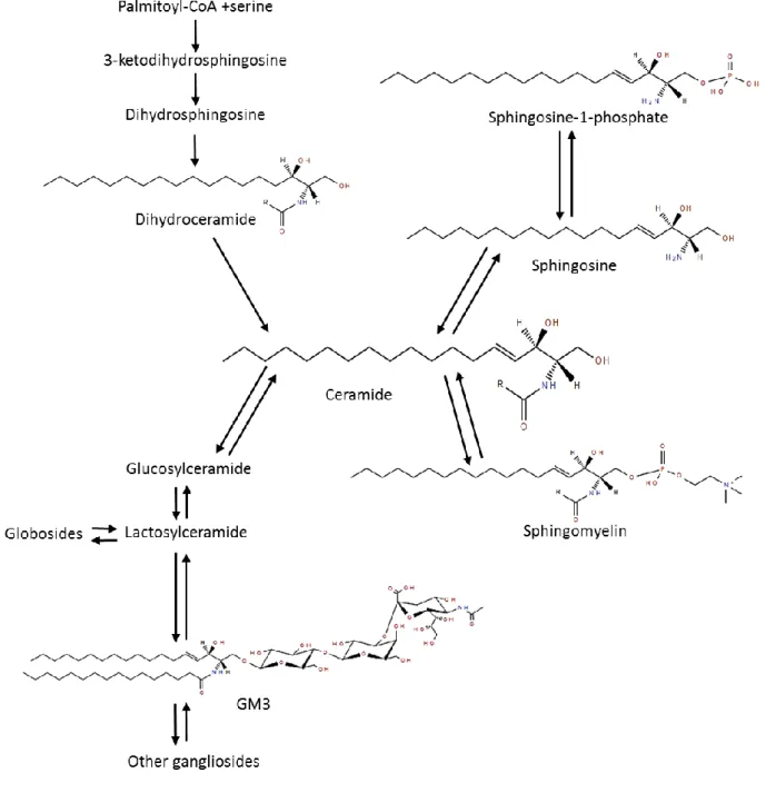
36
However, less is known about SL metabolism dysfunction in cells and tissues and how it may
37
impact the lipoprotein metabolism, its functionality and composition. In cardiometabolic
38
pathologies, the link between serum SL concentrations and alterations of their metabolism in
39
various organs and LD is still unclear. Pharmacological approaches have been developed in
40
order to activate or inhibit specific key enzymes of the SL metabolism, and to positively
41
modulate SL profile or related metabolic pathways. Nevertheless, little is known about the
42
long-term impact of such approaches in humans and the current literature still focuses on the
43
decomposition of the different parts of this complex system rather than performing an
44
integrated analysis of the whole SL metabolism. In addition, since SL can be provided from
45
exogenous sources, it is also of interest to evaluate their impact on the homeostasis of
46
endogenous SL metabolism, which could be beneficial in prevention or treatment of obesity
47
and related metabolic disorders.
48 49
Keywords: Obesity, metabolic diseases, sphingolipids, lipoproteins, lipid droplets
501. Introduction
52Worldwide incidence of obesity has increased exponentially in the last few years as well as
53
the prevalence of low-grade inflammation associated disorders [1] such as non-alcoholic fatty
54
liver disease (NAFLD) [2], type 2 diabetes (T2D) and cardiovascular diseases (CVD) [3].
55
Aside from traditional biomarkers of metabolic risks, progress in analytical technologies in
56
the last decades have facilitated the study of other molecules, especially bioactive lipids such
57
as phospholipids and sphingolipids (SL). SL are known as ubiquitous molecules that are
58
important components of cell membranes and lipoproteins [4]. They are involved in many
59
biological processes starting with cell growth, senescence and death as well as cell adhesion
60
and migration, but also inflammation, immune responses and nutrient uptake, among others
61
[5]. It is also starting to emerge that SL could be involved in the pathophysiology of obesity
62
and cardiometabolic risk factors. Indeed, changes in plasma or serum SL concentrations are
63
associated with metabolic disorders as well as accumulation of some SL species in
insulin-64
sensitive tissues, such as the liver [6]. SL could also be involved in the accumulation of an
65
emerging cell organelle that is the lipid droplets (LD), especially in non-adipose tissue [7]. In
66
addition to the endogenous SL metabolism, exogenous SL present in the diet also need to be
67
considered, especially regarding their potential effect on the intestine and the gut microbiota -
68
other important actors of metabolic homeostasis [8]. Another non-negligible aspect is that the
69
liver and the gut are organs that are largely involved in and impacted by metabolic
70
abnormalities. Considering that they are the producers of lipoproteins that carry a great
71
amount of both endogenous and exogenous SL, it is also highly relevant to study the possible
72
effect of an altered SL metabolism on these particles. Indeed, lipoproteins are already known
73
to be highly involved in various metabolic diseases from T2D to atherosclerosis and coronary
74
heart disease but the impact of their SL composition remains largely unknown.
75
In this review, we will describe the involvement of SL in different pathophysiological
76
mechanisms related to lipid metabolism that could be reflected on systemic SL concentrations
77
and associated with the development of metabolic diseases.
78 79
2. Overview of sphingolipids metabolism
80SL represent a class of bioactive lipids that was first identified in the 19
thcentury [9]. Interest
81
for these particular lipids has markedly increased with the study of genetic SL disorders and
82
the identification of complex SL such as cerebrosides or gangliosides. Many elegant reviews
have already detailed SL metabolism to which the interested reader can refer [10–13],
84
therefore this section will provide a summary of the major pathways.
85 86
87
Figure 1. Summarized partial pathway of sphingolipid biosynthesis
88
In this partial metabolic pathway of SL synthesis, general structures of the classes discussed
89
in this review are represented, taking into account that each SL class contains numerous
90
molecular species according to the length of their acyl chain. This Figure contains molecular
91
structures created using the Lipid Structure Drawing Tools from LIPID MAPS online tools
92
for lipid research. Fahy E, Sud M, Cotter D and Subramaniam S. Nucleic Acids Research 35,
93
W606-12 (2007).
95
Briefly, the biosynthesis of SL starts in the endoplasmic reticulum from the condensation of
96
serine and palmitoyl-CoA, and leads to the production of ceramides (Cer) by the introduction
97
of a double bond into its precursor dihydroceramide [14]. Cer represent the basic structure of
98
SL with the presence of a sphingosine base and an acyl chain that can be of various lengths.
99
Each SL class present numerous molecular species according to the number of carbon of this
100
acyl chain (C16, C18, C20, etc.). Sphingomyelin (SM) can be synthetized from Cer by
101
condensation of phosphocholine to a Cer. Of note, SM is also classified as a phospholipid.
102
Finally, more complex molecules comprised in the glycosphingolipid family, such as GM3
103
ganglioside for example, are constituted of a Cer backbone and various type and number of
104
sugar molecules (Figure 1).
105
In addition to de novo synthesis, the generation of Cer from hydrolysis of SM or breakdown
106
of glycosphingolipids can also lead to its conversion into other SL classes or bioactive
107
derivatives), which makes Cer a central molecule of the SL biosynthesis pathway. Cer can be
108
converted back to sphingosine for this base to be recycled into SL synthesis again or
109
phosphorylated to produce sphingosine-1-phosphate (S1P) [15]. SL metabolism is a highly
110
regulated process that can lead to thousands of molecules for which the functions will vary
111
according to their molecular composition and structure (see further details in [16,17,15,18].
112 113
3. Plasma alterations of SL metabolism in metabolic diseases
114In the bloodstream, SL are present in the outer cell membranes, in lipoproteins and bound to
115
albumin as well. In healthy individuals, plasma SL are mostly represented by SM (87% of
116
total SL) [4], whereas glycosphingolipids contribute to 9-10% and Cer to 3% of total plasma
117
SL. With few exceptions, detected SL mainly belong to C16, C18 and C24 species in human
118
plasma and tissues [19]. SM is co-localized with cholesterol in cell membranes and all classes
119
of lipoproteins [20] while Cer are equally distributed between ApoB-containing lipoproteins
120
and
HDL
[19]
(Table
1).
Glycosphingolipids,
mostly
glucosylceramides
and
121
lactosylceramides, are also present on VLDL, LDL and HDL. Another SL found in plasma
122
and carried in part by lipoproteins is S1P, which is the most abundant SL in HDL [4].
123 124
Table 1. General classification of lipoproteins and distribution of major circulating lipid
125species.
126VLDL
LDL
HDL
Total
Ref.
Density (g/ml)
0.93-1.006
1.019-1.063
1.063-1.210
[21]Distribution of major lipid content (% of total content carried by circulating lipoproteins)
TG
66
26
7
100
[22]Cholesterol esters
20
57
23
100
[22]Unesterified cholesterol
7
69
24
100
[22]Phospholipids
7
31
62
100
[23]Distribution of minor lipid content (% of total content carried by circulating lipoproteins)
Phosphatidylcholine
5-8
30-39
56-62
100
[22,23]Sphingomyelin
3-7
33-50
43-64
100
[19,23]Ceramides
9-16
40-60
24-51
100
[19,23]S1P*
1-2
4-6
90-95
100
[19,24]Glycosphingolipids
6-9
42-50
42-52
100
[24]Protein composition
Apolipoproteins
Apo B-100,
Apo E,
Apo C
Apo B-100
Apo A-I,
A-II, Apo C,
Apo E
[21]*An important part of total plasma S1P is carried by albumin (30-40%)
127 128
3. 1. Ceramides and their precursors
129
In obesity and cardiometabolic diseases, many organs are affected and thus involved in the
130
development of IR and low-grade inflammation, and this can be reflected in systemic
131
circulation [25]. Among SL classes that could be potential biomarkers for metabolic diseases,
132
Cer are the most studied. It is now well established that Cer metabolism is altered in the
133
context of T2D. Many studies have shown an elevation of circulating Cer [26,27] and a
134
positive association with insulin resistance [28] as well as a role of Cer in β-cell apoptosis
135
[29]. Higher plasma concentrations of Cer, as compared to healthy individuals, have even
136
been reported in obese female children and adolescents with T2D and it was correlated with a
137
deleterious metabolic profile [30]. Cer increased plasma levels have also been described in
138
other metabolic diseases or cardiovascular risk factors, such as NAFLD [31], visceral obesity
[32,33], atherosclerosis [34], and are a predictor of cardiovascular mortality [35]. In addition,
140
Peterson et al. [36] recently demonstrated that a decreased Cer C24:0/C16:0 ratio could be a
141
more potent predictive marker of coronary heart disease incidence and all-cause mortality,
142
than alterations in specific Cer species.
143
There is also an increasing interest for other SL classes in the context of metabolic diseases,
144
especially Cer precursors such as dihydroceramides. Indeed, very recent studies suggest that
145
dihydroceramides are not inert molecules as it was first thought, but are also involved in
146
metabolic disturbances [14]. Plasma level of dihydroceramides could even be considered as a
147
potential predictive biomarkers for the development of T2D [37]. For instance, two different
148
clinical studies corroborated results obtained in mouse models by showing a specific increase
149
in plasma long-chain fatty acids (FA)-containing dihydroceramides in individuals who
150
develop metabolic disorders related to obesity and T2D [37,38]. In vitro experiments also
151
demonstrated that the accumulation of dihydroceramides inhibits autophagy-related pathways,
152
which in turn could lead to fibrosis and thus plays a role in the progression of NAFLD [39]. It
153
was also revealed that levels of sphingosine and sphinganine are elevated in plasma of
154
patients suffering from T2D, compared to healthy individuals [40]. Regarding S1P, its
155
involvement in cardiometabolic diseases is quite controversial; starting with the fact that a
156
large part (50-70 %) of S1P in circulation is comprised in HDL, which are particles known to
157
have anti-atherogenic effect. However, low S1P concentrations in HDL from T2D or coronary
158
artery disease patients are associated with HDL dysfunctions [41,42]. In the development of
159
atherosclerosis, it is not really defined yet if S1P has beneficial or deleterious effects [43].
160
Indeed, a recent review from Kurano and Yatomi highlighted that S1P bound to HDL via
161
apolipoprotein M displays atheroprotective properties, while when bound to albumin it exerts
162
both beneficial and harmful effects [44]. Same as for the involvement of S1P in T2D and
163
obesity since it has been reported that S1P plasma levels are increased in these two
164
populations compared to healthy individuals [45,46] are associated with visceral fat
165
accumulation in T2D patients [47]. On the other hand, S1P is thought to have favorable
166
effects in models of lipotoxicity and hyperlipemia as well as for β-cell survival, among others
167
[48].
168
Interestingly, several studies also reported that serine–palmitoyl–CoA transferase (SPT), a
169
rate-limiting enzyme involved in de novo SL synthesis which basically conjugates L-serine
170
and palmitoyl-CoA, is also able to conjugates other amino acid substrates such as L-alanine
171
producing 1-deoxysphingolipids [49–51]. Recent papers demonstrated that this specific SL
172
class could be used as potential predictor of metabolic disorders, such as T2D, since
deoxysphingolipids accumulate in systemic circulation and cells, exerting toxic and
174pathological effects [51–53].
175 1763. 2. Complex sphingolipids
177In addition to Cer precursors, complex SL have been of interest when studying
178
cardiometabolic diseases physiopathology. Altered SM plasma levels have also been observed
179
in T2D as well as coronary artery disease and obesity [34,54–56]. Indeed, SM level is
180
considered as an independent risk factor for coronary heart disease [34,57]. Regarding the
181
development of insulin resistance, growing evidence suggests that GM3 is also involved. In
182
fact, the first data about GM3 and insulin response were reported in the early 1990 and a
183
negative correlation was established between GM3 concentrations and insulin responsiveness
184
in leukemic cell lines [58]. Almost two decades later, Sato et al. analyzed GM3
185
concentrations in individuals with hyperlipemia, T2D patients with or without hyperlipemia
186
and controls. They observed that serum GM3 levels were elevated in all groups compared to
187
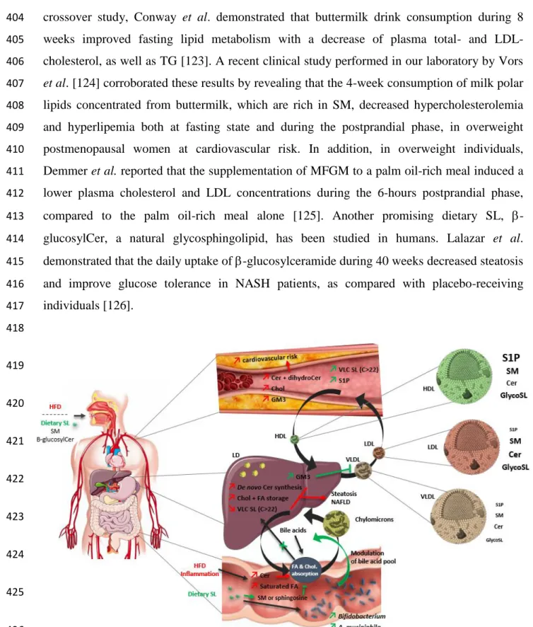
controls [59]. Lastly, a recent study confirmed a higher concentrations of some GM3
188
molecular species in subjects with visceral fat accumulation and metabolic disease and
189
showed that hydroxylated GM3 species concentrations in human serum were also increased
190
and correlated with risk factors of cardiometabolic diseases [60].
191 192
When examining the relation between SL plasma levels and metabolic diseases, it is hard to
193
draw a clear line in the associations between all the different SL classes and each metabolic
194
disorder. Moreover, it is now necessary to have a better understanding of how SL metabolism
195
is altered in tissues of interest and at the cellular level, notably in LD.
196 197
4. Modulation of cholesterol and lipid metabolism by sphingolipids in the
198gut-liver axis
199Growing evidence supports that the gut-liver axis plays an essential role in nutrient absorption
200
and NAFLD development [61,62]. Indeed, the development of metabolic disorders has been
201
widely associated with impaired gut homeostasis, gut microbiota dysbiosis and increased
202
intestinal permeability [63]. Recent reviews described how SL modulate metabolic and
203
inflammatory pathways in the gastrointestinal tract [64,65]. However, once ingested,
204
metabolites produced after digestion by enzymes from host and microbial cells present in the
gut, reach the liver through the portal vein and could also exert some physiological or
206
pathological effects.
207 208
4. 1. Interplay between intestinal absorption of cholesterol and sphingomyelin
209
In metabolic diseases, lipid metabolism is altered at several levels from intestinal absorption
210
to endogenous biosynthesis and lipid species distribution, and could participate to the
211
worsening of cardiometabolic disorders. In obese patients with T2D, intestinal absorption of
212
cholesterol is lower than in healthy subjects [66], whereas in normal metabolic conditions,
213
there is already an inter-individual variation of 30 to 80% [67]. Moreover, in metabolic
214
diseases, an altered bile acid profile was associated with hypercholesterolemia and defects in
215
pathways related to lipid metabolism [68]. SL are complex molecules that cannot passively
216
diffuse into enterocytes. Consequently, they need to be hydrolyzed by bile acids and enzymes
217
at the surface of epithelial cells and in the lumen [69]. Once absorbed, SL influence the
218
enterocyte lipid flux: SM is hydrolyzed to sphingosine which is incorporated into triglycerides
219
(TG) after conversion and acetylation, and then reach the lymphatic circulation in
220
chylomicrons [70]. Agren et al. demonstrated that postprandial lipid profiles and lipoprotein
221
composition depend on the efficiency of cholesterol absorption, which was determined
222
according to a higher baseline serum cholestenol/cholesterol ratio [71]. In fact, compared to
223
low cholesterol absorbers, high cholesterol absorption efficiency results in a higher
224
concentration of triglycerides, cholesterol and SM in VLDL during the postprandial phase,
225
and a higher free cholesterol/PC ratio in chylomicrons [71]. In rats, dietary supplementation
226
of a mixture of SM and cholesterol demonstrated a mutual inhibitory effect on their
227
absorption, inducing an increased presence of Cer in the colon [72]. In Caco-2 cells, the
228
incorporation of SM in apical bile salt-cholesterol micelles inhibits cholesterol absorption,
229
which could be explained by the decrease of ABC-A1 and -G1 gene expression [73].
230 231
4. 2. Ceramide metabolism in liver and its relation with metabolic diseases
232
In metabolic diseases, excess of lipids (e.g. saturated FA) is reflected in the systemic
233
circulation but also in tissues such as liver, which in turn leads to de novo Cer synthesis [26].
234
Liver plays a critical role in the organism homeostasis since it directly participates to
235
detoxification as well as metabolism, biosynthesis and storage. In addition, this important
236
organ has the capacity to repair itself and regenerate. A recent review by Nojima et al.
237
focused on the role of SL in both liver injury and regeneration [74]. Moreover, several in vivo
and clinical studies reported that the impact of SL on the liver could be linked to their
acyl-239
chain length and composition [75–77]. Indeed, Montgomery et al. compared the hepatic
240
profile of Cer and SM in 5 different strains of mice submitted to a high-fat diet. In the 4 mice
241
strains that developed glucose intolerance, the results revealed a marked decrease of very long
242
chain Cer and SM species (C>22) and a significant increase of C16-C22 Cer and SM species
243
in the liver [77]. Interestingly, it was correlated with the specific decrease of Cer synthase 2
244
(CerS2) expression and activity. Park et al. demonstrated that in CerS2 null mice, unable to
245
synthetize Cer C22-C24, the activity of key proteins involved in FA uptake was dysregulated
246
in favor of an increase of CD36 [76]. In clinical studies, a similar pattern was reported with a
247
significant decrease of serum Cer C24 in patients suffering from cirrhosis [75]. In addition,
248
Grammatikos et al. also reported an upregulation of serum acid sphingomyelinase in NAFLD
249
patients, which was also associated with an increase of circulating sphingosine and
250
dihydroceramides species, correlated to elevated cholesterol levels [78]. Another possible
251
pathway involves bile acids which plays an important role in the enterohepatic pathway of
252
cholesterol and thus in lipid metabolism. It has been demonstrated that some toxic bile acids
253
(e.g. chenodeoxycholic acid, deoxycholic acid, lithocholic acid) are endogenous activators of
254
farnesoid X receptors (FXR) involved in glucose intolerance and insulin resistance through
255
hepatic de novo Cer production [79,80]. Besides, elevated plasma concentration of GM3 has
256
been associated with metabolic diseases [59,60], however Choi et al. reported that plasma
257
membrane associated GM3 inhibits the secretion of TG-enriched ApoB containing
258
lipoproteins (i.e. VLDL and LDL) and could contribute to lower the development of
259
atherosclerosis [81].
260 261
As mentioned above, when evaluating the role of the gut-liver axis, it is also important to
262
consider the gut microbiota as an additional metabolically active “organ”, whose composition
263
and functions are altered in metabolic diseases [82–84]. Thus, the composition of the human
264
gut microbiota may also have an impact on circulating SL, since it has been shown that it
265
contains SL-producing bacteria. Same as for mammalian SL, bacterial SL may be detected
266
and used by intestinal endothelial cells and then potentially modulate lipid metabolism [69].
267 268
5. Involvement of sphingolipids in lipoprotein metabolism and cell lipid
269droplets
270Another major aspect to consider when addressing the question of SL systemic alterations in
271
metabolic diseases is their important role in lipid and lipoprotein synthesis and regulation.
272
Among many others, Meikle and Summers recently published a state of the art about the
273
involvement of dyslipidemia, particularly related to SL metabolism disruptions, in metabolic
274
disorders associated with obesity [85]. Indeed, SL can modulate genes involved in lipid
275
biosynthesis in mice [86] and their involvement in cholesterol transport has also been
276
demonstrated [87]. In addition, there is a growing interest for the lipoprotein composition in
277
SL that can be modified in metabolic diseases. The development of mass spectrometry-based
278
lipidomics also allowed to identify and quantify many lipid molecular species in lipoproteins
279
[24,88–91]. These particles have an important role in the transport of endogenous and dietary
280
lipids including SL. They are a complex of lipids and proteins all assembled following the
281
same pattern with a central hydrophobic core of TG and cholesterol esters that is surrounded
282
by apolipoproteins phospholipids, SL and unesterified cholesterol forming a membrane and
283
contributing to maintaining the shape of the particles [21,92] (Table 1).
284 285
5. 1. Sphingolipids in lipoprotein composition and function
286
Modifications in the lipidome of some lipoprotein classes were observed in cardiovascular
287
diseases and T2D [93,94]. This modified lipid composition could affect lipoprotein clearance
288
from plasma and certain enzymes related to lipoprotein metabolism. A study by Redgrave et
289
al. where rats were injected with chylomicron-like TG emulsions showed that when the
290
emulsion was stabilized with 100% SM or 50% SM combined with egg yolk
291
phosphatidylcholine, the clearance of CM emulsions from plasma was negligible and very
292
slow, respectively [95]. In comparison, when the emulsion was stabilized with egg yolk
293
phosphatidylcholine alone, clearance was much more important. This study also showed that
294
the liver uptake of triolein and cholesteryl oleate was significantly reduced when the emulsion
295
contained 50% and 100% SM compared to dioleoyl phosphatidylcholine-enriched emulsions
296
[95]. These results were supported by Arimoto et al. who also showed delayed clearance of
297
triolein-SM (2:1 molar ratio) lipid emulsion injected in rats. In addition, the incorporation of
298
SM to the emulsion reduced the binding of ApoE, ApoC-II and ApoC-III to the surface lipids
299
of the emulsion [96], which can have functional consequences that would deserve to be
300
investigated.
301
Modifications in the lipid composition of lipoproteins are of interest since they could
302
potentially affect the functionality of the particles, especially in the context of metabolic
303
diseases. Most studies have focused on LDL and HDL particles, since it is well established
that the first have atherogenic properties and the second are cardioprotective. Ståhlman et al.
305
found that major classes of surface lipids including SM and Cer were diminished in HDL
306
from dyslipidemic insulin resistant and T2D women [97]. Interestingly, Denimal et al.
307
observed reductions in SM and d18:1 S1P, but not Cer concentrations in HDL2 and HDL3 of
308
patients with metabolic syndrome without insulin resistance [93]. This suggest that HDL
309
composition is already altered in the presence of metabolic syndrome, but insulin resistance
310
and T2D could alter other specific classes of SL in these particles. Boon et al. observed an
311
elevation in Cer content of LDL from T2D individuals compared to lean non-diabetic subjects
312
that was reversed with weight loss [98]. The lipid composition of LDL particles can also
313
affect their aggregability, which is an important aspect in the development of atherosclerosis
314
and cardiovascular complications. A recent study showed that there is a strong association
315
between the surface lipid LDL composition and their susceptibility to aggregate. LDL prone
316
to aggregation are enriched in SM and Cer and contained less lysoPC and PC [99]. On a
317
general point of view, changes of SL composition in lipoproteins are mostly observed in the
318
representation of each SL class rather than in the variation of particular molecular species and
319
this particular question has to be explored.
320 321
5. 2. Sphingolipids in cell lipid droplets
322
In addition to lipoproteins, it is now important to consider the role of LD in cells which are
323
recognized as dynamic organelles also involved in lipid metabolism as well as many cellular
324
mechanisms [100]. LD are present in almost all type of cells and tissues and their excessive
325
accumulation in various non-adipose tissues has been associated with many metabolic
326
alterations and diseases. Interestingly, Deevska et al. have recently described the importance
327
of SL in LD biogenesis and metabolism in a very elegant review [7]. In addition to cell
328
membranes, SL also are found in very low concentrations in LD with cholesterol and TG. In
329
fact, it is now well-admitted that under obesogenic conditions, ectopic fat accumulation in
330
metabolic tissues such as liver or skeletal muscle causes lipotoxicity and is associated with the
331
development of insulin resistance [26,101], meaning that SL in LD could also play a role in
332
lipid metabolism at the tissue level. Recently, Senkal et al. elegantly demonstrated the
333
generation of acylceramide from ceramide in LD of HCT116 cell lines and showed that in the
334
liver of mice fed an oleate-enriched high fat diet, acylceramides were generated from Cer and
335
specifically accumulated in LD and this was accompanied by steatosis development [102].
336
Nevertheless, in the current literature, there is very few data about SL and LD, since SL
profiles and related metabolic pathways are mostly studied at the whole tissue level and not
338
specifically in LD or cell membranes.
339 340
6. Towards the prevention of metabolic diseases by the modulation of
341sphingolipid metabolism
342Despite the reported deleterious role of some SL, particularly Cer, it is important to keep in
343
mind that it is not the presence of SL itself that could be harmful for metabolism, but rather
344
the modulation of the quantity and length of the acyl chain of the represented species [103]. In
345
this context, several approaches have been developed in order to positively modify the SL
346
metabolism, with the aim of reducing metabolic disorders.
347 348
6. 1. Modulation of enzymes of the sphingolipid metabolic pathway
349
Many studies reported that modulating the activity of key enzymes involved in SL
350
metabolism – such as ceramidase or CerS activity – via pharmacological or dietary
351
approaches, could have beneficial impacts on SL metabolism and thus improve metabolic
352
disorders induced in a context of obesity or T2D [104,105]. For instance, the specific
knock-353
down of CerS6 in an obese insulin resistant mice model induced an efficient reduction of Cer
354
C16:0 concentration in plasma and hepatic tissue which was associated with an improvement
355
of glucose tolerance and insulin sensitivity [33]. Xia et al. developed a transgenic model of
356
mice able to inducibly overexpress acid ceramidase in the liver and demonstrated that
357
activating this pathway prevented hepatic steatosis and insulin sensitivity, associated with a
358
lower hepatic Cer concentration [106]. In ApoE-KO mice, it has been demonstrated that the
359
oral administration of myriocin, a specific inhibitor of SPT, induced a significant decrease of
360
plasma total cholesterol, SM concentrations and atherosclerotic lesions, combined with an
361
increase in plasma HDL cholesterol [107,108]. Another possible pathway involves
362
adiponectin which acts as a regulator of insulin sensitivity and induces ceramidase activity
363
through its receptors AdipoR1/R2, both depending on AMPK, an important kinase involved
364
in energy metabolism and activated by the increase of S1P [109]. The authors demonstrated
365
that the decrease of Cer via the activation of AdipoR1/R2 in hepatocytes or adipocytes
366
(especially Cer C16:0 but also the other Cer species according to the activated receptor
367
isoform) was associated with the improvement of hepatic steatosis, glucose tolerance and
368
insulin sensitivity in mice fed with a high-fat diet [109]. Another potential mechanism
369
concerns the suppression of the intestinal FXR signaling, which in turn induced a decrease of
plasma Cer by reducing the expression of genes of Cer synthesis, thus leading to a decrease of
371
hepatic gluconeogenesis [80].
372
However, in the literature, both in vivo and clinical studies do not reflect the complexity of the
373
SL metabolism since the authors selected and analyzed specific classes of SL or pathways
374
without considering the impact of their intervention on the overall metabolism. In fact, since
375
SL classes and species are differentially distributed among cells, tissues and systemic
376
circulation, it is quite reductive to only describe the modulation of few SL species in one or
377
two compartments of the individual without keeping in mind that the intervention could have
378
an effect in SL profile in other metabolic tissues, specific classes of lipoproteins or in LD at
379
the cellular level. Indeed, further in vitro studies are needed to demonstrate potential
380
mechanisms or to suggest an integrative model.
381 382
6. 2. Beneficial effects of dietary sphingolipid supplementation in metabolic
383
diseases
384
SL are ubiquitous in many plant and animal organisms, explaining that they are found in
non-385
negligible amounts in our diet. Therefore, the consumption of dietary SL may modulate
386
plasma and tissue levels of SL and thus could impact the endogenous SL metabolism. Indeed,
387
Norris and Blesso recently published a state of the art concerning the potential beneficial
388
effects of dietary SL in (i) the regulation of inflammatory signaling pathways, (ii) the decrease
389
of dyslipidemia focusing on hepatic lipid accumulation and also (iii) the positive modulation
390
of the gut microbiota associated to a decrease of metabolic endotoxemia in mice [110,111].
391
The major dietary sources of SL studied are chicken eggs, meat and dairy products [112]. The
392
particularity of milk and dairy products is that the milk fat presents a specific profile of
393
bioactive lipids, rich in SM and Cer, and organized in milk fat globule membrane (MFGM)
394
[113–115]. In addition, the consumption of dairy products has been associated with a decrease
395
of metabolic syndrome prevalence or improvements of metabolic disorders related to obesity
396
and CVD [116–118]. Buttermilk is a dairy product particularly rich in milk polar lipids and
397
our group recently revealed its SL composition [119]. In fact, previous in vivo studies
398
demonstrated that the consumption of milk polar lipid-rich extract (derived from buttermilk)
399
prevented hepatic TG and cholesterol accumulation normally induced by a high-fat diet [120],
400
and addition of SM in the diet decreased cholesterol absorption in mice [121]. More recently,
401
our group confirmed that milk polar lipids significantly enhance in vitro digestive lipolysis
402
and improve postprandial lipid metabolism in mice, compared to soy polar lipids [122]. In a
crossover study, Conway et al. demonstrated that buttermilk drink consumption during 8
404
weeks improved fasting lipid metabolism with a decrease of plasma total- and
LDL-405
cholesterol, as well as TG [123]. A recent clinical study performed in our laboratory by Vors
406
et al. [124] corroborated these results by revealing that the 4-week consumption of milk polar
407
lipids concentrated from buttermilk, which are rich in SM, decreased hypercholesterolemia
408
and hyperlipemia both at fasting state and during the postprandial phase, in overweight
409
postmenopausal women at cardiovascular risk. In addition, in overweight individuals,
410
Demmer et al. reported that the supplementation of MFGM to a palm oil-rich meal induced a
411
lower plasma cholesterol and LDL concentrations during the 6-hours postprandial phase,
412
compared to the palm oil-rich meal alone [125]. Another promising dietary SL,
-413
glucosylCer, a natural glycosphingolipid, has been studied in humans. Lalazar et al.
414
demonstrated that the daily uptake of
-glucosylceramide during 40 weeks decreased steatosis
415
and improve glucose tolerance in NASH patients, as compared with placebo-receiving
416
individuals [126].
417 418 419 420 421 422 423 424 425 426Figure 2. Schematic representation of potential sphingolipid metabolism alterations in
427
cardiometabolic diseases. In obesity and cardiometabolic diseases, many tissues are affected
428
and undergo profound metabolic dysfunction. Among them, the metabolism of major lipids
429
like triglycerides can be altered, but the involvement of minor bioactive lipids like SL in these
430
processes is starting to emerge. SL are involved in lipid metabolism regulation in organs such
as the liver and the gut, but is also an important constituent of cell lipid droplets and
432
lipoproteins. Plasma SL concentrations could be a reflection of all the SL metabolism
433
alterations in the gut-liver axis as well as changes in lipoprotein composition in obesity and
434
metabolic diseases. Cer: ceramides, Chol: cholesterol, FA: fatty acids, glycoSL:
435
glycosphingolipids, HFD: high fat diet, LD: lipid droplets, NAFLD: non-alcoholic fatty liver
436
disease, SL: sphingolipids, SM: sphingomyelin, S1P: sphingosine-1-phosphate, VLC: very
437
long chain. This Figure contains pieces of artwork that are derivatives of artworks kindly
438
provided by the Servier Medical Art database licensed under a Creative Commons Attribution
439
3.0 Unported License.
440 441
6. 3. Modulations of the gut microbiota composition and cholesterol absorption
442
by sphingolipids
443
In addition, since the gut microbiota is involved in the development of low-grade
444
inflammation and metabolic disorders [127,128], the use of pharmacological or dietary
445
molecules in order to positively modulate its composition or function has gained more
446
attention. For example, in a rat model of fructose-induced metabolic disorders, Crescenzo et
447
al. [129] demonstrated that an antibiotic treatment reversed gut microbiota dysbiosis in
448
fructose-fed rats by positively modulating its composition, and decreases hepatic Cer content
449
and insulin resistance in both liver and visceral adipose tissues. Norris et al. reported that milk
450
SL supplementation induced an increase of Bifidobacterium in the colon of high fat-fed mice
451
and lowered serum total cholesterol and lipopolysaccharide [130]. In a recent in vivo study
452
performed in our laboratory, Milard et al. demonstrated that dietary supplementation with
453
milk polar lipids (1.1-1.6% of diet corresponding to 0.25-0.36% of SM) prevented the
454
development of obesity without impacting caloric intake, but was associated with changes in
455
gut microbiota populations [131]. Under a more mechanistical approach realized both in vitro
456
and in vivo, our results demonstrated that SM could induce beneficial effects at the intestinal
457
barrier level through the increase of plasma IL-8, an interleukin involved in immunity, leading
458
to an improvement of gut integrity [132]. These results from Milard et al. also suggested that
459
some effects could be mediated by sphingosine, which is liberated during the digestion of SM
460
from both egg and milk [132]. In fact, since SM co-localize with cholesterol, it could explain
461
that dietary SM is associated with a decrease in cholesterol absorption in animal models
462
[121]. However, in vitro experiments realized by Garmy et al. also demonstrated that
sphingosine forms lipid complexes with cholesterol and this specific interaction inhibits the
464
intestinal transport of micellar cholesterol [133].
465 466
6. 4. Sphingolipid metabolism in specific bacterial species
467
In the previous section, we demonstrated that SL and the gut microbiota could be
468
interconnected since specific dietary SL may modulate the gut microbiota composition.
469
However, it has also been reported that specific bacterial species are able to produce or
470
metabolize SL. In fact, it was previously established that SL were exclusively found in
471
eukaryote cells until their discovery in prokaryotes [134]. For example, whereas most
472
bacterial membranes only contain glycerol-based PL, few of them possess both PL and SL
473
[135]. Among the SL-containing bacteria, the genus Bacteroides has been mainly studied
474
because of its specific PL membrane, which is composed of 40-70% of SL [136]. An et al.
475
demonstrated that SL-rich membrane of Bacteroides plays an important role in the survival of
476
this genus in the gastro-intestinal tract [135]. Wieland Brown et al. also reported that
-477
galactosylceramides produced by Bacteroides species can modulate natural killer T cell
478
activity and thus influence the host immune response [136]. In addition, the genus
479
Bacteroides is mainly deprived from key SL metabolism-related enzymes as
480
sphingomyelinases whereas they are found in some pathogens such as Staphylococcus aureus
481
[137]. More recently, promising studies demonstrated that, among all the known mechanisms,
482
the anti-inflammatory effects of certain probiotics could be the result of the SL metabolism
483
modulations. Angulo et al. reported that Lactobacillus brevis and Streptococcus thermophilus
484
species could influence the mucosal immune cells by inducing cell apoptosis through the
485
activation of a neutral SMase and the production of Cer [138]. In addition, Soo et al.
486
demonstrated that the supplementation with the probiotic cocktail VSL#3 induced a
487
significant increase of alkaline SMase levels in the intestinal sections of both IL-10 deficient
488
mice and humans with colitis, resulting in a decrease of intestinal inflammation [139].
489 490
7. Conclusion
491In this review, we realized a focused state of the art about the possible alterations of the SL
492
metabolism in the context of obesity and associated metabolic diseases. In fact, all the data
493
discussed suggest that SL metabolism is highly involved in the regulation of lipid
494
homeostasis. In addition, changes in both the quantity and profile of represented SL species
495
and classes at the molecular, cellular and tissue levels could also be reflected in circulation
(Figure 2). As described above, the most studied pathway mainly concerns the metabolism of
497
Cer whereas few papers now also included the analysis of SM, dihydroceramide, ganglioside
498
and other glucosylceramide species. In clinical studies, the difficult access to biopsies may
499
also be a limitation in the study of the SL metabolism in obese and T2D patients for example,
500
partly explaining why lipidomic analysis are most of the time only realized in systemic
501
circulation. In this review, we discussed about how plasma SL alterations can reflect the
502
accumulation of perturbations in tissue, lipoprotein and LD metabolism. In addition, the last
503
part of this review demonstrated that more and more studies now describe the beneficial
504
impact of metabolic disorder treatments on SL profile and metabolism. These preventive or
505
curative treatments concern pharmacological drugs but also dietary bioactive molecules or
506
extracts. In addition, such as highlighted by Bellini et al. [140], it is unthinkable to
non-507
specifically inhibit key enzymes involved in the production of specific Cer species to improve
508
T2D-related metabolic disorders because all SL are part of a plethora of physiological
509
pathways. There is also growing evidence in vivo that the dietary supplementation with
510
specific SL could exert beneficial effects on both metabolic disorders and SL metabolism.
511
Therefore, it confers an advantage to prevent or attenuate metabolic disorders development,
512
and to improve overall SL metabolism homeostasis.
513 514
Author contributions: MLB, MMB: performed literature review and wrote the original draft.
515
CC, DC, MCM: revised the manuscript for important intellectual content. All: read and
516
approve the final manuscript.
517 518
Funding: This research did not receive any specific grant from funding agencies in the
519
public, commercial, or not-for-profit sectors. MM Boulet received a doctoral contract from
520
the Ministère de l'Enseignement supérieur et de la Recherche and M Le Barz acknowledged a
521
postdoctoral grant from Société Francophone du Diabète.
522 523
Conflict-of-interest/financial disclosure statement: MCM received other research fundings
524
on other topics from Sodiaal-Candia R&D, the Centre National Interprofessionnel de
525
l’Economie Laitière (CNIEL, French Dairy Interbranch Organization) and Nutricia Research
526
and has consultancy activities for food & dairy companies. These activities had no link with
527
the present study. MCM is coordinator of a research project funded by the National Research
528
Agency on the valuing and health properties of buttermilk and milk polar lipids (VALOBAB,
ANR-2011-0007), in which DC and MLB participate; the present review is not part of this
530
project. Other authors have no conflict of interest to declare.
531 532
References
533[1] S. O’Neill, L. O’Driscoll, Metabolic syndrome: a closer look at the growing epidemic and its 534
associated pathologies, Obes Rev. 16 (2015) 1–12. doi:10.1111/obr.12229. 535
[2] R. Loomba, A.J. Sanyal, The global NAFLD epidemic, Nat Rev Gastroenterol Hepatol. 10 536
(2013) 686–690. 537
[3] J.-P. Després, Body Fat Distribution and Risk of Cardiovascular Disease, Circulation. 126 538
(2012) 1301. doi:10.1161/CIRCULATIONAHA.111.067264. 539
[4] J. Iqbal, M.T. Walsh, S.M. Hammad, M.M. Hussain, Sphingolipids and Lipoproteins in Health 540
and Metabolic Disorders., Trends Endocrinol Metab. 28 (2017) 506–518. 541
doi:10.1016/j.tem.2017.03.005. 542
[5] Y.A. Hannun, L.M. Obeid, Sphingolipids and their metabolism in physiology and disease., 543
Nat Rev Mol Cell Biol. 19 (2018) 175–191. doi:10.1038/nrm.2017.107. 544
[6] S. Choi, A.J. Snider, Sphingolipids in High Fat Diet and Obesity-Related Diseases., Mediators 545
Inflamm. 2015 (2015) 520618. doi:10.1155/2015/520618. 546
[7] G.M. Deevska, M.N. Nikolova-Karakashian, The expanding role of sphingolipids in lipid 547
droplet biogenesis., Biochim Biophys Acta Mol Cell Biol Lipids. 1862 (2017) 1155–1165. 548
doi:10.1016/j.bbalip.2017.07.008. 549
[8] F. Fava, L. Rizzetto, K.M. Tuohy, Gut microbiota and health: connecting actors across the 550
metabolic system., Proc Nutr Soc. (2018) 1–12. doi:10.1017/S0029665118002719. 551
[9] J. Thudichum, A treatise on the chemical constitution of the brain, London, Bailliere, Tindall 552
and Cox, 1884. 553
[10] J. Iqbal, M.T. Walsh, S.M. Hammad, M.M. Hussain, Sphingolipids and Lipoproteins in Health 554
and Metabolic Disorders., Trends Endocrinol Metab. 28 (2017) 506–518. 555
doi:10.1016/j.tem.2017.03.005. 556
[11] C.R. Gault, L.M. Obeid, Y.A. Hannun, An overview of sphingolipid metabolism: from 557
synthesis to breakdown., Adv Exp Med Biol. 688 (2010) 1–23. 558
[12] A.H.J. Merrill, De novo sphingolipid biosynthesis: a necessary, but dangerous, pathway., J 559
Biol Chem. 277 (2002) 25843–25846. doi:10.1074/jbc.R200009200. 560
[13] R. Pralhada Rao, N. Vaidyanathan, M. Rengasamy, A. Mammen Oommen, N. Somaiya, M.R. 561
Jagannath, Sphingolipid metabolic pathway: an overview of major roles played in human diseases., J 562
Lipids. 2013 (2013) 178910. doi:10.1155/2013/178910. 563
[14] M.M. Siddique, Y. Li, B. Chaurasia, V.A. Kaddai, S.A. Summers, Dihydroceramides: From 564
Bit Players to Lead Actors., J Biol Chem. 290 (2015) 15371–15379. doi:10.1074/jbc.R115.653204. 565
[15] N. Bartke, Y.A. Hannun, Bioactive sphingolipids: metabolism and function., J Lipid Res. 50 566
Suppl (2009) S91-96. doi:10.1194/jlr.R800080-JLR200. 567
[16] A.H. Futerman, Y.A. Hannun, The complex life of simple sphingolipids., EMBO Rep. 5 568
(2004) 777–782. doi:10.1038/sj.embor.7400208. 569
[17] Y.A. Hannun, L.M. Obeid, Principles of bioactive lipid signalling: lessons from 570
sphingolipids., Nat Rev Mol Cell Biol. 9 (2008) 139–150. doi:10.1038/nrm2329. 571
[18] T. Kolter, A view on sphingolipids and disease., Chem Phys Lipids. 164 (2011) 590–606. 572
doi:10.1016/j.chemphyslip.2011.04.013. 573
[19] S.M. Hammad, J.S. Pierce, F. Soodavar, K.J. Smith, M.M. Al Gadban, B. Rembiesa, R.L. 574
Klein, Y.A. Hannun, J. Bielawski, A. Bielawska, Blood sphingolipidomics in healthy humans: impact 575
of sample collection methodology., J Lipid Res. 51 (2010) 3074–3087. doi:10.1194/jlr.D008532. 576
[20] J.P. Slotte, B. Ramstedt, The functional role of sphingomyelin in cell membranes, European 577
Journal of Lipid Science and Technology. 109 (2007) 977–981. doi:10.1002/ejlt.200700024. 578
[21] M. Ana Jonas, Lipoprotein struture, in: Biochemistry of Lipods, Lipoproteins and Membranes, 579
Fifth edition, Elsevier, 2008. 580
[22] J. Serna, D. Garcia-Seisdedos, A. Alcazar, M.A. Lasuncion, R. Busto, O. Pastor, Quantitative 581
lipidomic analysis of plasma and plasma lipoproteins using MALDI-TOF mass spectrometry., Chem 582
Phys Lipids. 189 (2015) 7–18. doi:10.1016/j.chemphyslip.2015.05.005. 583
[23] P. Wiesner, K. Leidl, A. Boettcher, G. Schmitz, G. Liebisch, Lipid profiling of FPLC-584
separated lipoprotein fractions by electrospray ionization tandem mass spectrometry., J Lipid Res. 50 585
(2009) 574–585. doi:10.1194/jlr.D800028-JLR200. 586
[24] M. Scherer, A. Bottcher, G. Schmitz, G. Liebisch, Sphingolipid profiling of human plasma 587
and FPLC-separated lipoprotein fractions by hydrophilic interaction chromatography tandem mass 588
spectrometry., Biochim Biophys Acta. 1811 (2011) 68–75. doi:10.1016/j.bbalip.2010.11.003. 589
[25] S.A. Summers, D.H. Nelson, A role for sphingolipids in producing the common features of 590
type 2 diabetes, metabolic syndrome X, and Cushing’s syndrome., Diabetes. 54 (2005) 591–602. 591
[26] B. Chaurasia, S.A. Summers, Ceramides - Lipotoxic Inducers of Metabolic Disorders., Trends 592
Endocrinol Metab. 26 (2015) 538–550. doi:10.1016/j.tem.2015.07.006. 593
[27] J.M. Haus, S.R. Kashyap, T. Kasumov, R. Zhang, K.R. Kelly, R.A. Defronzo, J.P. Kirwan, 594
Plasma ceramides are elevated in obese subjects with type 2 diabetes and correlate with the severity of 595
insulin resistance., Diabetes. 58 (2009) 337–343. doi:10.2337/db08-1228. 596
[28] D.I. Kuzmenko, T.K. Klimentyeva, Role of Ceramide in Apoptosis and Development of 597
Insulin Resistance., Biochemistry (Mosc). 81 (2016) 913–927. doi:10.1134/S0006297916090017. 598
[29] S. Galadari, A. Rahman, S. Pallichankandy, A. Galadari, F. Thayyullathil, Role of ceramide in 599
diabetes mellitus: evidence and mechanisms., Lipids Health Dis. 12 (2013) 98. doi:10.1186/1476-600
511X-12-98. 601
[30] X. Lopez, A.B. Goldfine, W.L. Holland, R. Gordillo, P.E. Scherer, Plasma ceramides are 602
elevated in female children and adolescents with type 2 diabetes., J Pediatr Endocrinol Metab. 26 603
(2013) 995–998. doi:10.1515/jpem-2012-0407. 604
[31] T. Kasumov, L. Li, M. Li, K. Gulshan, J.P. Kirwan, X. Liu, S. Previs, B. Willard, J.D. Smith, 605
A. McCullough, Ceramide as a mediator of non-alcoholic Fatty liver disease and associated 606
atherosclerosis., PLoS One. 10 (2015) e0126910. doi:10.1371/journal.pone.0126910. 607
[32] B. Choromanska, P. Mysliwiec, H. Razak Hady, J. Dadan, H. Mysliwiec, A. Chabowski, A. 608
Miklosz, Metabolic Syndrome is Associated with Ceramide Accumulation in Visceral Adipose Tissue 609
of Women with Morbid Obesity., Obesity (Silver Spring). (2019). doi:10.1002/oby.22405. 610
[33] S. Raichur, B. Brunner, M. Bielohuby, G. Hansen, A. Pfenninger, B. Wang, J.C. Bruning, P.J. 611
Larsen, N. Tennagels, The role of C16:0 ceramide in the development of obesity and type 2 diabetes: 612
CerS6 inhibition as a novel therapeutic approach, Molecular Metabolism. (2019). 613
doi:10.1016/j.molmet.2018.12.008. 614
[34] X.C. Jiang, F. Paultre, T.A. Pearson, R.G. Reed, C.K. Francis, M. Lin, L. Berglund, A.R. Tall, 615
Plasma sphingomyelin level as a risk factor for coronary artery disease., Arterioscler Thromb Vasc 616
Biol. 20 (2000) 2614–2618. 617
[35] J.W. Meeusen, L.J. Donato, S.C. Bryant, L.M. Baudhuin, P.B. Berger, A.S. Jaffe, Plasma 618
Ceramides., Arterioscler Thromb Vasc Biol. 38 (2018) 1933–1939. 619
doi:10.1161/ATVBAHA.118.311199. 620
[36] L.R. Peterson, V. Xanthakis, M.S. Duncan, S. Gross, N. Friedrich, H. Volzke, S.B. Felix, H. 621
Jiang, R. Sidhu, M. Nauck, X. Jiang, D.S. Ory, M. Dorr, R.S. Vasan, J.E. Schaffer, Ceramide 622
Remodeling and Risk of Cardiovascular Events and Mortality., J Am Heart Assoc. 7 (2018). 623
doi:10.1161/JAHA.117.007931. 624
[37] L. Wigger, C. Cruciani-Guglielmacci, A. Nicolas, J. Denom, N. Fernandez, F. Fumeron, P. 625
Marques-Vidal, A. Ktorza, W. Kramer, A. Schulte, H. Le Stunff, R. Liechti, I. Xenarios, P. 626
Vollenweider, G. Waeber, I. Uphues, R. Roussel, C. Magnan, M. Ibberson, B. Thorens, Plasma 627
Dihydroceramides Are Diabetes Susceptibility Biomarker Candidates in Mice and Humans., Cell 628
Rep. 18 (2017) 2269–2279. doi:10.1016/j.celrep.2017.02.019. 629
[38] A. Mousa, N. Naderpoor, N. Mellett, K. Wilson, M. Plebanski, P.J. Meikle, B. de Courten, 630
Lipidomic profiling reveals early-stage metabolic dysfunction in overweight or obese humans., 631
Biochim Biophys Acta Mol Cell Biol Lipids. 1864 (2019) 335–343. doi:10.1016/j.bbalip.2018.12.014. 632
[39] A.Y. Lee, J.W. Lee, J.-E. Kim, H.J. Mock, S. Park, S. Kim, S.-H. Hong, J.-Y. Kim, E.-J. Park, 633
K.-S. Kang, K.P. Kim, M.-H. Cho, Dihydroceramide is a key metabolite that regulates autophagy and 634
promotes fibrosis in hepatic steatosis model., Biochem Biophys Res Commun. 494 (2017) 460–469. 635
doi:10.1016/j.bbrc.2017.10.110. 636
[40] M. Gorska, A. Dobrzyn, M. Baranowski, Concentrations of sphingosine and sphinganine in 637
plasma of patients with type 2 diabetes., Med Sci Monit. 11 (2005) CR35-38. 638
[41] K. Sattler, M. Gräler, P. Keul, S. Weske, C.-M. Reimann, H. Jindrová, P. Kleinbongard, R. 639
Sabbadini, M. Bröcker-Preuss, R. Erbel, G. Heusch, B. Levkau, Defects of High-Density Lipoproteins 640
in Coronary Artery Disease Caused by Low Sphingosine-1-Phosphate Content, Journal of the 641
American College of Cardiology. 66 (2015) 1470. doi:10.1016/j.jacc.2015.07.057. 642
[42] T. Vaisar, E. Couzens, A. Hwang, M. Russell, C.E. Barlow, L.F. DeFina, A.N. Hoofnagle, F. 643
Kim, Type 2 diabetes is associated with loss of HDL endothelium protective functions., PLoS One. 13 644
(2018) e0192616. doi:10.1371/journal.pone.0192616. 645
[43] M. Maceyka, K.B. Harikumar, S. Milstien, S. Spiegel, Sphingosine-1-phosphate signaling and 646
its role in disease., Trends Cell Biol. 22 (2012) 50–60. doi:10.1016/j.tcb.2011.09.003. 647
[44] M. Kurano, Y. Yatomi, Sphingosine 1-Phosphate and Atherosclerosis., J Atheroscler Thromb. 648
25 (2018) 16–26. doi:10.5551/jat.RV17010. 649
[45] S. Ito, S. Iwaki, K. Koike, Y. Yuda, A. Nagasaki, R. Ohkawa, Y. Yatomi, T. Furumoto, H. 650
Tsutsui, B.E. Sobel, S. Fujii, Increased plasma sphingosine-1-phosphate in obese individuals and its 651
capacity to increase the expression of plasminogen activator inhibitor-1 in adipocytes., Coron Artery 652