Two types of auditory neglect
12
0
0
Texte intégral
(2) Two types of auditory neglect order mechanisms such as attentional processes (Kimura, 1967; Sparks and Geschwind, 1968). In that conception, extinction is not a direct outcome of the spatial origin of the sound. Competition between the contradictory messages received by each ear would favour an inhibition of the weaker ipsilateral auditory pathway by the stronger contralateral pathway. As a consequence, in that particular situation, each hemisphere would deal with the input of the opposite ear; a unilateral lesion would prevent the processing of the contralateral ear’s input, and a callosal lesion the processing of the left ear input if the task is verbal. Both the attentional and the structural explanation have received experimental support (e.g. Nicholls, 1998; Schu¨ eli et al., 1999). An ear asymmetry might be a perceptual extinction in some patients and an inattention effect in others (Hugdahl and Wester, 1994). Nevertheless, the nature of the deficit in a given case remains impossible to establish unequivocally with the standard procedure of dichotic listening (Beaton and McCarthy, 1995). Neglect phenomena have also been suggested to play a role in auditory mislocalization following right hemispheric lesions. Several studies described localization deficits restricted to the contralesional hemispace and considered them either as hemineglect manifestations (Pinek et al., 1989; Pinek and Brouchon, 1992) or as spatial processing deficits (Sanchez-Longo and Forster, 1958; Klingon and Bontecou, 1966; Efron et al., 1983; Poirier et al., 1994; Zatorre et al., 1995). These deficits in sound localization in the contralesional space were reported following right or left hemispheric lesions and may not reflect attentional deficits as in hemineglect. The occurrence of specific errors was, however, related unanimously to attentional mechanisms. Several authors have described systematic directional errors to the ipsilesional side (Altman et al., 1979; Bisiach et al., 1984; Vallar et al., 1995; Haeske-Dewick et al., 1996; Sterzi et al., 1996; Soroker et al., 1997). This spatial bias was observed in the whole field. In the case of contralesional stimuli, shifts to the other part of the median line, called alloacusis, were frequent. These alloacusis are reminiscent of similar allochiria phenomena in other modalities, commonly associated with hemispatial neglect (Halligan et al., 1992). Systematic directional errors and alloacusis were found exclusively or predominantly among right hemispheredamaged patients. The fact that the stimuli were not omitted and that the directional shifts were found over the whole space instead of being restricted to the contralesional hemifield ensures that in the present case, perceptive impairments cannot account for the deficits. Furthermore, a recent EEG study on neglect patients with right hemispheric lesions has shown an N1 of normal magnitude in response to auditory stimuli coming from the left hemispace, whereas the mismatch negativity elicited by deviance occurring in that hemifield was reduced (Deouell et al., 2000). Currently published investigations of neglect patients report either on auditory localization or on dichotic listening. The error profile is very different (spatial bias in one case,. 677. unilateral omissions in the other case) and it remains unclear whether these two types of deficits reflect the same functional disturbance. To address this issue, we have developed a new task of bilateral spatial presentation in which the hemispatial omissions cannot be attributed to perceptive extinction.. Methods Subjects Four right-handed patients were included in this study, according to the following criteria: (i) unilateral right hemispheric damage with no history of previous neurological illness; (ii) left ear extinction or significant asymmetry on a dichotic listening task; (iii) normal hearing thresholds at the tonal audiometry and ⬍10 dB difference between the ears averaged from all the frequencies; and (iv) right-handedness. All four patients were investigated and treated in the Division of Neuropsychology. Informed consent of the patients and control subjects was obtained according to the declaration of Helsinki. The study was approved by the Ethical Committee for Clinical Research, University of Lausanne. J.C.N. was a 40-year-old man who sustained right insula and basal ganglia haemorrhage secondary to high blood pressure. In the acute stage, the most striking neuropsychological deficit was a severe visual hemispatial neglect, apparent on cancellation tasks [only the three rightmost bells cancelled (Vanier et al., 1990), omission of the left third of the page on line cancellation (Albert, 1973)], line bisection (14–40% deviation to the right), picture description, copy, writing (confined on the right side of the paper) and at reading of sentences or words. The auditory testing described in the Results was performed 2.5 months after the haemorrhage. At this stage, the neglect symptoms receded, but he still omitted isolated elements on cancellation tasks (especially when the situation was new), switched the midline 3–8% to the right on line bisection and showed partial left visual, tactile and auditory extinction to the simultaneous stimulation. Further investigations revealed visuoconstructive, visuospatial memory and executive deficits. Language and calculation as well as ideomotor praxias, verbal memory and reasoning were within normal limits. M.B. was a 56-year-old man who sustained a haemorrhage affecting the basal ganglia on the right side. In the acute stage, he presented a mild visual hemispatial neglect at cancellation tasks, line bisection and writing. The description of the Cookie Theft Picture from the Boston Diagnostic Aphasia Examination (Goodglass and Kaplan, 1983) was correct but initiated from the right. Within 2 months, the neglect symptoms had disappeared. At the moment of the auditory testing, 2 years after the infarction, the neuropsychological examination showed the persistence of visuospatial and executive impairments. Language, calculation, constructional and ideomotor praxias as well as verbal and visuospatial memory were within the range of normal performance..
(3) 678. A. Bellmann et al.. A.J. was a 58-year-old man who sustained a frontotemporoparietal ischaemic infarction subsequent to occlusion of the internal carotid artery. In the acute stage, he presented severe visual hemispatial neglect. At the line bisection task, the midline was switched 12.5–25% to the right and four lines on the left part of the page were omitted. Neglect was also present on cancellation tasks, description of the Cookie Theft Picture, reading, writing and drawing from memory. At the moment of the auditory testing, 4 months post-onset, the patient still presented hemispatial neglect. His trunk and head were oriented rightwards, he described the Cookie Theft Picture, cancelled the lines of the Albert’s task and copied the Necker cube from right to left, and omitted two targets at a letter cancellation task. Moreover, the neuropsychological examination revealed mild topographical disorientation and visuoconstructive difficulties, as well as executive and visuospatial memory impairments. Language functions, oral calculation, ideomotor praxias and verbal memory were normal. E.S. was a 64-year-old woman who suffered the rupture of right middle cerebral artery aneurysma with subarachnoid haemorrhage and consecutive arterial spasms. MRI performed 16 months later revealed a right frontotemporoparietal lesion. In the acute stage, the patient presented signs of mild visual neglect (cancellation task initiated from the right, with two left-sided omissions) which resorbed rapidly. At the time of the auditory testing, 2 months later, the patient still presented visuoconstructional, executive and visuospatial memory impairments. Language, calculation, ideomotor praxis and verbal memory were normal. The control population (CTRL), comprising 60 subjects, 30 male and 30 female, aged between 20 and 85 years, served as controls for the tests reported in the Results. Twenty subjects were aged 20–34 years, 20 were aged 35–49 years and 20 were over 50 years old.. left ear and by a lateralization index. The latter was calculated from the number of correctly reported words from the right ear minus the left ear, divided by right plus left ear, the whole multiplied by 100. A monaural right and left presentation of 10 items each followed the dichotic condition. In the control population, the right ear average score was 29.2 (SD ⫽ 1.685) and the left ear average score was 28.85 (SD ⫽ 2.74). Paired t tests between right and left ear scores revealed no statistically significant right ear advantage (P ⫽ 0.1004). The average lateralization index was 0.986 (SD ⫽ 4.45).. ITD diotic task This task consisted of 30 pairs of words (same items as in the dichotic listening task). Both ears received both items of each pair at the same intensity level, but one of them was lateralized in the left hemispace and the other in the right hemispace. The spatial lateralizations were simulated by interaural time differences (ITDs) of 1 ms (Fig. 1). A similar procedure was applied previously to a population of normal subjects (Morais and Bertelson, 1975). The ITD diotic task was perceived subjectively by the controls as identical to the dichotic test. They reported hearing word pairs of which one was presented on the left and the other on the right. As in the dichotic task, the subjects were instructed to report both items. We have assessed the performance as number of correctly repeated words on the right side, or left side, and by the lateralization index (right minus left side, divided by right plus left side, multiplied by 100). The right side average score for the whole normal population was 26.15 (SD ⫽ 4.632) and the left side average score was 24.867 (SD ⫽ 5.02). Paired t tests between right and left side scores revealed a statistically significant right side advantage (P ⬍ 0.0001). The average lateralization index was 3.521 (SD ⫽ 5.96).. Auditory localization Material The three tests described below were digitally constructed on a Power Macintosh 8100 equipped with an audio-media card and the software Protools Powermix and Sound Designer II. The patients sat in a quiet room in front of the examiner. The stimuli were played through earphones directly linked to the computer, and set at the volume judged comfortable by the patients.. Dichotic listening task This task was made up of 30 pairs of simultaneous disyllabic words, one transmitted exclusively to the left ear, the other to the right ear, through earphones. The onset of the stimuli was synchronized. The patients were instructed that they would be presented with two simultaneous words. They were asked to concentrate equally on both words and to repeat both of them if possible. Performance was assessed as the number of correctly repeated words presented to the right or. This task has already been described elsewhere (Clarke et al., 2000). The test consisted of 60 2-s broadband ‘bumblebee’ sounds, shaped with 100 ms rising and falling times, and presented through earphones. Five different azimuthal (intracranial) positions were simulated by varying the ITD. One central (no ITD) and four lateral positions, two in each hemispace, were created. For the lateral positions, the ITD was 300 µs or 1 ms, with either the left or the right ear leading. These values were chosen according to other data of the literature which showed that fused acoustic images were perceived for ITDs of up to 2 or 2.5 ms, with the most lateral positions reported for ITDs between 800 µs and 1 ms (Walsh, 1957; Jones et al., 1991). The 60 normal subjects confirmed that they heard single sound sources. The average angular perceived positions were 60.1° (SD ⫽ 13.0°) on the left and 62.9° (SD ⫽ 12.5°) on the right for a 1 ms ITD, 37.8° (SD ⫽ 13.8°) on the left and 40.5° (SD ⫽ 14.2°) on the right for a 300 µs ITD and 0.09° (SD ⫽ 4.5°) on the left for a 0 ITD. The patients were asked to indicate the perceived.
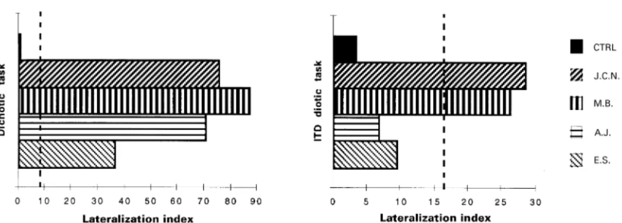
(4) Two types of auditory neglect. 679. Fig. 1 Dichotic and ITD diotic listening tasks. In both cases the subjects reported hearing ‘souris’ (mouse) on the left and ‘cheval’ (horse) on the right. In the dichotic listening task, the spatial dimension is provided by the ear of input. In the ITD diotic task, each ear receives both words at the same intensity level; the spatial dimension is simulated by an interaural time difference of 1 ms (∆t).. position on their head with their ipsilesional hand (same procedure as used in Altman et al., 1979; Bisiach et al., 1984). A graduated half circle fixed on the headphones was used to determine the angular value of the position (from 0° at the vertex to 90° at each ear). As a measure of overall performance, the relative positions attributed to two consecutive stimuli were compared; a response was counted as correct when a stimulus was correctly placed to the left or the right of the previous stimulus in correspondence with the difference in ITD or within ⫾10° of the previous location for identical ITDs. Two measures were used to quantify directional bias: (i) the deviation of the midline position, i.e. the difference between the theoretical 0° and the actual mean angular response given for the stimuli with no ITD; and (ii) the index of response asymmetry for the 48 lateralized stimuli, i.e. the number of pointings to the right minus number of pointings to the left, irrespective of the correctness of the replies. The number of alloacusis was also recorded independently. The 60 normal subjects achieved on average 57.15 (SD ⫽ 1.79) correct responses for the global score. They located the central stimuli (no ITD) at 0.09° to the left (SD ⫽ 4.5°). The index of response bias for the 48 lateralized stimuli was 0.00 (SD ⫽ 0.74) and alloacusis never occurred.. Analysis The three tests described above were part of a wider battery of auditory tasks and were administered in two different sessions. Patients M.B., E.S. and half of the control subjects performed the dichotic listening task on the first session and the sound localization task and spatial diotic 1 week later, whereas J.C.N., A.J. and the other half of the normal subjects performed the ITD diotic and the localization test on the first. session and the dichotic listening task 1 week later. Individual performances of the patients on the different measures described above were compared with the means and standard deviations computed from the control population. The cutoff score was set at 2 SD below the mean. Comparison of anatomical data collected in different subjects may be compromised by brain size and shape variations. To overcome this problem, we have adopted the normalized coordinate system of Talairach and Tournoux (Talairach and Tournoux, 1988) as in our previous neuropsychological and anatomical studies (Clarke et al., 1997, 1998, 2000; Di Virgilio and Clarke, 1997; Rivier and Clarke, 1997). The lesions as apparent on MRI (for M.B. and E.S.) or CT scan (for A.J. and J.C.N.) were determined within the corresponding horizontal, coronal and parasagittal planes of the proportional grid and represented on the standard brain.. Results Dichotic listening task All four patients had a marked left ear disadvantage: the lateralization index was 75.8 for J.C.N., 87.1 for M.B., 70.6 for A.J. and 36.6 for E.S; it was outside the 2 SD limit for all cases (Figs 2 and 3). In the four patients, the right ear score was normal (either 28 or 29) whereas the left ear score was greatly reduced (ranging from 2 to 13). No errors were noted in the monaural condition.. ITD diotic task Two of the four patients presented a significant hemispatial asymmetry in disfavour of the left side (Fig. 2). The.
(5) 680. A. Bellmann et al.. Fig. 2 Lateralization indexes for the dichotic and the ITD diotic tasks. The lateralization index corresponds to: 100 ⫻ (Rtot – Ltot)/ (Rtot ⫹ Ltot); ‘R/Ltot’ ⫽ total number of correct responses to either the right/left ear or the right/left side of space. The maximal asymmetry corresponds to 100. The mean value from the control population (CTRL) and the individual index for each patient are represented. The dashed line indicates the limit of normal performance set at 2 SD outside the mean lateralization index of control subjects.. Fig. 3 Reports of stimuli presented to the left or right ear in the dichotic task (left panel) and in the left or right hemispace in the ITD diotic task (right panel). Mean control data (CTRL) and patients’ scores are represented by bars; the maximum number of correct responses is 30. The dashed line indicates the limit of normal performance set at 2 SD below the mean score of control subjects.. lateralization index of J.C.N. and M.B. was well above the 2 SD limit (28.6 and 26.3, respectively). This asymmetry was due to abnormally low reporting of the left-sided stimuli (10 and 14, respectively), whereas reporting of the rightsided stimuli was within the normal range (18 and 24). Single trials were errorless. J.C.N. and M.B. thus failed to report a significant proportion of stimuli presented within left auditory hemispace, while they reported normally simultaneously presented stimuli on the right. A different result was found for patients A.J. and E.S. (see Fig. 2). Their lateralization index was within normal limits (6.7 and 9.5, respectively). Their left (21 and 19) and right side scores (24 and 23) were both within the normal range (Fig. 3). This absence of asymmetry in disfavour of the left hemispace stands in marked contrast to the left ear extinction presented by these patients at the standard dichotic listening task. This dissociation was not due to possible differences in the difficulty of the dichotic and diotic tasks. Patients J.C.N. and A.J. had a very similar lateralization index on the dichotic. listening task (75.8 and 70.6, respectively), but performed very differently on the ITD diotic task: J.C.N. was clearly deficient with a lateralization index of 28.6, while A.J. was within normal limits with 6.7 (Fig. 2).. Sound localization Two patients presented a spatial bias towards the ipsilesional side. A.J.’s global score of localization was severely deficient (35 out of 59). He located the central position 27.5° to the right, a deviation which stands 5.3 SDs below the mean of normal subjects. Moreover, his response bias index of 27 indicated a marked propensity to point to the right side of the head even when the sound originated from the left hemispace. From the 13 alloacusis recorded, 11 concerned extreme left (1 ms ITD) sound sources (see Fig. 4). E.S. also showed a severely reduced global score (25 out of 59, 17.9 SD below the mean of the controls), a significant deviation of the central position, 55.8° to the right, and an.
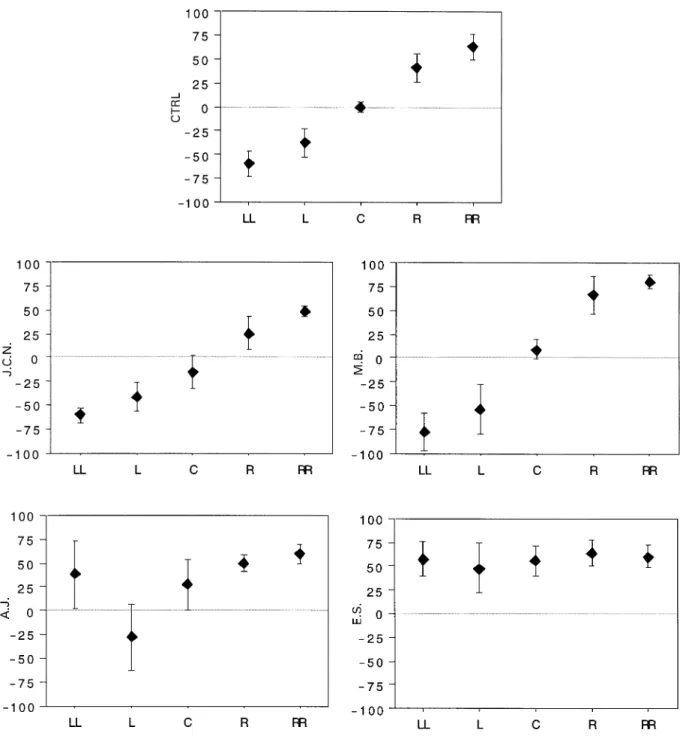
(6) Two types of auditory neglect. 681. Fig. 4 Performance in auditory localization of normal subjects (CTRL) and patients (identified by initials). Mean angular values of pointing responses for each ITD (negative for left), with 1 SD. For control data, the inter-subjects standard deviation is represented. ‘LL’ and ‘RR’ correspond to the 1 ms ITD favouring either the left or the right ear, ‘L’ and ‘R’ correspond to the 0.3 ms ITD and ‘C’ corresponds to the central position (no ITD).. extreme response bias to the right. Indeed, she located all the 24 left-sided sound sources to the right, except one which was located at 0°. This profile of results, although indicating an unquestionable rightwards spatial bias, differs from A.J.’s performance in that all the stimuli were perceived at the same subjective position, whereas A.J. differentiated the five. positions from one another and almost selectively translocated the extreme left sound sources to the right hemispace. The two other patients, J.C.N. and M.B., showed no spatial asymmetry on this test. Their global scores of 57 and 58, respectively, were well within the normal range. J.C.N. deviated the central position slightly (14°), but to the left of.
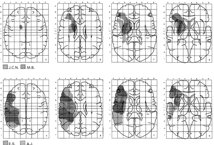
(7) 682. A. Bellmann et al.. Fig. 5 Cerebral lesions of J.C.N. and M.B. (top row), and E.S. and A.J. (bottom row). M.B. and E.S. had MRI, A.J. and J.C.N. had a CT scan. J.C.N. and M.B.’s lesions were mainly subcortical, involving the basal ganglia, the prefrontal white matter and the anterior insula. A.J. and E.S.’s lesions were cortical and involved the inferior frontal, the pre- and postcentral gyri, the insula, the superior temporal gyrus (with Heschl’s gyrus for E.S.) and the inferior parietal area including the supramarginalis gyrus. Right is to the left.. the midline, thus in the direction opposed to that expected with an auditory hemispatial neglect, and opposed to the rightwards bias presented by this patient in the two versions of the dichotic tasks. M.B. located the central position 8.8° to the right of the midline, which remains in the normal range (1.9 SD below the mean). None of these patients exhibited any alloacousis. Their response bias indexes of 0 were perfectly normal.. Lesions analysis Patients J.C.N. and M.B. presented a hemispatial asymmetry on the ITD diotic task with normal sound localization, while A.J. and E.S. presented a rightward bias on sound localization without asymmetry on the ITD diotic task. These two different profiles of auditory neglect were associated with different sites of lesion. Figure 5 displays the superimposed lesions of J.C.N. and M.B. (top row) and of A.J. and E.S. (bottom row). These lesions have been rescaled according to the Talairach proportional system and drawn on the standard brain in the horizontal plane at levels 5–8 of the Talairach space. J.C.N.’s lesion included the insula and extended. medially to the putamen and dorsal part of the caudate nucleus. The patient also had a small paraventricular lesion. M.B.’s lesion involved the basal ganglia (putamen, pallidum and head of caudate), the anterolateral part of the thalamus, the anterior insula, and extended rostrally to the white matter of the inferior frontal gyrus. The temporal, parietal and occipital cortices were intact in J.C.N. and M.B. E.S. had a large cortical temporofrontoparietal lesion. The supratemporal region was destroyed (with a lateral band of apparently normal but most probably disconnected tissue), as well as the dorsal part of the middle temporal gyrus. The frontal lesion was mainly dorsolateral and extended to the ventral portion of the inferior gyrus. The inferior parietal lobule was completely destroyed. There was also a lesion of the callosal pathway just anterior to the splenium, where the auditory information is believed to cross the callosum (Damasio and Damasio, 1979; Musiek et al., 1985; Alexander and Warren, 1988; Pujol et al., 1991). The basal ganglia and thalamus were not affected. A.J. had a cortical temporofrontoparietal lesion. It involved the insular cortex, the superior temporal gyrus (probably sparing the transverse gyrus), the inferior frontal gyrus and most of the inferior parietal lobule, including.
(8) Two types of auditory neglect the supramarginal but not the angular gyrus. The lesion extended medially to the body of the lateral ventricle and was likely to interrupt callosal pathways conveying auditory information. The basal ganglia and thalamus seemed to be unaffected by the lesion.. Discussion Four patients with unilateral right hemispheric damage were tested for dichotic listening and for two spatial attentional tests: an ITD diotic task, simulating spatial bilateral simultaneous stimulation without any interaural intensity or content differences and a sound localization task. The four patients had a significant asymmetry with lower report of stimuli presented to the left ear on the dichotic listening task. In two of them (J.C.N. and M.B.), this asymmetry was also found in the purely spatial (diotic) version of the test, where hemispatial origin is dissociated from input ear. These two patients performed normally on the sound localization task. The other two patients (A.J. and E.S.) had the opposite profile: they did not show hemispatial asymmetry in the report of auditory targets on the spatial diotic task, but presented severe spatial bias to the ipsilesional side in auditory localization. The present findings contribute to our understanding of auditory neglect in two ways. First, they suggest that in some patients (like A.J. and E.S.) an ear asymmetry in the standard dichotic listening task may depend crucially on the ear of entry of each stimulus, instead of being driven by purely spatial factors: this suggests that the dichotic listening test alone is not appropriate to assess auditory neglect. Secondly, they provide evidence for two distinct types of auditory neglect: hemispatial inattention as revealed by our new ITD diotic task, and spatial bias in auditory localization.. Ear extinction and auditory hemispatial neglect Currently, auditory neglect tends to be assessed by auditory double stimulation, either clinically with finger clicking or with the dichotic listening task. It remains, however, controversial as to whether an ear asymmetry in the dichotic listening test is due to a structural–perceptive or spatial– attentional mechanism. To dissociate these two mechanisms, we have designed the ITD diotic task. In this task, both ears receive the same acoustic stimuli at the same intensity level. Spatial positions are simulated with ITD; omissions to report items presented in the left hemispace cannot be accounted for by ear extinction and reflect a genuine attentional spatial deficit. Previous attempts to disentangle ‘ear of entry’ and auditory hemispace used loudspeakers positioned on each side of the subjects (for normal subjects see Morais and Bertelson, 1973; Hublet et al., 1976; Pierson et al., 1983; for brain-damaged patients see Tweedy et al., 1980; Soroker et al., 1997). These studies showed a right side advantage for verbal material. However, in the free-field condition, each ear still receives. 683. the sound source of the corresponding side of space with more intensity. Morais and Bertelson have proposed an ITD diotic test similar to ours, using consonant–vowels syllables (Morais and Bertelson, 1975). It was, however, administered only to normal subjects, for whom a slight right side advantage for verbal stimuli was found. We have found a similar statistically significant right side advantage in our control population. Much greater asymmetry was found in cases with specific unilateral right hemispheric lesions. Patients J.C.N. and M.B. presented both an ear asymmetry and a hemispatial asymmetry, characterized by left ear omissions on the dichotic test and left side omissions on the ITD diotic test. These results strongly suggest a deficit in the spatial allocation of attention and not a simple left ear extinction. A very different profile was found in patients A.J. and E.S., who presented an asymmetry on the dichotic listening task and performed normally on the ITD diotic task. Their deficit can be explained by ‘perceptual extinction’ (Hugdahl and Wester, 1994), i.e. deficit in accessing or processing the input of the contralesional ear. This interpretation is supported further by the fact that E.S. had a lesion of the right primary auditory cortex, as well as a lesion of the isthmus of the corpus callosum, where auditory fibres are believed to cross (Damasio and Damasio, 1979; Musiek et al., 1985; Alexander and Warren, 1988; Pujol et al., 1991). In A.J., the Heschl gyrus seemed to be intact, but the output of the primary auditory area on its way to the left hemisphere for verbal processing may have been interrupted, due to a partial lesion of the right associative temporoparietal junction extending dorsally and medially to the body of the lateral ventricle.. Sound localization and auditory hemispatial neglect Several studies adopted sound localization for the assessment of auditory hemispatial neglect. Systematic directional errors towards the ipsilesional side and alloacusis are considered to be neglect phenomena (Altman et al., 1979; Bisiach et al., 1984; Vallar et al., 1995; Haeske-Dewick et al., 1996; Sterzi et al., 1996; Soroker et al., 1997). Two of our patients, A.J. and E.S., presented a strong directional bias and alloacusis. A.J.’s auditory localization was subject to very specific spatial distortions. He discriminated well the different positions of the stimulus, including the two left positions, but he located the leftmost targets to the right hemispace. In his case, alloacousis concerned stimuli with the largest ITD (1 ms), i.e. stimuli which should be easiest to localize from the psychophysical point of view. The ipsilesional shift is likely to reflect distortions of a higher level spatial representation. Similar profiles were observed in visual hemispatial neglect (Halligan and Marshall, 1991; Kinsbourne, 1993). E.S. presented the directional bias to the ipsilesional side of space; in addition, she did not discriminate individual positions and placed them all in the right hemifield. Performance with.
(9) 684. A. Bellmann et al.. similar systematic directional errors and/or alloacusis have been described in free-field conditions, either clinically (Wortis and Pfeffer, 1948; Diamond and Bender, 1965; Altman et al., 1979) or experimentally (Vallar et al., 1995; Haeske-Dewick et al., 1996; Soroker et al., 1997). It may be argued that the spatial bias of A.J. and E.S. was due to a directional hypokinesia or hypometria rather than to a perceptual auditory neglect. Indeed, it is known that right-damaged patients are often reluctant to cross the midline with their ipsilesional limb (Heilman et al., 1985) and a premotor form of auditory neglect has been reported recently (Sterzi et al., 1996). Three observations speak against this hypothesis. First, we found as much alloacousis (and even more for A.J.) in another version of the test, where the patients had to indicate with a laser pointer the position on the wall corresponding best to the perceived intra-cranial location of the sound. In that response condition, patients did not have to cross the midline with their arm. They only inclined slightly the hand holding the laser pointer. Secondly, we asked A.J. and E.S. to give a verbal response for some supplementary items, and found alloacousis in that condition too. Thirdly, E.S. was given a discrimination task with two successive spatial stimuli. In that condition, where she had to answer ‘same’ or ‘different’, she performed at chance level. These observations thus speak against the directional hypokinesia account and the two last observations also rule out the hypothesis that the alloacusis might reflect motor perseverations.. Two distinct forms of auditory hemispatial neglect Previous studies reported dissociations between perceptual and premotor components of auditory neglect (Sterzi et al., 1996) and between auditory neglect in the front versus back space (Vallar et al., 1995). Our results suggest dissociation of auditory spatial attention and auditory spatial representation within the neglect syndrome. The division of attention between two simultaneous stimuli, that we have assessed with the ITD diotic task, made use of spatial information without requiring a precise representation of space. Auditory hemispatial neglect in patients J.C.N. and M.B. may be interpreted as attentional failure to targets within the contralesional hemispace or, alternatively, from an over-attention to the ipsilesional side. This asymmetry between the attention allocated to the right versus the left hemispaces may be combined with a reduction in processing speed or capacity and account for extinction phenomena (Driver et al., 1997). In the visual modality, extinction to double simultaneous stimulation was found in the absence of any other manifestation of visuospatial neglect, such as contralesional omissions in cancellation tasks or rightwards bias in line bisection (Di Pellegrino and De Renzi, 1995). Extinction was proposed to result from a failure to activate a sensory–attentional emergency system which is able to. detect and to respond to brief lateralized stimuli and which may have the characteristics of Kinsbourne’s dynamic directional bias model (Di Pellegrino and De Renzi, 1995). The cerebral lesions of J.C.N. and M.B. were confined to the basal ganglia, insular cortex (mainly anterior) and white matter of the inferior and middle frontal gyri and anterior cingulate cortex. Other studies have reported contralesional auditory extinction to clinically assessed double stimulation following basal ganglia infarctions (Damasio et al., 1980; Ferro et al., 1987). The ear asymmetry was attributed to an attentional deficit for the stimuli originating from the contralesional hemispace. The involvement of basal ganglia in spatial attention was documented in non-human primates (Boussaoud and Kermadi, 1997) and in man (Mesulam, 1990; Filoteo et al., 1997; Gitelman et al., 1999; Koski et al., 1999). Other evidence suggests that basal ganglia lesions may lead to a reduction of processing resources (Brown and Marsden, 1991), or impair reallocation of attention, once it has been committed to the analysis of one object (Husain et al., 1997). The insula, also damaged in our patients, has been proposed to be involved in the selection of relevant auditory information (Habib et al., 1995). The localization task required spatial representation obtained by matching the sound source to body position. We propose that auditory hemispatial neglect in patients A.J. and E.S. was the result of a systematic bias in auditory space representation with respect to the body. This interpretation is in agreement with previously formulated concepts of visual hemispatial neglect (Karnath, 1997) as deficient transformation of sensory input coordinates into the egocentric coordinate system. The latter allows accurate orientation of the body and guidance of limb or eye movements within space. Manipulations of neck muscle proprioception or vestibular input were shown to compensate visual neglect at the cancellation task or body orientation within space, but not visual extinction to double stimulation (Karnath, 1994, 1995). These observations and the occurrence of visual neglect in the absence of visual extinction (Barbieri and De Renzi, 1989) suggest different mechanisms for visual extinction and ipsilesional bias in the neglect syndrome. In the auditory modality, eye position, head-to-trunk position and transcutaneous vibration of the posterior neck muscles were shown to influence auditory localization in normal subjects (Lewald and Ehrenstein, 1996, 1998; Lewald et al., 1999), whereas it had no influence on the ear asymmetry presented by normal subjects (Asbjornsen et al., 1990; Struthers et al., 1992) or by two right-damaged patients (Vuilleumier et al., 1999) on dichotic listening. The cerebral lesions of A.J. and E.S., whose deficits were compatible with a distorted auditory spatial representation, involved the whole (E.S.) or most of the right inferior parietal lobule (A.J.), as well as large parts of the prefrontal and superior temporal cortices. In other studies, right parietal lesions were associated with systematic directional errors and/or alloacusis in sound localization (Bisiach et al., 1984; Cornelisse and Kelly, 1987). Electrophysiological recordings in monkeys have shown that.
(10) Two types of auditory neglect auditory spatially tuned cells of the posterior parietal cortex (LIP), frontal eye field and other parts of the prefrontal cortex present plurimodal properties, are influenced by eye or head position, or discharge only when an active motor response to the target is required (Vaadia et al., 1986; Russo and Bruce, 1994; Mazzoni et al., 1996; Stricanne et al., 1996). These data further support the idea that these inferior parietal and prefrontal structures may be involved in the building up of a representation of auditory space following an egocentric frame of reference, and once lesioned may give rise to the systematic bias observed in our patients A.J. and E.S.. Conclusion Our data demonstrate two types of auditory neglect characterized by (i) left-sided disadvantage to report contralesional targets when they are presented together with an ipsilesional target; or (ii) a spatial bias to the ipsilesional side of space in active localization of single auditory targets. The first type of auditory neglect is interpreted as imbalance between the attentional load allocated to the left and right hemispaces, due to basal ganglia and insular lesions. The second type of auditory neglect is interpreted as systematic bias of auditory egocentric space representation, due to inferior parietal and frontal dysfunction.. Acknowledgements We wish to thank Professor G. Assal for encouragement throughout this work and Dr C. Bindschaedler for neuropsychological evaluation of the four patients and helpful comments on the manuscript. This study was supported by the Swiss National Science Foundation grants 3100-52718.97 and 3231-41607.94 (‘Score A’ fellowship) to S.C.. 685. Beaton A, McCarthy M. On the nature of auditory neglect: a reply to Hugdahl and Wester. Brain Lang 1995; 48: 351–8. Bisiach E, Cornacchia L, Sterzi R, Vallar G. Disorders of perceived auditory lateralization after lesions of the right hemisphere. Brain 1984; 107: 37–52. Boussaoud D, Kermadi I. The primate striatum: neuronal activity in relation to spatial attention versus motor preparation. Eur J Neurosci 1997; 9: 2152–68. Brown RG, Marsden CD. Dual task performance and processing resources in normal subjects and patients with Parkinson’s disease. Brain 1991; 114: 215–31. Clarke S, Lindemann A, Maeder P, Borruat FX, Assal G. Face recognition and posterior-inferior hemispheric lesions. Neuropsychologia 1997; 35: 1555–63. Clarke S, Walsh V, Schoppig A, Assal G, Cowey A. Colour constancy impairments in patients with lesions of the prestriate cortex. Exp Brain Res 1998; 123: 154–8. Clarke S, Bellmann A, Meuli RA, Assal G, Steck AJ. Auditory agnosia and auditory spatial deficits following left hemispheric lesions: evidence for distinct processing pathways. Neuropsychologia 2000; 38: 797–807. Cornelisse LE, Kelly JB. The effect of cerebrovascular accident on the ability to localize sounds under conditions of the precedence effect. Neuropsychologia 1987; 25: 449–52. Damasio H, Damasio A. ‘Paradoxic’ ear extinction in dichotic listening: possible anatomic significance. Neurology 1979; 29: 644–53. Damasio AR, Damasio H, Chui HC. Neglect following damage to frontal lobe or basal ganglia. Neuropsychologia 1980; 18: 123–32. Deouell LY, Bentin S, Soroker N. Electrophysiological evidence for an early (pre-attentive) information processing deficit in patients with right hemisphere damage and unilateral neglect. Brain 2000; 123: 353–65. De Renzi E, Gentilini M, Pattacini F. Auditory extinction following hemisphere damage. Neuropsychologia 1984; 22: 733–44.. References Albert ML. A simple test of visual neglect. Neurology 1973; 23: 658–64. Alexander MP, Warren RL. Localization of callosal auditory pathways: a CT case study. Neurology 1988; 38: 802–4. Altman JA, Balonov LJ, Deglin VL. Effects of unilateral disorder of the brain hemisphere function in man on directional hearing. Neuropsychologia 1979; 17: 295–301. Asbjornsen A, Hugdahl K, Hynd GW. The effects of head and eye turns on the right ear advantage in dichotic listening. Brain Lang 1990; 39: 447–58. Barbieri C, De Renzi E. Patterns of neglect dissociation. Behav Neurol 1989; 2: 13–24. Beaton A, McCarthy M. ‘Auditory neglect after right frontal lobe and right pulvinar thalamic lesions’: comment on Hugdahl, Wester, and Asbjornsen (1991) and some preliminary findings. Brain Lang 1993; 44: 121–6.. Diamond SP, Bender MB. On auditory extinction and alloacusis. Trans Am Neurol Assoc 1965; 90: 154–7. Di Pellegrino G, De Renzi E. An experimental investigation on the nature of extinction. Neuropsychologia 1995; 33: 153–70. Di Virgilio G, Clarke S. Direct interhemispheric visual input to human speech areas. Hum Brain Mapp 1997; 5: 347–54. Driver J, Mattingley JB, Rorden C, Davis G. Extinction as a paradigm measure of attentional bias and restricted capacity following brain injury. In: Thier P, Karnath H-O, editors. Parietal lobe contributions to orientation in 3D space. Berlin: Springer-Verlag; 1997. p. 401–29. Efron R, Crandall PH, Koss B, Divenyi PL, Yund EW. Central auditory processing. III. The ‘cocktail party’ effect and anterior temporal lobectomy. Brain Lang 1983; 19: 254–63. Ferro JM, Kertesz A, Black SE. Subcortical neglect: quantitation, anatomy, and recovery. Neurology 1987; 37: 1487–92. Filoteo JV, Delis DC, Salmon DP, Demadura T, Roman MJ, Shults CW. An examination of the nature of attentional deficits in patients.
(11) 686. A. Bellmann et al.. with Parkinson’s disease: evidence from a spatial orienting task. J Int Neuropsychol Soc 1997; 3: 337–47.. Kimura D. Functional asymmetry of the brain in dichotic listening. Cortex 1967; 3: 163–78.. Gitelman DR, Nobre AC, Parrish TB, LaBar KS, Kim Y-H, Meyer JR, et al. A large-scale distributed network for covert spatial attention. Further anatomical delineation based on stringent behavioural and cognitive controls. Brain 1999; 122: 1093–106.. Kinsbourne M. Hemi-neglect and hemisphere rivalry. Adv Neurol 1977; 18: 41–9.. Goodglass H, Kaplan E. The assessment of aphasia and related disorders. 2nd edn. Philadelphia: Lea & Febiger; 1983. Habib M, Daquin G, Milandre L, Royere ML, Rey M, Lanteri A, et al. Mutism and auditory agnosia due to bilateral insular damage— role of the insula in human communication. Neuropsychologia 1995; 33: 327–39. Haeske-Dewick H, Canavan AG, Ho¨ mberg V. Sound localization in egocentric space following hemispheric lesions. Neuropsychologia 1996; 34: 937–42. Halligan PW, Marshall JC. Spatial compression in visual neglect: a case study. Cortex 1991; 27: 623–9. Halligan PW, Marshall JC, Wade DT. Left on the right: allochiria in a case of left visuo-spatial neglect. J Neurol Neurosurg Psychiatry 1992; 55: 717–9. Heilman KM, Valenstein E. Auditory neglect in man. Arch Neurol 1972; 26: 32–5. Heilman KM, Bowers D, Coslett HB, Whelan H, Watson RT. Directional hypokinesia: prolonged reaction times for leftward movements in patients with right hemisphere lesions and neglect. Neurology 1985; 35: 855–9. Hublet C, Morais J, Bertelson P. Spatial constraints on focused attention: beyond the right-side advantage. Perception 1976; 5: 3–8. Hugdahl K, Wester K. Auditory neglect and the ear extinction effect in dichotic listening: a reply to Beaton and McCarthy (1993). Brain Lang 1994; 46: 166–73. Hugdahl K, Wester K, Asbjornsen A. Auditory neglect after right frontal lobe and right pulvinar thalamic lesions. Brain Lang 1991; 41: 465–73. Husain M, Shapiro K, Martin J, Kennard C. Abnormal temporal dynamics of visual attention in spatial neglect patients. Nature 1997; 385: 154–6. Jones SJ, Pitman JR, Halliday AM. Scalp potentials following sudden coherence and discoherence of binaural noise and change in the inter-aural time difference: a specific binaural evoked potential or a ‘mismatch’ response? Electroencephalogr Clin Neurophysiol 1991; 80: 146–54. Karnath H-O. Subjective body orientation in neglect and the interactive contribution of neck muscle proprioception and vestibular stimulation. Brain 1994; 117: 1001–12. Karnath H-O. Transcutaneous electrical stimulation and vibration of neck muscles in neglect. Exp Brain Res 1995; 105: 321–4. Karnath H-O. Neural encoding of space in egocentric coordinates?— Evidence for and limits of a hypothesis derived from patients with parietal lesions and neglect. In: Thier P, Karnath H-O, editors. Parietal lobe contributions to orientation in 3D space. Berlin: Springer-Verlag; 1997. p. 497–520.. Kinsbourne M. Orientational bias model of unilateral neglect: evidence from attentional gradients within hemispace. In: Robertson IH, Marshall JC, editors. Unilateral neglect: clinical and experimental studies. Hove (UK): Lawrence Erlbaum; 1993. p. 63–86. Klingon GH, Bontecou DC. Localization in auditory space. Neurology 1966; 16: 879–86. Koski L, Paus T, Hofle N, Petrides M. Increased blood flow in the basal ganglia when using cues to direct attention. Exp Brain Res 1999; 129: 241–6. Lewald J, Ehrenstein WH. The effect of eye position on auditory lateralization. Exp Brain Res 1996; 108: 473–85. Lewald J, Ehrenstein WH. Influence of head-to-trunk position on sound lateralization. Exp Brain Res 1998; 121: 230–8. Lewald J, Karnath H-O, Ehrenstein WH. Neck-proprioceptive influence on auditory lateralization. Exp Brain Res 1999; 125: 389–96. Mazzoni P, Bracewell RM, Barash S, Andersen RA. Spatially tuned auditory responses in area LIP of macaques performing delayed memory saccades to acoustic targets. J Neurophysiol 1996; 75: 1233–41. Mesulam MM. Large-scale neurocognitive networks and distributed processing for attention, language, and memory. [Review]. Ann Neurol 1990; 28: 597–613. Morais J, Bertelson P. Laterality effects in diotic listening. Perception 1973; 2: 107–11. Morais J, Bertelson P. Spatial position versus ear of entry as determinant of the auditory laterality effects: a stereophonic test. J Exp Psychol Hum Percept Perform 1975; 1: 253–62. Musiek FE, Reeves AG, Baran JA. Release from central auditory competition in the split-brain patient. Neurology 1985; 35: 983–7. Nicholls ME. Support for a structural model of aural asymmetries. Cortex 1998; 34: 99–110. Pierson JM, Bradshaw JL, Nettleton NC. Head and body space to left and right, front and rear—I. Unidirectional competitive auditory stimulation. Neuropsychologia 1983; 21: 463–73. Pinek B, Brouchon M. Head turning versus manual pointing to auditory targets in normal subjects and in subjects with right parietal damage. Brain Cogn 1992; 18: 1–11. Pinek B, Duhamel J-R, Cave´ C, Brouchon M. Audio-spatial deficits in humans: differential effects associated with left versus right hemisphere parietal damage. Cortex 1989; 25: 175–86. Poirier P, Lassonde M, Villemure J-G, Geoffroy G, Lepore F. Sound localization in hemispherectomized patients. Neuropsychologia 1994; 32: 541–53. Pujol J, Junque´ C, Vendrell P, Garcı`a P, Capdevila A, Martı`-Vilalta JL. Left-ear extinction in patients with MRI periventricular lesions. Neuropsychologia 1991; 29: 177–84..
(12) Two types of auditory neglect Rivier F, Clarke S. Cytochrome oxidase, acetylcholinesterase, and NADPH-diaphorase staining in human supratemporal and insular cortex: evidence for multiple auditory areas. Neuroimage 1997; 6: 288–304.. 687. Tweedy JR, Rinn WE, Springer SP. Performance asymmetries in dichotic listening: the role of structural and attentional mechanisms. Neuropsychologia 1980; 18: 331–8.. Russo GS, Bruce CJ. Frontal eye field activity preceding aurally guided saccades. J Neurophysiol 1994; 71: 1250–3.. Vaadia E, Benson DA, Hienz RD, Goldstein MH Jr. Unit study of monkey frontal cortex: active localization of auditory and of visual stimuli. J Neurophysiol 1986; 56: 934–52.. Sanchez-Longo LP, Forster FM. Clinical significance of impairment of sound localization. Neurology 1958; 8: 119–25.. Vallar G, Guariglia C, Nico D, Bisiach E. Spatial hemineglect in back space. Brain 1995; 118: 467–72.. Schu¨ eli H, Henn V, Brugger P. Vestibular stimulation affects dichotic lexical decision performance. Neuropsychologia 1999; 37: 653–9.. Vanier M, Gauthier L, Lambert J, Pepin EP, Robillard A, Dubouloz CJ, et al. Evaluation of left visuospatial neglect: norms and discrimination power of two tests. Neuropsychology 1990; 4: 87–96.. Soroker N, Calamaro N, Glicksohn J, Myslobodsky MS. Auditory inattention in right-hemisphere-damaged patients with and without visual neglect. Neuropsychologia 1997; 35: 249–56. Sparks R, Geschwind N. Dichotic listening in man after section of neocortical commissures. Cortex 1968; 4: 3–16. Sterzi R, Piacentini S, Polimeni M, Liverani F, Bisiach E. Perceptual and premotor components of unilateral auditory neglect. J Int Neuropsychol Soc 1996; 2: 419–25. Stricanne B, Andersen RA, Mazzoni P. Eye-centered, head-centered, and intermediate coding of remembered sound locations in area LIP. J Neurophysiol 1996; 76: 2071–6. Struthers G, Charlton S, Bakan P. Lateral orientation, eye movements and dichotic listening. Int J Neurosci 1992; 66: 189–95. Talairach J, Tournoux P. Co-planar stereotaxic atlas of the human brain. Stuttgart: Thieme; 1988.. Vuilleumier P, Valenza N, Mayer E, Perrig S, Landis T. To see better to the left when looking more to the right: effects of gaze direction and frames of spatial coordinates in unilateral neglect. J Int Neuropsychol Soc 1999; 5: 75–82. Walsh EG. An investigation of sound localization in patients with neurological abnormalities. Brain 1957; 80: 222–50. Wortis SB, Pfeffer AZ. Unilateral auditory-spatial agnosia. J Nerv Ment Dis 1948; 108: 181–6. Zatorre RJ, Ptito A, Villemure J-G. Preserved auditory spatial localization following cerebral hemispherectomy. Brain 1995; 118: 879–89.. Received October 17, 2000. Revised November 21, 2000. Accepted November 29, 2000.
(13)
Figure

Documents relatifs