British Journal of Rheumatology 1992;31:531-534
THE PREVALENCE OF PALPABLE FINGER JOINT NODULES IN
DIFFUSE IDIOPATHIC SKELETAL HYPEROSTOSIS (DISH). A
CONTROLLED STUDY
BY P. SCHLAPBACH*, CH. BEYELER*, N. J. GERBER*, SJ. VAN DER LINDEN*, U. BURGIf, W. A. FUCHSt AND H. EHRENGRUBER§
Departments of * Rheumatology, flnternal Medicine, tRadiology, and §Data Processing, University of Berne, Inselspital, 3010 Berne, Switzerland
SUMMARY
The presence of clinically palpable finger joint nodules (Heberden's and Bouchard's nodes) was documented in 123 con-secutive cases with diffuse idiopathic skeletal hyperostosis (DISH) of the thoracic spine and 191 matched DISH negative controls. The prevalence of palpable finger joint nodules was almost twice as frequent in cases with spinal DISH compared to controls (46% versus 31%, x2 = 7.67, P<0.01; multivariate adjusted odds ratio OR = 1.84; 95% CI: 1.14-2.98). This
increase was most marked at the proximal interphalangeal joint, in males and in patients up to the age of 65 years. In addi-tion and independent of other variables such as hyperostotic features, age and sex, the prevalence of palpable finger joint nodules was about twice as high in probands with a history of physically heavy work compared to those without (43% ver-sus 26%, x = 9.18, P<0.005; multivariate adjusted odds ratio OR = 2.10; 95% CI: 1.26-3.52). From these results we con-clude that DISH should be considered as an independent risk factor in the development of finger joint nodules.
KEY WORDS: Heberden's nodes, Diffuse idiopathic skeletal hyperostosis (DISH), Controlled study.
DIFFUSE idiopathic skeletal hyperostosis (DISH) is a
well defined systemic condition, characterized by spinal and extraspinal hyperostotic calcification and ossification of ligaments, tendons and joint capsules [1-5]. Radiological changes in the hands include broadened and arrowhead distal phalangeal tufts, increased cortical width of tubular bones, prominent enthesopathy of the proximal phalanges, exostoses and new bone formation in joint capsules [6, 7]. In addi-tion, clinically palpable finger joint nodules have been described [2,3,5,8,9]. However, the prevalence of fin-ger nodules in cases with DISH and their association with osteoarthritic nodules remains unknown. As part of a controlled study of hospitalized patients with or without spinal DISH, we examined the prevalence of palpable finger joint nodules in both groups. The main objective was to confirm our hypothesis, that DISH is associated with the development of palpable finger joint nodules, irrespective of age, sex and the history of strenuous manual labour.
MATERIAL AND METHODS
One hundred and twenty-three consecutive unselected cases with spinal DISH, based on routine lateral chest X-ray, were compared with 191 age- and sex-matched controls without spinal DISH. All were inpatients of two departments of internal medicine and one of cardiovascular surgery, referred to for problems not related to the locomotor system. They were inter-viewed for an occupational history during the previous 6 months and before by two blinded rheumatologists.
Submitted 6 February; revised version accepted 30 August 1991.
Correspondence to P. Schlapbach, Department of Rheuma-tology, University of Berne, Inselspital, CH-3010 Berne, Switzerland.
0263-7103/92/080531 + 04 $03.00/0
Occupational activities were classified as physically heavy or light by consensus of the two interviewers. Palpable finger joint nodules, defined as fixed bony expansions at the dorsolateral and dorsomedial aspect of the distal and proximal interphalangeal joints were documented by two observers blind to the X-ray find-ings. The lateral chest X-rays were graded as follows: Grade 0: No ossification
Grade I: Prevertebral and/or prediscal ossification at one or two vertebral bodies of the thor-acic spine or one bridging ossification between vertebrae
Grade II: Continuous flowing prevertebral and/or prediscal ossification along three or more vertebral bodies of the thoracic spine or two bridging ossifications
Grade III: At least three bridging ossifications along the thoracic spine
As proposed by Resnick et al. [2], the intervertebral discs of the hyperostotic segments showed no degener-ative, dysplastic or inflammatory abnormalities. Pro-bands with grades 0 and I were classified as spinal DISH negative (controls), those with grades II and III as spinal DISH positive (cases).
Intra- and interobserver reliability of X-ray grading was documented as previously described [10]. Statis-tical calculations were based on the chi-square test for dichotomous variables and the Student's r-test for continuous variables. Multiple regression analyses were performed with the Statistical Analysis System (SAS Institute Inc., Cary, NC, USA) under licence of the University of Berne. Logistic procedures with step-down regression analyses of four independent vari-ables were used. Calculation of odds ratios (OR), multivariate adjusted odds ratios and 95% confidence © 1992 British Society for Rheumatology
532 BRITISH JOURNAL OF RHEUMATOLOGY VOL. XXXI NO. 8
TABLE I
DEMOGRAPHIC DATA AND PREVALENCE OF PALPABLE FINGER JOINT NODULES
Spinal DISH cases (n = 123) DISH negative controls (n = 191) Mean age, years (± SD)
Males Females
History of heavy work DIP joint nodules PIP joint nodules DIP or PIP joint nodules
71 ±9.4 96 (78%) 27 (22%) 79 (64%) 42(34%) 40 (33%) 57 (46%) 68 ± 9 . 9 126 (66%) 65 (34%) 120 (63%) 47 (25%) 36 (19%) 59 (31%) <0.05 <0.05 NS NS <0.01 <0.01
DIP, distal interphalangeal joint; PIP, proximal interphalangeal joint.
intervals (95% CI) were carried out according to stan-dard procedures [11,12]. The level of statistical signifi-cance was set at P = 0.05.
RESULTS
Demographic data of the 314 probands studied are shown in Table I. Palpable finger joint nodules were significantly more prevalent in spinal DISH positive cases than in DISH negative controls (OR = 1.93; 95% CI; 1.21-3.09; Table I). Analysing the distal and prox-imal interphalangeal joints separately, the difference was marked at the proximal joint level (OR = 2.08; 95% CI: 1.23-3.50) but only slight at the distal joint level (OR = 1.59; 95% CI: 0.97-2.61). This variation resulted from an increased prevalence of distal over proximal interphalangeal joint nodules in DISH nega-tive controls, whereas no difference was detectable in DISH positive cases (Table I).
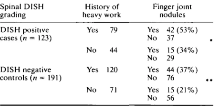
The prevalence of palpable finger joint nodules was significantly higher in probands with a history of physi-cally heavy work compared to those without (43% ver-sus 26%; OR = 2.16; 95% CI: 1.31-3.56). This increase was similar in DISH positive cases (OR = 2.19; 95% CI: 1.02-4.71) and DISH negative controls (OR = 2.16; 95% CI: 1.09-4.27; Table II). These results underline the contribution of mechanical fac-tors to the development of finger joint nodules.
The prevalence of palpable finger joint nodules was similar in females and males (41% versus 35%; OR = 1.30; 95% CI: 0.79-2.14). However, taking spinal DISH rating into account, an interesting difference was revealed. Whereas in DISH positive cases finger
TABLE II
PREVALENCE OF PALPABLE FINGER JOINT NODULES IN PROBANDS WITH AND WITHOUT A HISTORY OF HEAVY WORK
Spinal DISH grading History of heavv work Finger joint nodules
joint nodules were found as often in females as in males (OR = 0.91; 95% CI: 0.38-2.14), in DISH negative controls finger nodules were found more frequently in females than males (OR = 1.88; 95% CI: 1.0-3.55; Table III).
The prevalence of palpable finger joint nodules increased with age (21% in probands up to 55 years, 31 % in 56-65 years old, 41% in 66-75 years old, 42% in 76-85 years old and 50% in over 85 years old respect-ively). It was higher in DISH positive cases than DISH negative controls in most age groups (Table IV). Because of a considerable interaction between age, sex, occupational history and finger nodules, multiple regression analyses were performed (Table V). All the results were in agreement with the univariate analyses presented above. The multivariate adjusted odds ratios for the presence versus the absence of palpable finger joint nodules were 1.84(95% CI: 1.14-2.98) for DISH and 2.10 (95% CI: 1.26-3.52) for a history of heavy work. Finally, there was a strong association between distal and proximal interphalangeal joint nodules (x2 =
64.44, P<0.0005).
DISCUSSION
In DISH the presence of palpable finger nodules at the distal and proximal interphalangeal joints has been described in about one-third of the examined cases ([2, 3, 5, 8, 9]; Table I). Our data support the hypothesis that DISH is associated with the development of pal-pable finger joint nodules. The prevalence of nodules at the distal or proximal interphalangeal joints was almost twice as high in spinal DISH positive cases
com-TABLE III
PREVALENCE OF PALPABLE FINGER JOINT NODULES IN FEMALES AND MALES Spinal DISH grading Sex Finger joint nodules DISH positive cases (n = 123) DISH negative controls (n = 191) •x 2 = 4.13, P<0.05. • V = 5.05, P<0.25. Yes No Yes No 79 44 120 71 Yes No Yes No Yes No Yes No 42 (53%) 37 15 (34%) 29 44 (37%) 76 15(21%) 56 DISH positive cases(n = 123) DISH negative controls (n = 191) V = 0.05, NS. **X23.83.NS. Females Males Females Males 27 96 65 126 Yes No Yes No Yes No Yes No 12 (44%) 15 45 (47%) 51 26 (42%) 39 33 (26%) 93
SCHLAPBACH ETAL.: PALPABLE FINGER JOINT NODULES IN DISH 533
Spinal DISH grading
DISH positive cases
(n = 123)
DISH negative controls (n = 191) PREVALENCE OF Finger joint nodules Yes No Yes No TABLE IV
PALPABLE FINGER JOINT NODULES IN
Age up to 55 3 (50%) 3 3 (13%) 20 (OR = 6.67) 55-65 15 (44%) 19 12 (23%) 40 (OR = 2.63) X2 = 4.23 P<0.05
DIFFERENT AGE GROUPS
66-75 16 (40%) 24 28(41%) 40 (OR = 0.95) X2 = 0.01 NS 76-85 20(51%) 19 15 (34%) 29 (OR = 2.04) X2 = 2.52 NS 86 and older 3 (75%) 1 1 (25%) 3
pared to spinal DISH negative controls, irrespective of the history of heavy work at present or in the past. However, this difference was prominent only at the proximal interphalangeal joint level, in males and up to the age of 65 years. This could be due to mixed aetio-pathogenetic mechanisms for the development of nod-ules at the distal interphalangeal joint level, in females and the elderly. Indeed, it has been shown that finger joint osteoarthrosis is more common in females than in males over the age of 55 years, mainly affecting the dis-tal finger joints [13-17]. Further, our data support the hypothesis that heavy physical work is associated with the development of palpable finger joint nodules. The prevalence of nodules at the distal or proximal inter-phalangeal joints was about twice as high in patients with a history of heavy work at present or in the past, irrespective of spinal DISH grading, sex and age. We are well aware that this relationship has to be interpre-ted with caution. It is difficult to judge objectively whether work has to be considered as strenuous or not. In addition, time factors might play an important role. However, our findings are in agreement with previous studies, suggesting occupational and traumatic factors such as repetitive impulsive loading [18,19] to be aetio-pathogenetically relevant for the development of fin-ger joint nodules [13-15], as shown for judoka [20] and cotton mill workers [21].
From the presented results we conclude that DISH is
a separate but discrete risk factor for the development of finger joint nodules. It has to be considered as aetio-pathogenetically relevant especially in situations uncommon for osteoarthritic changes such as in young males without heavy occupational activities. In addi-tion, our data indicate that mechanical factors may play a role in the formation of finger nodules.
ACKNOWLEDGEMENTS
We thank E. Hachler, MD, for examining patients and all colleagues of the Departments of Internal Medicine (Prof. P. W. Straub, Prof. H. Studer, Prof. T. Hess) and Cardiovascular Surgery (Prof. U. Althaus), Inselspital Berne for allowing us access to their patients.
REFERENCES
1. Resnick D, Shaul SR, Robins JM. Diffuse idiopathic skeletal hyperostosis DISH: Forestier's disease with extraspinal manifestations. Radiology 1975;115:513-24.
2. Resnick D, Shapiro RF, Wiesner KB, Niwayama G, Utsinger PD, Shaul SR. Diffuse idiopathic skeletal hyperostosis (DISH) (ankylosing hyperostosis of Forestier and Rotes-Querol). Semin Arthritis Rheum 1978;7:153-87.
3. Utsinger PD, Resnick D, Shapiro RF. Diffuse skele-tal abnormalities in Forestier's disease. Arch Intern Med 1976;136:763-8.
4. Utsinger PD. Diffuse idiopathic skeletal hyperosto-sis. Clin Rheum Dis 1985;11:325-51.
TABLE V
MULTIPLE LOGISTIC REGRESSION ANALYSES
Independent variable
DIP joint nodules
PIP joint nodules
DIP or PIP joint nodules
Dependent variable DISH Sex Age History of work Sex History of work Age DISH Sex Age DISH History of work Parameter estimate beta -0.030 -0.712 -0.587 -0.034 -0.673 -0.031 -0.611 -0.744
x
2 2.40 2.59 4.94 6.50 0.06 3.87 5.49 6.09 0.56 5.67 6.17 8.06 Probability of xP 0.122 0.108 0.026 0.011 0.803 0.049 0.019 0.014 0.455 0.017 0.013 0.005534 BRITISH JOURNAL OF RHEUMATOLOGY VOL. XXXI NO. 8
5. Arlet J, Mazieres B. La maladie hyperostotique. Rev Med Interne 1985;6:553-64.
6. Littlejohn GO, Urowtiz MB, Symthe HA, Keystone EC. Radiographic features of the hand in diffuse idiopathic skeletal hyperostosis (DISH). Radiology 1981;140:623-9.
7. Fischer E. Exo- und endomarginale Reaktionen an der Hand bei der diffusen idiopathischen Skele-tthyperostose, ihre Quantifizierung und Altersab-hangigkeit. Fortschr Rontgenstr 1985;142:447-54. 8. Henrard JC, Bennett PH. Etude 6pid6miologique de
l'hyperostose vert6brale. Enquete dans une popula-tion adulte d'indiens d'am^rique. Rev Rhum 1973;40:581-91.
9. Harris J, Carter AR, Glick WN, Storey GO. Anky-losing hyperostosis. Clinical and radiological fea-tures. Ann Rheum Dis 1974;33:210-15.
10. Schlapbach P, Beyeler Ch, Gerber NJ et al. Diffuse idiopathic skeletal hyperostosis (DISH) of the spine: a cause of back pain? Br J Rheumatol 1989;28:299-303.
11. Kirkwood BR. Essentials of medical statistics. Oxford: Blackwell Scientific Publications, 1988. 12. Schlesselmann JJ. Case-control studies. Oxford:
Oxford University Press, 1982.
13. Stecher RM. Heberden's nodes: the incidence of
hypertrophic arthritis of the fingers. N Engl J Med 1940;222:300-8.
14. Stecher RM. Heberden Oration. Heberden's nodes. A clinical description of osteoarthritis of the finger joints. Ann Rheum Dis 1955;14:1-10.
15. Kellgren JH, Lawrence JS. Osteoarthrosis and disk degeneration in an urban population. Ann Rheum Dis 1958; 17: 388-97.
16. Kellgren JH, Lawrence JS, Bier F. Genetic factors in generalized osteoarthrosis. Ann Rheum Dis
1963;22:237-55.
17. Lawrence JS, Brenner JM, Bier F. Osteoarthrosis. Prevalence in the population and relationship between symptoms and X-ray changes. Ann Rheum Dis 1966;25:l-24.
18. Radin EL, Parker HG, Paul IL. Pattern of degener-ative arthritis. Preferential involvement of the distal finger joints. Lancet 1971 ;i:377-9.
19. Radin EL, Paul IL, Rose RM. Role of mechanical factors in the pathogenesis of primary osteoarthritis. Lancet 1972;i:519-22.
20. Frey A, Miiller W. Heberden-Arthrosen bei Judo-Sportlern. Schweiz Med Wschr 1984;114:40-8. 21. Lawrence JS. Rheumatism in cotton operatives. Br J