DIFFUSE IDIOPATHIC SKELETAL HYPEROSTOSIS
(DISH) OF THE SPINE: A CAUSE OF BACK PAIN? A
CONTROLLED STUDY
BY P. SCHLAPBACH*, CH. BEYELER', N. J. GERBER*, SJ. VAN DER LINDEN*, U. BURGIf, W. A. FUCHS* AND H. EHRENGRUBER§
Departments of * Rheumatology, t Internal Medicine, t Radiology and § Electronic Data Processing, University of Berne, Inselspital, CH-3010 Berne, Switzerland
SUMMARY
This is the first controlled study of the frequency of back pain in a European Caucasian population with diffuse idiopathic skeletal hyperostosis (DISH).
Elderly patients admitted to hospital for reasons other than back pain were assessed for the presence of spinal DISH using the routine lateral chest radiograph films. A total of 106 probands (82 males, 24 females) with a mean age of 70 years fulfilled the criteria for DISH as defined previously. One hundred and seventy-eight patients (117 males, 61 females) not meeting these criteria were used as controls. The prevalence of back pain was assessed by a blinded interviewer using a structured questionnaire. Our primary hypothesis was that spinal DISH positive probands had not had back pain more often than controls. This controlled study showed no statistically significant difference in pain frequency between spinal DISH positive probands and controls at any spinal level.
We conclude that back pain does not occur more often in radiographically defined DISH positive probands than in controls. The radiological finding of spinal DISH, as far as it does not lead to stenosis of the spinal canal or dysphagia, thus seems to be a finding without clinical relevance.
KEY WORDS: Spine, Radiographs, Pain, Osteoarthritis, Forestier's disease, Ankylosing vertebral hyperostosis.
DIFFUSE idiopathic skeletal hyperostosis (DISH) of the spine is a frequent radiological finding. The condition is characterized by prevertebral and prediscal ossification, involving mainly liga-ments and entheses. Typically, the hyperostotic ossification is located along the anterolateral aspect of the thoracic spine, but can also be found fa the cervical and lumbar spine [1-4]. The disorder has been known for several decades under different synonyms, e.g. Forestier's disease, hyperostotic spondylosis, senile ankylosing hyperostosis of the spine or ankylos-ing vertebral hyperostosis. The term diffuse idiopathic skeletal hyperostosis was proposed by Resnick et al. [5], based on the observation of hyperostotic ossification at extraspinal skeletal sites.
Population studies by Julkunen el al. [6] in Finland revealed an overall prevalence of spinal DISH in 3.8% of males and 2.6% of females over the age of 40 years, the prevalence rates rising with increasing age. In male Pima Indians over 40 years of age, the prevalence of radiographic spinal hyperostosis is 25%, and in
Submitted 18 August 1988; revised version accepted 2 January 1989.
Correspondence to Dr. Schlapbach.
females 5% [7]. Routine autopsies show signs of spinal DISH in 6-28% [4,8,9].
The aetiology [3,10-12] and clinical relevance of this condition are unknown. The objective of this controlled study was to clarify the relevance of spinal DISH to back pain.
MATERIAL AND METHODS A controlled study was carried out on patients hospitalized for reasons other than back pain. The subjects were recruited as follows: unselected consecutive lateral chest radiographs done on admission to two departments of inter-nal medicine and one department of cardio-vascular surgery identified probands who fulfilled the criteria for spinal DISH (PS). For each spinal DISH positive proband we assigned DISH negative controls. Name, date of birth and room-number of DISH positive probands and controls were reported to a rheumatologist (CH.B., E.H.), who blindly collected data on the clinical symptoms in the past by interview using a structured questionnaire. The questions, concerning the prevalence of back pain in the past 6 months as well as the prevalence of back pain prior to the last 6 months (Table I) were explained to all probands during the interview. The presence or absence of extraskeletal causes
300 BRITISH JOURNAL OF RHEUMATOLOGY VOL. XXVIII NO. 4
TABLE I
PRINCIPAL QUESTIONS OF THE APPLIED STRUCTURED QUESTIONNAIRE
1. Were you admitted to hospital due to —lumbar pain?
—pain in the thoracic spine? —cervical pain?
2. Have you felt pain in the lumbar region during the past 6 months?
3. Have you felt pain in the lumbar region earlier than in the past 6 months?
4. Have you felt pain in the thoracic spine in the past 6 months?
5. Have you felt pain in the thoracic spine earlier than in the past 6 months?
6. Have you felt pain in the cervical region in the past 6 months?
7. Have you felt pain in the cervical region earlier than in the past 6 months?
Other questions not mentioned here.
of back pain, such as malignancies, inflamma-tory, metabolic, or other internal medical dis-orders were noted on the basis of the medical report by an independent blinded physician (U.B.) Patients from orthopaedic, neurological, neurosurgical or rheumatological departments were not included in the study.
Lateral chest films were graded as follows: Grade 0 = No ossifications.
Grade I = Prevertebral and/or prediscal ossification at one or two vertebral bodies of the thoracic spine or one bridging prediscal ossification. Grade II = Flowing continuous prevertebral
and/or prediscal ossification along three or more vertebral bodies of the thoracic spine or two bridging prediscal ossifications.
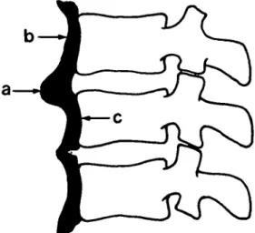
Grade III = At least three bridging prever-tebral and/or prediscal ossifica-tions along the thoracic spine. Prediscal and prevertebral ossifications are shown schematically in Fig. 1.
In accordance with the criteria of Resnick et
al. [4], the intervertebral discs of the
hyperosto-tic segments showed no degenerative, dysplashyperosto-tic or inflammatory abnormalities.
Probands with grades 0 and I were considered as DISH negative, probands with grades II and III as DISH positive. We considered grade II as DISH positive, in order to be able to include probands with developing, but not yet ankylos-ing DISH.
The intra- and interobserver reliability of radiograph grading was assessed by comparing
samples of lateral chest films with the corre-sponding lateral views of the thoracic spine of the main probands being studied. We calculated
Po (observed proportion of agreement) and kappa (possible proportion of agreement).
kappa (K) =
1-Pc [13]
Where Pc = expected proportion of agreement, Po = observed proportion of agreement.
Statistical calculations were based on the chi-squared test for dichotomous variables and Stu-dent's /-test for continuous variables. The level of statistical significance was set at p = 0.05.
Multiple logistic regression analysis was carried out to determine the relevance of various variables (age, sex, degenerative lesions of the thoracic spine, DISH grade) for back pain.
The study was approved by the ethical com-mittee of the university.
RESULTS
A total of 314 DISH positive probands and controls were selected, but 30 had to be excluded due to malignancies with skeletal pain, leaving 106 spinal DISH positive probands and 178 spinal DISH negative controls for evaluation of the association between radiographic DISH of the spine and back pain.
FIG. 1.—Schematic drawing of two spinal segments show-ing prediscal (a) and prevertebral (b) ossification with characteristic radioluccnt (c) areas between the vertebral bodies and prevertebral ossification.
TABLE n
CUN1CAL AND RADIOLOGICAL CHARACTERISTICS OP THE SPINAL D I S H POSITTVE PKOBANDS AND D I S H
Mean age (±SD)
Males Females
History of heavy work: in last 6 months
previously
Degenerative lesions at other thoracic segments (assessment of lateral chest film)
Degenerative lesions at other thoracic segments (assessment of lateral radiograph of thoracic spine n=74) CONTROLS Spinal DISH positive probands (n=106) 71±9.4 77.4% (82/106) 22.6% (24/106) 13% (14/106) 61% (65/106) 11.3% (12/106) 15.6% (5/32) Controls (n-178) 68+9.8 65.7% (117/178) 34.3% (61/178) 16% (29/178) 63% (112/178) 36.5% (65/178) 21.4% (9/42) NEGATIVE p<0.02 p<0.05 p<0.05 NS NS p<0.001 NS
Demographic data (Table II)
The mean age (± SD) of the spinal DISH positive probands was 71 ± 9.4 years, ai.M that of the controls 68 ± 9.8 years (p < 0.02). The DISH negative control group contained more females than the DISH positive probands (34.3% and 22.6% respectively, p< 0.05). There was no difference in the frequency of previous heavy physical work between the groups. Degenera-tive lesions at non-hyperostotic thoracic spinal segments were significantly more frequent in controls (36.5%, 65/178) than in DISH positive probands (11.3%, 12/106; p< 0.01) when the lateral chest films were used for assessment. In contrast, using the lateral films of the thoracic spine for assessment, there was no relevant difference in frequency of degenerative lesions at the non-hyperostotic spinal segments between both groups.
Intra- and interobserver reliability of radiograph grading (Table III)
Using the aforementioned criteria, the intra-and interobserver reliability for thoracospinal grading was good. There was no significant difference in grading reliability between rheu-matologists (P.S., N.J.G.) and radiologist (W.A.F.). Agreement between grading of lat-eral chest films and corresponding latlat-eral views of the thoracic spine was satisfactory (Po = 0.85, K = 0.70, n = 87), showing a slight tendency of undergrading in the lateral chest films.
Back pain frequency (Table IV)
History of back pain in the 6 months preceding evaluation. Complete information was available
for 104 DISH positive probands and 178 con-trols, two DISH positive probands had to be
excluded because of incomplete questionnaires. There was no significant difference in frequency of back iain between the compared groups at any spinal level.
History of back pain prior to the last 6 months preceding evaluation. Complete information was
available for 104 DISH positive probands and 178 controls, two DISH positive probands had to be excluded due to incomplete questionnaires. Again, there was no difference in frequency of back pain between the compared groups at any spinal level.
Using multiple logistic regression analysis we found no correlation between back pain and age, sex, history of heavy work, internal medical dis-orders or interviewer.
DISCUSSION
These are the results of the first controlled study of the frequency of back pain in European Caucasian spinal DISH positive probands. They demonstrate that there is no significant differ-ence in the frequency of back pain at any spinal level between DISH positive probands and con-trols. This holds both for the prevalence of recent back pain (i.e. pain within the past 6 months) and for the prevalence of back pain in
T A B L E III
OBSERVER RELIABILITY OF IDENTIFICATION OF SPINAL DISH POSITIVE PROBANDS AND DISH NEGATIVE CONTROLS BASED ON THE LATERAL CHEST FILMS
Observer comparison Po (K value) P S ^ P S P S ^ N J G P S ^ W A F NJG ^ WAF 0.90 (0.80) n=60 0.95 (0.90) n=60 0.96 (0.92) n=55 0.95 (0.90) n=55
302 BRITISH JOURNAL OF RHEUMATOLOGY VOL. XXVIII NO. 4 FREQUENCY OF Cervical spine Thoracic spine Lumbar spine TABLE IV
BACK PAJN IN SPINAL DISH POSITIVE PROBANDS AND SPINAL DISH NEGATIVE CONTROLS
Back pain ir DISH positive probands (n=104) 28% (29/104) 8.7% (9/104) 33% (34/104) i last 6 months Controls (n=178) 21% (37/178) NS 6.7% (12/178) NS 41% (73/178) NS
Back pain earlier than in last 6 months DISH positive probands (n=104) 27% (28/104) 11.5% (12/104) 56% (58/104) Controls (n=178) 31% (55/178) NS 9% (16/178) NS 61% (109/178) NS
the past (i.e. pain prior to the last 6 months). Our results are in agreement with one previous controlled study [7]. The selected probands of the latter study, however, were Pima Indians, and not comparable with our population due to different genetic, ethnic and socioeconomic backgrounds. Julkunen etal. [6] also carried out a controlled study analysing the subjective mus-culoskeletal complaints of DISH positive pro-bands. However, the authors failed to differentiate between back and joint pain in their results, so that the true frequency of back pain in DISH positive probands and controls remained undefined. In comparison to other previously published studies (Table V), the fre-quency of back pain of the examined DISH posi-tive probands and controls in this study did not differ. Previous studies [1,3,5,9,14,15] were uncontrolled or may have suffered from a selec-tion bias, in that probands were collected from specialized departments for locomotor diseases. Both facts (i.e. the failure to differentiate between back and joint pain and the selection of probands from a department for locomotor dis-eases) have certainly led to an overestimation of DISH as a cause of back pain. While this study
shows that back pain does not occur more fre-quently in spinal DISH positive probands than in controls, it is well known that spinal DISH may occasionally lead to stenosis of the spinal canal [16-19] and cause dysphagia [20,21].
When using lateral chest films to assess the thoracic spine, degenerative lesions at non-hyperostotic segments of the thoracic spine were significantly more frequent in controls than in spinal DISH positive probands (p< 0.001). This difference could not be confirmed by assessing lateral radiographs of the thoracic spine. This may have been due to the smaller number of assessed films of the thoracic spine (74 versus 284 lateral chest films). On the other hand, it may well be that we are victims of a selection bias, in that diffuse idiopathic skeletal hyperostosis pro-tects against the development of degenerative lesions thus leading to the lower frequency of degenerative lesions of the thoracic spine in DISH positive probands.
We conclude that the radiological finding of spinal DISH lacks clinical relevance as a cause of back pain; the frequency of back pain in spinal DISH positive probands is not higher than in spinal DISH negative controls.
TABLE V
SYNOPSIS OF CUNICAL STUDIES EVALUATING BACK PAIN IN PATIENTS WITH SPINAL DISH
Author Resnick etal. [1] Utsinger et al. [3] Resnick et al. [5] Julkunen et al. [6) Henrard et al. [7] Forestier et al. [9] Harris et al. [14] Utsinger et al. [15) * Joint or back pain;
Method of study Uncontrolled Uncontrolled Uncontrolled Case-control Case-control Uncontrolled Uncontrolled Uncontrolled "aching spinal stiffness; NM,
Number of probands 40 (series B) 200 21 61 61 controls 46 35 controls 245 34 30 not mentioned. Average age of probands (years) 67 63 66 NM 61 56 88% over 50 67 67 Frequency of back pain in the past (%) 57-67 72 76 70* 77 10.9 5.7 NM 85 7 7 "
ACKNOWLEDGEMENTS
We thank Dr. Esther Hachler for questioning probands, Dr. Christoph Minder for statistical assistance, Madeleine Kummer and Esther Gerny for the preparation of the manuscript.
REFERENCES
1. Resnick D, Shapiro RF, Wiesner KB, et al. Diffuse idiopathic skeletal hypcrostosis (DISH) (ankylosing hyperostosis of For-estier and Rotes-Querol). Semin Arthritis Rheum 1978;7:153-87.
2. Forestier J, Rotes-Querol J. Senile ankylosing hyperostosis of the spine. Ann Rheum Dis 1950;9:321-30.
3. Utsinger PD. Diffuse idiopathic skeletal hyper-ostosis. Clin Rheum Dis 1985;11:325-51. 4. Resnick D, Niwayama G. Radiographic and
pathologic features of spinal involvement in diffuse idiopathic skeletal hyperostosis (DISH). Radiology 1976;119:559-68. 5. Resnick D, Shaul SR, Robins JM. Diffuse
idio-pathic skeletal hyperostosis (DISH): For-estier's disease with extraspinal manifesta-tions. Radiology 1975;115:513-24.
6. Julkunen H, Heinonen OP, Knekt P, etal. The epidemiology of hyperostosis of the spine together with its symptoms and related mor-tality in a general population. ScandJ Rheu-matol 1975;4:23-7.
7. Henrard JC, Bennett PH. Etude e"pide"miologi-que de l'hyperostose verte'brale: ene"pide"miologi-queue dans une population adulte d'indiens d'ame'rique. Rev Rhum 1973;40:581-91. 8. Boachie-Adjei O, Bullough PG. Incidence of
ankylosing hyperostosis of the spine (For-estier's disease) at autopsy. Spine 1987;12:739-43.
9. Forestier J, Lagier R. Ankylosing hyperostosis of the spine. Clin Orthop 1971;74:65-83. .10. Littlejohn GO, Smythe HA. Marked
hyperin-sulinemia after glucose challenge in patients
with diffuse idiopathic skeletal hyperostosis. J Rheumatol 1981;8:965-8.
11. Littlejohn GO, Hall S. Diffuse idiopathic skeletal hyperostosis and new bone forma-tion in male gouty subjects. Rheumatol Int
9 8 2 8 3 £
12. Smythe HA. Osteoarthritis, insulin and bone density. J Rheumatol 1987;14(Suppl 14):91-3.
13. Kelsey JL, Thompson WD, Evans AS. Methods in observational epidemiology. Monographs in epidemiology and biostatis-tics, Vol. 10. Oxford: Oxford University Press, 1986: 288-93.
14. Harris J, Carter RA, Glick EN, et al. Ankylos-ing hyperostosis. Clinical and radiological features. Ann Rheum Dis 1974;33:210-15. 15. Utsinger PD, Resnick D, Shapiro R. Diffuse
skeletal abnormalities in Forestier disease. Arch Intern Med 1976;136:763-8.
16. Meinen A. Neurokompression bei Diffuser Idiopathischer Skelettaler Hyperostose (DISH) der Wirbelsaule. Thesis, University of Berne, Switzerland, 1987.
17. Arlet J, Abiteboul M, Mazieres B, Adam Ph, Roulleau J. Ste'nose acquise des canaux lom-baires et hyperostose vertebral. Rev Rhum 1983;50:635-^l.
18. Ono K, Ota H, Tada K, Hamada H, Takaoka K. Ossified posterior longitudinal ligament: a clinicopathologic study. Spine 1977;2:126-38.
19. Pouchot J, Watts CS, Esdaile JM, Hill RO. Sudden quadriplegia complicating ossifica-tion of the posterior longitudinal ligament and diffuse idiopathic skeletal hyperostosis. Arthritis Rheum 1987;30:1069-72.
20. Gamache FW, Voorhies RM. Hypertrophic cervical osteophytes causing dysphagia. J Neurosurg 1980;53:338-44.
21. FahrerH,MarkwalderT. Dysphagia caused by diffuse idiopathic skeletal hyperostosis. Case report. Clin Rheumatol 1987;7:117-21.