Short Conceptual Overview
Roger Schneiter* and Antonio Di Pietro*
The CAP protein superfamily: function in sterol
export and fungal virulence
Abstract: CAP superfamily proteins, also known as sperm-coating proteins, are found in all kingdoms of life and have been implicated in a variety of physiological contexts, including immune defense in plants and mammals, sperm maturation and fertilization, fungal virulence, and toxicity of insect and reptile venoms as well as prostate and brain cancer. CAP family members are mostly secreted glycopro-teins that are highly stable in the extracellular fluid. All members of the superfamily share a common CAP domain of approximately 150 amino acids, which adopts a unique α-β-α sandwich fold. The conserved structure suggests that CAP proteins exert fundamentally similar functions. How-ever, the molecular mode of action of this protein family has remained enigmatic. The budding yeast
Saccharomy-ces cerevisiae has three CAP family members designated
Pry (pathogen related in yeast), and recent evidence indi-cates that they act as sterol-binding and export proteins. Expression of the mammalian CAP protein CRISP2, which binds sterols in vitro, complements the sterol export defect of a yeast pry mutant, suggesting that sterol binding and export is conserved among different CAP family members. Collectively, these observations suggest that CAP family members constitute a novel class of secreted extracellular sterol-binding proteins. A ligand-binding activity of the CAP domain could explain many of the biological activities attributed to these proteins. For example, the strong induc-tion of plant pathogenesis-related 1 protein upon exposure to pathogens may serve to inhibit pathogen proliferation by extracting sterols from the pathogen membrane. Simi-larly, the presence of these proteins in the venom of toxic insects and reptiles or in the secretome of pathogenic fungi might inflict damage by sequestering sterols or related small hydrophobic compounds from the host tissue. Keywords: fungal virulence; Fusarium oxysporum; mouse infection model; pathogenesis-related (PR-1); sterol detoxification.
*Corresponding authors: Roger Schneiter, Division of Biochemistry,
Department of Biology, University of Fribourg, Chemin du Museé 10, 1700 Fribourg, Switzerland, e-mail: roger.schneiter@unifr.ch;
Antonio Di Pietro, Departamento de Genetica, Facultad de
Ciencias and Campus de Excelencia Internacional Agroalimentario, Universidad de Cordoba, 14071 Cordoba, Spain,
e-mail: ge2dipia@uco.es
Introduction
The CAP protein superfamily (pfam PF00188) was named after the three founding members cysteine-rich secre-tory protein, antigen 5, and pathogenesis-related 1, and comprises more than 4500 known members in over 1500 species from all kingdoms of life. CAP proteins have been implicated in a wide variety of biological processes, including immune defense in mammals and plants, pathogen virulence, sperm maturation and fertilization, venom toxicity, and prostate and brain cancer. Almost all CAP proteins are secreted glycoproteins that exhibit a high stability in the extracellular fluid over a wide range of environmental conditions. The overall structural con-servation within the CAP superfamily is likely to result in fundamentally similar functions for the CAP domains in the different members, whereas the diversity outside of this core region may alter target specificity and thus mod-ulate physiological responses. For example, mammalian CAP proteins have been classified into nine subfamilies that differ in regions flanking the conserved CAP domain (1). CAP proteins are an intensely studied class of proteins; however, their mode of action has remained elusive [for reviews, see (1, 2)].
The first founding member of the CAP superfamily is the plant pathogenesis-related protein-1 (PR-1), which was identified in 1970 among the proteins induced in tobacco leaves upon viral infection (3). Plant pathogenesis-related (PR) proteins are classified into 17 families according to their molecular mass, PR-1 being the smallest one with 14 kDa. Subsequent studies revealed that other PR pro-teins, such as glucanase (PR-2), chitinase (PR-3, 4, 8, 11), protease inhibitor (PR-6), peroxidase (PR-9), or ribonu-clease activity (PR-10), have different biochemical activi-ties, serving as a direct line of defense against fungal and
bacterial pathogens (4, 5). Intriguingly, even though it is the most abundant PR protein, PR-1 remains the only member for which no biochemical function is known. Some PR-1 proteins display antimicrobial activity against fungal and oomycete pathogens, but their mechanism of action is unclear (6, 7).
Antigen 5 (Ag5) is the second founding member of the CAP superfamily, an abundant protein present in the venom-secretory ducts of stinging insects that elicits a strong allergenic response. Ag5 proteins form part of a cocktail of salivary proteins that are believed to function either in suppression of the host immune system or in prevention of blood clotting to prolong feeding (8). The biochemical activity of Ag5 is unknown, but a related CAP protein from hookworm, neutrophil inhibitory factor (NIF), was shown to abrogate the adhesion of neutrophils to endothelial cells by binding to and thereby blocking integrin action (9).
Cysteine-rich secretory proteins (CRISPs) constitute the third founding members of the CAP superfamily. They are highly enriched in the mammalian reproductive tract and in the venom-secretory ducts of snakes, lizards, and other vertebrates. CRISPs are two-domain proteins containing an N-terminal CAP domain and a C-terminal cysteine-rich domain (CRD) with a conserved spacing of up to 16 Cys residues. This modular structure is likely to result in a dual-function protein with distinct activities associated with the N-terminal CAP domain and the C-ter-minal CRD domain (1, 10). Recent data suggest that the CRD adopts a fold similar to potassium channel inhibitors and may thus modulate the activity of cyclic nucleotide-gated ion channels (see the following) (11).
Structure of CAP proteins
CAP proteins share only limited sequence identity, includ-ing two signature PROSITE-recognized motifs referred to as CRISP motifs (http://expasy.ch/prosite) (Figure 1A). The NMR structure of plant PR-1 protein and the crystal struc-tures of several CAP proteins revealed that the conserved CAP sequence motifs are present in a small and structur-ally conserved 17- to 21-kDa CAP domain, which adopts a unique α-β-α sandwich fold. The tight packing of the α-helices on both sides of the central β-sheet results in a compact, bipartite molecular core, which is stabilized by hydrophobic interactions, multiple hydrogen bonds, and two highly conserved disulfide bonds (Figure 1B). These features are thought to provide the high thermal, pH, and proteolytic stability reported for CAP proteins, consistent
with the structural requirements of an extracellular func-tion (11–14).
A seminal study used the NMR structure of plant PR-1 as template to model the structure of the human glioma pathogenesis-related protein 1 (GLIPR1), revealing the presence of four conserved partially surface exposed residues, two histidines, and two glutamic acids. The high structural conservation of the CAP domain immedi-ately suggested a common mode of action of these pro-teins in plant pathogen defense and brain immune cells (13). These surface residues were later suggested to form part of a putative active-site triad of a CAP protein with a reported in vitro protease activity, Tex31 (15). However, although a large number of proteins within the superfam-ily contain a CAP domain in isolation (e.g., Ag5 and PR-1), many others contain additional N- or C-terminal exten-sions (1).
Proposed functions of CAP proteins
A 28-kDa CAP family member, Tex 31, was purified from the venom duct of the marine cone snail Conus textile and found to have proteolytic activity in vitro. This activ-ity was abolished by serine protease inhibitors and stimulated by addition of Ca2+ (15). Modeling of the Tex31
sequence to the structure of PR-1 and Ag5 was consistent with a possible catalytic role of the conserved surface-exposed histidine and glutamic acid residues (15). The lack of a conserved serine in this putative active-site triad led others to propose that dimerization of CAP proteins is required to complete the formation of the active site (14). However, subsequent studies failed to detect pro-tease activity with purified CAP family members, and a conclusive demonstration of the protease activity for a mammalian, fungal, or plant CAP protein is still lacking (11, 16, 17). Apart from a possible catalytic activity, the CAP domain has been suggested to form a stable scaf-fold for biological interactions with other proteins (18), although no interactions of plant PR-1 with other proteins have been detected (19).
CRISP subfamily CAP proteins harboring a C-termi-nal CRD have been associated with modulation of the activity of ion channels, which might account for their role in sperm maturation and venom toxicity (11, 20, 21). However, whether the CRD acts directly on the ion channel or whether CRISP proteins affect the channel activity indi-rectly, for example, through concomitant alterations of the lipid composition of the plasma membrane, remains to be established.
The role of CAP proteins in humans
and their implication in cancer
Transcriptional deregulation of two CAP superfamily members in malignant cells suggested a possible function of these proteins in cancer development or progression. GLIPR1 and GLIPR2 (also known as RTVP1/GAPR1) are among the most highly up-regulated transcripts in human gliomas, i.e., astrocyte-derived brain tumors, which account for over 65% of all human primary brain tumors (22). Interestingly, GLIPR1 has pro-apoptotic activity and acts as a tumor suppressor in prostate cancer (23). On the other hand, the CRISP family member CRISP3 is expressed in different cell types of the immune system including
pre-B cells, and is believed to function in innate immu-nity. Transcription of CRISP3 is dramatically increased in prostate cancer and serves as a potential biomarker (24). These observations raise the possibility that CAPs may be used as diagnostic agents and perhaps, ultimately, as therapeutic targets (1).
Yeast Pry proteins function in sterol
binding and export
New insight into the function of the CAP family of pro-teins comes from recent studies in budding yeast. The
S. cerevisiae genome codes for three members of this
A
B
C
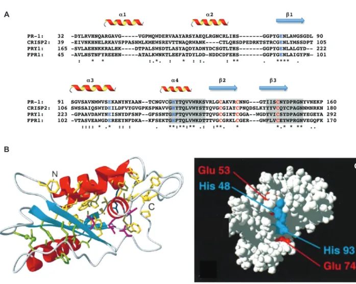
Figure 1 Conservation and structure of the CAP domain.
(A) Partial sequence alignment of a core set of CAP proteins, PR-1 (Arabidopsis thaliana), CRISP2 (Homo sapiens), Pry1 (S. cerevisiae), Fpr1 (F. oxysporum). Secondary-structure elements are indicated and conserved residues of a putative active site are in blue; conserved cysteines are in red. The two CRISP motifs are shaded. (B) Ribbon drawing of the CAP domain of PR-1. α-Helices are shown in red, β-strands in cyan. Amino acid side chains that form a large hydrophobic cluster are shown in yellow; those forming a smaller hydrophobic cluster are green. Disulfide bonds are shown as ball-and-stick models. (C) Space-filling representation of PR-1 with the partially surface exposed histidinyl and glutamyl residues depicted in blue and red, respectively. (B) and (C) are adapted from (13) (© 1998, National Academy of Sciences, USA).
protein superfamily, termed Pry. Pry1 and Pry2 are secreted glycoproteins, whereas Pry3 is associated with the cell wall and contains a signal for attachment of a glycosylphosphatidylinositol anchor (25, 26). Pry1 and Pry2 share a redundant function in the export of acety-lated cholesterol, a lipid intermediate that accumulates in cells lacking a corresponding lipid deacetylase (27). Double mutants lacking both Pry1 and Pry2 accumulate cholesteryl acetate in the endoplastic reticulum mem-brane, whereas in the presence of a wild- type copy of either Pry1 or Pry2 cholesteryl acetate is secreted from the cells and accumulates in the culture media (26, 27). Purified Pry1 and Pry2 bind both free cholesterol and cholesteryl acetate in a dose-dependent manner, con-sistent with a role of Pry proteins in binding and solu-bilizing sterols and possibly other small hydrophobic compounds.
Importantly, the sterol binding and export function of the yeast CAP proteins Pry1 is confined to the CAP domain because expression of the CAP domain alone is sufficient to rescue the sterol export phenotype of a pry1Δ pry2Δ double mutant, and the CAP domain of Pry1 alone binds sterols in vitro. Evidence for the specificity of the protein-lipid interaction comes from the finding that it is pre-vented by mutation of a highly conserved cysteine residue known to form a disulfide bridge. Most importantly, the lipid-binding and export function of the Pry proteins appears to be conserved among the members of the CAP protein superfamily because expression of the human CAP protein CRISP2 relieves the lipid export block of a yeast pry1Δ pry2Δ double mutant, and purified CRISP2 binds sterols in vitro (26).
Cells lacking PRY1 and PRY2 are hypersensitive to the plant oil eugenol, and this phenotype is also rescued by expression of human CRISP2. Eugenol is a member of the allylbenzene class of compounds present in clove oil, nutmeg, cinnamon, and bay leaf that is used as local antiseptic and anesthetic and has antifungal and bac-teriostatic activity (28). Pry proteins bind eugenol and confer dose-dependent resistance against this potential membrane-perturbing compound (26, 27). Thus, members of the CAP protein superfamily may exert a wide variety of physiological functions through binding to lipids and related small hydrophobic compounds that affect the integrity of cellular membranes.
In the light of these results, it is conceivable that the function of plant PR-1 proteins in pathogen defense involves a sterol binding and sequestration mechanism. Capturing sterols from the surface membrane of patho-gens could exert a growth inhibitory effect or even kill the invaders. Sterols are not only an essential lipid constituent
in eukaryotic plasma membranes, but also bacteria syn-thesize hopanoid and tetrahymenol compounds that are structurally and functionally related to sterols (29–31). Drugs that target ergosterol, the fungus-specific mem-brane sterol, or products like azoles that inhibit specific steps in ergosterol biosynthesis, are widely used in anti-fungal treatments of plants, animals, and humans (32). The sterol-binding mechanism of CAP proteins could therefore be exploited as a novel mode of action to target invasive microbial infections.
CAP proteins act as fungal virulence
determinants
CAP proteins have recently emerged as novel virulence factors in pathogenic fungi. The RBT4 gene encodes a predicted secreted CAP protein from the human patho-gen Candida albicans that was originally identified in a search for transcripts under control of a morphogenetic regulator, Tup1. Candida mutants lacking RBT4 had reduced virulence both in a rabbit cornea and in a sys-temic mouse infection model (33). Rbt4, together with a close homolog, Rbe1, were recently detected in the fungal secretome and shown to be part of a family of five CaPRY proteins in C. albicans. Whereas single deletions of RBE1 or RBT4 resulted in a moderate attenuation in a mouse model for disseminated candidiasis, the rbe1Δ rbt4Δ double mutant was dramatically reduced, suggesting that different CAP family proteins have partially redundant roles in virulence (34).
Another CAP family protein, Fpr1, was reported in the ascomycete Fusarium oxysporum, a ubiquitous fungal pathogen that causes vascular wilt disease on a wide range of plant species and can produce life-threat-ening infections in immunocompromised humans (17). Fpr1 is an extracellular protein with an N-terminal secre-tion signal, a serine/proline-rich region of unknown function, and a C-terminal CAP domain including the highly conserved His and Glu residues. Western blot analysis of culture supernatants revealed that Fpr1 is secreted by F. oxysporum as a precursor migrating at 40 kDa and rapidly converted by proteolytic cleavage into the major 30-kDa form. Strikingly, transcription of the fpr1 gene was rapidly induced during growth in human blood, and immunodepressed mice infected with fpr1Δ mutants exhibited significantly lower mor-tality rates than those infected with the wild-type strain. Importantly, the CAP domain was required for the func-tion of Fpr1 in virulence because complementafunc-tion with
a native fpr1 allele restored virulence of the fpr1Δ mutant to wild-type levels, whereas an allele in which two of the predicted active-site residues had been mutated (fpr1H170A,E177A) did not (17).
Evidence for expansion of CAP
proteins in fungal pathogens
A survey of predicted CAP proteins in sequenced fungal genomes revealed that most fungi contain two members of the protein family that cluster into well-separated clades. The presence of these ‘core’ CAP proteins in non-pathogens including yeast, suggests they must fulfill functions unrelated to pathogenicity, such as binding and export of sterols and related small hydrophobic com-pounds (26). However, some fungal pathogens display a remarkable increase in the number of CAP family pro-teins. C. albicans has five members, all of which cluster within a separate Hemiascomycete-specific clade, sug-gesting that this CAP gene family has expanded recently during evolution. Even more strikingly, the plant patho-gens F. oxysporum, Fusarium graminearum, and
Magna-porthe grisea contain a new family of CAP proteins that
appears phylogenetically closer to plant PR-1 proteins than to the fungus-specific clades, raising the intrigu-ing possibility that their ancestor might have originated from a horizontal gene transfer event. Interestingly, the new clade also includes the F. oxysporum virulence determinant Fpr1 (17). Similarly, the genome of
Monili-ophthora perniciosa, a basidiomycete fungus that causes
witches’ broom disease in cacao, was shown to contain 11 CAP family genes some of which were highly and spe-cifically expressed during the interaction with the host plant (35). Collectively, these findings suggest a possible link between the evolutionary expansion of fungal CAP proteins and pathogenicity.
Possible function of CAP proteins
during fungal infection
Although the role of CAP proteins in fungal virulence remains unclear, the capacity of the yeast homologs to bind sterols and related small hydrophobic compounds suggests a parallel between the ligand-binding activity and their function during infection. This question is of particular interest because some CAP proteins from the
pathogen-specific clades lack several of the conserved cysteine residues involved in intramolecular disulfide bridges, suggesting distinct ligand-binding properties and/or protein stabilities (17).
Fungal infections, particularly in humans, are diffi-cult to control because, as eukaryotes, their metabolism closely resembles ours. As a result, most current fungi-cides and medical antifungals are based on only a handful of active principles. Sterol metabolism is a known Achil-les heel of fungi and acts as a major fungicide target in medicine and agriculture (36). Intriguingly, plants have exploited ergosterol binding in the fungal membrane as a mechanism of antifungal phytoalexins such as the tomato glycoalkaloid tomatine (37). The finding that secreted CAP proteins are conserved fungal virulence factors suggests that they could serve as potential targets to reduce fungal infection.
Outlook
The breakthrough finding that the yeast CAP family members Pry1 and Pry2 are required for exporting cho-lesteryl acetate and that these proteins directly bind the lipid has provided a new lead to address the molecular function of CAP family members in other organisms. It is conceivable that CAP proteins exert their multitude of bio-logical tasks through a common mechanism of binding/ sequestering sterols and/or related small hydrophobic compounds. Thus, sterol sequestration from the host tissue by fungal pathogens might facilitate tissue penetra-tion, consistent with the role of CAP proteins in virulence. Meanwhile, targeting of sterols from microbial invaders by host CAP proteins could account for their function in innate immunity of plants and mammals. Finally, inter-action with lipids might also contribute to the multi-ple roles of human CAP proteins associated with cancer development.
Acknowledgments: We thank L. Falquet for help with the figure. Work in the two laboratories is supported by the Swiss National Science Foundation (grant 31003A_134742) to RS, and the Spanish Ministerio de Innovación y Compet-itividad (MINECO; grants BIO2010-15505, BIO2008-04479, and EUI2009-03942) and the EU (grant FP7-PEOPLE-ITN-237936) to ADP.
References
1. Gibbs GM, Roelants K, O’Bryan MK. The CAP superfamily: cysteine-rich secretory proteins, antigen 5, and pathogenesis-related 1 proteins – roles in reproduction, cancer, and immune defense. Endocr Rev 2008; 29: 865–97.
2. Cantacessi C, Campbell BE, Visser A, Geldhof P, Nolan MJ, Nisbet AJ, Matthews JB, Loukas A, Hofmann A, Otranto D, Sternberg PW, Gasser RB. A portrait of the “SCP/TAPS” proteins of eukaryotes – developing a framework for fundamental research and biotechnological outcomes. Biotechnol Adv 2009; 27: 376–88. 3. van Loon LC, van Kammen A. Polyacrylamide disc
electro-phoresis of the soluble leaf proteins from Nicotiana tabacum var. “Samsun” and “Samsun NN”. II. Changes in protein constitution after infection with tobacco mosaic virus. Virology 1970; 40: 190–211.
4. Van Loon LC, Van Strien EA. The families of pathogenesis-related proteins, their activities, and comparative analysis of PR-1 type proteins. Physiol Mol Plant Pathol 1999; 55: 85–97. 5. van Loon LC, Rep M, Pieterse CM. Significance of inducible
defense-related proteins in infected plants. Annu Rev Phytopathol 2006; 44: 135–62.
6. Niderman T, Genetet I, Bruyere T, Gees R, Stintzi A, Legrand M, Fritig B, Mosinger E. Pathogenesis-related PR-1 proteins are antifungal. Isolation and characterization of three 14-kilodalton proteins of tomato and of a basic PR-1 of tobacco with inhibitory activity against Phytophthora infestans. Plant Physiol 1995; 108: 17–27.
7. Rauscher M, Adam AL, Wirtz S, Guggenheim R, Mendgen K, Deising HB. PR-1 protein inhibits the differentiation of rust infection hyphae in leaves of acquired resistant broad bean. Plant J 1999; 19: 625–33.
8. King TP, Spangfort MD. Structure and biology of stinging insect venom allergens. Int Arch Allergy Immunol 2000; 123: 99–106. 9. Lo SK, Rahman A, Xu N, Zhou MY, Nagpala P, Jaffe HA, Malik AB.
Neutrophil inhibitory factor abrogates neutrophil adhesion by blockade of CD11a and CD11b β2 integrins. Mol Pharmacol 1999; 56: 926–32.
10. Yamazaki Y, Morita T. Structure and function of snake venom cysteine-rich secretory proteins. Toxicon 2004; 44: 227–31. 11. Guo M, Teng M, Niu L, Liu Q, Huang Q, Hao Q. Crystal structure
of the cysteine-rich secretory protein stecrisp reveals that the cysteine-rich domain has a K+ channel inhibitor-like fold. J Biol Chem 2005; 280: 12405–12.
12. Fernandez C, Szyperski T, Bruyere T, Ramage P, Mosinger E, Wuthrich K. NMR solution structure of the pathogenesis-related protein P14a. J Mol Biol 1997; 266: 576–93.
13. Szyperski T, Fernandez C, Mumenthaler C, Wuthrich K. Structure comparison of human glioma pathogenesis-related protein GliPR and the plant pathogenesis-related protein P14a indicates a functional link between the human immune system and a plant defense system. Proc Natl Acad Sci USA 1998; 95: 2262–6. 14. Serrano RL, Kuhn A, Hendricks A, Helms JB, Sinning I, Groves
MR. Structural analysis of the human Golgi-associated plant pathogenesis related protein GAPR-1 implicates dimerization as a regulatory mechanism. J Mol Biol 2004; 339: 173–83. 15. Milne TJ, Abbenante G, Tyndall JD, Halliday J, Lewis RJ. Isolation
and characterization of a cone snail protease with homology to CRISP proteins of the pathogenesis-related protein superfamily. J Biol Chem 2003; 278: 31105–10.
16. Wang YL, Kuo JH, Lee SC, Liu JS, Hsieh YC, Shih YT, Chen CJ, Chiu JJ, Wu WG. Cobra CRISP functions as an inflammatory modulator via a novel Zn2+- and heparan sulfate-dependent transcriptional regulation of endothelial cell adhesion molecules. J Biol Chem 2010; 285: 37872–83.
17. Prados-Rosales RC, Roldan-Rodriguez R, Serena C,
Lopez-Berges MS, Guarro J, Martinez-del-Pozo A, Di Pietro A. A PR-1-like protein of Fusarium oxysporum functions in virulence on mammalian hosts. J Biol Chem 2012; 287: 21970–9. 18. Kirby TW, Mueller GA, DeRose EF, Lebetkin MS, Meiss G,
Pingoud A, London RE. The nuclease A inhibitor represents a new variation of the rare PR-1 fold. J Mol Biol 2002; 320: 771–82.
19. van Loon LC, Gerritsen YAM, Ritter CE. Identification, purification, and characterization of pathogenesis-related proteins from virus-infected Samsun NN tobacco leaves. Plant Mol Biol 1987; 9: 593–609.
20. Wang J, Shen B, Guo M, Lou X, Duan Y, Cheng XP, Teng M, Niu L, Liu Q, Huang Q, Hao Q. Blocking effect and crystal structure of natrin toxin, a cysteine-rich secretory protein from Naja atra venom that targets the BKCa channel. Biochemistry 2005; 44: 10145–52.
21. Gibbs GM, Orta G, Reddy T, Koppers AJ, Martinez-Lopez P, Luis de la Vega-Beltran J, Lo JC, Veldhuis N, Jamsai D, McIntyre P, Darszon A, O’Bryan MK. Cysteine-rich secretory protein 4 is an inhibitor of transient receptor potential M8 with a role in establishing sperm function. Proc Natl Acad Sci USA 2011; 108: 7034–9.
22. Murphy EV, Zhang Y, Zhu W, Biggs J. The human glioma pathogenesis-related protein is structurally related to plant pathogenesis-related proteins and its gene is expressed specifically in brain tumors. Gene 1995; 159: 131–5.
23. Li L, Ren C, Yang G, Fattah EA, Goltsov AA, Kim SM, Lee JS, Park S, Demayo FJ, Ittmann MM, Troncoso P, Thompson TC. GLIPR1 suppresses prostate cancer development through targeted oncoprotein destruction. Cancer Res 2011; 71: 7694–704. 24. Kosari F, Asmann YW, Cheville JC, Vasmatzis G. Cysteine-rich
secretory protein-3: a potential biomarker for prostate cancer. Cancer Epidemiol Biomarkers Prev 2002; 11: 1419–26. 25. Yin QY, de Groot PW, Dekker HL, de Jong L, Klis FM, de Koster
CG. Comprehensive proteomic analysis of Saccharomyces cerevisiae cell walls: identification of proteins covalently attached via glycosylphosphatidylinositol remnants or mild alkali-sensitive linkages. J Biol Chem 2005; 280: 20894–901. 26. Choudhary V, Schneiter R. Pathogen-Related Yeast (PRY)
proteins and members of the CAP superfamily are secreted sterol-binding proteins. Proc Natl Acad Sci USA 2012; 109: 16882–7.
27. Tiwari R, Koffel R, Schneiter R. An acetylation/deacetylation cycle controls the export of sterols and steroids from S. cerevisiae. EMBO J 2007; 26: 5109–19.
28. Bakkali F, Averbeck S, Averbeck D, Idaomar M. Biological effects of essential oils – a review. Food Chem Toxicol 2008; 46: 446–75.
29. Rohmer M, Bouvier P, Ourisson G. Molecular evolution of biomembranes: structural equivalents and phylogenetic precursors of sterols. Proc Natl Acad Sci USA 1979; 76: 847–51.
30. Ji YH, Moog C, Schmitt G, Luu B. Polyoxygenated sterols and triterpenes: chemical structures and biological activities. J Steroid Biochem 1990; 35: 741–4.
31. Haines TH. Do sterols reduce proton and sodium leaks through lipid bilayers? Prog Lipid Res 2001; 40: 299–324.
32. Gupte M, Kulkarni P, Ganguli BN. Antifungal antibiotics. Appl Microbiol Biotechnol 2002; 58: 46–57.
33. Braun BR, Head WS, Wang MX, Johnson AD. Identification and characterization of TUP1-regulated genes in Candida albicans. Genetics 2000; 156: 31–44.
34. Rohm M, Lindemann E, Hiller E, Ermert D, Lemuth K, Trkulja D, Sogukpinar O, Brunner H, Rupp S, Urban CF, Sohn K. A family of secreted pathogenesis-related
proteins in Candida albicans. Mol Microbiol 2013; 87: 132–51.
35. Teixeira PJ, Thomazella DP, Vidal RO, do Prado PF, Reis O, Baroni RM, Franco SF, Mieczkowski P, Pereira GA, Mondego JM. The fungal pathogen Moniliophthora perniciosa has genes similar to plant PR-1 that are highly expressed during its interaction with cacao. PLoS One 2012; 7: e45929.
36. Odds FC, Brown AJ, Gow NA. Antifungal agents: mechanisms of action. Trends Microbiol 2003; 11: 272–9.
37. Simons V, Morrissey JP, Latijnhouwers M, Csukai M, Cleaver A, Yarrow C, Osbourn A. Dual effects of plant steroidal alkaloids on Saccharomyces cerevisiae. Antimicrob Agents Chemother 2006; 50: 2732–40.