Structure and reactivity of small arteries in aging
Pierre Moreau
1, Livius V. d’Uscio, Thomas F. Luscher
)¨
Cardiology, CardioÕascular Research, UniÕersity Hospital, Bern, SwitzerlandInstitute of Physiology, University Hospital, Zurich, Switzerland Received 14 April 1997; accepted 6 August 1997
Abstract
Objective: Increased pulse pressure has been observed in aging subjects, but the impact on the structure and reactivity of small
arteries has been scarcely evaluated. Methods: This study presents the modifications of vascular structure and function observed in female rats of 5, 18 and 32 months of age, and their relation to the prevailing hemodynamic status. Geometry and reactivity of perfused and pressurized basilar and mesenteric small arteries were analyzed in vitro using a video dimension analyzer. Results: Mean arterial pressure was similar in the three age groups, and only pulse pressure was increased in the oldest group. Media thickness and cross sectional area increased in basilar and mesenteric arteries of the oldest rats and these structural abnormalities were positively related to pulse pressure but not to mean, systolic or diastolic arterial pressure. Only minor changes of vascular reactivity were noted with age: there was a decreased contraction to angiotensin II in mesenteric arteries and an enhanced contraction to endothelin-1 in the basilar arteries.
Conclusion: In conclusion, aging is associated with increased pulse pressure and hypertrophy of basilar and mesenteric resistance
arteries, suggesting that this hemodynamic variable may influence cerebral and peripheral vascular structure in aging. q 1998 Elsevier Science B.V.
Keywords: Aging; Basilar artery; Mesenteric artery; Vascular remodeling; Pulse pressure; Rat
1. Introduction
With age, the vascular wall of large peripheral vessels undergoes structural changes that may contribute to the
w x
age-related increase in systolic arterial pressure 1 . These include increased stiffness, thickening of the media and
w x
enlargement of the lumen diameter 2 . Few studies have directed their attention at the structure of small arteries in
w x
the context of advanced physiological aging 3 . By differ-ent means of investigation, an increased wall thickness has
w x
been reported in the hindquarter of normotensive rats 4 ,
w x
small muscular and ear arteries of rabbits 5 and small arteries from several human vascular beds studied
post-w x
mortem 6,7 . In contrast, one study reported an atrophy of cerebral arterioles associated with a decreased
distensibil-)
Ž . Ž .
Corresponding author. Tel. q41-1 255 2121 or 2177; Fax q41-1 255 4251; E-mail: 100771.1237@compuserve.com
1
Present affiliation: Faculty of Pharmacy, Universite de Montreal,´ ´
Montreal, Quebec, Canada.´ ´
w x
ity of the vessel wall in aged rats 8 . However, in most studies, the structural alterations have not been related to the hemodynamic status prevailing in aging.
Similar to vascular structure, reports on vascular reac-tivity of small arteries in the context of aging are also scarce and none are available for the female sex. Indeed,
w x
most of the studies were performed in large arteries 3 and it is known that resistance vessels differs in several impor-tant aspects, most probably due to their different function
w x9 . Even in conduit arteries, regional differences have
been noted. Indeed, in a recent study, the release of nitric
Ž .
oxide NO was reduced with age in the rat aorta, but not
w x
in pulmonary circulation 10 . Since hemodynamic or vas-cular structural changes present in aging may alter reactiv-ity of resistance arteries, it will be evaluated in relation to the structural and hemodynamic conditions prevailing.
Since pulsatile stretch has been shown to increase
vas-Time for primary review 37 days.
0008-6363r98r$19.00 q 1998 Elsevier Science B.V. All rights reserved.
Ž . PII S 0 0 0 8 - 6 3 6 3 9 7 0 0 2 2 5 - 3
w x
cular smooth muscle cell proliferation 11 , it is of particu-lar interest to determine if long term increased pulse pressure can have an impact on the structure of smaller resistance vessels. In addition, pulse pressure has been
Ž
associated with an increased cross-sectional area CSA, an
.
index of vascular wall hypertrophy of pial arterioles in the
w x
context of hypertension 12 , but it is not known if this can happen independently of changes in mean arterial pressure.
Ž .
In this study, we examined the effect of aging 18 months
Ž .
and advanced aging 32 months , characterized by a selec-tive increase in pulse pressure, on vascular remodeling and reactivity of the rat basilar and small mesenteric arteries, as directly assessed in perfused and pressurized in vitro conditions.
2. Materials and methods
Rats of the RORO strain were purchased from
Biologi-Ž .
cal Research Laboratories Fullinsdorf, Switzerland at 5
¨
Žadult , 18 old and 32 very old months of age n s 7. Ž . Ž . Ž .
per age group; female sex . These rats, originally of the Wistar strain, were outbred for twenty years in Hoffmann-La Roche laboratories. Their life expectancy is
approxi-Ž
mately 36 months. The rats were anesthetized thiopental,
.
50 mgrkg, intra peritoneal and a short polyethylene
Ž .
catheter internal diameter: 0.58 mm was inserted in the left femoral artery and connected to a pressure transducer
ŽLetica PRI 256r2, Letica SrA, Hospitalet, Spain. to allow for the determination of systolic, diastolic arterial pressure and heart rate. During the anesthesia, the average of a 15 min recording was used to calculate mean and pulse pressure. All these procedures were approved by the Commission for Animal Research of the canton of Bern, and conform with the Guide for the care and use of laboratory animals of the NIH.
The animals were then decapitated and the basilar artery as well as a segment of a fourth branch of the mesenteric
Ž .
arterial bed closest segment to the ileum were isolated under a dissecting microscope in cold Krebs solution of
Ž .
the following composition in mmolrl; control solution : NaCl 118.6, KCl 4.7, CaCl2 2.5, KH PO 1.2, MgSO2 4 4
1.2, NaHCO 25.1, edetate calcium disodium 0.026, glu-3
cose 10.1. The arteries were then inserted and sutured on two small glass cannula positioned in a vessel chamber
ŽLiving Systems Instrumentation, Burlington, VT, USA.
and superfused with control solution maintained at 378C
Ž .
and oxygenated 95% O , 5% CO . The vessel perfusion2 2
chamber was positioned on the stage of an inverted
micro-Ž .
scope Nikon, TSM-F and the amplified image was trans-mitted, by a video camera, to a monitor and a video
Ž .
dimension analyzer V91, Living Systems Instrumentation , allowing for the measurements of lumen diameter and wall thickness. With this technique it is possible to distinguish
between the adventitia and the media, and the latter was used for calculations and comparisons. Longitudinal stretch was controlled by adjusting the vessel length to a value slightly superior to the one required to produce a small
w x
bending of the vessel 13 .
The mesenteric arteries were allowed to equilibrate for 60 min with a perfusion of control solution containing 1% bovine serum albumin at a constant and optimal perfusion
w x
pressure of 30 mmHg 14 and their resting lumen diame-ter and media thickness were recorded. The basilar ardiame-teries were equilibrated for 60 min in a calcium free control solution to prevent myogenic tone. The perfusion pressure was then increased from 25 to 55 mmHg in 10 mmHg steps and the efferent pressure was adjusted to maintain a constant flow. In basilar arteries, the vascular structure was determined at each of the four pressure steps in maximally
Ž
relaxed conditions confirmed by the inefficacy of
pa-.
paverine to further relax the artery .
In the functional experiments, all drugs were applied extraluminally and each section of the protocol was pre-ceded by a washout period of 45 min. In the mesenteric
Ž .
artery, the following protocol was performed: 1 a single
Ž y7 . Ž .
dose of angiotensin II Ang II, 10 molrl , 2 a
concen-Ž y9
tration–response curve to norepinephrine NE, 10 –3 =
y5 . Ž .
10 molrl , 3 a concentration–response curve to
Ž y9 y5 .
acetylcholine Ach, 10 –10 molrl after a 40%
pre-Ž .
contraction of the vessel with norepinphrine, 4 similar
Ž y1 0 y6
experiment with sodium nitroprusside SNP, 10 –10
. Ž .
molrl and 5 a concentration–response curve to
endothe-Ž y1 1 y8 .
lin-1 ET-1, 10 –10 molrl . In the basilar artery,
Ž y1 1 y8
only a concentration–response curve to ET-1 10 –10
.
molrl was studied since contractions to serotonin were very weak.
All the drugs were obtained from Sigma Chemicals
ŽBuchs, Switzerland , except for ET-1 which was obtained. Ž
from Calbiochem-Novabiochem Laufelfingen, Switzer-
¨
.
land . The CSA and the growth index were calculated
w x Ž
according to the formulas previously described 15,16 see
.
legend of Table 2 . Since CSA does not change with changes in pressure, a mean of the values obtained at four
Ž .
different pressures see above was calculated for the
Ž .
basilar artery Fig. 2 . The distensibility of the basilar artery is expressed as mm changes per mmHg of pressure increase and represents the slope of the pressure–lumen diameter curve. Contractions are expressed as the percent-age of decrease in lumen diameter from the baseline diameter. Relaxations are expressed as the percentage of increase in lumen diameter from the extent of precontrac-tion. For each individual concentration–response curve, the maximum response and the half maximum effective
Ž .
concentration expressed as negative logarithm, pD2 were calculated by non-linear regression. Values are expressed as mean " S.E.M, except for correlation analysis which show the actual data. Statistical evaluation was done by one-way ANOVA with Bonferroni’s correction for
multi-w x Ž
Table 1
Characteristics of the adult, old and very old rats studied
Ž . Ž . Ž .
Adult 5 months Old 18 months Very old 32 months
Number of rats 7 7 7
) )
Ž .
Body weight g 222"5 316"5 294"4
Ž .
Mean arterial pressure mmHg 86"5 103"8 88"6
)
Ž .
Pulse pressure mmHg 21"1 21"1 25"1
Ž .
Heart rate beatrmin 311"12 330"11 303"11
Arterial pressure and heart rate were measured in anesthetized conditions.)
P - 0.05 as compared to 5 months old rats.
Table 2
Morphological characteristics of basilar and small mesenteric arteries in adult, old and very old rats
Ž . Ž . Ž .
Adult 5 months Old 18 months Very old 32 months
( ) Basilar artery 35 mmHg Ž . Lumen diameter mm 302"5 316"14 316"14 a Ž . Media thickness mm 24.8"1.0 29.5"2.5 35.6"1.7 a Ž . MediarLumen ratio % 8.2"0.3 9.6"1.1 11.4"0.8 b b Ž .
Growth index % from control - 24.1"8.7 54.2"9.7
Mesenteric artery Ž . Lumen diameter mm 233"9 271"8 279"21 a Ž . Media thickness mm 16.4"1.0 17.1"1.3 20.2"0.6 Ž . MediarLumen ratio % 7.0"0.3 6.4"0.5 7.5"0.6 b Ž .
Growth index % from control - 19.3"10.4 46.3"12.3
For the basilar artery, the data is presented only at 35 mmHg of perfusion pressure. Similar results and statistics were obtained at other pressures. The
Ž . Ž .
growth index is calculated as a ratio of the difference between the treatment cross-sectional area CSAt, Fig 2 and the control CSA CSAc over CSAc
ŽCSA -CSA rCSA . Thus, the control group has a growth index of zero. P - 0.05 as compared to 5 months old rats ANOVA . The 95% Confidencet c c. a Ž .b
interval does not include 0.
.
index . Pearson’s correlation coefficients were calculated by linear regression. P - 0.05 was considered significant.
3. Results 3.1. Animals
Ž .
Body weight was higher in old 18 months and very
Ž . Ž .
old 32 months rats than in adult animals Table 1 . Probably due to their very old age, the 32 months old rats were lighter than 18 months old animals and seemed to
Ž .
have reduced general activity subjective observation . Mean arterial pressure tended to be higher in rats of 18
Ž .
months of age Table 1 , as did systolic and diastolic
Ž .
arterial pressures n.s., data not shown . Pulse pressure was significantly enhanced only in very old rats and was
Ž .
similar in the 5 and 18 months old groups Table 1 . Heart rate was not different among the groups.
3.2. Vascular structure
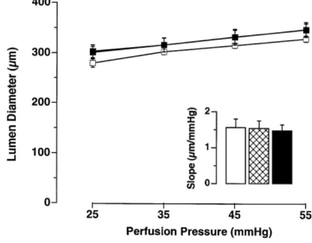
Aging had no influence on the lumen diameter of the basilar or small mesenteric arteries, although there was a tendency for mesenteric arteries to have a larger lumen
Ž .
with age Table 2 . Different perfusion pressures applied to the basilar arteries gave similar increments in the lumen
Ž
diameter for adult, old and very old rats, respectively Fig.
.
1 . Indeed, the slope of the relationship between perfusion pressure and lumen diameter was very similar among the
Ž .
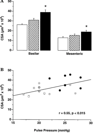
groups Fig. 1, inset . The media thickness and media CSA were significantly increased in both vascular beds of very old rats, but the mediarlumen ratio was augmented only in
Ž .
the basilar artery Fig. 2A, Table 2 . Indeed, in the
mesen-Fig. 1. Change of lumen diameter as a function of in vitro perfusion
Ž . Ž .
pressure in basilar arteries from 5 months I , 18 months G and 32
Ž . Ž .
months B old rats. Please note that the curves from old 18 months
Ž .
and very old rats 32 months are superimposed. The inset represents the
Ž .
slope mmrmmHg obtained from the linear regression of the curves shown in the graphic. The order of the bars is the same as the symbol description above. There was no statistical difference between the three groups.
. Ž .
Fig. 2. A Cross-sectional area CSA of the basilar and small mesenteric
Ž . Ž . Ž
arteries of adult 5 months, I , old 18 months, G and very old 32
. Ž .
months, B female rats ns 7rgroup . The CSA was calculated with the media thickness and not with the total wall thickness. )
P - 0.05 as .
compared to 5 months old rats. B Relationship between the mean CSA
Ž . Ž .
of the basilar artery and in vivo pulse pressure of 5 ` , 18 ( and 32
Žv. months old rats. Similar results were obtained in the mesenteric
arteries.
teric artery, the increase in lumen diameter prevented the mediarlumen ratio to be different in very old as compared to adult rats. The growth index was significant in both 18 and 32 months old groups for the mesenteric artery, but only in the very old group for the basilar artery.
There were positive correlations between pulse pressure
Ž .
and CSA r s 0.55, p - 0.05, Fig. 2B or media thickness
Žr s 0.53, p - 0.05 in basilar arteries. Similar findings. Ž
were obtained in mesenteric arteries r s 0.47 and r s 0.55
Ž . Ž .
Fig. 3. Maximal relaxation A and sensitivity B of preconstricted
Ž .
mesenteric arteries stimulated with acetylcholine Ach and sodium
nitro-Ž . Ž . Ž .
prusside SNP in adult 5 months, I , old 18 months, G and very old
Ž32 months, B female rats ns 7rgroup .. Ž .
.
respectively, p - 0.05 . However, pulse pressure was not
Ž
correlated with mediarlumen ratio basilar: r s 0.45, p s
.
0.05; mesenteric: r s 0.36, p s 0.12 . Structural parame-ters were not related to any other hemodynamic variable such as mean, systolic or diastolic arterial pressure.
3.3. Vascular reactiÕity
Maximal endothelium-dependent relaxations to Ach and endothelium-independent relaxations to SNP of mesenteric arteries were not significantly different among the groups, but a similar tendency for reduced relaxations with aging
Ž .
were noted with both agents Fig. 3 . In terms of sensitiv-ity, the concentration–response curve to SNP was shifted
Table 3
Reactivity of small mesenteric arteries to vasoconstrictors in adult, old and very old rats
Ž . Ž . Ž .
Adult 5 months Old 18 months Very old 32 months
y7 ) Ž . Angiotensin II 10 M 67.6"4.5 53.8"5.3 50.5"5.8 Norepinephrine Max 85.1"2.2 80.2"5.8 85.9"1.5 pD2 6.02"0.08 6.19"0.10 6.23"0.14 Endothelin-1 Max 84.1"0.9 83.0"3.2 84.5"2.1 pD2 9.29"0.07 9.12"0.09 9.13"0.04
Max.: Maximum contraction; pD : negative log of the concentration producing half of the maximal contraction. Both were calculated for each animal2
using non linear regression and the mean"s.e.m. is presented for each group. Due to tachyphylaxis, only one dose of angiotensin II was applied to the
)
Ž .
Ž .
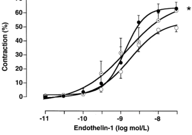
Fig. 4. Concentration-response curves to endothelin-1 ET-1 of basilar
Ž . Ž . Ž
arteries in adult 5 months, ` , old 18 months, ( and very old 32
. Ž . )
months, v female rats ns 7rgroup . The maximum contraction in the very old group is significantly different from the 5 months old rats.
Ž . Ž .
to the right less sensitive only in the old rats Fig. 3 and
Ž .
relaxations to Ach followed a similar pattern n.s. . In mesenteric arteries, there was a selective decrease in contractions to Ang II in very old rats, while NE and ET
Ž
concentration–response curves were not modified Table
.
3 . In contrast, however, the maximal contraction to ET-1
Ž .
were enhanced with age in the basilar artery Fig. 4 . There were no relationships between reactivity and either
Ž . Ž
vascular structure CSA or hemodynamic changes pulse
.
pressure .
4. Discussion
Advanced age in rats was associated with increased pulse pressure without changes in MBP. There was a tendency for MBP to rise in 18 months old rats, but this subsided in older animals. A similar hemodynamic pattern
w x w x
has been observed with aging in rabbits 5 , rats 4 and
w x
man 3 , although MBP remained significantly elevated in the animal species. Our study, therefore, offers the advan-tage of having a selective increase in pulse pressure in comparison to previous studies, thus making it possible to isolate the effect of this important hemodynamic parameter on vascular structure and reactivity.
Advanced aging was also associated with an increased media thickness in both vascular beds studied, as well as an increased mediarlumen ratio in the basilar artery. These structural changes do not appear to be the result of
w x
eutrophic remodeling 16 as there was no reduction of lumen diameters. Furthermore, the media CSA was signifi-cantly increased indicating hypertrophic remodeling. Changes in vascular geometry cannot be imputed on changes in distensibility, as this parameter remained un-changed in basilar arteries at the study pressures. Our results, therefore, add precision in terms of nature of vascular remodeling during aging to the previous studies
reporting weight increase of arterial segments in old
rab-w x
bits 5 , as well as increased vessel wall thickness of small arteries evaluated at autopsy from a heterogeneous human
w x
population 7 . However, they are at variance with a study of 24–27 months old Fisher 344 rats, showing a relative
w x Ž .
atrophy of the cerebral arterioles 8 see below . The intermediate results obtained in the 18 months old group confirms modest changes of the geometry of resistance arteries in the hindquarter of 21 months old rats as
com-w x
pared to younger controls 4 . It is noteworthy that,
al-w x
though there exists exceptions such as Fisher 344 rats 8 , a
w x
study by Burek and Hollander 18 , suggested that rats older than 30 months seem to better represent old age
Ž) 70 years of age in man..
Vascular CSA and media thickness were positively related to pulse pressure and not to any other pressure parameter in this study of advanced aging. A simple correlation does not necessarily imply any causal
relation-w x
ship 19 . However, it is well accepted that hemodynamic changes can induce modification of the vascular structure
w16,20 . Accordingly, at least two studies in experimentalx
models of hypertension that have used different ap-proaches to alter the local or systemic hemodynamic con-ditions have suggested that pulse pressure could be an important factor to induce adaptive changes in the vascular
w x
geometry 12,21 . In addition, pulsatile stretching has been shown to promote growth of vascular smooth muscle cells in culture, again lending support for a role of pulse
pres-w x
sure to induce hypertrophy of the vessel wall 11 . Further-more, Fisher 344 rats showed a relative atrophy of cerebral
w x
arterioles and pulse pressure was slightly reduced 8 . It is therefore reasonable to suspect a causal link between pulse pressure and the vascular hypertrophy. It must be noted,
Ž .
however, that correlation coefficients r around 0.55
sug-Ž 2.
gest that 30% r of the changes of CSA could be
w x
explained by the variability of pulse pressure 19 . Thus, it is not possible to exclude the participation of other factors in the hypertrophy of small arteries that we observed. Alternative or additional explanations include a decreased force generated by vascular smooth muscle cells with age requiring a thicker media, or a slight reduction in cardiac
w x
output requiring a greater peripheral resistance 3 . On the other hand, body weight does not appear to contribute to the alterations, since the old rats had a greater body weight
Ž .
than the very old rats p - 0.05 , without marked changes in their vascular structure. One limitation of our study is the fact that pulse pressure was measured at the level of the femoral artery, but not directly at the level of smaller arteries. However, in the very old rats, it is tempting to assume that the form and velocity of the pressure wave reaching the small arteries is altered, as it has been
gener-w x
ally suggested in aging 3,22 .
Maximal relaxations generated by endogenous
forma-Ž .
tion of NO or other EDRFs with Ach or by exogenous
Ž .
application of NO with SNP as well as sensitivity to
Ž
sensi-.
tivity to SNP , but with a consistent pattern. These obser-vations, more apparent in old rather than in very old rats, suggest that the alterations may result from a decreased responsiveness of vascular smooth muscle cells to NO, but not to a decreased production of NO or other EDRFs by endothelial cells. This is in contrast to a report using 14 months old rats and showing reduced sensitivity to a
w x
muscarinic agonist but not to SNP 23 . However, consis-tent with our results, most studies in resistance arteries do not support a marked alteration of endothelial function
w x
with age 1,24,25 , in contrast to studies looking at conduit
w x
arteries 23,26 . It must be noted, however, that resistance vessels seem to depend more on an endothelium-derived
Ž .
hyperpolarizing factor EDHF, also stimulated by Ach for
w x
tonic vasorelaxation than do conduit arteries 27,28 . This may help to explain the discrepancy between small and
w x
large vessels in the context of aging 23 .
There was regional heterogeneity in the responsiveness of small arteries to exogenous ET-1. Indeed, contractions were enhanced in the basilar, but not in the mesenteric arteries. Contractions to ET-1 have also been reported to
w x
be increased in the coronary circulation with age 24 . In contrast, previous experiments in mesenteric arteries from aging Fischer 344 rats showed a reduced sensitivity to
w x
ET-1 29 . In most studies on small arteries, including the present, stimulation of the a1-adrenoceptors have not
w x
demonstrated any difference in contraction 23,25,30 . In
w x
contrast to previous studies 24,30 , however, contractions to Ang II were reduced in very old rats. This discrepancy may relate to the sex of the animals as contractions to the
Ž .
peptide are markedly greater in adult female 68% than in
Ž . w x
male rats 30% 31 .
We have previously shown in Nv-nitro-L-arginine
Ž .
methyl ester L-NAME -induced hypertension that systolic arterial pressure per se was responsible for the increased mediarlumen ratio of the basilar artery through eutrophic
w x
remodeling 15 . Our present results suggest that a selec-tive pulse pressure increase, in the context of physiological advanced aging, is associated with vascular hypertrophy of small arteries of the peripheral and cerebral circulations with very limited alterations of vascular reactivity. The
Ž .
process eutrophic or hypertrophy remodeling involved in the adaptation of the vessel wall may therefore depend on the nature of the hemodynamic changes to which the vessels are exposed. However, there is a common goal; that is to minimize the impact of hemodynamic alterations, and associated changes in wall tension, on small artery function.
Acknowledgements
This work was supported by a grant from the Swiss
Ž .
National Research Foundation grant Nr. 32-32541.91 . Pierre Moreau holds a fellowship from the Medical Re-search Council of Canada and Livius V. d’Uscio receives a
stipend from the Intermedia Foundation, Bern, Switzer-land.
References
w x1 Dohi Y, Kojima M, Sato K, Luscher TF. Age-related changes in¨
vascular smooth muscle and endothelium. Drugs and Aging 1995;7:278–291.
w x2 Michel JB, Heudes D, Michel O. Effect of chronic ANG
I-convert-i n g e n z y m e i n h i b i t i o n o n a g i n g p r o c e s s e s
Ž .
II. Large arteries. Am J Physiol 1994;267 1 Pt2 :R124–R135.
w x3 Folkow B, Svanborg A. Physiology of cardiovascular aging. Physiol
Rev 1993;73:725–764.
w x4 Folkow B, Karlstrom G. Age- and pressure-dependent changes of¨
systemic resistance vessels concerning the relationship between geo-metric design, wall distensibility, vascular reactivity and smooth muscle sensitivity. Acta Physiol Scand 1984;122:17–33.
w x5 Owen TL. Effect of age on blood pressure and small vessels
reactivity in male rabbits. Blood Vessels 1986;23:271–278.
w x6 Nagasawa S, Handa H, Okumara A, Naruo Y, Morikate K, Hayashi
K. Mechanical properties of human cerebral arteries: effects of age and vascular smooth muscle activation. Surg Neurol 1979;12:297– 304.
w x7 Auerbach O, Hammond EC, Garfin L. Thickening of walls of
arterioles and small arteries in relation to age and smoking habits. N Engl J Med 1968;278:980–984.
w x8 Hajdu MA, Heistad DD, Siems JE, Baumbach GL. Effects of aging
on mechanics and composition of cerebral arterioles. Circ Res 1990;66:1747–1754.
w x9 Daemen MJAP, De Mey JGR. Regional heterogeneity of arterial
structural changes. Hypertension 1995;25:464–473.
w10 Tschudi MR, Barton M, Bersinger NA, Moreau P, Cosentino F, Nollx
G, et al. Effect of age on kinetics of nitric oxide release in rat aorta and pulmonary artery. J Clin Invest 1996;98:899–905.
w11 Yang Z, Noll G, Luscher TF. Calcium antagonists differently inhibitx ¨
proliferation of human coronary smooth muscle cells in response to pulsatile stretch and platelet-derived growth factor. Circ Res 1993;88:832–838.
w12 Baumbach GL, Siems JE, Heistad DD. Effects of local reduction inx
pressure on distensibility and composition of cerebral arterioles. Circ Res 1991;68:338–351.
w13 Moreau P, d’Uscio L, Takase H, Shaw S, Barton M, Luscher TF.x ¨
Angiotensin II increases tissue endothelin and induced vascular hypertrophy in vivo: reversal by ETA-receptor antagonist, Circula-tion 1997;in press.
w14 Takase H, Moreau P, Kung CF, Nava E, Luscher TF. Antihyperten-x ¨ ¨
sive therapy improves the endothelial function of resistance arteries in nitric oxide deficient hypertension: Effect of verapamil and trandolapril. Hypertension 1996;27:25–31.
w15 Moreau P, Takase H, Kung CF, van Rooijen M-M, Schaffner T,x ¨
Luscher TF. Structure and function of the rat basilar artery during¨
chronic nitric oxide synthase inhibition. Stroke 1995;26:1922–1929.
w16 Heagerty AM, Aalkjaer C, Bund SJ, Korsgaard N, Mulvany MJ.x
Small artery structure in hypertension: dual process of remodelling and growth. Hypertension 1993;21:391–397.
w17 Wallenstein S, Zucker CL, Fleiss JL. Some statistical methods usefulx
in circulation research. Circ Res 1980;47:1–9.
w18 Burek JD, Hollander CF. Experimental gerontology. editors. Thex
laboratory rat. New York: Academic, 1980:149-159.
w19 Brown RA, Swanson Beck J. A non-algebraic guide to their appro-x
priate use in biomedical research and pathology laboratory practice 4. Correlation and regression. J Clin Pathol 1988;42:4–12.
w20 Baumbach GL. Is pulse pressure a stimulus for altered vascularx
w21 Christensen KL. Reducing pulse pressure in hypertension may nor-x
malize small artery structure. Hypertension 1991;18:722–727.
w22 Berne RM, Levy MN. Cardiovascular physiology. 5th ed. St-Louis:x
The C.V. Mosby Company, 1986.
w23 Husken BCP, Hendriks MGC, Pfaffendorf M, van Zwieten PA.x ¨
Effects of aging and hypertension on the reactivity of isolated conduit and resistance vessels. Microvasc Res 1994;48:303–315.
w24 Tschudi M, Luscher TF. Age and hypertension differently affectx ¨
coronary contractions to endothelin-1, serotonin and angiotensins. Circulation 1995;91:2415–2422.
w25 Haidet GC, Wennberg PW, Rector TS. Aging and vasoreactivity —x
in vivo responses in the beagle hindlinb. Am J Physiol 1995;37:H92–H99.
w26 Kung CF, Luscher TF. Different mechanism of endothelial dysfunc-x ¨ ¨
tion with aging and hypertension in rat aorta. Hypertension 1995;25:194–200.
w27 Hwa JJ, Ghibaudi L, Williams P, Chatterjee M. Comparison ofx
acetylcholine-dependent relaxation in large and small arteries of rat mesenteric vascular bed. Am J Physiol 1994;266:H952–H958.
w28 Vargas F, Sabio JM, Luna JD. Contribution of endothelium-derivedx
relaxing factors to acetylcholine-induced vasodilatation in the rat kidney. Cardiovasc Res 1994;28:1373–1377.
w29 Dohi Y, Luscher TF. Aging differentially affects direct and indirectx ¨
actions of endothelin-1 in perfused mesenteric arteries of the rat. Br J Pharmacol 1990;100:889–893.
w30 Lang M, Noll G, Luscher TF. Effect of aging and hypertension onx ¨
contractility of resistance arteries: modulation by endothelial factors. Am J Physiol 1995;38:H837–H844.
w31 Moreau P, Takase H, Luscher TF. Blood pressure and vascularx ¨
effects of endothelin blockade in chronic nitric oxide-deficient hypertension. Hypertension 1997;29:763–769.