Ribozymes: the characteristics and properties of catalytic RNAs
N. Kyle Tanner *
De¨partement de Biochimie Me¨dicale, Centre Me¨dical Universitaire, 1, rue Michel-Servet, 1211 Geneva 4, Switzerland Received 11 November 1998; revised 20 November 1998; accepted 20 November 1998
Abstract
Ribozymes, or catalytic RNAs, were discovered a little more than 15 years ago. They are found in the organelles of plants and lower eukaryotes, in amphibians, in prokaryotes, in bacteriophages, and in viroids and satellite viruses that infect plants. An example is also known of a ribozyme in hepatitis delta virus, a serious human pathogen. Additional ribozymes are bound to be found in the future, and it is tempting to regard the RNA component(s) of various ribonucleoprotein complexes as the catalytic engine, while the proteins serve as mere scaffolding ^ an unheard-of notion 15 years ago! In nature, ribozymes are involved in the processing of RNA precursors. However, all the characterized ribozymes have been converted, with some clever engineering, into RNA enzymes that can cleave or modify targeted RNAs (or even DNAs) without becoming altered themselves. While their success in vitro is unquestioned, ribozymes are increasingly used in vivo as valuable tools for studying and regulating gene expression. This review is intended as a brief introduction to the characteristics of the different identified ribozymes and their properties. ß 1999 Federation of European Microbiological Societies. Published by Elsevier Science B.V. All rights reserved.
Keywords: Ribozyme; Catalytic RNA; Self-cleaving RNA; Self-splicing intron; Gene therapy; Review
Contents
1. Introduction . . . 258
2. General characteristics . . . 258
3. General properties of introns . . . 259
3.1. Group I introns . . . 261
3.2. Group II introns . . . 263
4. RNase P . . . 264
5. General properties of small catalytic RNAs . . . 266
5.1. Hammerhead ribozyme . . . 266
5.2. Hairpin ribozyme . . . 269
5.3. VS RNA ribozyme . . . 269
5.4. HDV ribozyme . . . 270
6. Concluding comments . . . 272
Acknowledgements . . . 272 References . . . 272
1. Introduction
Ribozymes, or catalytic RNAs, were ¢rst discov-ered in the laboratory of Tom Cech, at the Univer-sity of Colorado, in 1982. His research team found that the ribosomal RNA precursor from Tetrahyme-na thermophila contained an intron, a nonencoding sequence that interrupts the gene, that was capable of excising itself, in vitro, without any protein or external energy source [1]. Shortly thereafter, Sid Altman's group, at Yale University, showed that the RNA component of RNase P, M1 RNA, from Escherichia coli was likewise able to process tRNA precursors without any protein factors [2]. Cech and Altman shared the Nobel Prize in Chemistry in 1989 for this work.
This seminal work ushered in a major research activity, and RNA catalysis was soon found to be widespread in nature, occurring in plants, bacteria, viruses, and lower eukaryotes. Although ribozymes are rare in vertebrates, one is found in humans. Sev-en distinct catalytic RNAs are idSev-enti¢ed in nature. This number is probably not exhaustive, and addi-tional catalytic RNAs are bound to be found. It appears that the RNA components of some, perhaps many, ribonucleoprotein complexes may have cata-lytic activity; for example, highly deproteinized ribo-somes can still catalyze a peptidyl transfer reaction [3,4], and nuclear mRNA splicing may be RNA-driv-en (see below). Moreover, new ribozymes are being generated, de novo, in the laboratory through com-binatorial screening of randomized RNA sequences. The fact that RNA can function as both a reser-voir of genetic information and a catalyst of biolog-ical reactions has inevitably raised the supposition that RNA was the primordial biological molecule, and there is much written both for and against this idea ^ often based on the same experimental obser-vations! The in vitro selection of catalytic RNAs, as well as the demonstration that ribozymes can cata-lyze polymerization reactions, has provided evidence for this ([5] and references therein). The di¤culty of synthesizing discrete nucleotide building blocks under what is considered to be prebiotic conditions,
much less the di¤culty of forming speci¢c polymers from them, provides evidence against it (e.g., [6]). In the absence of a time machine, such speculations are bound to be rich sources of discussion for some time to come.
This article is intended as an overview of ribo-zymes, their structures, and their properties. How-ever, a thorough discussion could easily ¢ll a book; therefore, I will limit my presentation to more gen-eral properties of the molecules. Extensive reference will be made to recent reviews, and I will generally avoid citing articles that can be found through other, more recent, sources. As this review is intended to emphasize the structural features, as well as the bio-logical functions, of ribozymes, I will not make ex-tensive reference to the therapeutic or prophylactic aspects nor will I discuss strategies for delivering and expressing ribozymes in vivo. The reader is referred to some extensive reviews [7^9]. An excellent review of arti¢cial ribozymes can also be found elsewhere [5].
2. General characteristics
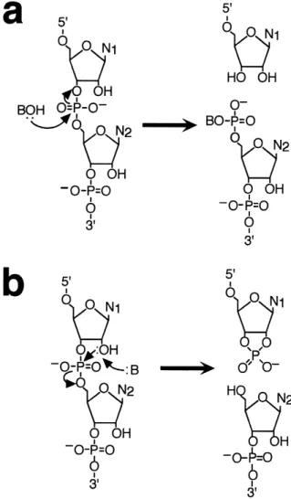
Catalytic RNAs are broadly separated into two classes based on their size and reaction mechanisms (reviewed by [10^14]). The large catalytic RNAs con-sist of RNase P, and the group I and group II in-trons. These molecules range in size from a few hun-dred nucleotides to around 3000. They catalyze reactions that generate reaction intermediates and products with 3P hydroxyls and 5P phosphates (Fig. 1a). The small catalytic RNAs include the hammer-head, the hairpin (or paperclip), hepatitis delta and VS RNA. These molecules range in size from V35 to V155 nucleotides. They use the 2P hydroxyl of the ribose sugar as a nucleophile, and they generate products with a 2P,3P-cyclic phosphate and a 5P hy-droxyl (Fig. 1b). The relationship between the size and the reaction mechanism of these molecules has raised intriguing questions about their origins and evolution. It may be that the reaction mechanism and the size of the large ribozymes are needed to
bring often very distal elements of the substrate into close proximity. The small, self-cleaving, RNAs are not faced with this constraint and perhaps this per-mitted them to evolve smaller catalytic centers. It remains possible, however, that the relationship be-tween the size and reaction mechanism is simply for-tuitous.
With one exception, all these RNAs catalyze reac-tions that modify themselves. Hence, they cannot be considered true enzymes or catalysts. The exception is RNase P, which processes the 5P end of tRNA precursors. It is the only known example of a natu-rally occurring RNA-based enzyme. However, all these molecules can be converted, with some clever engineering, into true RNA enzymes that modify other RNAs in trans without becoming altered them-selves.
Ribozymes increase reaction rates by up to 1011
-fold and have reaction e¤ciencies, kcat/Km, up to 108
M31 min31, which is in the range for
di¡usion-con-trolled duplex formation between oligonucleotides [10]. While impressive, the rate enhancements pro-vided by ribozymes are still V103-fold less than
those provided by protein enzymes catalyzing com-parable reactions [15]. Moreover, ribozymes cannot compare with proteins as multiple-turnover enzymes, mostly because product release is so slow that the catalytic site of the ribozyme is easily saturated. This may be an inherent limitation of RNA enzymes, but it could also re£ect evolutionary constraints, since ribozymes generally catalyze intramolecular, single-turnover, reactions in nature. An exhaustive comparison of the enzymatic mechanistics of protein and RNA enzymes has recently been made [15].
All known ribozymes have an absolute require-ment for a divalent cation, which is generally Mg2. Some, notably within the large catalytic
RNAs, require divalent cations for proper assembly of the tertiary structures as well. On this basis, cata-lytic RNAs are considered to be metalloenzymes, and a general two-metal-ion reaction mechanism has been proposed for the large catalytic RNAs, based on analogy with the properties of protein met-alloenzymes [16]. The role of divalent cations for the small catalytic RNAs is less clear, but they are gen-erally considered to be essential for catalysis (see [17] for alternative view).
3. General properties of introns
Introns, or intervening sequences (IVS), are non-encoding sequences that interrupt the coding, exon,
Fig. 1. Reactions catalyzed by RNAs. a: The reaction mecha-nism of the large catalytic RNAs. The nucleophile BOH is the 3P hydroxyl of a guanosine cofactor in the ¢rst step of group I splicing, the 2P hydroxyl of a nucleotide, generally A, within the intron in the ¢rst step of group II splicing, and a water molecule in the RNase P-catalyzed reaction. b: The reaction mechanism of the small catalytic RNAs. The reaction is most likely initiated by the activation of the 2P hydroxyl on the ribose located at the scissile bond, and it results in a product with a 2P,3P-cyclic phos-phate. The nature of the activation is still poorly understood, but it most likely involves a coordinated metal hydroxide. Notice in both (a) and (b) that the net number of bonds is conserved throughout the reaction.
sequences. These introns must be removed, at the RNA level, in order for the gene to be expressed functionally. There are ¢ve major categories of in-trons and splicing mechanisms. These consist of nu-clear tRNA introns (reviewed by [18]), archaeal in-trons (reviewed by [19]), nuclear mRNA inin-trons (reviewed by [20]), and the group I and group II introns (reviewed by [10,21]). Of these introns, some members of the group I and group II are clearly capable of catalyzing their own excision, in vitro, in an RNA-catalyzed fashion. However, genet-ic analyses have revealed protein factors that are often essential for group I and group II splicing in vivo [22]. The nuclear tRNA and archaeal introns clearly require protein factors (endonuclease and li-gase). Nuclear mRNA introns may represent an in-termediate state. These introns are spliced within large (40^60S) ribonucleoprotein particles (RNP), which consist of a number of small nuclear RNAs (snRNAs) and proteins [20]. However, it is increas-ingly thought that it is the snRNAs that are the
chemical engines of the complex as well as the deter-minants of the splice sites [23].
One of the oddities of group I and group II in-trons, which makes the term `intron' something of an oxymoron, is that they sometimes encode proteins that are required in vivo either for splicing activity or for intron mobility (reviewed by [22]). The former are intron maturases, which can be highly speci¢c for the intron that encodes it. The latter are endonu-cleases with reverse-transcriptase properties. These proteins are involved in intron `homing', where crosses in yeast (the best studied genetically) result in the transfer of the intron into the intronless allele of the gene. There is also intron transposition (or `retrohoming'), where the intron is inserted into oth-er alleles. The widespread, but scattoth-ered, distribution of introns ^ especially introns with high sequence similarity occupying the same location in di¡erent organisms ^ suggests the possibility for horizontal transfer of introns; thus these introns may act some-what akin to infectious agents. In group I introns,
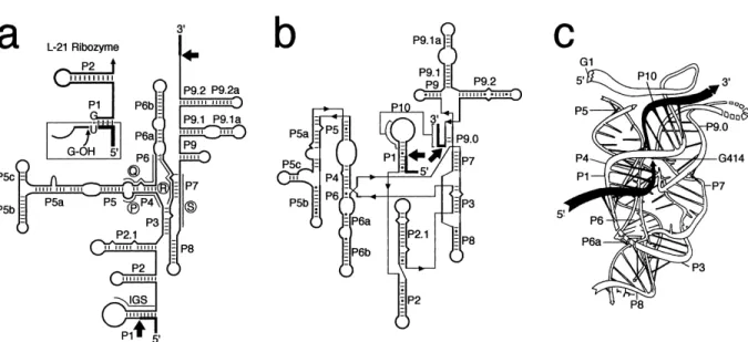
Fig. 2. The structure of group I introns. a: The `classical' depiction of the secondary structure of the rRNA intron from T. thermophila, showing the phylogenetically conserved sequences P, Q, R, and S (circled) and the conserved pairings P1^P9. Nonconserved elements are designated by additional numbers or letters (e.g., P9.1 and P5c). The IGS sequence, which forms P1 and P10, is as indicated. The exon sequences are indicated as heavy lines and the splice sites by the arrows. The insert shows the 5P end of the L-21 ribozyme, which is used in various reactions in trans, with a bound substrate. b: The new secondary structure depiction that more accurately represents the rela-tionships of the di¡erent structural elements. The heavy lines are exon sequences and the very light ones are used to bridge the di¡erent elements, which are very close in tertiary space. c: A computer-generated model of the catalytic core of the Tetrahymena intron. The exon sequences are shown in solid and the intron as outline. This ¢gure shows the intron after the ¢rst reaction, where the nonencoded guanosine, G1, is attached to the 5P end of the intron and the 3P hydroxyl of the 5P exon is posed to make a nucleophilic attack at the 3P splice site. This ¢gure is reprinted with permission from [27].
mobility is apparently catalyzed by proteins alone and a functional (active) intron is not required. In group II, the intron itself is essential for activity and the intron RNA actually becomes inserted into the double-stranded DNA substrate [24,25].
3.1. Group I introns
Group I introns range in size from a few hundred nucleotides to around 3000. They are abundant in fungal and plant mitochondria, but they are also found in nuclear rRNA genes, chloroplast DNA (ctDNA), bacteriophage, eukaryotic viruses, and in the tRNA of ctDNA and eubacteria. In short, they are widely found except in higher eukaryotes (i.e., vertebrates; reviewed by [10,21,26]). The various group I introns have little sequence similarity, but they are characterized by four short conserved se-quence elements, called P, Q, R, and S (Fig. 2a). P can always partially base-pair with Q, and R can always partially base-pair with S. In addition, all group I introns can fold into distinctive, phylogeneti-cally conserved, secondary structures consisting of 10 paired segments (P1^P10; Fig. 2). Additional se-quences, including large open reading frames (ORF), and structures are often found, but they do not disrupt the catalytic core, which consists of P3, P4, P6 and P7. However, their presence is used to subdivide the introns into various subgroups [27,28]. The secondary structure was originally deduced from computer modeling based on phylogenetic comparisons and, in the case of the Tetrahymena intron, on limited structural probing [29^31]. Addi-tional evidence for this structure comes from enzy-matic and chemical probing studies, additional phy-logenetic analyses and from physical approaches (reviewed by [10,21,26,28]). These studies have con-¢rmed the overall structure and revealed other im-portant interactions, which have resulted in a new secondary structure depiction that more accurately re£ects the spatial relationship of the di¡erent ele-ments (Fig. 2b) [32].
The exon sequences are oriented relative to the catalytic core by base pairings with an intron se-quence called the internal guide sese-quence (IGS), which constitutes P1 and P10 in the structure [29]. An additional pairing, called P9.0, further aligns the intron-3P exon splice site [33,34]. It was so named
because it occurs between P8 and P9 in the second-ary structure, the latter being named before the interaction was discovered. The 5P exon contains a highly conserved uridine on its 3P end, which forms a functionally important UcG base pair with the IGS, and the intron contains a conserved guanosine on its 3P end. Finally, there is a speci¢c binding site for the guanosine cofactor that initiates the reaction. This site involves a conserved G-C base pair in P7 [35].
Michel and Westhof [27] have made a very clever, computer-generated, three-dimensional model of the catalytic core based on careful phylogenetic compar-isons (Fig. 2c). This model brings all the relevant elements into proximity, and it has provided a sound basis for additional studies. A crystal structure was solved to 2.8 Aî for a 160-nucleotide fragment of the Tetrahymena intron, consisting of P4^P6 [36]. While this structure contains less than half of the catalytic core of the enzyme and it tells us little about the reaction mechanism, it has revealed several exciting features. These include a tetraloop, GNRA (N is any
Fig. 3. Splicing mechanisms of group I and group II introns. In both cases, a series of trans-esteri¢cation reactions are used to excise the intron and ligate the exons. The net number of bonds remains the same throughout. The reaction is initiated by a gua-nosine cofactor in group I introns and by an internal adegua-nosine in group II. The splicing reaction of nuclear pre-mRNAs follows the same pathway as group II introns, but it occurs on a large ribonucleoprotein complex. Details are described in the text.
base and R is purine), docking with its receptor, several adenosine platforms, which are derived from adjacent adenosines forming a pseudo-base pair within the helix, and a ribose zipper, which in-volves a network of hydrogen bonds between the 2P-OH groups of ribose and acceptor groups of bases within the shallow groove of helices [36,37].
Recently, a 5.0-Aî crystal structure has been solved for a 247-nucleotide-long fragment of the Tetrahyme-na intron, consisting of helices P3^P9 [38]. This new structure shows the previously characterized P4^P6 domain (helices P4, P5, and P6) largely unchanged and the P3^P9 domain (helices P3, P7, P8 and P9) wrapped around it. The close packing of the two domains creates a shallow cleft into which the P1 helix, containing the 5P splice site, could ¢t. The structure also creates a particularly tight binding site in the P7 helix for the guanosine cofactor that initiates the splicing reaction. Unfortunately, the res-olution is too low to visualize details within the structure, and the crystal structure is still missing the P1^P2 domain containing the splice site, so little can be determined about the reaction mechanism. However, it is clear that the computer generated model for the catalytic core [27,28] is largely consis-tent with this crystal structure. A triumph for com-puter modeling of RNA structures!
The splicing reaction of group I introns was ¢rst worked out for Tetrahymena thermophila (reviewed by [10,21,26]), and it will form the basis for my fur-ther discussion. Nevertheless, all characterized group I introns follow essentially the same pathway. The Tetrahymena intron is excised from the precursor rRNA by a two-step transesteri¢cation reaction (Fig. 3). The reaction is initiated by the nucleophilic attack of the 3P hydroxyl of a guanosine cofactor at the 5P splice site. The exon-intron phosphodiester bond is cleaved and the guanosine forms a 3P,5P-phosphodiester bond at the 5P end of the intron. The now free 3P hydroxyl of the 5P exon then makes a nucleophilic attack at the 3P splice site to form the ligated exons and release the intron with the non-encoded guanosine.
Nucleotides close to the 5P end of the intron are realigned on the IGS and the highly conserved 3P terminal guanosine of the intron makes a nucleo-philic attack at a phosphodiester bond between nu-cleotides 15 and 16 or between nunu-cleotides 19 and 20
within the intron in a reaction that is analogous to the ¢rst step of splicing. The intron is circularized (C-15 or C-19) and a small fragment containing the nonencoded guanosine is released. The circular prod-uct is also found in vivo, and its formation presum-ably helps to drive the reaction to completion. Although the circularization reaction is not univer-sally conserved among group I introns, it is very common. Each step is essentially the forward or re-verse of the same reaction, and the total number of phosphodiester bonds is conserved. Each step is fully reversible, and no external energy source is needed. The intron can fully reintegrate into rRNAs both in vitro and in vivo ([39] and reference therein). The phosphodiester bonds are inverted during the reac-tion, which is consistent with an SN2, in-line,
reac-tion mechanism (see [10,26]).
The circular product is normally considered the end product of the reaction, but the released intron retains catalytic activity and substrate speci¢city. For T. thermophila, a linear form of the intron lacking the ¢rst 21 and the last ¢ve nucleotides (L-21 ScaI) will catalyze a wide range of reactions on substrates added in trans. These include sequence-speci¢c endo-nuclease, nucleotidyltransferase, ligase and phospha-tase activities ([10,21,26]). Substrate speci¢city is changed by altering the IGS sequence. Because the reactions are catalyzed using the 3P hydroxyl, they will work on both RNA and DNA substrates, although the latter has a much lower binding a¤nity and hence less activity. Since the L-21 ScaI RNA is unaltered in the reaction, it is considered a true RNA-based enzyme.
The properties of group I introns lend themselves to a number of di¡erent applications. The L-21 ScaI version of the Tetrahymena intron has been commer-cially sold as an RNA restriction enzyme. Circularly permutated precursor RNA, which contains end-to-end fused exons inserted within the middle of the intron sequence, is used to generate circular exon molecules by an `inverse' splicing reaction [40]. A variant of this technique is used to generate circular, trans-cleaving, HDV ribozymes in vitro [41] and in vivo [42], which are more resistant to nucleases. A trans-splicing reaction can repair a truncated lacZ transcript in Escherichia coli [43] and in the cyto-plasm of mammalian cells [44]. A trans-splicing in-tron can also change mutant L-globin transcripts, in
sickle cell anemia, into mRNAs coding for antisick-ling Q-globin in human erythroid lineage cells [45]. 3.2. Group II introns
Group II introns range in size from several hun-dred to around 2500 nucleotides. Although they are much less widely distributed than group I introns, they are found in fungal and plant mitochondria, in chloroplasts of plants, in algae, in eubacteria and especially in the chloroplasts of the protist Eu-glena gracilis (reviewed by [10,21,46,47]). Most are present in mRNAs, but a few also occur in tRNA and rRNA genes. In contrast to the extensively an-alyzed group I introns, much less is known about group II introns. This is partly because of their more limited distribution but also because very few of them are found to be self-splicing in vitro. Those that are autocatalytic require reaction conditions that are far from physiological (e.g., 100 mM MgCl2,
500 mM (NH4)2SO4 and 45³C; [48]).
The secondary structure of group II introns was originally deduced from phylogenetic comparisons and computer modeling [30,49]. It is normally
de-picted as six helical domains (I^VI) radiating as spokes from a central wheel (Fig. 4). On the basis of the structural features, group II introns are di-vided into two major subclasses, although some in-trons ¢t into neither class. Some contain long ORFs. Important structural features have been di¤cult to elucidate because of their poor reactivity in vitro and the di¤culty of working with organelle-speci¢c mol-ecules. However, it appears that only domains I and V are indispensable [50]. In addition, domain V tains most of the relatively few phylogenetically con-served nucleotides found in group II introns and it might constitute the reaction center [46,47,51]. Do-main VI contains the highly conserved adenosine that is generally used to initiate the splicing reaction. Except for domains I and V, the domains can be modi¢ed or deleted and the intron will retain some catalytic activity. Optional ORFs are frequently lo-cated in the loop of domain IV.
Degenerated forms of group II introns are found in plant chloroplasts and mitochondria that often lack recognizable cognates of the various domains. These are called group III introns (reviewed by [52]); they may require factors in trans for activity or be assembled from parts of group II introns. These de-generate introns provide a feasible pathway between group II and nuclear mRNA splicing, where more and more of the role of the intron is supplanted by trans-acting factors [23,47]. This possibility is made more plausible by the observation that some group II and group III introns occur within other group II or group III introns (called twintrons; [52]) and that others are discontinuous. Segments of these latter introns are transcribed within two or even three sep-arate molecules from distant regions of the genome and the exons are assembled by a trans-splicing re-action (reviewed by [53]).
The 5P exon is aligned by interactions between two intron binding sequences (IBS1 and IBS2), located near the 3P end of the 5P exon, and two exon binding sequences (EBS1 and EBS2), which are located in domain I of the intron (Fig. 4). The 5P splice site is further de¢ned by O-OP interactions. These interac-tions are important both before and after 5P cleav-age. The 3P splice site has multiple determinants, comprising N-NP, Q-QP, and other as yet unidenti¢ed interactions. As yet, no tertiary model has been pro-posed.
Fig. 4. The secondary structure of group II introns. This cartoon is a generalization from a number of di¡erent introns. The exon sequences are indicated as heavy lines and the splice sites by the arrows. The characteristics of the central wheel with the radiating domains is conserved, but the characteristics of the individual do-mains vary considerably. Tertiary interactions are formed be-tween IBS1-EBS1, IBS2-EBS2, K-KP, O-OP, N-NP and Q-QP; the con-nections between these elements are not shown for clarity. The conserved adenosine used to initiate the splicing reaction is indi-cated in domain VI. Additional tertiary interactions have been identi¢ed, which vary with the intron (not shown).
Unlike group I introns, where the ¢rst and second steps are thought of as forward and reverse steps of the same reaction, implying a single catalytic site, group II introns use two di¡erent nucleophiles (2P and 3P hydroxyls) and both steps show the same stereospeci¢city ([46,47] and references therein). This suggests that there are two independent reaction centers or that there is a single site that switches substrates [16].
The splicing reaction is generally initiated by the nucleophilic attack of the 2P hydroxyl of a highly conserved adenosine in domain VI to form a distinc-tive structure, called a lariat, containing 3P-5P and 2P-5P phosphodiester bonds at the adenosine branch site (Fig. 3). The now free 3P hydroxyl of the 5P exon then makes a nucleophilic attack at the 3P splice site to form the ligated exons and release the intron, still as a lariat. The reaction can also be initiated, both in vitro and in vivo, by the nucleophilic attack of water [48,54], although this is not a typical route. In this case, the intron is released as a linear mole-cule. As mentioned above, these reactions can also be catalyzed in trans from separate transcripts. The splicing mechanism and the lariat product are re-miniscent of those found in nuclear mRNA splicing,
and it provides an additional evolutionary link be-tween the two splicing reactions [23,47].
The applications of group II introns are more lim-ited than those of group I. Nevertheless, it can cata-lyze the cleavage of ligated precursors using the en-ergy of a phosphoanhydride bond, ligate RNA to DNA, and cleave single-stranded DNA substrates [55]. It is also used to circularize exon sequences [56], as described above for group I introns. How-ever, the fact that the intron RNA becomes inserted into double-stranded DNA during intron homing [24,25] suggests that other applications will soon be found.
4. RNase P
RNase P is a ubiquitous enzyme in all character-ized organisms that processes the 5P termini of tRNA precursors (reviewed by [10,57^60]). In eubacteria, RNase P exists as a ribonucleoprotein complex, con-sisting of a large RNA of about 350^400 nucleotides (M1 RNA in E. coli) and a small basic protein of V14 kDa (C5 in E. coli). The basic protein is essen-tial for activity in vivo, however the RNase P RNA,
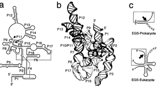
Fig. 5. Characteristics of RNase P RNA. a: The proposed secondary structure of M1 RNA, the RNA component of RNase P from E. coli. b: The computer-modeled tertiary structure of (a). The black spheres represent invariant nucleotides at the catalytic site and con-served nucleotides in the T-loop recognition site. Note that these concon-served residues are clustered close to each other in what forms the catalytic core of the molecule. This ¢gure is modi¢ed from [71]. c: Synthetic substrates for RNase P from prokaryotes and eukaryotes. The EGS, which is used to de¢ne the target speci¢city, is shown bound to the substrate (boxed) and the cleavage site is indicated by an arrow.
by itself, can catalyze the reaction in vitro [2]. This reaction requires high salt concentrations (e.g., 1 M K or NH
4 and 10 mM MgCl2; [60]), which
sug-gests that the basic protein component acts only as an electrostatic shield to promote binding between the RNA enzyme and the RNA substrate. However, the protein also a¡ects cleavage-site speci¢city and turnover, so its full role in the reaction is still unclear (reviewed by [58,60]). RNase P from E. coli will also process other substrates that partially resemble tRNAs (e.g., 4.5S RNA [61]), but these reactions appear to be relatively minor. RNase P RNA is the only characterized ribozyme that, unmodi¢ed, acts in trans on multiple substrates, and hence it is considered the only true, naturally occurring, RNA enzyme.
RNase P from the nuclei and mitochondria of eu-karyotes also exist as ribonucleoprotein complexes, although they generally have much higher protein contents (ca. 50^70% versus ca. 10% for eubacteria [57]). Moreover, while the RNA component is inevi-tably found to be essential, the RNase P RNA from eukaryotes has never been shown to have catalytic activity. Archaebacteria have many properties that make them more similar to the eukaryotes than to the eubacteria, and they likewise are not known to have RNA-alone catalytic activity [60].
There is evidence that another ribonuclease in eu-karyotes, RNase MRP, is closely related, and per-haps homologous (i.e., evolutionarily related), to RNase P (reviewed by [62,63]). It is a ribonucleopro-tein complex that participates in nucleolar pre-rRNA processing. The RNA component can fold into sim-ilar secondary structures as RNase P RNA and, in yeast, it shares common protein components [64]. However, like the eukaryotic RNase P, the RNase MRP RNA has not been shown to have catalytic activity by itself.
Although there is little sequence conservation, all the eubacterial RNase P RNAs can be folded into similar, although not identical, secondary structures on the basis of comparative sequence analyses [60,65,66]. The E. coli M1 RNA consists of 18 paired helices (P1^P18; Fig. 5a). The Bacillus subtilis M1 RNA analog is more diversi¢ed from E. coli than many of the other eubacteria. It folds into a similar structure, but P6, P13, P14, P16 and P17 are missing and it contains extra helices P5.1, P10.1, P15.1, and
P19 (not shown). These two RNAs are the most extensively studied, but through a careful compara-tive analysis of the di¡erent eubacteria, it is possible to derive a common core structure consisting of heli-ces P1^P5, P7^P12 and P15 [60]. However, the activ-ity of this `minimal' structure has not yet been dem-onstrated.
Native RNase P RNAs di¡er from the phyloge-netic minimum by having extra stems and stem-loop structures. While probably not needed for catalysis, these structures nevertheless lower the ionic strength requirements and enhance their thermal stability [60]. Such elements may be redundant in the sense that any single one can be deleted or modi¢ed without signi¢cantly altering the activity, but it is not possi-ble to simultaneously delete or modify all of them. The RNase P RNA from eukaryotes and archaebac-teria have little sequence similarity to their eubacte-rial counterparts, but they can often be folded into a universally conserved core structure as well [67]. Missing RNA elements from the structures may be supplanted by the protein component(s), but this has not been characterized.
Three-dimensional models of the E. coli M1 RNA have been made by Westhof and Altman [68] and by Harris et al. [69], and both groups have recently re¢ned these models [70,71] (Fig. 5b). Three-dimen-sional models for the B. subtilis RNase P RNA are also available [70,71]. These are computer-derived models based on data obtained from phylogenetic comparisons, mutational analyses, chemical probing and from crosslinking studies. It is beyond the scope of this review to discuss these models, especially in light of their speculative nature, but generally the models have a good ¢t to the experimental data. Both groups' models have similar overall structures and both provide a pocket or cleft into which the tRNA substrate will ¢t. However, many of the spe-ci¢c interactions vary; thus these models are ex-pected to undergo continual re¢nement as additional experimental data become available.
RNase P uses water as a nucleophile to cleave the phosphodiester bond (Fig. 1a). The exact mechanism by which the tRNA precursor is bound is still un-clear. The 3P half of the acceptor stem is thought to function as an external guide sequence (EGS), but it does not uniquely de¢ne the cleavage site. The pri-mary sequence does not seem to be important nor
does any single element uniquely de¢ne the cleavage site. Instead, recognition could result from several redundant factors, including the distance along the coaxially stacked T stem-loop and acceptor stem, the 3P terminal CCA sequence, the EGS alignment, and a conserved guanosine 3P to the cleavage site (re-viewed by [58,59]). Thus, recognition is largely, if not entirely, based on tertiary interactions with the substrate.
From studies using small substrates, it is possible to design synthetic EGSs that can target any RNAs for cleavage by RNase P in vitro or in vivo (Fig. 5c; reviewed by [72]). The basis for this is that the EGS binds to the target RNA and makes it look like a tRNA substrate. In eubacteria, the minimal require-ment is that it forms a short stem with a free NCCA (Fig. 5c). The target sequence can be virtually any-thing. In eukaryotes, the EGS-substrate complex must more closely resemble a tRNA (Fig. 5c). This reaction works in vitro, in bacteria and in human cells [72]. A variant of this technique is to add the appropriate EGS (here called an internal guide se-quence or IGS) to the 3P end of the RNase P RNA. This increases the e¤ciency of the reaction, and it is used to inactivate thymidine kinase mRNA from herpes simplex virus in cell lines [73]. Yet, while the therapeutic potential of RNase P has been dem-onstrated, it has not been widely used in therapeutic applications.
5. General properties of small catalytic RNAs Self-cleaving RNAs are generally found in small (V220 to V460 nucleotides long) RNA pathogens of plants known as viroids, virusoids and linear sat-ellite viruses (reviewed by [13,74]). However, they are also found within satellite RNAs of salamanders, Neurospora, and within another pathogenic satellite virus found in man. The viroid and satellite RNAs are generally replicated by an RNA-dependent roll-ing-circle mechanism, and the catalytic domains are thought to process the linear concatemers that are generated into unit-length progeny.
The linear, unit-length progenies produced during replication in vivo are subsequently ligated to form closed-circular molecules that are used in the next round of rolling-circle replication. It is reasonable
to expect that this is catalyzed by the ribozyme as well since, mechanistically, it represents the reverse of the cleavage reaction, and it would be analogous to the splicing reaction carried out by the group I and group II introns. However, in vitro only the hairpin ribozyme shows signi¢cant ligation activity. Protein factors may be involved in vivo or other, as yet unidenti¢ed, RNA elements may be required.
Four motifs are characterized (described below), and they all catalyze reactions that generate products with 2P,3P-cyclic phosphates and 5P hydroxyls (re-viewed by [13,74]; Fig. 1b). As with the previous ribozymes, they all require a divalent metal ion, nor-mally Mg2, for activity.
The intramolecular self-cleaving activity is con-verted into a trans-cleaving activity by making the `substrate' and `ribozyme' into separate molecules. However, the `substrate' remains an integral part of the structure of the active ribozyme, and hence the ribozyme is often simply de¢ned as the unmodi¢ed portion of the molecule and the substrate is the cleaved portion. Hence, the `ribozyme' may consist of di¡erent sequence elements, depending on the construct. The catalytic domains of these ribozymes are small and relatively well characterized, and they are more widely used in therapeutic applica-tions. Each has characteristics that confer speci¢c advantages and disadvantages as therapeutic agents. The in vitro and ex vivo activity of cis-cleaving forms of three of these self-cleaving ribozymes (ham-merhead, hairpin and HDV) have been compared [75].
5.1. Hammerhead ribozyme
The hammerhead ribozyme is probably the most extensively studied of all the ribozymes, and it is the motif most commonly found in the viroids and sat-ellite RNAs (reviewed by [13]). Currently 16 ham-merhead motifs are known in the plus and minus strands of these plant pathogens. Three other ham-merhead motifs are found in the satellite 2 RNAs from the salamanders, Triturus vulgaris, Ambystoma talpoideum and Amphiuma tridactylum ([13] and references therein). This ribozyme was so named be-cause its Australian discoverers found the secondary structure, as originally drawn, to be reminiscent of the head of a hammerhead shark. It is the smallest of
the naturally occurring self-cleaving RNAs, at 40^50 nucleotides in length.
Analyses of the hammerhead ribozyme are volu-minous (reviewed by [9,76^78]). It consists of three helical regions, which are variable, and three single-stranded regions that contain most of the highly con-served nucleotides (Fig. 6a). The length of the helical arms can be quite variable, and helix II can be re-duced to two base pairs. Mutating any of the con-served residues markedly reduces activity; conse-quently, important functional groups are often identi¢ed by incorporating synthetic nucleotide ana-logs into the RNA (e.g., inosine for guanine; re-viewed by [76]).
Cleavage occurs after an NUH triplet, where N is any nucleotide, and H is any nucleotide except gua-nosine. The most e¡ective triplet is GUC, but other triplet combinations will work nearly as well; their relative activities have been compared, although the ordering can vary depending on the method of anal-ysis (reviewed by [8,9,77]). The reaction mechanism is extensively studied, and it appears to involve a metal-coordinated hydroxide, which probably di-rectly activates the 2P hydroxyl ([9,76,78] and refer-ence therein). The reaction products are consistent with an SN2 (in-line) reaction mechanism; this was
suggested by an inversion of the phosphate at the scissile linkage.
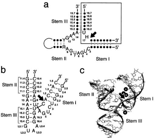
Fig. 6. Characteristics of the hammerhead ribozyme. a: The secondary structure of the hammerhead ribozyme showing the conserved se-quence and structure. The dots represent nucleotides that can be anything, Y is a pyrimidine, R is a purine and H is any nucleotide ex-cept guanosine. The arrow indicates the self-cleavage site. The boxed region shows the portion that is normally the substrate in trans-cleaving versions of the ribozyme. The numbering is based on standardized nomenclature [104]. b: A new secondary structure drawing that more accurately re£ects the spatial relationships of the di¡erent elements. This structure di¡ers from (a) in that it shows a loop in stem III rather than in stem II. c: The solved crystal structure of the sequence shown in (b). This is an RNA-only structure infused with Mg2, shown as spheres. Only the Mg2ion close to the scissile bond is generally accepted as being functionally relevant (see text). This
The hammerhead ribozyme was the ¢rst catalytic RNA for which the complete X-ray crystal structure was solved. There are now a number of such struc-tures available with resolutions ranging from 2.6 Aî to 3.1 Aî, as well as a structure derived from £uores-cence resonance energy transfer (FRET; reviewed by [76,78,79]). The molecule has a Y shape (Fig. 6b,c), with stem I and stem II at the arms and stem III at the base. Recently, the crystallized structure of a re-action intermediate was determined using a tallo-5P-C-methyl-ribose-modi¢ed ribozyme that is kinetically blocked for the ¢nal cleavage reaction [80]. This lat-ter structure is more compatible with an SN2
reac-tion mechanism. This is in contrast to the previously solved structures that showed the ribozyme in a ground state that was incompatible with such a mechanism. A second Mg2 binding site has
poten-tially been identi¢ed in another crystal structure that could be involved in stabilizing the pentacoordinated phosphate transition state [81]. However, the exist-ence of this site is still debated (see [78]). Moreover, there are other incompatibilities between the
exper-imental data and the crystalline structures [78], and clearly additional work will be needed.
The hammerhead ribozyme is divided into sepa-rate `ribozyme' and `substsepa-rate' in several ways, but the one shown in Fig. 6a is the most commonly used because most of the conserved residues are contained within the ribozyme rather than in the substrate. The hybridizing arms are varied to opti-mize ribozyme activity and substrate speci¢city. Nor-mally hybridizing arms of six or seven base pairs are considered optimal, but for variable arms it is better to have a long stem III and short stem I than the reverse (reviewed by [9]). Often, a tetraloop (frequently GAAA) is used for loop II and stem II is GC-rich to further increase the stability of the stem-loop. The malleability of the hammerhead ribozyme makes it the most commonly used ribo-zyme for in vivo studies, and there are many success-ful examples (reviewed by [7^9]). A trans-cleaving hammerhead ribozyme is approved for phase II clin-ical trials against HIV-1 by Ribozyme Pharmaceut-icals (RPI).
Fig. 7. Secondary structure of the hairpin ribozyme. a: The minus strand of sTRSV (numbering is that of the full-length virus). The ar-row shows the cleavage site. b: The consensus sequence and structure, where dots are any nucleotide, Y is a pyrimidine and R is a pu-rine. The boxed region represents the portion that is normally the substrate in trans-cleaving reactions. The substrate is numbered relative to the cleavage site and the `ribozyme' relative to the 5P end. This ¢gure is modi¢ed from [83] and it incorporates recent experimental data from [105].
5.2. Hairpin ribozyme
The hairpin ribozyme is found in three pathogenic, plant, satellite viruses, although the one found in the satellite virus associated with tobacco ring spot virus (sTRSV) is the best characterized. It consists of four stem regions that, when lined up coaxially, some-what resemble a hairpin; interestingly, it was also originally named `paperclip,' which may, in fact, bet-ter represent its overall three-dimensional shape (see [13]; Fig. 7a). It consists of two noncontiguous se-quences of 50 and 14 nucleotides within the minus strand of sTRSV (reviewed by [13,17,82^84]. The secondary structure was determined based on com-puter-aided modeling, limited phylogenetic compari-sons, mutational analyses and by in vitro selection.
The other hairpin ribozymes are found in the sat-ellite viruses of arabis mosaic virus (sARMV) and chicory yellow mottle virus (sCYMV), and they mostly di¡er from the sequence shown in Fig. 7a by nucleotide changes within the helical regions that maintain the structure as shown. Indeed, the mutational and in vitro selection analyses show that the helical regions are structural elements that can largely be changed, as long as the integrity of the helices is maintained. Most of the conserved nucleo-tides occur within the single-stranded regions. The guanosine 3P to the cleavage site is essential, but altering the other conserved positions can dramati-cally reduce the activity as well. The bulged region between helices III and IV contain a conserved mo-tif, called a UV-loop momo-tif, that is found in a diverse group of RNAs, including viroids, 5S rRNA and the sarcin-ricin loop of 28S rRNA [82]. However, the role this motif plays in catalysis is still unknown. The consensus sequence and structure are shown in Fig. 7b.
Recently, a computer-generated tertiary model was made that was based on preexisting structural data and on the spatial distance of tolerated, inter-domain, aryl-disul¢de crosslinks [85]. Additional in-formation was also obtained by Walker et al. [86] using FRET data. The current model shows helix I coaxially stacked on helix II and helix IV coaxially stacked on helix III. These two extended helices are then bent so that helix II and helix III, and helix I and helix IV are positioned side by side. This places the two highly conserved bulged regions in
proxim-ity, and they could thus form the catalytic core. However, this tertiary model is still preliminary and additional data are required before the details of the catalytic site are known.
A major advantage of the hairpin ribozyme lies in its ability to catalyze both cleavage and ligation re-actions e¤ciently in vitro; this has greatly facilitated in vitro selection experiments because new sub-strates, with the appropriate PCR primer sites, are easily generated ([82] and references therein). The RNA-catalyzed ligation reaction is also thought to be relevant in vivo, in that the RNA can both cleave the linear multimers generated during rolling-circle replication and ligate them to form the circular RNA progeny. However, as with the other catalytic motifs used in viroid replication, the cleavage-liga-tion reaccleavage-liga-tion must be carefully regulated in vivo to prevent inappropriate cleavage, or ligation, of the resulting progeny. The mechanism by which this is accomplished is still unknown. Like the other cata-lytic RNAs, the hairpin ribozyme reaction requires a divalent cation. However, it appears that, unlike the hammerhead ribozyme, the hydrated cation (usually Mg2) is not directly coordinated to the phosphate;
moreover, the essential role of divalent cations has recently been called into question (see [17]).
Since the `substrate' portion of the cis-cleaving hairpin ribozyme is discontinuous with the rest of the molecule, it is obvious where to separate the two domains (boxed region in Fig. 7b). The substrate should contain the sequence RYN*GUC, where R is a purine, Y is a pyrimidine and N is any nucleotide. Cleavage occurs at the position indicated by a *. Stem II should be four base pairs, but stem I can be signi¢cantly extended (e.g., [87]), as can stem IV [88]. Substrate speci¢city is changed by altering the nonconserved residues within the base-paired region. The hairpin ribozyme is used to target HIV-1 RNA in cell culture, and it is currently approved for clin-ical trials (see [84]). However, its e¡ective use as a trans-cleaving ribozyme in vivo is still rather limited; the reasons for this are unclear.
5.3. VS RNA ribozyme
The mitochondria of certain strains of Neurospora contain the Varkud plasmid (a retroplasmid), which encodes a reverse transcriptase, and a small,
unre-lated, RNA (VS RNA). The VS RNA is transcribed from circular or multimeric VS plasmid DNA by a mitochondrial RNA polymerase, and the resulting transcripts are subsequently site-speci¢cally cleaved and ligated to form circular, 881 nucleotides long, RNA monomers [89]. These monomers are then re-verse transcribed and made double stranded to form the mature VS plasmid.
In vitro transcribed VS RNA precursors are cleaved and ligated by the RNA itself and this is presumed to occur in vivo as well ([90] and referen-ces therein). Of all the self-cleaving RNAs, the cata-lytic properties of VS RNA are the most poorly understood. At 154 nucleotides long it is also the largest. The minimal sequence that retains catalytic activity contains one nucleotide 5P and 153 nucleo-tides 3P to the cleavage site. However, this struc-ture can be reduced to 121^126 nucleotides by mak-ing internal deletions within the helices [91]. An RNA secondary structure is proposed, but except for a tertiary interaction between loop I and loop V, little is known about its overall conformation (Fig. 8).
The catalytic domain of VS RNA is converted into a trans-cleaving ribozyme by using a 144-nucleotide fragment of the VS RNA from 640 to 881 (VS RNA numbering; [92]). The minimal substrate consists of one nucleotide 5P and 19 nucleotides 3P to the cleav-age site, and it forms a short stem-loop structure. As with RNase P, the ribozyme seems to recognize the structure of the substrate largely as a helical domain.
The minimal sequence requirement 5P to the cleavage site is a characteristic shared only with the HDV ribozyme (see below) and it could make this ribo-zyme suitable for 3P end trimming of RNAs ex-pressed in vitro or in vivo. However, the uncertainty in the substrate requirements and the lackadaisical activity in trans have limited its application, although recent experiments have improved its activity [93]. 5.4. HDV ribozyme
The hepatitis delta virus (HDV) is a viroid-like satellite virus of the hepatitis B virus (HBV), and it is the sole example of such a virus in mammalian systems (reviewed by [94,95]). It is widespread and can cause severe fulminant hepatitis in infected pa-tients. It is about 1700 nucleotides long, and it enc-odes a single protein that is expressed in two forms due to an RNA editing event. Both the genomic, infectious strand, and the antigenomic strand have self-cleaving domains (reviewed by [96^99]). Despite previous pronouncements, no biologically relevant, RNA-catalyzed, ligation reaction has been observed in vitro, although the integrity of the RNA catalytic domains is clearly essential for both the cleavage and ligation reaction in vivo ([99] and references therein). A possible mechanism for the biological control of these reactions, to prevent inappropriate cleavage or ligation, has been proposed [96].
The minimal domain containing self-cleaving ac-tivity has one nucleotide 5P and 84 nucleotides 3P to
Fig. 8. Secondary structure of the VS RNA ribozyme. The arrow shows the cleavage site and numbering is that of the full-length VS RNA. The trans-cleaving form consists of nucleotides 640^881 and the substrate is shown boxed.
the cleavage site for both domains. Despite the se-quence di¡erences, both sese-quences fold into similar secondary structures, of which the pseudoknotted structure shown in Fig. 9a is now the most widely accepted. It consists of four stem regions; three of these stems (I, II and IV) are largely structural ele-ments, while the speci¢c sequences in hairpin III and in the junctions I/IV and IV/II are more important. The catalytic domains of HDV are known for their ability to retain cis-cleaving activity at high temper-atures and in the presence of denaturants (see [97] and references therein). The tertiary structures have been computer modeled for the genomic and antige-nomic forms of the pseudoknot model ([100,101]; Fig. 9b) and for the antigenomic axehead variant [102].
Recently, a 2.3-Aî crystal structure has been solved for the genomic HDV ribozyme [103]. This was ac-complished by replacing hairpin IV with a small hairpin structure that binds tightly to the protein U1A, a spliceosomal protein. By co-crystallizing
the RNA with the protein, the authors were better able to obtain highly structured crystals that dif-fracted to a high resolution. It also greatly facilitated heavy metal substitution that is necessary for obtain-ing crystal phasobtain-ing. This structure is very similar to the computer-predicted model, but it revealed some unexpected results. There is an additional pseudo-knot structure derived from base pairs between C21 and C22, in loop III with G39 and G38, at the base of helix I. These speci¢c interactions were not previously predicted, although the importance of the nucleotides were correctly derived [100] In addi-tion, A43 and G74 stack on the end of helix IV to form noncanonical base pairs and G10 forms an ex-tension to helix II as previously predicted [97,98]. These interactions create a structure where helix IV is rotated relative to the computer model and hairpin III is more compressed. Nevertheless, the two struc-tures are otherwise very similar. The crystal structure provides an organized, almost protein-like, crevice for the active site. Unfortunately, it reveals little
Fig. 9. Characteristics of the HDV ribozyme. a: The genomic and antigenomic ribozymes from HDV. Numbering of the nucleotides is relative to the cleavage sites, indicated by arrows. Helical domains are separated by lines to facilitate the presentation. The length of stem I is critical, although there is no evidence that the 31 base pair is needed. The typical trans-cleaving forms are separated in junction I/II, where the substrates are shown boxed. The identities of the nonbase-paired residues in the substrate (boxed) are not important for trans-cleaving activity. Additional base-pair interactions, that were recently derived for the genomic ribozyme, are shown as dashed lines. b: Computer-generated three-dimensional model of the genomic catalytic domain. This ¢gure is modi¢ed from [100]. Recently, the crystal structure for the genomic HDV ribozyme was obtained [103]. This structure is similar to that shown here except that there is an addition-al pseudoknot interaction between C21 and C22 with G38 and G39. Moreover, G10 is stacked on helix II and A43 and G74 stack on hairpin IV.
about the reaction mechanism or the role of the Mg2 ions.
As with the other ribozymes, the cis-cleaving ac-tivity of the HDV ribozymes can be converted into a trans-cleaving activity (reviewed by [97^99]). The most common form is indicated in Fig. 9a. However, other permutations are possible, including a large substrate consisting of sequences 35 to V60 (num-bering in Fig. 9a) and a small ribozyme consisting of sequences from V60 to +84. Since most of the con-served elements are contained within the substrate, the practical utility of this latter form is somewhat limited. A completely closed-circular variant of the trans-cleaving ribozyme shown in Fig. 9a also has been generated; a short loop was used to close the end of stem II [41]. The absence of free ends makes this ribozyme particularly resistant to the exonu-cleases found in serum and the cellular environment. It is possible to change the substrate binding se-quence to target other RNAs. In theory, most sub-strate sequences are possible, although a guanosine at the 31 position, relative to the cleavage site, is inhibitory and a purine-pyrimidine base pair at posi-tion +1 is preferred. However, in practice many of the changes in the substrate-binding sequence have unpredictable e¡ects.
Up to now, the HDV ribozyme has a rather lim-ited use in therapeutic applications, largely because of the di¤culty of obtaining high activity with the trans-cleaving forms. However, because of the mini-mal sequence requirements 5P to the cleavage site, the cis-cleaving activity is useful for generating discrete 3P ends of RNA. This has applications, for example, in processing ribozyme cassettes (transcripts contain-ing multiple ribozyme units), for generatcontain-ing homoge-neous ends on in vitro transcribed RNA or for the expression of discrete viral RNA transcripts o¡ plas-mid DNA ([99] and references therein).
6. Concluding comments
In this review I have attempted to summarize brie£y the important features of the di¡erent cata-lytic RNAs that have so far been identi¢ed. The ribozyme ¢eld has advanced far in an incredibly short time. There are seven ribozymes identi¢ed in nature, and all of them have been engineered to
cleave or modify other RNAs in trans. Other ribo-zymes have been created de novo, and they can cat-alyze a variety of reactions. Moreover, other cellular processes have signi¢cant RNA components (e.g., the spliceosome and ribosomes) where RNA cataly-sis may play an important role. Additional ribo-zymes are bound to be discovered in the future. Fi-nally, the disappointment that many earlier researchers had when working with ribozymes in vivo has now opened up to new opportunities as people have discovered new ways of dealing with the intracellular environment. Ribozymes are now an important component of future developments in gene regulation. They are worth keeping an eye on. Acknowledgements
This summary does not do justice to the immense amount of work done by a large number of people who contributed to the advancement of this ¢eld. As a consequence, I have been forced to be selective in the work I presented here. I apologize to those whose work was not included. I thank Didier Kress-ler, Alexander Richardson and Josette Banroques for reading through the text and for helpful criticism. I am especially grateful to Patrick Linder, Costa Geor-gopoulos and to members of the Department of Medical Biochemistry for their support. This work was supported in part by a grant from the Roche Research Foundation.
References
[1] Kruger, K., Grabowski, P.J., Zaug, A.J., Sands, J., Gottsch-ling, D.E. and Cech, T.R. (1982) Self-splicing RNA: autoex-cision and autocyclization of the ribosomal RNA intervening sequence of Tetrahymena. Cell 31, 147^157.
[2] Guerrier-Takada, C., Gardiner, K., Marsh, T., Pace, N. and Altman, S. (1983) The RNA moiety of ribonuclease P is the catalytic subunit of the enzyme. Cell 35, 849^857.
[3] Noller, H.F., Ho¡arth, V. and Zimniak, L. (1992) Unusual resistance of peptidyl transferase to protein extraction proce-dures. Science 256, 1416^1419.
[4] Nitta, I., Kamada, Y., Noda, H., Ueda, T. and Watanabe, K. (1998) Reconstitution of peptide bond formation with Escheri-chia coli 23S ribosomal RNA domains. Science 281, 666^669. [5] Jaeger, L. (1997) The New World of ribozymes. Curr. Opin.
[6] Joyce, G.F. and Orgel, L.E. (1993) Prospects for understand-ing the origin of the RNA world. In: The RNA World (Geste-land, R.F. and Atkins, J.F., Eds.), pp. 1^25. Cold Spring Harbor Laboratory Press, Cold Spring Harbor, NY. [7] Marschall, P., Thomson, J.B. and Eckstein, F. (1994)
Inhib-ition of gene expression with ribozymes. Cell. Mol. Neurobiol. 14, 523^538.
[8] Kijima, H., Ishida, H., Ohkawa, T., Kashani-Sabet, M. and Scanlon, K.J. (1995) Therapeutic applications of ribozymes. Pharmacol. Ther. 68, 247^267.
[9] Birikh, K.R., Heaton, P.A. and Eckstein, F. (1997) The struc-ture, function and application of the hammerhead ribozyme. Eur. J. Biochem. 245, 1^16.
[10] Cech, T.R. (1993) Structure and mechanism of the large cata-lytic RNAs: group I and group II introns and ribonuclease P. In: The RNA World (Gesteland, R.F. and Atkins, J.F., Eds.), pp. 239^269. Cold Spring Harbor Laboratory Press, Cold Spring Harbor, NY.
[11] Symons, R.H. (1994) Ribozymes. Curr. Opin. Struct. Biol. 4, 322^330.
[12] Scott, W.G. and Klug, A. (1996) Ribozymes: structure and mechanism in RNA catalysis. Trends Biochem. Sci. 21, 220^ 224.
[13] Symons, R.H. (1997) Plant pathogenic RNAs and RNA cat-alysis. Nucleic Acids Res. 25, 2683^2689.
[14] Tanner, N.K. (1998) Ribozymes: caracte¨ristiques et applica-tions. Virologie 2, 127^137.
[15] Narlikar, G.J. and Herschlag, D. (1997) Mechanistic aspects of enzymatic catalysis: lessons from comparison of RNA and protein enzymes. Annu. Rev. Biochem. 66, 19^59.
[16] Steitz, T.A. and Steitz, J.A. (1993) A general two-metal-ion mechanism for catalytic RNA. Proc. Natl. Acad. Sci. USA 90, 6498^6502.
[17] Walter, N.G. and Burke, J.M. (1998) The hairpin ribozyme: structure, assembly and catalysis. Curr. Opin. Chem. Biol. 2, 24^30. (correction Curr. Opin. Chem. Biol. 2, 303)
[18] Abelson, J., Trotta, C.R. and Li, H. (1998) tRNA splicing. J. Biol. Chem. 273, 12685^12688.
[19] Lykke-Andersen, J., Aagaard, C., Semionenkov, M. and Gar-rett, R.A. (1997) Archaeal introns: splicing, intercellular mo-bility and evolution. Trends Biochem. Sci. 22, 326^331. [20] Moore, M.J., Query, C.C. and Sharp, P.A. (1993)Splicing of
precursors to mRNA by the spliceosome. In: The RNA World (Gesteland, R.F. and Atkins, J.F., Eds.), pp. 303^ 357. Cold Spring Harbor Laboratory Press, Cold Spring Har-bor, NY.
[21] Saldanha, R., Mohr, G., Belfort, M. and Lambowitz, A.M. (1993) Group I and group II introns. FASEB J. 7, 15^24. [22] Lambowitz, A.M. and Belfort, M. (1993) Introns as mobile
genetic elements. Annu. Rev. Biochem. 62, 587^622. [23] Newman, A. (1997) RNA splicing: out of the loop. Curr. Biol.
7, R418^R420.
[24] Grivell, L.A. (1996) Transposition: Mobile introns get into line. Curr. Biol. 6, 48^51.
[25] Matsuura, M., Saldanha, R., Ma, H., Wank, H., Yang, J., Mohr, G., Cavanagh, S., Dunny, G.M., Belfort, M. and Lam-bowitz, A.M. (1997) A bacterial group II intron encoding
reverse transcriptase, maturase, and DNA endonuclease activ-ities: biochemical demonstration of maturase activity and in-sertion of new genetic information within the intron. Genes Dev. 11, 2910^2924.
[26] Cech, T.R. (1990) Self-splicing of group I introns. Annu. Rev. Biochem. 59, 543^568.
[27] Michel, F. and Westhof, E. (1990) Modelling of the three-dimensional architecture of group I catalytic introns based on comparative sequence analysis. J. Mol. Biol. 216, 585^610. [28] Lehnert, V., Jaeger, L., Michel, F. and Westhof, E. (1996) New loop-loop tertiary interactions in self-splicing introns of subgroup IC and ID: A complete 3D model of the Tetrahy-mena thermophila ribozyme. Chem. Biol. 3, 993^1009. [29] Davies, R.W., Waring, R.B., Ray, J.A., Brown, T.A. and
Scazzocchio, C. (1982) Making the ends meet: a model for RNA splicing in fungal mitochondria. Nature 300, 719^724. [30] Michel, F., Jacquier, A. and Dujon, B. (1982) Comparison of
fungal mitochondrial introns reveals extensive homologies in RNA secondary structure. Biochimie 64, 867^881.
[31] Cech, T.R., Tanner, N.K., Tinoco Jr., I., Weir, B.R., Zuker, M. and Perlman, P.S. (1983) Secondary structure of the Tet-rahymena ribosomal RNA intervening sequence: Structural homology with fungal mitochondrial intervening sequences. Proc. Natl. Acad. Sci. USA 80, 3903^3907.
[32] Cech, T.R., Damberger, S.H. and Gutell, R.R. (1994) Repre-sentation of the secondary and tertiary structure of group I introns. Nature Struct. Biol. 1, 273^280.
[33] Burke, J.M., Esherick, J.S., Burfeind, W.R. and King, J.L. (1990) A 3P splice site-binding sequence in the catalytic core of a group I intron. Nature 344, 80^82.
[34] Michel, F., Netter, P., Xu, M.-Q. and Shub, D.A. (1990) Mechanism of 3P splice site selection by the catalytic core of the sunY intron of bacteriophage T4: the role of a novel base-pairing interaction in group I introns. Genes Dev. 4, 777^788. [35] Michel, F., Hanna, M., Green, R., Bartel, D.P. and Szostak, J.W. (1989) The guanosine binding site of the Tetrahymena ribozyme. Nature 342, 391^395.
[36] Cate, J.H., Gooding, A.R., Podell, E., Zhou, K., Golden, B.L., Kundrot, C.E., Cech, T.R. and Doudna, J.A. (1996) Crystal structure of a group I ribozyme domain: principles of RNA packing. Science 273, 1678^1685.
[37] Cate, J.H., Gooding, A.R., Podell, E., Zhou, K., Golden, B.L., Szewczak, A.A., Kundrot, C.E., Cech, T.R. and Doud-na, J.A. (1996) RNA tertiary structure mediation by adeno-sine platforms. Science 273, 1696^1699.
[38] Golden, B.L., Gooding, A.R., Podell, E.R. and Cech, T.R. (1998) A preorganized active site in the crystal structure of the Tetrahymena ribozyme. Science 282, 259^264.
[39] Roman, J. and Woodson, S.A. (1998) Integration of the Tet-rahymena group I intron into bacterial rRNA by reverse splic-ing in vivo. Proc. Natl. Acad. Sci. USA 95, 2134^2139. [40] Puttaraju, M. and Been, M.D. (1992) Group I permuted
in-tron-exon (PIE) sequences self-splice to produce circular exons. Nucleic Acids Res. 20, 5357^5364.
[41] Puttaraju, M., Perrotta, A.T. and Been, M.D. (1993) A circu-lar trans-acting hepatitis delta virus ribozyme. Nucleic Acids Res. 21, 4253^4258.
[42] Puttaraju, M. and Been, M.D. (1996) Circular ribozymes gen-erated in Escherichia coli using group I self-splicing permuted intron-exon sequences. J. Biol. Chem. 271, 26081^26087. [43] Sullenger, B.A. and Cech, T.R. (1994) Ribozyme-mediated
repair of defective mRNA by targeted, trans-splicing. Nature 371, 619^622.
[44] Jones, J.T., Lee, S.W. and Sullenger, B.A. (1996) Tagging ribozyme reaction sites to follow trans-splicing in mammalian cells. Nature Med. 2, 643^648.
[45] Lan, N., Howrey, R.P., Lee, S.W., Smith, C.A. and Sullenger, B.A. (1998) Ribozyme-mediated repair of sickle beta-globin mRNAs in erythrocyte precursors. Science 280, 1593^1596. [46] Michel, F. and Ferat, J.L. (1995) Structure and activities of
group II introns. Annu. Rev. Biochem. 64, 435^461. [47] Jacquier, A. (1996) Group II introns: elaborate ribozymes.
Biochimie 78, 474^487.
[48] Jarrell, K.A., Peebles, C.L., Dietrich, R.C., Romiti, S.L. and Perlman, P.S. (1988) Group II intron self-splicing. Alternative reaction conditions yield novel products. J. Biol. Chem. 263, 3432^3439.
[49] Schmelzer, C., Schmidt, C. and Schweyen, R.J. (1982) Identi-¢cation of splicing signals in introns of yeast mitochondrial split genes: mutational alterations in intron bI1 and secondary structures in related introns. Nucleic Acids Res. 10, 6797^ 6808.
[50] Koch, J.L., Boulanger, S.C., Dib-Hajj, S.D., Hebbar, S.K. and Perlman, P.S. (1992) Group II introns deleted for multiple substructures retain self-splicing activity. Mol. Cell. Biol. 12, 1950^1958.
[51] Qin, P.Z. and Pyle, A.M. (1998) The architectural organiza-tion and mechanistic funcorganiza-tion of group II intron structural elements. Curr. Opin. Struct. Biol. 8, 301^308.
[52] Copertino, D.W. and Hallick, R.B. (1993) Group-II and group-III introns of twintrons ^ potential relationships with muclear pre-messenger RNA introns. Trends Biochem. Sci. 18, 467^471.
[53] Bonen, L. (1993) Trans-splicing of pre-mRNA in plants, ani-mals, and protists. FASEB J. 7, 40^46.
[54] Podar, M., Chu, V.T., Pyle, A.M. and Perlman, P.S. (1998) Group II intron splicing in vivo by ¢rst-step hydrolysis. Na-ture 391, 915^918.
[55] Morl, M., Niemer, I. and Schmelzer, C. (1992) New reactions catalyzed by a group II intron ribozyme with RNA and DNA substrates. Cell 70, 803^810.
[56] Mikheeva, S., Hakim-Zargar, M., Carlson, D. and Jarrell, K. (1997) Use of an engineered ribozyme to produce a circular human exon. Nucleic Acids Res. 25, 5085^5094.
[57] Darr, S.C., Brown, J.W. and Pace, N.R. (1992) The varieties of ribonuclease P. Trends Biochem. Sci. 17, 178^182. [58] Altman, S., Kirsebom, L. and Talbot, S. (1993) Recent studies
of ribonuclease P. FASEB J. 7, 7^14.
[59] Kirsebom, L.A. (1995) RNase P ^ A Scarlet Pimpernel. Mol. Microbiol. 17, 411^420.
[60] Pace, N.R. and Brown, J.W. (1995) Evolutionary perspective on the structure and function of ribonuclease P, a ribozyme. J. Bacteriol. 177, 1919^1928.
[61] Peck-Miller, K.A. and Altman, S. (1991) Kinetics of the
pro-cessing of the precursor to 4.5S RNA, a naturally occurring substrate for RNase P from Escherichia coli. J. Mol. Biol. 221, 1^5.
[62] Karwan, R. (1993) RNase MRP/RNase P: a structure-func-tion relastructure-func-tion conserved in evolustructure-func-tion? FEBS Lett. 319, 1^4. [63] Morrissey, J.P. and Tollervey, D. (1995) Birth of the
snoRNPs: the evolution of RNase MRP and the eukaryotic pre-rRNA-processing system. Trends Biochem. Sci. 20, 78^82. [64] Chamberlain, J.R., Lee, Y., Lane, W.S. and Engelke, D.R. (1998) Puri¢cation and characterization of the nuclear RNase P holoenzyme complex reveals extensive subunit overlap with RNase MRP. Genes Dev. 12, 1678^1690.
[65] James, B.D., Olsen, G.J., Liu, J.S. and Pace, N.R. (1988) The secondary structure of ribonuclease P RNA, the catalytic ele-ment of a ribonucleoprotein enzyme. Cell 52, 19^26. [66] Brown, J.W. and Haas, E.S. (1996) Ribonuclease P structure
and function in Archaea. Mol. Biol. Rep. 22, 131^134. [67] Chen, J.L. and Pace, N.R. (1997) Identi¢cation of the
univer-sally conserved core of ribonuclease P RNA. RNA 3, 557^ 560.
[68] Westhof, E. and Altman, S. (1994) Three-dimensional work-ing model of M1 RNA, the catalytic RNA subunit of ribonu-clease P from Escherichia coli. Proc. Natl. Acad. Sci. USA 91, 5133^5137.
[69] Harris, M.E., Nolan, J.M., Malhotra, A., Brown, J.W., Har-vey, S.C. and Pace, N.R. (1994) Use of photoa¤nity cross-linking and molecular modeling to analyze the global archi-tecture of ribonuclease P RNA. EMBO J. 13, 3953^3963. [70] Chen, J.L., Nolan, J.M., Harris, M.E. and Pace, N.R. (1998)
Comparative photocross-linking analysis of the tertiary struc-tures of Escherichia coli and Bacillus subtilis RNase P RNAs. EMBO J. 17, 1515^1525.
[71] Massire, C., Jaeger, L. and Westhof, E. (1998) Derivation of the three-dimensional architecture of bacterial ribonuclease P RNAs from comparative sequence analysis. J. Mol. Biol. 279, 773^793.
[72] Altman, S. (1995) RNase P in research and therapy. BioTech-nology 13, 327^329.
[73] Liu, F. and Altman, S. (1995) Inhibition of viral gene expres-sion by the catalytic RNA subunit of RNase P from Esche-richia coli. Genes Dev. 9, 471^480.
[74] Symons, R.H. (1992) Small catalytic RNAs. Annu. Rev. Bio-chem. 61, 641^671.
[75] Chowrira, B.M., Pavco, P.A. and McSwiggen, J.A. (1994) In vitro and in vivo comparison of hammerhead, hairpin, and hepatitis delta virus self-processing ribozyme cassettes. J. Biol. Chem. 269, 25856^25864.
[76] McKay, D.B. (1996) Structure and function of the hammer-head ribozyme: an un¢nished story. RNA 2, 395^403. [77] Stage-Zimmermann, T.K. and Uhlenbeck, O.C. (1998)
Ham-merhead ribozyme kinetics. RNA 4, 875^889.
[78] Wedekind, J.E. and McKay, D.B. (1998) Crystallographic structures of the hammerhead ribozyme: relationship to ribo-zyme folding and catalysis. Annu. Rev. Biophys. Biomol. Struct. 27, 475^502.
[79] Doudna, J.A. and Cate, J.H. (1997) RNA structure: crystal clear? Curr. Opin. Struct. Biol. 7, 310^316.
[80] Murray, J.B., Terwey, D.P., Maloney, L., Karpeisky, A., Us-man, N., BeigelUs-man, L. and Scott, W.G. (1998) The structural basis of hammerhead ribozyme self-cleavage. Cell 92, 665^ 673.
[81] Scott, W.G., Murray, J.B., Arnold, J.R.P., Stoddard, B.L. and Klug, A. (1996) Capturing the structure of a catalytic RNA intermediate: the hammerhead ribozyme. Science 274, 2065^ 2069.
[82] Burke, J.M. (1996) Hairpin ribozyme: current status and fu-ture prospects. Biochem. Soc. Trans. 24, 608^615.
[83] Earnshaw, D.J. and Gait, M.J. (1997) Progress toward the structure and therapeutic use of the hairpin ribozyme. Anti-sense Nucleic Acid Drug Dev. 7, 403^411.
[84] Hampel, A. (1998) The hairpin ribozyme: discovery, two-di-mensional model, and development for gene therapy. Prog. Nucleic Acid Res. Mol. Biol. 58, 1^39.
[85] Earnshaw, D.J., Masquida, B., Muller, S., Sigurdsson, S.T., Eckstein, F., Westhof, E. and Gait, M.J. (1997) Inter-domain cross-linking and molecular modelling of the hairpin ribo-zyme. J. Mol. Biol. 274, 197^212.
[86] Walter, N.G., Hampel, K.J., Brown, K.M. and Burke, J.M. (1998) Tertiary structure formation in the hairpin ribozyme monitored by £uorescence resonance energy transfer. EMBO J. 17, 2378^2391.
[87] Moosbauer, J. and Tabler, M. (1997) A helix 1-extended hair-pin ribozyme exhibits altered cleavage behavior in vitro. Anti-sense Nucleic Acid Drug Dev. 7, 79^87.
[88] Sargueil, B., Pecchia, D.B. and Burke, J.M. (1995) An im-proved version of the hairpin ribozyme functions as a ribonu-cleoprotein complex. Biochemistry 34, 7739^7748.
[89] Kennell, J.C., Saville, B.J., Mohr, S., Kuiper, M.T., Sabourin, J.R., Collins, R.A. and Lambowitz, A.M. (1995) The VS cat-alytic RNA replicates by reverse transcription as a satellite of a retroplasmid. Genes Dev. 9, 294^303.
[90] Beattie, T.L. and Collins, R.A. (1997) Identi¢cation of func-tional domains in the self-cleaving Neurospora VS ribozyme using damage selection. J. Mol. Biol. 267, 830^840. [91] Rastogi, T. and Collins, R.A. (1998) Smaller, faster ribozymes
reveal the catalytic core of Neurospora VS RNA. J. Mol. Biol. 277, 215^224.
[92] Guo, H.C. and Collins, R.A. (1995) E¤cient trans-cleavage of a stem-loop RNA substrate by a ribozyme derived from neu-rospora VS RNA. EMBO J. 14, 368^376.
[93] Olive, J.E. and Collins, R.A. (1998) Spermine switches a
Neu-rospora VS ribozyme from slow Cis cleavage to fast trans cleavage. Biochemistry 37, 6476^6484.
[94] Taylor, J.M. (1992) The structure and replication of hepatitis delta virus. Annu. Rev. Microbiol. 46, 253^276.
[95] Lai, M.M.C. (1995) The molecular biology of hepatitis delta virus. Annu. Rev. Biochem. 64, 259^286.
[96] Lazinski, D.W. and Taylor, J.M. (1995) Regulation of the hepatitis delta virus ribozymes to cleave or not to cleave? RNA 1, 225^233.
[97] Tanner, N.K. (1995) The catalytic RNAs from hepatitis delta virus: structure, function and applications. In: The Unique Hepatitis Delta Virus (Dinter-Gottlieb, G., Ed.), pp. 11^31. R.G. Landes, Austin, TX.
[98] Been, M.D. and Wickham, G.S. (1997) Self-cleaving ribo-zymes of hepatitis delta virus RNA. Eur. J. Biochem. 247, 741^753.
[99] Tanner, N.K. (1998) Biochemistry of the hepatitis delta virus catalytic RNAs. In: Ribozymes in Gene Therapy of Cancer (Scanlon, K.J. and Kashani-Sabet, M., Eds.). R.G. Landes, Austin, TX.
[100] Tanner, N.K., Scha¡, S., Thill, G., Petit-Koskas, E., Crain-Denoyelle, A.M. and Westhof, E. (1994) A three-dimensional model of hepatitis delta virus ribozyme based on biochemical and mutational analyses. Curr. Biol. 4, 488^498.
[101] Bravo, C., Lescure, F., Laugaa, P., Fourrey, J.L. and Favre, A. (1996) Folding of the HDV antigenomic ribozyme pseu-doknot structure deduced from long-range photocrosslinks. Nucleic Acids Res. 24, 1351^1359.
[102] Branch, A.D. and Polaskova, J.A. (1995) 3-D models of the antigenomic ribozyme of the hepatitis delta agent with eight new contacts suggested by sequence analysis of 188 cDNA clones. Nucleic Acids Res. 23, 4180^4189.
[103] Ferre-D'Amare, A.R., Zhou, K. and Doudna, J.A. (1998) Crystal structure of a hepatitis delta virus ribozyme. Nature 395, 567^574.
[104] Hertel, K.J., Pardi, A., Uhlenbeck, O.C., Koizumi, M., Oht-suka, E., Uesugi, S., Cedergren, R., Eckstein, F., Gerlach, W.L., Hodgson, R. and Symons, R.H. (1992) Num-bering system for the hammerhead. Nucleic Acids Res. 20, 3252.
[105] Siwkowski, A., Shippy, R. and Hampel, A. (1997) Analysis of hairpin ribozyme base mutations in loops 2 and 4 and their e¡ects on cis-cleavage in vitro. Biochemistry 36, 3930^ 3940.


![Fig. 9b) and for the antigenomic axehead variant [102].](https://thumb-eu.123doks.com/thumbv2/123doknet/14922007.663297/15.816.115.708.119.442/fig-b-antigenomic-axehead-variant.webp)
